#fMRI visual system
Explore tagged Tumblr posts
Visit Tumblr Blog
Explore Tumblr blogs with no restrictions, modern design and the best experience.
Last Seen Tumblr Blogs


Fun Fact
The “We are the 99%” Tumblr blog became the slogan for the Occupy Wall Street movement.
Text
fMRI Explained: Decoding the Brain’s Visual Circuitry
fMRI is a word often heard in discussions about brain research, psychology, and medical imaging. But what is fMRI exactly? And what makes it such a pioneering instrument for comprehending not just the appearance of the brain but also its functioning?
Functional Magnetic Resonance Imaging, or fMRI for short, is a non-invasive imaging technique that lets researchers and medical professionals view what the brain is doing in real-time. It displays dynamic activity by showing how various brain areas “light up” in reaction to ideas, pictures, emotions, and actions. fMRI goes beyond the static anatomical photographs of a conventional MRI. Knowledge of fMRI is not limited to researchers. fMRI is an incredible step towards understanding the inner workings of the human mind, and it can transform our understanding of mental health and memory.
How fMRI Works: Brain Activity via Blood Flow
A basic yet potent principle is at the heart of all fMRI systems: the brain requires more oxygen when a portion of it is engaged. In response, the body increases blood flow to the affected region. fMRI uses a technique known as BOLD (Blood Oxygen Level Dependent) opposition to monitor these minute variations in blood oxygen levels.
fMRI provides a movie-like perspective revealing patterns of cerebral activity throughout time, in contrast to standard brain imaging, which provides a still image. Your brain activity may be examined when solving a math problem, gazing at a picture, or experiencing anxiety.
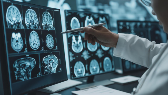
Using fMRI to Explore the Brain’s Visual World
fMRI visual system investigation is among the most intriguing uses of this technology. The brain uses a complicated network of regions to interpret information when perceiving a face, colour, or moving items.
Scientists can see how the brain reacts to various visual inputs through fMRI scans of the visual system. The scans cover anything from spotting an object’s edge to identifying a well-known face. The visual centres of our brains nevertheless function in strikingly comparable ways to real-world sight, even when we visualise scenes (such as a sunset or a loved one’s smile). fMRI is brilliant because it allows us to monitor perception and imagination.
The Department of Biotechnology’s National Brain Research Centre has developed SWADESH, India’s first large-scale multimodal neuroimaging database tailored for the Indian population.
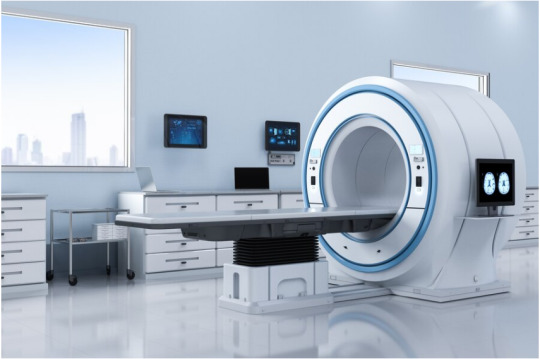
The Workings of an fMRI System in the Lab
A complete fMRI system comprises more than simply the scanner. It usually consists of:
A strong magnet (often 3 Tesla or greater)
Radiofrequency coils for collecting brain impulses
Data processing software for BOLD contrast
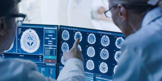
fMRI monitor used in a stimulus delivery system
The function of the fMRI monitor is crucial. It displays pictures, movies, audio, or text to participants during scanning. For example, a participant may see a sequence of emotive facial expressions or read words on a screen while the scanner records which brain areas are active. Even little timing differences between the monitor presentation and capture might distort findings. Therefore, engineers design fMRI monitors for ultra-high accuracy and MRI safety.
The Prospects of fMRI: More Intelligent, Quicker, and Tailored
fMRI technology is rapidly advancing. We observe improvements in:
Time resolution: Recording brain activity on increasingly more precise time scales
Spatial accuracy: Accurately identifying activity down to the minutest cortical layers in space
Giving patients real-time feedback: Using neurofeedback to teach them how to control their brain states
AI integration: Using machine learning to decipher ideas, intents, or even forecast behaviour from fMRI data.
Consider a scenario in which a neurologist can use fMRI to diagnose Alzheimer’s disease decades before symptoms appear, or a therapist can monitor a patient’s emotional control in real time. The future isn’t as far as it previously appeared.
fMRI in the Real World: Practical and Clinical Uses
Most people think of fMRI as being used in high-tech labs or for academic research, yet its practical uses are expanding rapidly, particularly in clinical settings.
Doctors currently utilise fMRI devices for:
Pre-operatively map the brain function of patients with epilepsy or tumours to assist neurosurgeons in avoiding key regions while doing surgery.
Evaluate brain damage following a stroke or trauma to inform recovery strategies.
Track the healing process of brain injury patients by observing changes in their functional abilities.
Researchers use fMRI in forensic and judicial settings to investigate memory, dishonesty, and truth-telling-though the method remains experimental and contentious. fMRI can become a personal health tool for anybody interested in brain function. Experts may use it to track cognitive ageing, monitor mental well-being, or enhance learning and performance.
A study by IIT Delhi using fMRI found significant changes in brain functional connectivity during Yoga Nidra practice, highlighting its impact on emotion regulation and awareness.
What’s Next for fMRI: Looking Deeper Into Ourselves
In many respects, fMRI is among the most audacious scientific advances made in attempting to address the perennial query: Who are we? We discover more about the formation of ideas, the formation of memories, the flow of emotions, and our connection to the outside world. We start to realise the true complexity of our exquisitely designed brains.
Therefore, fMRI is your invitation to pursue that journey- to go past the obvious and genuinely understand what is going on within the mind, whether you are a neuroscience student, a patient seeking a diagnosis, or just someone interested in the inner workings of the brain. And as technology advances, so will our understanding not only of the brain but of the entire human experience.
Conclusion
fMRI has completely changed our understanding of the brain by providing real-time information on our thoughts, emotions, and perceptions of the outside world. It has proven to be an effective tool in therapeutic and scientific contexts by establishing a connection between brain activity, behaviour, and emotion. By creating new avenues for the diagnosis, treatment, and eventual comprehension of the intricacies of the human mind, its contributions will continue to influence neuroscience and medicine in the future.
#fmri visual system#fmri system#Functional Magnetic Resonance Imaging#brain activity#brains visual#neuroscience
0 notes
Text
A dog’s eye view: Exploring the visual system from the kennel to the clinic
A dog’s eye view: Exploring the visual system from the kennel to the clinic gene therapyblindnessconesdogrodscolor blindnesscolor visiondog fMRIdog visionvisual system organization

View On WordPress
#blindness#color blindness#color vision#cones#Dog#dog fMRI#dog vision#gene therapy#rods#visual system organization
0 notes
Text
The ‘Pain of Paying’: How Spending Feels and Why Tracking Helps

In today’s digital economy, spending money has become easier than ever. With one-click purchases, contactless cards, and auto-renewing subscriptions, we can make transactions in seconds — and forget about them just as quickly.
But beneath this convenience lies a powerful psychological force: the pain of paying.
This emotional response plays a surprising role in shaping our financial habits, and if you understand how it works, you can use it to your advantage — especially with the help of money-tracking tools like Mevolve.
In this blog, we’ll explore:
What the pain of paying is and how it works in the brain
How modern spending dulls that pain — and why that’s risky
How tracking expenses revives financial awareness
How Mevolve’s Money Tracker helps you take control
Let’s dive in.
What Is the Pain of Paying?
The “pain of paying” is a term from behavioral economics that describes the negative feeling we experience when we part with money. It’s not a metaphor — it’s real emotional discomfort.
The term was popularized by MIT professor Dr. Drazen Prelec, whose research found that our brains process spending similarly to physical pain.
When you pay with cash, your brain registers the outflow in a tangible, almost painful way. This pain serves a purpose: it makes you think twice before spending.
What Happens in the Brain When We Spend?
Neuroscience research, including fMRI scans, shows that:
The insula, a region linked to pain and emotion, activates when we anticipate or make a purchase.
The nucleus accumbens, associated with pleasure, lights up when we desire something.
This creates a mental tug-of-war:
“I want it” vs. “Ouch, that’s expensive!”
This is what makes financial decisions so emotional — not just logical.
How Technology Has Numbed the Pain
Here’s the problem: modern payment systems have muted our pain of paying.
Why?
Credit cards separate the action of buying from the act of paying (you’ll “feel it” later).
Digital wallets and tap-to-pay make payments so fast, they feel weightless.
Subscriptions and autopay remove visibility — you’re not actively involved in the transaction.
This phenomenon is called decoupling — the disconnection between consumption and payment. And it reduces spending resistance, making us more likely to overbuy.
According to a study by MIT and Carnegie Mellon, people spend up to 100% more when using cards instead of cash.
Why the Pain of Paying Is Good (When Controlled)
You might think pain is a bad thing. But in finance, it’s protective.
It’s your brain’s natural alert system saying:
“Are you sure this is worth it?”
“Is this aligned with your goals?”
“Will you regret this tomorrow?”
When dulled or bypassed, this system fails to prevent impulsive or emotional spending.
That’s where expense tracking comes in.
How Expense Tracking Brings Awareness Back
Tracking expenses manually or with an app like Mevolve brings back the awareness we lose in digital payments. Here’s how:
1. It Creates Conscious Reflection
Every time you log an expense, you’re forced to think about it. This moment of reflection reactivates the emotional circuits your payment method skipped.
2. It Makes Spending Visual
When your expenses are visualized — by date, category, or chart — your brain processes your behavior more clearly. You begin to see patterns, not just feel them.
3. It Builds Self-Control Over Time
Studies show that self-monitoring increases financial discipline. Tracking allows you to course-correct before things spiral out of control.
How Mevolve’s Money Tracker Supports Financial Awareness
The Mevolve Money Tracker is built to bring clarity, control, and calm to your finances — without complexity.
Here’s how it helps:
Instant Logging
Add expenses or income in seconds. No clutter, no confusion.
Visual Reports
View your spending by date, category, or tag — using monthly calendars or clean charts.
Tagging
Label transactions for groceries, subscriptions, travel, or custom categories. Discover hidden spending trends.
When Tracking Becomes a Habit, You Spend Smarter
Most people don’t overspend because they lack discipline — they overspend because they lack visibility.
With consistent tracking:
You reconnect with your emotional money triggers
You make more intentional choices
You save more — without cutting joy
It’s not about being strict. It’s about being smart.
Conclusion
The pain of paying is a natural, intelligent part of how your brain protects you. But modern life is designed to numb it.
By tracking your expenses, you restore your brain’s ability to feel, reflect, and decide — all while building long-term financial habits.
With Mevolve, you’re not budgeting in spreadsheets. You’re building a relationship with money that feels human — clear, calm, and under control.
Don’t just spend. Spend with awareness.
0 notes
Text
How the Brain Works: A Beginner’s Guide to Neuroscience | Lahouaria Hadri, Mount Sinai

The human brain is the most complex structure in the known universe. It allows us to think, feel, remember, move, and even question our own existence. Neuroscience—the study of the nervous system, including the brain—helps us understand what makes us human, how our bodies function, and how we perceive the world. From memory and emotion to consciousness and behavior, neuroscience explores the mechanics behind what we experience every day.
What is Neuroscience?
At its core, neuroscience is the scientific study of the nervous system, which includes the brain, spinal cord, and a vast network of nerves that branch throughout the body. It combines biology, chemistry, physics, and even computer science to explore how neurons (nerve cells) communicate and how these interactions produce everything from motor skills to moods.
Neuroscience isn’t just academic—it plays a vital role in understanding and treating disorders like Alzheimer’s disease, Parkinson’s disease, depression, anxiety, stroke, and many more.
The Building Blocks: Neurons and Neurotransmitters
The brain contains around 86 billion neurons, each connected to thousands of others, forming a dense communication network. These neurons don’t physically touch; instead, they communicate via tiny chemical messengers called neurotransmitters. Some key neurotransmitters include:
Dopamine – involved in reward, motivation, and pleasure.
Serotonin – regulates mood, appetite, and sleep.
Acetylcholine – essential for learning and memory.
GABA (gamma-aminobutyric acid) – reduces neural activity to prevent overstimulation.
The space between neurons where this communication happens is called a synapse. When a neuron “fires,” it sends an electrical signal down its axon, releasing neurotransmitters into the synapse, which are then absorbed by the receiving neuron’s receptors. This process forms the foundation of every thought, feeling, and action.
Brain Anatomy 101
The brain is divided into several regions, each with specialized functions:
Cerebrum: The largest part of the brain, responsible for reasoning, emotions, memory, and voluntary movement. It’s divided into four lobes:
Frontal lobe – decision-making, planning, personality.
Parietal lobe – sensory information like touch and spatial awareness.
Occipital lobe – visual processing.
Temporal lobe – auditory processing and memory.
Cerebellum: Coordinates balance, posture, and fine motor skills.
Brainstem: Connects the brain to the spinal cord and regulates vital functions like heartbeat and breathing.
Limbic system: Often called the “emotional brain,” it includes structures like:
Amygdala – processes emotions such as fear and pleasure.
Hippocampus – vital for forming new memories.
Hypothalamus – controls hunger, thirst, sleep, and hormones.
How Does Memory Work?
Memory isn’t like a video camera; it’s more like a jigsaw puzzle. Neuroscientists divide memory into three main stages:
Encoding – taking in information.
Storage – maintaining the information over time.
Retrieval – accessing the information when needed.
The hippocampus plays a crucial role in forming and organizing new memories. Over time, these memories are consolidated and stored in various parts of the cortex. Different types of memory (short-term, long-term, procedural, emotional) involve different neural circuits.
Consciousness and the Mind
One of the most profound questions neuroscience seeks to answer is: What is consciousness? How do physical neurons give rise to the subjective experience of being alive?
While we don’t fully understand consciousness, research has identified certain brain regions, like the prefrontal cortex and thalamus, as essential for self-awareness and higher-order thinking. Technologies like functional MRI (fMRI) and EEG allow researchers to study brain activity in real-time, offering clues into what happens in the brain when we make decisions, dream, or focus.
Brain Plasticity: The Brain That Adapts
The brain is not a fixed organ; it changes throughout life. This ability, known as neuroplasticity, allows the brain to reorganize itself by forming new neural connections. It’s how we learn, recover from injuries, or adapt to new environments.
For example, if someone suffers a stroke and loses the ability to speak, with therapy and time, other areas of the brain may take over those functions. Neuroplasticity is also why habits, both good and bad, can become deeply ingrained.
When the Brain Gets Sick
Understanding how the brain works also helps us recognize when it doesn’t. Disorders of the brain and nervous system can profoundly impact quality of life.
Alzheimer’s disease involves the buildup of toxic proteins that destroy neurons, particularly in memory-related areas.
Parkinson’s disease results from the loss of dopamine-producing neurons, affecting movement and coordination.
Depression and anxiety��are linked to imbalances in neurotransmitters and disrupted neural circuits.
Ongoing research is shedding light on the causes of these disorders and improving treatments. Innovations like brain-computer interfaces, deep brain stimulation, and gene therapy offer hope for future cures.
The Future of Neuroscience
Neuroscience is advancing rapidly, thanks in part to artificial intelligence, brain imaging technologies, and interdisciplinary collaboration. Some exciting developments include:
Brain-machine interfaces: Devices like Elon Musk’s Neuralink aim to connect the brain directly to computers, potentially restoring function to paralyzed patients or enhancing cognition.
Connectomics: Mapping all the connections in the brain (the “connectome”) to better understand how different areas interact.
Personalized medicine: Using genetic and neural data to create customized treatments for brain disorders.
As our understanding grows, so does our potential to unlock the brain’s mysteries, improve mental health, and enhance human capabilities.
Why Neuroscience Matters
Neuroscience doesn’t just belong in labs or hospitals—it affects our daily lives. It informs education, mental health, technology, and even how we relate to one another. By understanding how the brain works, we can improve learning methods, develop better treatments for disorders, and make more informed decisions about technology, ethics, and society.
Final Thoughts
The brain is astonishing—not just because of its power, but because it is the very thing that lets us understand it. Neuroscience is still young, but each discovery brings us closer to understanding who we are and how we can thrive.
Whether you’re curious about how memory works, passionate about mental health, or excited about futuristic brain tech, neuroscience has something for everyone. It’s a journey into the very center of what makes us human—and we’re only just getting started.
0 notes
Text
MRI Scan Cost in India
Magnetic Resonance Imaging (MRI) is a sophisticated, non-invasive diagnostic tool used by doctors to get detailed images of the internal organs, tissues, and skeletal system. It plays a crucial role in diagnosing a wide range of medical conditions, from brain tumors and spinal cord injuries to joint problems and organ diseases. While MRI scans are widely available across India, their cost can vary significantly depending on several factors. Understanding these factors can help patients make informed decisions when choosing where to get an MRI done.
What is an MRI Scan?
MRI uses powerful magnets, radio waves, and a computer to produce detailed images of the inside of the body. Unlike CT scans or X-rays, MRI does not use ionizing radiation, making it a safer option for many patients, including pregnant women in certain conditions.
It is commonly prescribed to investigate:
Brain and spinal cord abnormalities
Joint and musculoskeletal issues
Tumors and cancers
Liver and abdominal diseases
Heart and vascular conditions
Types of MRI Scans
The cost of an MRI scan depends largely on the type of MRI being conducted. Some common types include:
Brain MRI
Spine MRI (cervical, thoracic, lumbar)
Knee, shoulder, or other joint MRIs
Whole-body MRI
Contrast MRI (with dye for enhanced imaging)
Functional MRI (fMRI) for brain activity
MR Angiography (for blood vessels)
Each type requires different levels of expertise, imaging duration, and equipment usage, which contributes to variation in costs.
MRI Scan Cost in India: General Price Range
The cost of an MRI scan in India ranges from ₹2,000 to ₹25,000, depending on the region, type of scan, and healthcare facility.
Prices in metro cities like Delhi, Mumbai, Bengaluru, Hyderabad, and Chennai tend to be slightly higher due to advanced infrastructure and demand, while smaller cities and towns may offer more economical options.
Factors Affecting MRI Cost
Several elements influence how much you pay for an MRI scan:
Type of MRI Machine:
1.5 Tesla vs. 3 Tesla Machines: 3T machines provide higher resolution images and are generally more expensive.
Open vs. Closed MRI: Open MRI machines are more comfortable for claustrophobic patients but may cost more due to their specialized design.
With or Without Contrast: MRIs done with a contrast agent (usually gadolinium) enhance the image quality and are more expensive due to the added material and monitoring required.
Hospital or Diagnostic Center: Government hospitals and diagnostic centers often offer scans at subsidized rates, while private hospitals and imaging chains may charge a premium for faster service and advanced equipment.
City or Location: Urban areas with advanced medical infrastructure tend to have higher prices compared to semi-urban or rural areas.
Health Insurance: If covered under a medical insurance plan, the cost of MRI scans may be partially or fully reimbursed depending on the policy and reason for the scan.
Ways to Save on MRI Costs
Compare Prices: Use online diagnostic platforms to compare MRI scan prices across centers in your city.
Government Facilities: Look for state-run diagnostic centers which often provide MRI scans at reduced rates or for free under health schemes.
Health Camps and Packages: Some hospitals offer discounted packages during health camps or promotional periods.
Doctor Referrals: Ask your doctor for cost-effective lab referrals that maintain good quality and reliability.
Is MRI Worth the Cost?
While the cost of an MRI scan may seem high, especially for patients paying out-of-pocket, its diagnostic accuracy often makes it worth the investment. Unlike other imaging methods, MRI provides highly detailed visuals of soft tissues, brain structures, and internal organs, helping doctors detect and treat diseases at early stages.
Delaying an MRI due to cost concerns may lead to worsened conditions, longer treatments, or unnecessary procedures. Therefore, timely scanning, even at a slightly higher price, can be lifesaving.
MRI scans are an essential part of modern diagnostic medicine. In India, the cost of an MRI can vary widely depending on multiple factors, but patients now have more choices and flexibility due to the growing number of diagnostic centers. Whether through public healthcare systems or private providers, it is possible to access high-quality MRI scanning at a reasonable price with a little research and guidance.
Before undergoing an MRI, consult your doctor, understand the necessity of the scan, explore pricing options, and ensure the center you choose follows proper safety and hygiene protocols. In the end, prioritizing your health with accurate diagnosis can save both time and money in the long run.
Now avail hefty discounts on MRI scan cost in Delhi with Healthi India
0 notes
Text
Diagnostic Imaging Neuroradiology: A Comprehensive Guide
What is Diagnostic Imaging Neuroradiology?
Diagnostic imaging neuroradiology is a subspecialty of radiology focusing on the diagnosis and characterization of abnormalities of the central and peripheral nervous system, spine, and head and neck using neuroimaging techniques.
Key Imaging Modalities:
Magnetic Resonance Imaging (MRI): Provides detailed images of soft tissues, crucial for detecting brain and spinal cord anomalies.
Computed Tomography (CT): Offers quick imaging, essential in emergency settings like stroke evaluation.
Positron Emission Tomography (PET): Assesses metabolic activity, aiding in tumor detection and monitoring.
Angiography: Visualizes blood vessels, important for identifying aneurysms or vascular malformations.
Applications in Neurological Disorders
Neuroradiology plays a pivotal role in diagnosing various neurological conditions:
Stroke: Rapid imaging determines the type and extent, guiding timely interventions.
Brain Tumors: MRI and PET scans help in identifying tumor type, size, and response to therapy.
Multiple Sclerosis: MRI detects lesions and monitors disease progression.
Traumatic Brain Injuries: CT scans quickly assess injuries, crucial in acute settings.
Advanced Techniques Enhancing Diagnosis
Modern neuroradiology incorporates advanced imaging techniques:
Functional MRI (fMRI): Maps brain activity by detecting changes associated with blood flow.
Diffusion Tensor Imaging (DTI): Visualizes white matter tracts, aiding in assessing traumatic injuries.
Magnetic Resonance Spectroscopy (MRS): Analyzes chemical composition, assisting in tumor characterization.
The Role of uevolve.in in Neuroradiology
uevolve.in is at the forefront of integrating advanced neuroradiological practices:
Innovative Imaging Solutions: Offering state-of-the-art imaging modalities for accurate diagnoses.
Expert Collaboration: Bringing together specialists to interpret complex cases.
Patient-Centric Approach: Ensuring comfort and clarity throughout the diagnostic process.
Discover more about their contributions to neuroradiology at uevolve.in.
Future Perspectives
The field is continually evolving with:
Artificial Intelligence: Enhancing image analysis and diagnostic accuracy.
Telemedicine: Expanding access to neuroradiological expertise remotely.
Personalized Medicine: Tailoring diagnostics and treatments based on individual imaging profiles.
FAQs
Q1: How does neuroradiology differ from general radiology? Neuroradiology specializes in imaging the nervous system, focusing on the brain, spine, and related structures, whereas general radiology covers a broader range of body systems.
Q2: When is a neuroradiological assessment recommended? It’s advised when neurological symptoms are present, such as unexplained headaches, seizures, or after traumatic injuries.
Q3: Are there risks associated with neuroradiological imaging? While MRI is generally safe, CT scans involve radiation exposure. The benefits often outweigh the risks, but it’s essential to discuss concerns with your healthcare provider.
0 notes
Text
Hello! Neuroscience is a fascinating and multidisciplinary field devoted to the study of the nervous system, ranging from the molecular and cellular level to the complex systems that govern behavior and cognition.
Important Parts of Neuroscience:
Neuroscience can be divided into several areas of study, each with its own specific focus:
* Neuroanatomy: Dedicates itself to the study of the structure of the nervous system, including the brain, spinal cord, and peripheral nerves.
* Neurophysiology: Investigates the functioning of the nervous system, such as the electrical and chemical activity of nerve cells and how they communicate.
* Neurochemistry: Examines the chemical processes that occur in the nervous system, including neurotransmitters and other signaling molecules.
* Molecular and Cellular Neurobiology: Focuses on the molecular and cellular mechanisms that underlie neuronal function.
* Cognitive Neuroscience: Explores the neural basis of higher mental processes, such as perception, attention, memory, language, and reasoning.
* Neuroscience: Explores the neural basis of higher mental processes, such as perception, attention, memory, language, and reasoning.
* Neuroscience: * Behavioral Neuroscience: Investigates the relationships between the nervous system and behavior, including emotions, motivation, and social interactions.
* Clinical Neuroscience: Dedicated to the study of diseases and disorders of the nervous system, seeking to understand their causes, develop treatments, and improve diagnosis.
* Computational Neuroscience: Uses mathematical models and computer simulations to understand how the nervous system works.
Where Neuroscience Wants to Go:
Neuroscience has ambitious and high-impact goals:
* Understanding the normal functioning of the brain: Uncovering the mechanisms that enable cognition, emotion, consciousness, and behavior.
* Understanding the causes of neurological and psychiatric diseases: Identifying the biological processes that lead to conditions such as Alzheimer's, Parkinson's, depression, schizophrenia, and others, in order to develop more effective treatments.
* Developing new therapies and interventions: Creating innovative approaches to prevent, treat, and rehabilitate individuals with neurological and mental disorders.
* Advances in neurotechnology: Develop brain-machine interfaces, neural prosthetics, and other technologies to restore lost functions and enhance human capabilities.
* Apply neuroscience knowledge to other areas: Inform practices in education, law, marketing, and other disciplines, optimizing learning, decision-making, and social interaction.
Key Points of Neuroscience:
* Brain Plasticity: The discovery that the brain is malleable and capable of adapting and changing throughout life has revolutionized the understanding of learning and recovery from injury.
* Brain-Behavior Connection: Neuroscience increasingly demonstrates the intricate relationships between brain activity and our actions, thoughts, and feelings.
* Advances in Neuroimaging: Techniques such as functional magnetic resonance imaging (fMRI) and electroencephalography (EEG) allow us to visualize brain activity in real time, providing valuable insights into how the brain works. * Brain-Based Therapies: Neuroscientific knowledge is leading to the development of more targeted and effective therapeutic interventions, such as deep brain stimulation and neurofeedback.
* Neuroethics: As neuroscience advances, important ethical issues related to mental privacy, consent, and the use of neurotechnologies are increasingly being discussed.
In summary, neuroscience is a dynamic and essential field for understanding ourselves and for addressing challenges related to brain health and well-being. The ongoing search for knowledge promises significant transformations in many areas of science and society.
0 notes
Text
¶ … literacy specialist who works in Adult Education. You have noticed that reading is very difficult for people to learn in adulthood, and you wonder whether this might be related to brain plasticity. Gaillard, WH et al. (2002) Language dominance in partial epilepsy patients identified with an fMRI reading task, Neurology, 59, 2, 256-265 https://www.paperdue.com/customer/paper/literacy-specialist-who-works-in-adult-education-56222#:~:text=Logout-,LiteracyspecialistwhoworksinAdultEducation,-Length2pages Search words:reading + fMRI; Google Scholar; neuroscience. Poldrack, RA, Desmond, JE., Glover, GH & Gabrieli, JD. (1998) . The neural basis of visual skill learning: an fMRI study of mirror reading. Cereb. Cortex, 8, 1-10. Search words:reading + fMRI; Google Scholar; neuroscience. Guinevere F.E. et al. (2004) Neural Changes following Remediation in Adult Developmental Dyslexia, Neuron, 44, 3, 411-422 Search words: reading + fmri + adults + neuroplasticity; Google Scholar; neuroscience. Brain imaging studies have investigated the neural mechanisms of recovery in adults following acquired disorders and childhood developmental disorders. However, there are few to no studies that investigate the neural systems that underlie adult rehabilitation of neural-ingrained learning disorder (i.e. those from birth) despite their high incidence. In this study, researchers identified the differences in brain activity during a phonological manipulation task before and after a behavioral intervention in adults who had developmental dyslexia. Phonologically targeted training resulted in improvements in reading in the instructed participants as compared to the dyslexics who received no instruction, and fMRI showed concordant increases in activation in the bilateral parietal and right perisylvian cortices. Authors concluded that behavioral plasticity in adult developmental dyslexia involves two neural mechanisms (or regions in the brain) 24 participants were used. A sublecxical sound deletion task was employed where researchers tested for phonological processing. They tested 19 dyslexic patients who did not receive reading instruction with 19 dyslexic patients who had received instruction (112 hours of structured multi-sensory phonological intervention) and using FMRI identified regions of the brain that were activated during the study. . . Adults who received instruction benefited significantly in their phonological processing skills as compared to those who had not received instruction. This improved understanding of the phonological characteristics of language led, in turn, to improving other aspects of reading ability such as nonword decoding and oral paragraph reading. The study is helpful in that it provides enhanced understanding of the neural characteristics of adults with dyslexia as well as how to structure interventions that can improve their neural plasticity. Skalicky, AE. (2002) . The relationship between early visual processing and reading ability: investigation of temporal processing in adults and children . University of Georgia Theses and Dissertations . http://hdl.handle.net/10724/5745 Search words: reading + fmri + adults + neuroplasticity; Google Scholar; neuroscience. 5. Turkeltaub et al. (2003). Development of neural mechanisms for reading. Nature Neuroscience 6, 767-773 Search words: reading + fMRI in adults; Google Scholar; neuroscience Pediatric brain imaging has generally excluded studies that investigate the neural development of cognitive skills acquired during childhood. Researchers here compared pediatric brain development during reading tasks to those of adults using fMRI. Their task involved an implicit word-processing task that required detection of a visual feature (tall letters) within both words and matched false font strings. Reading was done using eye-motion. Researchers studied an unrelated amount of participants whose ages ranged from 6 to 22 years and found that learning to read is associated with two patterns of change in brain activity: increased activity in the left-hemisphere middle temporal and inferior frontal gyri and decreased activity in the right inferotemporal cortical areas. Activity in the left-posterior superior temporal sulcus of the youngest readers was associated with the maturation of their phonological processing abilities. These discoveries are relevant to the focus of this assignment's focus in that younger children may indeed have an easier time learning to read than adults. They also found that learning to read was associated with engagement of left inferior frontal and middle temporal cortices as well as disengagement of right inferotemporal cortex It remains to investigate whether neuroplasticity can be strengthened to make up for these natural conditions and whether it can be strengthened using mechanisms that would employ these neural regions. Read the full article
0 notes
Text
**Mechanism of Migraine with Aura: Step-by-Step Brain Tissue Changes**
Migraine with aura involves a cascade of neurological events, primarily driven by **cortical spreading depression (CSD)** and subsequent activation of pain pathways. Here’s a breakdown of the process:
---
### **1. Initiation of Cortical Spreading Depression (CSD)**
- **Trigger**: Neuronal hyperexcitability in the cortex (often genetic or due to ion channel dysfunction) leads to abnormal electrical activity.
- **Mechanism**: A sudden surge of neuronal depolarization (excessive firing) begins in a localized brain region, typically the **occipital lobe** (visual cortex).
- **Key Players**:
- **Glutamate**: Excess release of this excitatory neurotransmitter triggers depolarization.
- **Potassium (K⁺) ions**: Leak from neurons, propagating the wave.
---
### **2. Wave of Depolarization Spreads**
- **Process**: The depolarization wave spreads across the cortex at ~3–5 mm/minute, temporarily disrupting normal brain activity.
- **Symptoms**:
- **Visual aura**: Flashing lights, zigzag patterns (scintillations) or blind spots (scotoma) as the wave affects the visual cortex.
- **Sensory aura**: Tingling/numbness (parietal lobe involvement) or speech difficulties (Broca’s area).
---
### **3. Neurotransmitter & Ionic Shifts**
- **Glutamate Surge**: Sustained depolarization increases extracellular glutamate, overstimulating NMDA receptors and causing excitotoxicity.
- **Calcium Influx**: Neuronal calcium overload disrupts mitochondrial function, generating **reactive oxygen species (ROS)** and oxidative stress.
---
### **4. Cerebral Blood Flow Changes**
- **Initial Hyperemia**: Brief increase in blood flow (due to vasodilation) during depolarization.
- **Oligemia**: Prolonged reduced blood flow (vasoconstriction) following the wave, potentially contributing to hypoxia-like symptoms.
- **Imaging**: fMRI studies show a "spreading oligemia" matching the aura’s progression ([*Annals of Neurology*](https://doi.org/10.1002/ana.410280502)).
---
### **5. Trigeminovascular System Activation**
- **Trigeminal Nerve Stimulation**: CSD activates pain-sensitive trigeminal nerve terminals in the meninges (brain’s protective membranes).
- **Inflammatory Mediators**:
- **CGRP (Calcitonin Gene-Related Peptide)**: Released, causing vasodilation and neurogenic inflammation.
- **Substance P**: Enhances pain signaling and sensitizes blood vessels.
---
### **6. Central Sensitization & Pain**
- **Pain Pathways**: Trigeminal signals reach the thalamus and cortex, perceived as throbbing headache (often unilateral).
- **Sensitization**:
- **Allodynia**: Normal touch becomes painful (due to hyperexcitable neurons).
- **Photophobia/Phonophobia**: Brainstem nuclei (e.g., locus coeruleus) amplify sensitivity to light/sound.
---
### **7. Resolution & Postdrome**
- **Recovery**: Neurons repolarize, blood flow normalizes, and inflammatory mediators clear.
- **Postdrome**: Fatigue, cognitive fog, or residual sensitivity due to lingering neurotransmitter imbalances.
---
### **Key Factors Influencing Aura**
- **Genetics**: Mutations in ion channels (e.g., CACNA1A) increase neuronal excitability.
- **Hormones**: Estrogen fluctuations may modulate CSD susceptibility (common in women).
- **Triggers**: Stress, dehydration, or sleep changes lower the threshold for CSD.
---
### **Clinical Implications**
- **Preventive Therapies**: CGRP inhibitors, beta-blockers, or antiepileptics (e.g., topiramate) target CSD or trigeminal activation.
- **Acute Relief**: Triptans constrict dilated meningeal vessels and block CGRP release.
---
*Migraine with aura reflects a complex interplay of neuronal, vascular, and inflammatory processes. While CSD is central to aura, ongoing research aims to clarify genetic and molecular drivers.* 🧠⚡
0 notes
Text
Innovative MRI Technology With Stunning Life Impact
Magnetic Resonance Imaging (MRI) is a modern diagnostic healthcare standard. Its radiation-free, noninvasive imaging capabilities have transformed how physicians see and treat complex medical conditions. Yet, MRI is still out of reach for many people worldwide due to its high costs and unique requirements.
However, this limitation disproportionately affects low and middle-income countries, where MRI healthcare systems are preoccupied with resource balancing between advanced imaging technologies. A gap in accessibility is created because standard superconducting MRI scanners are limited by the availability of robust infrastructure, such as radio frequency (RF) shielding and high power consumption, for use. This article explores how the next wave of innovation in low-power and ultra-low field (ULF) MRI will go far in democratising life-saving diagnostic tools.
The Cost and Infrastructure Barriers
Conventional MRI systems typically require high magnetic fields (1.5 Tesla (T) to 7 T). However, these robust systems create high-resolution images at a steep price and operational demands like working in an RF shield room and consuming more than 25,000 watts of energy. On the other hand, recent advances in ULF MRI have brought a low-power, low-cost alternative to ULF MRI, as demonstrated by Yujiao Zhao and colleagues.
These innovative systems are operated utilising 0.05 T field strength and on standard wall power outlets with only 1,800 watts of a current draw during scanning. These ULF MRI systems solve electromagnetic interference problems using active sensing and deep learning technologies to provide image quality comparable to high-field MRI devices. The possibilities of such advancements enable their utilisation of smaller medical facilities or rural hospitals or clinics located in resource-constrained settings.
MRI Ambience for Enhancing Patient Experience
Technological advancements streamline costs and increase accessibility, but patient comfort in MR evaluations is fundamental to care. Improvements in patient experience have been made by innovations such as In-Bore MRI and MRI cinema for healthcare. These technologies integrate immersive audiovisual solutions, including MRI projectors and MRI-compatible displays, to create a soothing environment which reduces anxiety and increases cooperation during scans.
Patient relaxation virtual skylights, for example, create relaxing outdoor views in the confined MRI bore where the patient is situated. In addition, an MRI-compatible monitor prevents real-time visuals from interfering with the system’s magnetic field, providing patients with comfort and reassurance.
Functional MRI Systems Applications
Functional MRI systems measuring brain activity with blood flow changes have also undergone significant changes. Unlike traditional diagnostic MRI, advanced fMRI visual systems and fMRI monitors allow researchers and clinicians to perform detailed studies of neurological conditions through MRI. The MIT team demonstrates the adaptability of MRI technology to meet new healthcare and research needs with these innovations.
Portable MRI Solutions
Another game-changing development is the portability of low-field MRI systems. Their compact and lightweight designs require little logistical planning, and specialised reports are unnecessary. The MRI-compatible stretcher and wheelchair facilitate seamless interfacing with patient workflows, even in mobile or temporary medical locations.
MRI-compatible cameras and TVs further simplify operations by removing some of the obstacles that medical teams face in seeing, communicating, and doing their jobs, but without compromising the safety and operation of the MRI healthcare system.
Future Directions and Challenges
Even so, ULF MRI technology is in its infancy. Honest challenges are resolution refinement, noise reduction, and scalability. In addition, robust training programs for healthcare professionals are necessary to ensure effective use. Experts such as Udunna Anazodo and Stefan du Plessis point out that while low-field MRI has great promise, its success depends on the ability to prove its reliability and cheap enough for use in multiple clinical settings. The potential for ULF MRI to become a universally accessible diagnostic tool increases exponentially as these challenges are met.
Significant Changes with Small Impact
Healthcare developments include a shift towards patient-centred and environmentally sustainable practices by integrating new technologies, such as MRI-compatible displays and virtual skylights for hospitals. These advances improve diagnostics and address gaps in healthcare equity.
Kryptonite Solutions exemplifies this forward momentum. They can provide comprehensive MRI solutions that embrace innovation and the potential for accessibility. By focusing on supporting the advancement of healthcare technology, they are in the game to transform the way MRI systems serve global communities.
With this paradigm shift in MRI accessibility, comfort and innovation firmly underway, we are charting the future of a healthcare world that closes the diagnostic inequality gap.
#virtual skylights for healthcare#mri ambience#mri compatibles#fmri visual system#mri compatible products#mri compatible stretcher#mri compatible wheelchair#mri compatible#virtual skylights
0 notes
Text
Radiology Technology: Breaking New Ground with Modern Innovations
Radiology technology has made tremendous progress, delivering faster, safer, and more precise ways to diagnose and treat illnesses. With advancements in artificial intelligence (AI), imaging techniques, and portable devices, the role of radiology in healthcare has expanded beyond traditional methods. These innovations are transforming the way medical professionals detect diseases, manage treatments, and offer personalized care. This article delves into the most exciting developments reshaping radiology and their impact on the future of healthcare.
The Digital Revolution in Radiology
Gone are the days of film-based X-rays and analog systems. The shift toward digital radiology has introduced new efficiencies into medical imaging. Digital radiography (DR) allows healthcare providers to capture, view, and share high-resolution images in real-time. Radiologists no longer need to wait for physical films to be developed, speeding up diagnosis and treatment decisions.
Additionally, digital radiology reduces radiation exposure for patients, offering safer diagnostic procedures. Cloud storage systems make it easier for healthcare teams to access imaging data anytime, anywhere, enabling better collaboration among specialists.
AI and Machine Learning: The New Allies in Radiology
AI and machine learning (ML) are having a profound effect on radiology by automating tasks and boosting diagnostic accuracy. AI-powered tools like Computer-Aided Detection (CAD) systems are being used to flag subtle abnormalities that might be missed by human eyes. These tools play a crucial role in early detection of diseases like cancer and cardiovascular conditions.
AI also helps manage radiology workflows more efficiently. It can prioritize urgent cases by evaluating scan results, allowing radiologists to focus on critical patients. Machine learning algorithms continuously improve by learning from each case, ensuring enhanced performance and fewer errors over time.
New Frontiers in Imaging Techniques
Advancements in imaging technology have led to more detailed and functional images, improving diagnostic precision.
1. Hybrid Imaging Systems
Hybrid imaging, such as PET-CT and PET-MRI, provides a comprehensive view by combining two imaging methods into one system. This dual approach helps detect diseases at earlier stages and monitor treatment progress with greater accuracy. PET-CT is now a gold standard for diagnosing and staging cancers, while PET-MRI offers enhanced soft tissue visualization.
2. Functional MRI (fMRI)
Functional MRI (fMRI) takes imaging beyond anatomy by showing brain activity in real-time. This technology is invaluable in neurology, helping doctors understand brain functions and plan surgeries with precision. It is also being explored for use in mental health diagnostics, including conditions like depression and schizophrenia.
3. Low-Dose Imaging Techniques
The need for safer diagnostic tools has driven the adoption of low-dose imaging protocols. Low-dose CT (LDCT) scans are transforming lung cancer screening, enabling early detection with minimal radiation exposure. This innovation promotes preventive care, especially among high-risk patients, while minimizing health risks associated with radiation.
4. 3D and 4D Ultrasound Imaging
Ultrasound technology has advanced significantly, offering 3D and even 4D imaging capabilities. 3D ultrasound provides detailed visuals of organs, while 4D imaging allows real-time monitoring of fetal movements during pregnancy. These technologies are improving diagnostic precision and making ultrasound an indispensable tool for many medical fields.
Portable Imaging Devices: A New Era of Mobility
Portable imaging devices are changing the landscape of healthcare, especially in emergency care and rural settings. Mobile X-ray units, handheld ultrasound devices, and compact MRI systems enable healthcare professionals to perform scans at the patient’s location. This reduces delays in diagnosis and eliminates the need to transfer critically ill patients to imaging centers.
Point-of-care ultrasound (POCUS) devices have become essential tools in emergency departments and intensive care units. They allow quick evaluations of cardiac, abdominal, and vascular conditions, ensuring faster decision-making in critical situations. During the COVID-19 pandemic, portable ultrasound devices played a vital role in identifying lung abnormalities on-site.
Teleradiology and Cloud Integration: Imaging without Borders
The integration of teleradiology and cloud-based systems has unlocked new possibilities for radiology. Teleradiology allows radiologists to provide expert interpretation of scans from any location, making it possible to deliver diagnostic services 24/7. This is especially beneficial for hospitals and clinics in remote areas that lack access to specialized radiology expertise.
Cloud-based Picture Archiving and Communication Systems (PACS) streamline image sharing across healthcare networks, improving collaboration between medical teams. With instant access to imaging data, physicians can make informed treatment decisions more efficiently. The flexibility of cloud systems also supports radiology departments by reducing storage costs and maintenance challenges.
Interventional Radiology: Precision without Surgery
Interventional radiology (IR) has emerged as a game-changer, offering minimally invasive alternatives to surgical procedures. Using imaging guidance, radiologists can perform treatments like tumor ablation, embolization, and angioplasty through tiny incisions. These procedures minimize trauma, reduce recovery times, and decrease the risks associated with open surgeries.
Robotic-assisted interventions are the latest advancement in interventional radiology. Robotic systems enable radiologists to perform complex procedures with exceptional precision, making them highly effective for delicate tasks like stent placement. As robotic technology continues to improve, it will open new possibilities for treating previously inoperable conditions.
Radiomics: A Data-Driven Approach to Personalized Medicine
Radiomics is an emerging field that involves extracting vast amounts of data from medical images to uncover hidden patterns. By analyzing data beyond visual interpretation, radiomics can provide valuable insights into tumor characteristics and disease progression.
When combined with genomic data, radiomics offers a personalized approach to treatment. Oncologists can predict how a patient’s tumor will respond to specific therapies, enabling more targeted and effective treatment plans. This approach is transforming cancer care by improving outcomes and reducing unnecessary treatments.
Ensuring Safety: Radiation Dose Management
One of the key priorities in modern radiology is minimizing radiation exposure. New imaging systems are equipped with dose optimization features to ensure patients receive the lowest radiation necessary for accurate diagnosis. Pediatric imaging, in particular, benefits from these advances, as children are more sensitive to radiation.
Dose-tracking software also plays a vital role in patient safety by monitoring cumulative radiation exposure. Healthcare providers can use this data to make informed decisions about imaging frequency, balancing diagnostic needs with patient safety.
Radiology’s Role in Precision Oncology
Precision oncology relies heavily on advanced imaging for diagnosis, treatment planning, and monitoring. PET-CT and PET-MRI provide detailed insights into tumor behavior, helping oncologists personalize therapies based on individual patient profiles.
Liquid biopsies, which analyze tumor DNA from blood samples, complement imaging techniques by offering non-invasive diagnostic options. Together, radiology and molecular diagnostics provide a comprehensive picture of the patient’s condition, guiding treatment decisions and improving outcomes.
Challenges and Future Directions
While radiology technology offers exciting possibilities, there are challenges to address. Integrating AI systems requires extensive validation to ensure reliability. Additionally, adopting advanced equipment involves significant investment, making it difficult for smaller healthcare facilities to keep pace.
Data security is another pressing concern. As radiology departments increasingly rely on cloud-based platforms, safeguarding patient information from cyber threats is essential. Robust security measures are necessary to protect sensitive data while ensuring accessibility.
The future of radiology lies in continuous innovation. Researchers are exploring new ways to enhance imaging quality, improve AI algorithms, and unlock the full potential of radiomics. Collaboration between radiologists, data scientists, and medical researchers will be key to advancing the field further.
Radiology technology is undergoing a remarkable transformation, with innovations that enhance diagnostic accuracy, improve patient care, and support personalized treatment strategies. From AI-powered diagnostics to portable imaging devices and robotic interventions, the latest advancements are redefining the healthcare landscape.
As these technologies become more accessible, healthcare providers can offer faster, safer, and more precise care to patients. Radiology will remain at the forefront of medical innovation, ensuring that future generations benefit from improved diagnostics and more effective treatments. With ongoing research and collaboration, the possibilities for radiology are limitless, making it an essential pillar of modern healthcare.
0 notes
Text
Neurosurgery in the Digital Age: Leveraging Technology for Better Outcomes
In recent years, the field of neurosurgery has witnessed a remarkable transformation, largely driven by advancements in technology. This evolution has not only improved surgical techniques but has also redefined the way neurosurgeons interact with their patients and optimize outcomes. As we delve into the intricacies of this digital age, we will explore how these technological innovations impact minimally invasive procedures, enhance diagnostic capabilities, and ultimately lead to better patient care in the realm of neurosurgery.

The Rise of Minimally Invasive Techniques
One of the most significant advancements in neurosurgery is the shift towards minimally invasive techniques. Traditional open surgeries often require large incisions, resulting in longer recovery times and increased risk of complications. In contrast, minimally invasive procedures, such as those pioneered by leading Orange County neurosurgeon Robert Louis, MD, utilize advanced imaging and surgical tools to access the brain and spine through smaller openings.
These techniques emphasize the use of keyhole surgery and neuroendoscopy, allowing surgeons to perform complex procedures with minimal disruption to surrounding tissues. For instance, Dr. Louis specializes in endoscopic approaches for treating brain tumors, which significantly reduce postoperative pain and shorten hospital stays. By leveraging sophisticated technologies like the CyberKnife® Robotic Radiosurgery System, neurosurgeons can precisely target tumors with high-dose radiation while preserving healthy tissue. This approach exemplifies how technology not only enhances surgical precision but also fosters quicker recovery and improved patient outcomes.
Enhanced Diagnostic Capabilities
In addition to surgical techniques, technological advancements have revolutionized diagnostic capabilities in neurosurgery. Neuroimaging technologies, such as MRI and CT scans, have become indispensable tools for neurosurgeons, enabling them to visualize brain and spinal structures in remarkable detail. The advent of functional MRI (fMRI) allows surgeons to assess brain activity in real-time, providing critical information before and during surgery.
Moreover, the integration of machine learning and artificial intelligence in diagnostic processes has the potential to enhance accuracy and efficiency. Algorithms can analyze vast amounts of imaging data, identifying patterns that may be missed by the human eye. This capability is particularly crucial in diagnosing complex conditions, such as brain tumors or vascular malformations, where early detection can significantly influence treatment options and outcomes. By harnessing these advanced diagnostic tools, neurosurgeons like Dr. Louis are better equipped to develop personalized treatment plans that align with each patient's unique needs.
Patient-Centric Care Through Technology
The digital age has also fostered a shift towards patient-centric care in neurosurgery. The rise of telemedicine, mobile health applications, and online patient portals has transformed how patients interact with their healthcare providers. These technologies facilitate remote consultations, allowing patients to receive expert opinions without the need for extensive travel, which is particularly beneficial for individuals in Orange County and surrounding areas.
Moreover, patient education has become more accessible through online platforms. Educational videos and informational resources empower patients to understand their conditions and treatment options better. For instance, Dr. Louis's website offers a wealth of information on various neurologic conditions, including brain tumors and cervical spine disorders. This transparency not only alleviates patient anxiety but also fosters a sense of partnership between the patient and the neurosurgeon.
Robotic Assistance in Neurosurgery
The incorporation of robotic technology in neurosurgery marks another significant milestone in the digital age. Robotic systems assist surgeons in performing intricate procedures with unparalleled precision. For example, robotic-assisted surgery allows for improved dexterity and visualization in delicate operations, such as those involving the skull base or spinal cord.
Dr. Louis utilizes robotic systems to enhance the accuracy of minimally invasive surgeries, leading to reduced trauma and quicker recovery times for patients. The ability to navigate complex anatomical structures with robotic assistance is a game-changer, ensuring that even the most challenging surgeries can be executed with confidence and safety. This innovation exemplifies how technology enhances the surgeon's capabilities, ultimately translating into better outcomes for patients.
The Future of Neurosurgery: Data-Driven Decision Making
As we look to the future, the role of data analytics in neurosurgery will continue to expand. The collection and analysis of patient data can inform evidence-based practices, enabling neurosurgeons to make more informed decisions regarding treatment approaches. By leveraging data from various sources, including surgical outcomes and patient demographics, neurosurgeons can refine their techniques and improve overall patient care.
Incorporating predictive analytics into surgical planning allows neurosurgeons to anticipate potential complications and adjust their strategies accordingly. This proactive approach enhances patient safety and contributes to better overall outcomes. As the field of neurosurgery continues to evolve, the integration of data-driven decision-making will be paramount in shaping the future of patient care.
Conclusion
In conclusion, the digital age has ushered in a new era for neurosurgery, characterized by technological innovations that enhance surgical techniques, improve diagnostic capabilities, and promote patient-centric care. As highlighted by the expertise of Orange County neurosurgeon Robert Louis, MD, minimally invasive approaches and advanced imaging technologies are at the forefront of this transformation. By embracing these advancements, neurosurgeons are better equipped to deliver exceptional care, ensuring that patients experience optimal outcomes on their journey to recovery. As we move forward, the continued integration of technology in neurosurgery holds immense promise, paving the way for a future where patient care is more personalized, efficient, and effective.
0 notes
Text
The Future of Radiology: Innovations and Advancements
Radiology, the medical discipline that uses imaging to diagnose and treat diseases within the body, has witnessed remarkable innovations and advancements over the years. These advancements have revolutionized the way we understand and treat various health conditions, making radiology an indispensable part of modern medicine. As we look to the future, the landscape of radiology is set to evolve even further, driven by cutting-edge technologies and groundbreaking research. The Best Radiology Hospital in Jaipur exemplifies these advancements, consistently incorporating the latest technologies and research findings to provide superior diagnostic and treatment services. This article explores the future of radiology and highlights some of the most promising innovations and advancements shaping the field.
Artificial Intelligence in Radiology
Artificial Intelligence (AI) is at the forefront of transforming radiology. AI algorithms can analyze vast amounts of imaging data with unprecedented speed and accuracy, assisting radiologists in detecting abnormalities that may be missed by the human eye. Machine learning models are being trained to recognize patterns in medical images, aiding in early diagnosis and improving treatment outcomes. The integration of AI in radiology promises to enhance diagnostic precision, reduce human error, and optimize workflow efficiency.
Advanced Imaging Techniques
The development of advanced imaging techniques is another significant advancement in radiology. Techniques such as functional MRI (fMRI), positron emission tomography (PET), and dual-energy CT scans provide more detailed and comprehensive images of the body's internal structures. These techniques allow for better visualization of complex conditions, leading to more accurate diagnoses and tailored treatment plans. Furthermore, the advent of 3D and 4D imaging technologies offers dynamic views of organs and tissues, enabling real-time assessment of physiological functions.
Interventional Radiology
Interventional radiology is a rapidly growing subspecialty that utilizes minimally invasive procedures to diagnose and treat various medical conditions. Guided by imaging techniques such as fluoroscopy, ultrasound, and CT, interventional radiologists perform procedures like angioplasty, embolization, and tumor ablation with precision. These minimally invasive procedures often result in shorter recovery times, reduced risk of complications, and improved patient outcomes. The future of interventional radiology holds immense potential for further innovations, including the use of robotics and advanced imaging guidance systems.
Personalized Medicine and Radiogenomics
The concept of personalized medicine is gaining traction in radiology, with radiogenomics playing a crucial role. Radiogenomics involves the correlation of imaging features with genetic and molecular data to understand the underlying biology of diseases. This approach allows for the identification of specific biomarkers that can predict disease progression and response to treatment. By integrating radiogenomics into clinical practice, radiologists can provide personalized treatment plans tailored to each patient's unique genetic makeup, leading to more effective and targeted therapies.
The Role of the Best Radiology Hospital in Jaipur
In India, Jaipur stands out as a hub for advanced medical facilities, and the best radiology hospital in Jaipur is at the forefront of embracing these innovations. Equipped with state-of-the-art imaging technologies and staffed by highly skilled radiologists, this hospital is committed to providing top-notch diagnostic and interventional radiology services. By leveraging cutting-edge technologies and staying abreast of the latest advancements in the field, the best radiology hospital in Jaipur ensures that patients receive accurate diagnoses and optimal treatment outcomes.
Conclusion
The future of radiology is undeniably bright, with continuous innovations and advancements shaping the field. From the integration of AI and advanced imaging techniques to the rise of interventional radiology and personalized medicine, radiology is poised to play an even more critical role in healthcare. As we move forward, institutions like the best hospital in Jaipur will continue to lead the way, ensuring that patients benefit from the latest technological breakthroughs and receive the highest standard of care. The evolution of radiology promises a future where early detection, precise diagnosis, and effective treatment are the norm, ultimately improving patient outcomes and transforming the landscape of modern medicine.
#best hospital in jaipur#child hospital in jaipur#best orthopedic hospital in jaipur#best urology hospital in jaipur
0 notes
Text
INNOVATIONS IN DIAGNOSTIC IMAGING: WHAT THE FUTURE HOLDS
In the ever-evolving landscape of healthcare, diagnostic imaging stands at the forefront of technological advancements. As we move further into the 21st century, innovations in this field promise to revolutionize the way we detect, diagnose, and treat medical conditions. From artificial intelligence to molecular imaging, the future of diagnostic imaging holds exciting prospects that will enhance precision, reduce risks, and improve patient outcomes.
ARTIFICIAL INTELLIGENCE AND MACHINE LEARNING
One of the most transformative innovations in diagnostic imaging is the integration of artificial intelligence (AI) and machine learning. These technologies enable the analysis of vast amounts of imaging data with unprecedented speed and accuracy. AI algorithms can identify patterns and anomalies that may be imperceptible to the human eye, leading to earlier and more accurate diagnoses.
For instance, AI-powered tools are being developed to assist radiologists in detecting conditions such as cancer, cardiovascular diseases, and neurological disorders. These tools can highlight areas of concern, prioritize cases based on urgency, and even suggest potential diagnoses, thus streamlining the diagnostic process and reducing the likelihood of human error.
ADVANCED IMAGING TECHNIQUES
Advancements in imaging techniques are continuously pushing the boundaries of what is possible. Techniques such as functional MRI (fMRI), positron emission tomography (PET), and single-photon emission computed tomography (SPECT) are becoming more sophisticated, providing detailed insights into the functioning of organs and tissues.
Functional MRI, for example, measures brain activity by detecting changes in blood flow, offering valuable information about brain function and aiding in the diagnosis of neurological conditions like epilepsy, stroke, and Alzheimer's disease. Similarly, PET and SPECT imaging are instrumental in understanding metabolic processes and can be used to diagnose and monitor diseases like cancer and heart disease.
MOLECULAR IMAGING
Molecular imaging represents a significant leap forward in diagnostic imaging. Unlike traditional imaging methods that focus on the structure of organs and tissues, molecular imaging visualizes the biological processes at the molecular and cellular levels. This allows for the detection of diseases at their earliest stages, often before symptoms appear.
One of the most promising applications of molecular imaging is in oncology. By targeting specific molecules associated with cancer, molecular imaging can identify tumors with high specificity and sensitivity. This not only aids in early detection but also in monitoring the effectiveness of treatments, enabling personalized and adaptive therapy strategies.
HYBRID IMAGING SYSTEMS
Hybrid imaging systems, which combine two or more imaging modalities, are gaining traction in the medical field. Technologies like PET/CT and PET/MRI integrate the strengths of different imaging techniques, providing comprehensive information about both the anatomy and the metabolic activity of tissues.
PET/CT, for instance, combines the metabolic imaging capabilities of PET with the detailed anatomical imaging of CT. This allows for precise localization and characterization of lesions, improving diagnostic accuracy and aiding in treatment planning. PET/MRI, on the other hand, offers superior soft tissue contrast and reduced radiation exposure compared to PET/CT, making it particularly useful in pediatric and neurological imaging.
PORTABLE AND POINT-OF-CARE IMAGING
The future of diagnostic imaging also includes a shift towards portability and point-of-care solutions. Portable imaging devices, such as handheld ultrasound machines and compact MRI scanners, are making it possible to bring advanced imaging capabilities to remote and underserved areas. This democratization of imaging technology can significantly improve access to healthcare and facilitate timely diagnosis and treatment.
Point-of-care imaging, integrated with telemedicine, allows for real-time consultations and remote diagnostics. For example, a handheld ultrasound device connected to a smartphone can capture and transmit images to a specialist for immediate interpretation. This not only saves time but also reduces the need for patient transfers, making healthcare more efficient and accessible.
3D AND 4D IMAGING
The advent of 3D and 4D imaging is adding new dimensions to diagnostic imaging. 3D imaging provides a more accurate representation of anatomical structures, which is particularly useful in planning surgical procedures and guiding minimally invasive interventions. It also enhances the visualization of complex structures, such as the intricate networks of blood vessels in the brain or heart.
4D imaging, which incorporates the dimension of time, offers dynamic views of physiological processes. This is particularly valuable in cardiology, where 4D echocardiography can capture the motion of the heart in real-time, providing detailed information about cardiac function and aiding in the diagnosis of heart diseases.
PERSONALIZED MEDICINE AND THERANOSTICS
The future of diagnostic imaging is closely linked to the concept of personalized medicine. Advanced imaging techniques are enabling the development of theranostics, a field that combines diagnostics and therapeutics. Theranostics involves the use of imaging to guide targeted therapies, ensuring that treatments are tailored to the individual patient's condition.
For example, in oncology, theranostics can involve using molecular imaging to identify specific biomarkers on cancer cells and then delivering targeted therapies that bind to these markers. This approach not only enhances the effectiveness of treatments but also minimizes side effects by sparing healthy tissues.
As we look to the future, the innovations in diagnostic imaging promise to transform healthcare in profound ways. The integration of AI, the development of advanced and hybrid imaging techniques, the advent of molecular and portable imaging, and the rise of personalized medicine are just a few of the exciting trends shaping the field. These advancements will not only improve the accuracy and efficiency of diagnoses but also pave the way for more effective and personalized treatments, ultimately enhancing patient care and outcomes. The future of diagnostic imaging is bright, and its potential to revolutionize healthcare is immense.
Orignal Source: https://av-imaging.com/innovations-in-diagnostic-imaging-what-the-future-holds.html
0 notes
Text
What Is Neuroradiology? A Complete Guide to Brain and Spine Imaging
Introduction to Neuroradiology
Neuroradiology is a specialized branch of radiology focusing on diagnosing and treating disorders of the brain, spine, head, and neck using advanced imaging techniques. It plays a crucial role in identifying conditions such as strokes, tumors, aneurysms, and degenerative diseases like Alzheimer’s and Parkinson’s.
Key Imaging Techniques in Neuroradiology
Neuroradiologists employ various imaging modalities to obtain detailed views of the nervous system:
Magnetic Resonance Imaging (MRI): Provides high-resolution images of brain and spinal structures, aiding in the detection of tumors, multiple sclerosis, and other abnormalities.
Computed Tomography (CT): Offers rapid imaging, especially useful in emergency situations like acute strokes or traumatic injuries.
Functional MRI (fMRI): Measures brain activity by detecting changes associated with blood flow, assisting in pre-surgical planning.
Diffusion Tensor Imaging (DTI): Maps white matter tracts in the brain, useful in assessing traumatic brain injuries.
CT and MR Angiography: Visualizes blood vessels to identify aneurysms, blockages, or vascular malformations.
Conditions Diagnosed Through Neuroradiology
Neuroradiology is instrumental in diagnosing a range of neurological conditions:
Stroke: Identifies ischemic or hemorrhagic strokes, enabling timely intervention.
Brain and Spinal Tumors: Detects and monitors the progression of neoplasms.
Multiple Sclerosis: Reveals lesions in the central nervous system indicative of MS.
Aneurysms and Vascular Malformations: Assesses abnormalities in blood vessels that could lead to serious complications.
Degenerative Diseases: Aids in the evaluation of conditions like Alzheimer’s and Parkinson’s disease.
The Role of Neuroradiologists
Neuroradiologists are medical doctors with specialized training in interpreting neuroimaging studies. They collaborate closely with neurologists, neurosurgeons, and other specialists to provide accurate diagnoses and guide treatment plans. Their expertise ensures that subtle abnormalities are not overlooked, leading to better patient outcomes.
Advancements and Resources
With the rapid advancement of imaging technology, neuroradiology continues to evolve, offering more precise diagnostic capabilities. For those interested in exploring cutting-edge developments in this field, platforms like uevolve.in provide valuable insights and resources.
Conclusion
Neuroradiology stands at the forefront of diagnosing complex neurological conditions, leveraging advanced imaging techniques to provide critical insights into the brain and spine. As technology progresses, its role in patient care becomes increasingly significant. For more information and updates on neuroradiology and related topics, consider visiting uevolve.in.
#digital x-ray clinics reporting near me#best radiology center in mumbai#top ct scan reporting near me
0 notes
Text
Innovations in Neuroimaging: What Patients Need to Know

Neuroimaging technologies have transformed the field of neurosurgery, enabling more accurate diagnoses and improved surgical outcomes. As the best neurosurgeon doctor in Jaipur, Dr. Himanshu Gupta leverages these cutting-edge advancements to provide superior patient care. This article explores the latest innovations in neuroimaging and their impact on neurosurgical practices.
The Role of Neuroimaging in Neurosurgery
Neuroimaging is crucial for diagnosing neurological conditions and planning surgeries. Techniques such as MRI, CT scans, and PET scans provide detailed images of the brain and spinal cord, helping neurosurgeons like Dr. Himanshu Gupta to identify abnormalities and plan precise interventions.
Advanced Imaging Techniques
Functional MRI (fMRI): This technique measures brain activity by detecting changes in blood flow. fMRI helps in mapping brain functions and is essential for planning surgeries that avoid critical areas, thus preserving neurological function.
Diffusion Tensor Imaging (DTI): DTI visualizes white matter tracts in the brain, crucial for understanding brain connectivity. It aids in the assessment of conditions like brain tumors and traumatic brain injuries.
3D Imaging and Navigation: Advanced 3D imaging allows for the creation of detailed anatomical models. Combined with navigation systems, it enables Dr. Himanshu Gupta to perform minimally invasive surgeries with higher precision.
Benefits for Patients
Innovations in neuroimaging offer numerous benefits, including:
Improved Accuracy: Enhanced imaging techniques provide clearer, more detailed views of the brain and spine, leading to more accurate diagnoses.
Minimally Invasive Procedures: Better imaging supports less invasive surgical approaches, reducing recovery times and minimizing risks.
Personalized Treatment Plans: Detailed imaging allows for tailored treatment plans, improving overall outcomes for patients.
Dr. Himanshu Gupta: Pioneering Advanced Neuroimaging in Jaipur
As the best neurosurgeon doctor in Jaipur, Dr. Himanshu Gupta is at the forefront of integrating these neuroimaging innovations into clinical practice. His expertise and commitment to utilizing the latest technologies ensure that patients receive the highest standard of care.
Conclusion
Neuroimaging advancements have revolutionized neurosurgery, providing significant benefits to patients. With experts like Dr. Himanshu Gupta, the best neurosurgeon doctor in Jaipur, leading the way, patients can be assured of receiving top-notch care through the use of state-of-the-art imaging technologies.
0 notes