#formalin fixed paraffin embedded
Explore tagged Tumblr posts
Visit Tumblr Blog
Explore Tumblr blogs with no restrictions, modern design and the best experience.
Last Seen Tumblr Blogs


Fun Fact
The total number of visits Tumblr.com received during January 2021 is 327 million.
Text
Understanding the Value of FFPE Blocks in Modern Research
Biomedical research heavily depends on having good-quality, well-preserved tissue samples at their disposal. Of the most common types of specimens used are formalin fixed paraffin embedded (FFPE) blocks. These tissue samples give scientists an accessible, long-term means of performing multiple types of analyses, such as molecular profiling, histopathology, and biomarker discovery.
FFPE blocks are made by initially fixing tissues in formalin, a process that stabilizes cellular structures by crosslinking proteins. The fixed tissue is embedded in paraffin wax, and thin sectioning and long-term storage become possible. The process has been established as a gold standard in pathology laboratories globally because it is efficient, stable, and adaptable for various diagnostic and research uses.

Advantages and Applications of FFPE Block Collection
Having and collecting a strong FFPE block collection is essential for both academic and clinical institutions. These blocks have the distinct advantage of being able to be stored for years without loss of integrity. This makes it possible for researchers to go back and look at old cases for retrospective studies, so that historical information can still be used to make new scientific discoveries.
One of the major advantages of FFPE samples is their suitability for use with a broad array of methods, such as immunohistochemistry (IHC), in situ hybridization (ISH), and next-generation sequencing (NGS). Although formalin fixation may lead to some DNA and RNA fragmentation, improvements in extraction and amplification technologies have allowed for the recovery of quality nucleic acids from these samples. Consequently, FFPE tissue has emerged as an invaluable tool for genomic research and personalized medicine investigation.
Moreover, FFPE blocks also have an important function in cancer research. Paraffin-embedded tumor tissue can be examined for mutation profiling to help with the identification of targeted therapies. The reproducibility is provided by the uniform preparation process, which is critical when comparing findings between studies or confirming biomarkers.
How FFPE Blocks Support Global Bioresearch
Given the increasing demand for translational research, FFPE samples have become increasingly significant in global biorepositories. Such blocks allow researchers to obtain standardized high-quality tissue samples from around the globe, which are highly necessary for comparative studies and large-scale research. International collaborations usually rely on the presence of such specimens to provide uniformity to the outcomes of research.
Ethically obtained FFPE blocks, acquired under strict regulatory conditions, also improve the validity of published research. With access to varied and well-documented tissue repositories, researchers can investigate diseases among various populations and stages, improving the strength of their results.
Institutions with large FFPE archives make an important contribution to public health research, especially for the investigation of rare diseases or new health risks. FFPE samples from historical outbreaks or rare diseases provide an important window on disease progression, response to treatment, and long-term health status.
Reliable Access to FFPE Blocks for Your Research
Securing access to a reputable FFPE block collection can really speed up your research goals. When selecting a source, make sure to search for quality assurance, ethical acquisition, and thorough clinical annotation. These elements help ensure that the tissue samples are not only scientific grade but also rich in contextual information for your research.
If you want to acquire ethically sourced, high-quality formalin fixed paraffin embedded blocks or develop a quality FFPE block collection, choose to collaborate with a provider who puts emphasis on quality, compliance, and client service.
To explore a curated selection of FFPE tissue blocks for your next research project, connect with iBioSpecimen—your trusted partner in bioresearch excellence.
0 notes
Text
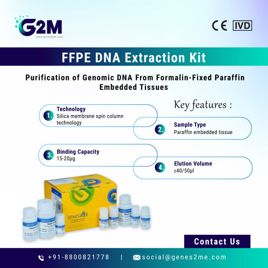
#G2M is thrilled to introduce you to our highly efficent #FFPE DNA Extraction kit which is designed for the purification of genomic #DNA from formalin-fixed paraffin-embedded (FFPE) tissue sections. Fixing of #tissues using formalin generates the cross-linking of the #NucleicAcids, proteins. This kit enables the partial reversing of the modification caused by formalin which results in high yield with good #quality.
Visit our website for more information https://www.genes2me.com/ffpe-dna-extraction-kit
For more details, Call us at +91-8800821778 or drop us an email at [email protected]
#extraction #Genes2me #kits #purification #solutions #genomics #technology #rtpcr #rna #madeinindia #india #manufacturer
#g2m#genes2me#ffpe#formalin fixed paraffin embedded#nucleic acid#quality#dna#extraction#purification#rna#tissues#rt pcr#kits
0 notes
Text
Formalin Fixed Paraffin Embedded Tissue: A Cornerstone in Histopathological Research
Introduction to Formalin Fixed Paraffin Embedded Tissue
Formalin fixed paraffin embedded tissue (FFPE) plays a crucial role in medical diagnostics and research. This widely used method preserves tissue samples for long-term storage, allowing detailed microscopic examination. By stabilizing tissue architecture and cellular morphology, FFPE ensures the integrity of biological samples, making it indispensable in pathology labs worldwide.
What is Formalin Fixed Paraffin Embedded Tissue?
Formalin fixed paraffin embedded tissue refers to tissue specimens preserved through a specific process involving two major steps: fixation and embedding. First, tissues are immersed in a 10% neutral-buffered formalin solution, which crosslinks proteins and halts biological decay. Following fixation, the samples undergo dehydration and are then embedded in paraffin wax, creating stable blocks that can be sectioned for microscopic analysis.

Benefits of Using Formalin Fixed Paraffin Embedded Tissue
One of the most significant advantages of formalin fixed paraffin embedded tissue is its long-term stability. Researchers and clinicians can store FFPE samples for years without compromising tissue morphology. This method also facilitates easier sectioning, staining, and analysis under a microscope. Moreover, FFPE blocks are compatible with various molecular techniques, including immunohistochemistry (IHC), in situ hybridization, and even next-generation sequencing with proper optimization.
Applications in Clinical and Research Settings
Formalin fixed paraffin embedded tissue is vital in both diagnostic and academic environments. In hospitals, it allows pathologists to examine biopsies and surgical specimens, providing insights into disease mechanisms, tumor classification, and infection detection. In research, FFPE samples are often used to study disease progression, validate biomarkers, and perform retrospective analyses on archived tissues.
Limitations and Challenges of FFPE Samples
Despite its many benefits, formalin fixed paraffin embedded tissue does come with limitations. The fixation process can sometimes lead to the degradation or crosslinking of nucleic acids, affecting the accuracy of downstream molecular assays. Furthermore, antigen masking during fixation may hinder antibody binding in immunohistochemistry. However, with advanced protocols and retrieval methods, many of these issues can be mitigated.
Optimizing Molecular Techniques for FFPE Samples
Molecular testing on formalin fixed paraffin embedded tissue requires specialized extraction techniques to recover high-quality DNA and RNA. Companies have developed commercial kits tailored for FFPE samples that help researchers overcome extraction challenges. In addition, antigen retrieval techniques, such as heat-induced epitope retrieval (HIER), are used to unmask proteins for accurate immunohistochemical analysis.
Storage and Archiving of FFPE Blocks
Another major advantage of formalin fixed paraffin embedded tissue is ease of storage. Once embedded in paraffin, tissue blocks can be archived at room temperature, occupying minimal space. This makes them highly accessible for future research, especially in large biorepositories and biobanks. Proper labeling and cataloging are essential to maintain the integrity and retrievability of stored samples.
FFPE in Personalized Medicine and Genomics
As precision medicine continues to advance, formalin fixed paraffin embedded tissue has found new importance in genomic studies. Archived FFPE samples are being used in whole-genome sequencing and transcriptomic analysis, provided the right protocols are in place. This allows researchers to study historical patient data and develop tailored therapeutic strategies based on molecular profiles.
Ethical and Regulatory Considerations
Handling formalin fixed paraffin embedded tissue requires adherence to strict ethical and regulatory guidelines. In clinical settings, informed consent is often necessary, especially when tissues are used for research beyond diagnosis. Regulatory bodies ensure that FFPE samples are handled, stored, and disposed of in accordance with biosafety and data protection standards.
Conclusion: The Enduring Value of FFPE Tissues
In conclusion, formalin fixed paraffin embedded tissue remains a gold standard in histopathology and biomedical research. Its ability to preserve tissue architecture, enable detailed analysis, and support modern molecular techniques makes it a cornerstone of both routine diagnostics and scientific discovery. Despite some technical limitations, continuous advancements in extraction and analysis methods are expanding the utility of FFPE tissues in cutting-edge research and personalized medicine.
1 note
·
View note
Text
Best Antibody Suppliers
BioGenex offers more than 600 primary antibodies with a specific emphasis on cancer and infectious diseases. All monoclonal and polyclonal antibodies are quality controlled and highly optimized to provide a maximum signal with the minimum background when used for immunohistochemical staining on formalin-fixed, paraffin-embedded (FFPE) tissue.
0 notes
Text
Redefining Molecular Testing: FLT3-TKD, BRAF, and H1N1 PCR Kits

Advancements in molecular diagnostics have significantly transformed the clinical paradigm for managing both oncological and infectious diseases. As medicine increasingly leans toward precision, the need for mutation-specific and pathogen-targeted testing grows stronger. Laboratories and clinicians now require tools that can detect mutations and pathogens quickly, accurately, and consistently. In this context, specialized kits like the BRAF PCR Kit, FLT3-TKD PCR Kit, and H1N1 PCR test have become essential in diagnostic workflows. Each of these kits is tailored for a distinct clinical purpose but shares a common commitment to analytical sensitivity, reliability, and standardization.
The FLT3-TKD PCR Kit helps detect changes in the tyrosine kinase part of the FLT3 gene, which is an important marker in diagnosing acute myeloid leukemia (AML).FLT3 mutations, especially internal tandem duplications (ITD) and TKD point mutations, are known to influence prognosis and therapy selection in AML patients. Identifying these mutations accurately enables hematologists to stratify risk and adjust treatment regimens, especially when considering tyrosine kinase inhibitors.
This FLT3-TKD PCR Kit uses allele-specific primers to detect common TKD mutations such as D835 and I836, Limiting false-negative results. It is configured for high throughput without compromising specificity, making it practical for routine clinical laboratories. The detection method, based on real-time PCR, ensures that clinicians receive quantitative, actionable data within a short time frame. It is in range of real-time PCR platforms, the FLT3-TKD PCR Kit delivers flexibility and operational convenience across lab environments.
In parallel with hematological applications, solid tumor diagnostics continue to depend heavily on molecular insights. The BRAF PCR Kit supports the detection of BRAF gene mutations, primarily the V600E variant, which is a defining marker in melanomas, colorectal cancers, and certain thyroid cancers. The presence of a BRAF mutation has direct therapeutic consequences, as several approved drugs target the BRAF V600E pathway specifically.
The BRAF Mutations Kit enables direct mutation detection from DNA samples using real-time PCR, without requiring extensive post-PCR processing. By applying validated primers and hydrolysis probes, the BRAF PCR Kit guarantees high sensitivity even in challenging samples, such as those from formalin-fixed paraffin-embedded (FFPE) tissues. This feature is crucial in oncology diagnostics where DNA quality may be compromised. The clinical utility of the BRAF Mutations Kit lies not just in detection, but in its ability to inform downstream treatment choices, supporting a personalized medicine framework.
The BRAF PCR Kit also minimizes operator-dependent variability by offering a ready-to-use master mix and detailed interpretation guidelines. This helps ensure consistency across laboratories, reducing discrepancies in patient diagnosis and improving the overall reliability of the results.
While oncology diagnostics dominate many molecular labs, infectious disease testing remains equally vital. Seasonal and pandemic viral infections require rapid, scalable solutions. The H1N1 PCR test, developed for the detection of the Influenza A H1N1 virus, addresses the need for early identification and containment of viral outbreaks. This is especially relevant in healthcare settings, where time-to-result can directly influence treatment decisions and isolation protocols.
The H1N1 PCR test employs a reverse-transcription PCR methodology to detect viral RNA with high sensitivity and specificity. The assay is optimized for respiratory specimens, and its closed-tube format reduces the risk of contamination. It detects both seasonal and pandemic H1N1 strains, making it useful during flu seasons or in the face of a suspected outbreak. With an intuitive workflow and minimal sample processing, the H1N1 PCR test offers laboratories a reliable diagnostic tool for fast and precise identification.
What makes these kits stand out is not only their diagnostic accuracy but also their relevance in different medical scenarios. The FLT3-TKD PCR Kit plays a pivotal role in managing leukemia patients by enabling targeted therapy choices. The BRAF Mutations Kit and BRAF PCR Kit provide essential insights in cancer subtyping and treatment planning, especially for patients with limited options. Meanwhile, the H1N1 PCR test continues to support infection control practices globally, where timely and accurate diagnosis is critical to managing influenza outbreaks.
All three kits align with the modern need for diagnostics that are not only accurate but also fast and scalable. With the increasing push toward decentralized diagnostics and smaller, on-site lab units, the format and functionality of these kits are highly suitable. Laboratories can implement them without extensive setup changes, and healthcare providers can trust the results for guiding clinical decisions.
In summary, the clinical importance of the BRAF PCR Kit, FLT3-TKD PCR Kit, and H1N1 PCR test cannot be overstated. Each addresses a specific medical challenge, whether it's oncogene mutation detection or viral pathogen identification. By focusing on analytical precision, ease of use, and rapid reporting, these kits serve as benchmarks in their respective domains. They demonstrate how focused diagnostic tools can support personalized care, reduce diagnostic delays, and ultimately lead to better patient outcomes.
0 notes
Text
Revolutionize IHC Workflows with Gene BioSolution’s Retrieval System
Bring speed, precision, and consistency to your lab with the Digital Antigen Retrieval System from Gene BioSolution. This powerful device is designed to unmask antigens effectively from formalin-fixed paraffin-embedded tissues—ensuring that your IHC results are both accurate and reproducible. Equipped with programmable settings, uniform heating technology, and safety-first operation, it’s the perfect solution for busy labs aiming to optimize their staining workflows. Whether you're conducting cancer diagnostics, antibody validation, or tissue profiling, this system delivers consistent results that meet the demands of modern pathology. Built with user-friendly features and industry-leading technology, it’s the ultimate upgrade for your histology lab.

0 notes
Text
FFPE Tissue Samples for Genomics Study & Analysis Market – Microarray Analysis Evolution
FFPE Tissue Samples for Genomics Study & Analysis Market: Trends, Drivers, and Future Outlook
The world of cancer diagnostics and research is undergoing a profound transformation driven by advances in genomics. Central to this evolution are formalin-fixed paraffin-embedded (FFPE) tissue samples, which have long been the gold standard for preserving histological architecture and molecular integrity. In 2023 alone, the global market for FFPE tissue samples in genomics study and analysis reached a valuation of US$ 891.1 million, and it is projected to surge past US$ 1.5 billion by 2034—propelled by a healthy compound annual growth rate (CAGR) of 5.1% over the 2024–2034 forecast period. In this blog post, we delve into the key factors shaping this market, the segmentation by product and application, regional dynamics, leading companies, recent technological breakthroughs, and what lies ahead for the industry.
The Aging Population and Rising Cancer Incidence
One of the most significant macro-trends fueling demand for FFPE tissue specimens is the global demographic shift toward older age groups. The risk of cancer increases substantially with age—nearly 87% of cancer-related deaths occur in individuals aged 50 or older, and almost half of those fatalities happen in people aged 70 and above. As life expectancies climb worldwide, the absolute number of new cancer diagnoses is expected to balloon: from an estimated 18.1 million new cases in 2018 to nearly 29.5 million by 2040. This burgeoning caseload creates an urgent need for reliable, high-quality tissue samples capable of supporting in-depth genomic analyses that inform both diagnostics and treatment planning. FFPE specimens, with their proven track record of maintaining both morphological detail and nucleic acid integrity over long storage periods, are uniquely positioned to meet this need, driving robust growth in sample procurement, processing, and analysis activities across research institutions, hospitals, and biotech companies.
Technological Advancements and Workflow Automation
While the foundational chemistry of formalin fixation and paraffin embedding has remained relatively consistent for decades, the downstream processes of sample preparation, extraction, and analysis have witnessed remarkable innovation. Automated tissue processors and sample‐prep systems now enable histopathology laboratories to handle greater volumes of specimens with enhanced precision and reproducibility. Newer bio-banking technologies ensure that FFPE blocks can be cataloged, stored, and retrieved efficiently for retrospective and prospective studies alike. On the analytics front, next-generation sequencing (NGS) platforms, digital PCR/qPCR instruments, microarray systems, and cutting-edge spatial genomics assays (such as MERFISH-based workflows) have been optimized for compatibility with FFPE inputs. This confluence of automation and analytical versatility not only shortens turnaround times and reduces human error, but also expands the scope of genomic applications—from transcriptome-wide profiling to DNA methylation mapping—cementing FFPE’s role as an indispensable resource in precision medicine.
Market Segmentation: Products, Sample Types, and Applications
The FFPE market landscape can be dissected along several axes, each reflecting unique value propositions and end-user needs:
Product Format:
FFPE Sections: Thinly sliced ribbons ideal for histological staining, immunohistochemistry, and laser-capture microdissection.
FFPE Arrays: Tissue microarrays that enable high-throughput analysis by consolidating dozens to hundreds of cores onto a single slide.
FFPE Panels: Curated sets of sections targeting specific anatomical sites or disease states, tailored for focused research studies.
FFPE Blocks: The cornerstone format, facilitating long-term storage and subsequent sectioning as analytical needs evolve. Blocks command the largest market share thanks to their versatility and superior preservation qualities.
Tissue Sample Type:
Normal Tissue Samples: Essential for establishing baseline genomic signatures and comparative analyses.
Diseased Tissue Samples: Predominantly cancerous specimens whose molecular profiling is critical for biomarker discovery, therapeutic target validation, and retrospective clinical studies. Diseased samples currently dominate the market, reflecting the intense focus on oncology research.
Application Areas:
DNA Extraction: Purifying genomic DNA from FFPE specimens for mutation analysis, copy-number assessment, and epigenetic studies.
RNA Extraction: Recovering often fragmented RNA for transcriptomic profiling, including RNA-seq and targeted gene expression assays.
Next-Generation Sequencing (NGS): Whole-exome, targeted panels, and even whole-genome sequencing directly from FFPE inputs are now routine in many laboratories.
PCR and qPCR: Sensitive detection and quantification of specific genetic alterations, gene fusions, and viral transcripts.
Microarray Analysis: High-density platforms for comparative genomic hybridization and expression profiling in archival specimens.
DNA Methylation Studies: Unraveling epigenetic modifications that inform cancer subtype classification, prognosis, and potential therapeutic interventions.
Others: Emerging modalities such as digital spatial profiling and proteogenomic assays that leverage FFPE architecture for multi-omic insights.
Each application carries its own set of technical requirements and cost considerations, but collectively they illustrate the multifaceted utility of FFPE tissues in modern genomics.
Regional Outlook: North America Takes the Lead
Geographically, North America stands at the forefront of the FFPE tissue samples market. The region’s leadership is anchored by a high concentration of biopharma companies, academic medical centers, and contract research organizations (CROs) that continuously invest in cutting-edge sample technologies. Notably, in December 2022, Vizgen launched its MERSCOPE FFPE solution, which combines high-multiplex spatial transcriptomics with peer-reviewed MERFISH chemistry, enabling investigators to simultaneously visualize hundreds of RNA targets at single-cell resolution within FFPE tissue sections. Such product innovations, along with robust reimbursement frameworks and established bio-banking infrastructures in the U.S. and Canada, ensure that North America will maintain its dominant position throughout the forecast period.
Europe follows closely, underpinned by government-funded cancer research initiatives and centralized pathology networks. Meanwhile, Asia Pacific is rapidly emerging—spurred by expanding healthcare budgets in China, India, Japan, and South Korea, growing public-private partnerships, and increasing emphasis on indigenous precision oncology programs. Latin America and the Middle East & Africa represent nascent but high-potential markets, where improvements in hospital infrastructure and regulatory harmonization could unlock new demand for FFPE‐based genomic assays.
Competitive Landscape and Key Players
The FFPE market is characterized by a mix of established life-science giants and innovative specialist providers. Key participants profiled in the latest industry report include:
Thermo Fisher Scientific, Inc. – Offering a broad portfolio of FFPE sections, blocks, and automated processors along with related extraction kits.
QIAGEN – Known for its robust nucleic acid purification products tailored for FFPE inputs, as well as companion diagnostics.
Merck KGaA – Providing specialty reagents and polymers for sample embedding and storage.
BioChain Institute, Inc., Amsbio, Cureline, Discovery Life Sciences, BioIVT, and OriGene Technologies, Inc. – Each with extensive human and animal FFPE tissue libraries.
Genoskin – Specializing in human skin FFPE panels for dermatology research.
TriStar Technology Group LLC and LifeNet Health LifeSciences – Serving translational medicine and surgical research markets.
SampleSmart Inc. and Audubon Bioscience – Pioneers in digital bio-banking and on-demand sample procurement.
These companies compete across multiple dimensions—sample quality and diversity, price, custom-sourcing capabilities, geographic reach, and value-added bioinformatics support.
Recent Technological Breakthroughs
Several landmark product launches and research collaborations over the past few years highlight the pace of innovation in FFPE workflows:
April 2024 – S2 Genomics’ Singulator 200+: The first fully automated single-cell isolation system for FFPE tissues, streamlining sample-prep for downstream single-cell genomics and reducing hands-on time by over 50%.
February 2023 – Bionano Genomics’ Modern Pathology Study: Johns Hopkins researchers published groundbreaking work demonstrating high-resolution optical genome mapping on FFPE blocks, enabling structural variation detection in archival cancer specimens.
June 2021 – 10x Genomics’ Visium Spatial Gene Expression for FFPE: This offering unlocked unbiased, whole-transcriptome spatial profiling in paraffin-embedded tissues, a capability previously limited to fresh‐frozen samples.
Looking ahead, integration of artificial intelligence–driven image analysis, advanced multiplex immunofluorescence, and digital pathology platforms promises even richer insights from FFPE archives.
Challenges, Opportunities, and Outlook
Despite the momentum, the FFPE market faces several headwinds. Long-term storage can lead to nucleic acid fragmentation, making certain downstream assays challenging. Regulatory requirements for human tissue procurement and use—particularly across multiple jurisdictions—can be complex to navigate. Additionally, high costs associated with cutting-edge spatial genomics and single-cell approaches may limit adoption in smaller research labs.
Nonetheless, the opportunity set remains vast. Innovative chemistries aimed at reversing formalin-induced crosslinks, enhanced extraction kits designed for ultra-low input yields, and streamlined digital procurement platforms are all unlocking new value. Emerging markets in Asia Pacific, where rapidly expanding healthcare infrastructure is creating fresh demand for diagnostic and research services, represent a major growth frontier. Moreover, the ongoing shift toward personalized medicine—encompassing targeted therapies, immuno-oncology, and companion diagnostics—will continue to drive the need for reliable, high-resolution molecular data from FFPE samples
Visit our report to discover a deeper understanding of the findings –
0 notes
Text
Understanding Tissue Samples at clinfinite solution

Introduction: What is a Tissue Sample?
A tissue sample is a little piece of live or non-living tissue that is taken from the body for scientific research, medical analysis, or diagnostic purposes. These samples offer priceless information about the composition and functionality of tissues, assisting researchers and medical professionals in making defensible choices in both clinical and scientific contexts.
Tissue Samples' Significance in Medical Research
Tissue samples are essential to the advancement of medical research because they enable researchers to examine the origins, onset, and course of a wide range of illnesses. Researchers can create more effective diagnostic methods, therapies, and treatments for conditions including cancer, neurological disorders, and infectious infections by examining tissue samples. Much of the advancement in contemporary medicine would not be feasible without tissue samples.
Types of Tissue Samples: Exploring Different Categories
Biopsy samples and surgical specimens are the two primary categories into which tissue samples can be generally divided. Each form of tissue sample has a distinct function in diagnosis and treatment.
Biopsy Samples
A biopsy involves removing a small piece of tissue from an organ or area of concern to detect abnormalities, such as cancer or infections. This minimally invasive procedure allows doctors to diagnose conditions early, leading to more effective treatments.
Surgical Specimens
Surgical specimens are larger tissue samples removed during surgical procedures, often to assess the extent of a disease or to guide further treatments. These samples are typically used in more advanced cases, where a diagnosis requires more extensive investigation.
Common Methods for Collecting Tissue Samples
Various techniques are used to collect tissue samples, including biopsy, fine needle aspiration, and surgical excision, depending on the nature of the tissue and the purpose of collection.
Needle Biopsy
A needle biopsy involves using a thin, hollow needle to remove a small tissue sample from deep within the body for analysis. This method is commonly used for detecting tumors in organs such as the liver or lungs.
Endoscopic Biopsy
Endoscopic biopsies involve the use of a flexible tube to view and collect tissue from organs such as the lungs, digestive tract, or bladder. This technique is minimally invasive and provides high accuracy.
Surgical Excision
In cases where larger tissue samples are required, surgical excision may be used to remove sections of tissue for comprehensive examination. This method is more invasive and is typically used when other sampling techniques are not feasible.
Tissue Sample Preparation: Ensuring Accuracy and Reliability
After collection, tissue samples must be properly prepared and preserved to ensure their integrity and accuracy during laboratory testing.
Fixation Methods
Fixation methods like formalin are essential to preserve tissue structure and prevent decay, ensuring accurate results during subsequent analysis.
Embedding in Paraffin
Once fixed, tissue samples are often embedded in paraffin wax to provide a stable base for cutting thin sections for examination under a microscope. This allows pathologists to observe cellular details necessary for diagnosing diseases.
The Role of Tissue Samples in Disease Diagnosis
Tissue samples are invaluable in diagnosing a range of conditions, from cancers to infections, by enabling pathologists to examine tissue structure and cellular abnormalities.
Detecting Cancer
In cancer diagnosis, tissue samples are crucial for identifying the type, stage, and severity of tumors, allowing for personalized treatment plans. The results of a biopsy can provide doctors with vital information to develop a targeted approach for cancer treatment.
Identifying Infections and Inflammatory Conditions
Tissue samples can also help identify infections or inflammatory diseases, enabling doctors to tailor treatments and interventions. For instance, a biopsy of an inflamed area may reveal the presence of bacterial or viral infections.
Ethical Considerations and Guidelines for Tissue Sampling
While tissue sampling is a vital tool for medical research and diagnostics, it raises ethical concerns regarding consent, privacy, and the potential for misuse of biological material.
Informed Consent
Obtaining informed consent from patients or donors is essential to ensure ethical and legal compliance during tissue collection and use. This ensures that individuals are aware of how their samples will be used and gives them the opportunity to make an informed decision.
Privacy and Confidentiality
Researchers and healthcare professionals must adhere to strict privacy regulations to safeguard the identity and sensitive data of individuals providing tissue samples.
The Future of Tissue Sampling: Innovations and Advancements
Advancements in technology and research are shaping the future of tissue sampling, promising improved accuracy, efficiency, and non-invasive methods of tissue collection.
Liquid Biopsy
Liquid biopsy, an innovative technique, allows for the collection of tissue-related information through blood or urine samples, potentially replacing more invasive methods. This approach is gaining traction for detecting cancers and genetic mutations without the need for traditional biopsies.
Advancements in Tissue Culture
Tissue culture technologies are rapidly evolving, offering the potential to grow tissues outside the body for more complex testing and personalized medicine. This could lead to breakthroughs in drug testing and disease modeling.
Conclusion: The Significance of Tissue Samples in Modern Medicine
Tissue samples remain at the heart of medical science, enabling accurate diagnoses, advancing research, and improving patient outcomes through personalized treatments. As technology progresses, the methods for collecting and analyzing tissue samples will continue to evolve, bringing new hope to patients and advancing the field of medicine.
0 notes
Text
Preparation Histology Sections: 1- Fixing (Fix samples one day with 10% Formalin or 75% ethanol alcohol or Carnoy’s fluid to prevent autolysis for possible living state to give suitable texture for staining) , 2- Dehydration (With series dilutions of ethanol alcohol (70%, 80%, 95% and 100%) to pull water from samples)) and Cleaning (Using Xylene or Chloroform or Benzene), 3- Embedding (By Pour fresh melted wax 60 degree Celsius in sample then allow wax settle and cool and solidify surrounding sample), 4- Sectioning (Using knife in microtome machine to cut (4-5) micron thick section to mount in glass microscope slide), 5- Staining (by remove paraffin from sample by dissolving with xylene the rehydrate section in descending series dilutions of ethanol (100% to 96% to 80%) for 1 minute next remove excess dye by tap water after that Dehydrate section in ascending series dilutions of ethanol (80% to 96% to 100%) for 1 minute and finally clear slide with xylene and enjoy examining) #geneticteacher
0 notes
Text

Unlock the Past: Your Key to Precision Medicine with FFPE Tissue Samples
Dive deep into the world of FFPE tissue samples. These invaluable biological archives hold the key to groundbreaking medical discoveries. Learn how these formalin-fixed, paraffin-embedded treasures are revolutionizing disease research, diagnostics, and personalized treatment plans. From cancer research to drug development, explore the potential of FFPE tissue samples and their impact on the future of healthcare.
#cerebral spinal fluid suppliers#clinical specimens#human biological material#human plasma & serum#aqueous fluid#blood whole#buffy coat#buffy coat blood#buffy coat plasma#cerebral fluid analysis
0 notes
Text
Reliable FFPE Blocks for Advanced Research | iBioSpecimen
Seeking quality FFPE blocks for research? iBioSpecimen provides an extensive collection of formalin-fixed paraffin-embedded blocks for molecular analysis. Perfect for clinical research and lab testing, our FFPE blocks come from reputable suppliers. From genetic studies to cancer research, we ensure the blocks adhere to strict quality control. Reach out to iBioSpecimen for affordable and reliable solutions.

1 note
·
View note
Text
Proteomics Landscape of Triple-Negative Apocrine Breast Carcinoma Reveals Molecular Mechanisms of Tumorigenesis and Characteristics of Chemo-insensitivity
Triple-negative apocrine breast carcinoma (TNAC) is a rare type of triple-negative breast cancer (TNBC) characterized by apocrine morphology. Due to its low clinical incidence, TNAC has not been well studied at the molecular level in terms of cellular mechanisms of tumorigenesis and treatment response. In this study, we collected formalin-fixed paraffin-embedded samples from 31 patients diagnosed with invasive TNAC. Mass spectrometry analysis and whole-exome sequencing were performed to systematically construct the proteome and mutation landscape of the TNAC. Comparing ductal carcinoma in situ (DCIS), invasive, and adjacent normal tissues, we observed increased cadherin binding and ligase activity during tumorigenesis, heightened extracellular matrix signaling at tumor initiation, and elevated GTPase activity during tumor progression. Besides, we discovered genes such as C3 and COL18A1 enriched in somatic mutations are also significantly dysregulated in invasive TNAC. Additionally, by assessing biopsies before neo-adjacent chemotherapy, we provided molecular evidence supporting clinical observations of chemotherapy insensitivity in TNAC patients. We further identified a protein panel (CAPN1, CORO1B, HK1, RAP1B, and VPS4B) differentiating TNAC from non-apocrine TNBC, which could potentially serve as diagnostic markers. Taken together, this study represents the first large-scale proteomics analysis of TNAC and may provide guidance on decisions regarding the chemotherapy treatment of TNAC patients. http://dlvr.it/TBJLJW
0 notes
Text
Comprehensive Analysis of the Spatial Mass Spectrometry Market By 2022 to 2032
The global spatial mass spectrometry market is on a trajectory of unprecedented growth, set to surge from its 2022 valuation of USD 254.8 million to a staggering USD 1 billion by 2032. This meteoric rise, characterized by a robust Compound Annual Growth Rate (CAGR) of 15.3%, underscores the immense potential and opportunities within the sector.
The remarkable expansion of the spatial mass spectrometry market mirrors a landscape of continual technological advancements and burgeoning demand across diverse industries. Esteemed for its precision and depth of analysis, this cutting-edge analytical technique has established a pivotal niche across pharmaceuticals, environmental studies, life sciences, and beyond.
Request A Report Sample Report: https://www.futuremarketinsights.com/reports/sample/rep-gb-15091
Fueled by relentless innovation and an unwavering pursuit of accuracy, the industry's growth is propelled by the convergence of technological breakthroughs, research and development endeavors, and an escalating need for high-resolution spatial analysis. As a result, the sector stands at the precipice of revolutionizing scientific exploration and application across multiple domains.
Key industry players are seizing this momentum, driving research and development initiatives to unlock the full potential of spatial mass spectrometry. Through strategic partnerships, pioneering research endeavors, and investments in state-of-the-art technologies, stakeholders are not only spurring market growth but also catalyzing a paradigm shift in the role of spatial analysis in scientific discoveries.
A primary driver of the spatial mass spectrometry industry is the escalating prevalence of cancer globally. With cancer emerging as a leading cause of mortality worldwide, the demand for advanced tools in cancer research and diagnostics is on the rise. Spatial mass spectrometry emerges as a valuable asset in this endeavor, enabling the identification and characterization of tumor cells, as well as facilitating the study of cancer treatments' effects.
Additionally, the burgeoning demand for omics-based clinical trials is propelling the spatial mass spectrometry market forward. Omics, encompassing the molecular-level study of biological systems, holds immense promise in identifying biomarkers for disease diagnosis and prognosis, as well as in formulating personalized treatment strategies. Spatial mass spectrometry emerges as a robust tool in omics-based research, facilitating the quantification and identification of diverse molecules in biological samples.
Moreover, the escalating investment in the pharmaceutical industry is serving as a catalyst for spatial mass spectrometry market growth. With pharmaceutical companies increasingly investing in cutting-edge technologies to expedite drug discovery and development processes, spatial mass spectrometry emerges as a valuable asset. It facilitates drug candidate screening for activity and toxicity, alongside offering insights into drug metabolism and distribution within the body.
While the spatial mass spectrometry market is still in its nascent stages of development, it is witnessing rapid expansion driven by burgeoning demand across diverse industries, including pharmaceuticals, academia, and clinical diagnostics.
Some of The Key Takeaways From The Report Include:
The spatial transcriptomics segment is expected to grow at the highest CAGR over the forecast period, owing to its increasing use in cancer research and drug development.
The formalin-fixed paraffin-embedded (FFPE) sample type segment is expected to account for the largest share of the market during the forecast period, due to its wide availability and ease of use.
North America is expected to remain the largest market for spatial mass spectrometry over the forecast period, followed by Europe and Asia Pacific.
The growth of the spatial mass spectrometry market is being driven by a number of factors, including:
Increasing demand for personalized medicine: spatial mass spectrometry is a valuable tool for understanding the spatial distribution of drugs and other molecules within cells and tissues. This information can be used to develop more personalized treatment plans for patients.
Growing prevalence of chronic diseases: The prevalence of chronic diseases such as cancer and Alzheimer's disease is increasing worldwide. spatial mass spectrometry is a valuable tool for diagnosing and monitoring these diseases.
Advances in technology: Technological advances are making spatial mass spectrometry systems more affordable and accessible. This is leading to increased adoption of spatial mass spectrometry in a wider range of applications.
The key players in the spatial mass spectrometry market include:
10x Genomics
Dovetail Genomics
S2 Genomics Inc
NanoString Technologies Inc
Seven Bridges Genomics
PerkinElmer, Inc.
Bio-Techne
Danaher Corporation
IonPath, Inc.
Market Segments Covered In Spatial mass spectrometry Market Analysis:
By Technology:
Spatial Transcriptomics
Spatial Genomics
Spatial Proteomics
By Product Type:
Instruments
Consumables
Software
By Workflow:
Sample Preparation
Instrumental Analysis
Data Analysis
By Sample Type:
FFPE
Fresh Frozen
By Region:
North America
Europe
Asia Pacific
Middle East and Africa
Latin America
0 notes
Text
Exploring Advanced Techniques in Drug Discovery and Research
In the realm of drug discovery and biomedical research, advanced techniques and specialized services are crucial for obtaining accurate and reliable data. Keywords like Non GLP Toxicology, Spatial Biology FFPE, Drug Discovery Histology, and Animal Tissue Histopathology represent cutting-edge methodologies that drive innovation and improve outcomes in research. This article delves into these key areas, highlighting their significance and applications in modern scientific investigations.
Non GLP Toxicology: Flexibility in Early-Stage Research
Non GLP Toxicology refers to toxicology studies conducted without the stringent compliance to Good Laboratory Practice (GLP) regulations. These studies are typically performed during the early stages of drug development to quickly assess the safety profile of new compounds. Non GLP toxicology offers greater flexibility and faster turnaround times, allowing researchers to make informed decisions about which compounds to advance into more rigorous GLP-compliant testing. This approach is cost-effective and efficient, providing valuable insights into the potential toxic effects of new drug candidates.
Spatial Biology FFPE: Unlocking Cellular Interactions
Spatial Biology FFPE (Formalin-Fixed, Paraffin-Embedded) is a technique that allows researchers to analyze the spatial organization of cells and molecules within preserved tissue samples. By applying spatial biology methods to FFPE samples, scientists can investigate the complex interactions and heterogeneity within tissues at a high resolution. This is particularly useful in oncology, where understanding the tumor microenvironment is critical for developing targeted therapies. Spatial biology FFPE combines the robustness of traditional histopathology with advanced imaging and molecular profiling techniques, providing a comprehensive view of cellular dynamics.
Drug Discovery Histology: Visualizing Cellular Changes
Drug Discovery Histology involves the microscopic examination of tissues to study the effects of new drug candidates. Histological analysis is essential for understanding the mechanisms of action, efficacy, and safety of potential therapies. By staining and examining tissue sections, researchers can identify pathological changes, assess tissue damage, and evaluate therapeutic effects. This detailed visualization of cellular changes helps in the identification of promising drug candidates and the optimization of treatment regimens. Drug discovery histology is a cornerstone of preclinical research, providing critical data to guide further development.
Animal Tissue Histopathology: Insights from Preclinical Models
Animal Tissue Histopathology plays a vital role in preclinical research by providing insights into the effects of drugs on various organs and tissues in animal models. Histopathological analysis of animal tissues helps researchers understand the safety and efficacy of new therapies before they are tested in humans. This process involves detailed examination of tissue samples to detect any adverse effects, tissue damage, or pathological changes induced by the drug. Animal tissue histopathology is crucial for ensuring that potential therapies are safe and effective, thereby reducing the risk of failure in clinical trials.
Integrating Advanced Techniques in Research
Integrating techniques such as non GLP toxicology, spatial biology FFPE, drug discovery histology, and animal tissue histopathology into research workflows enhances the quality and depth of scientific investigations. These methodologies provide comprehensive data that supports the development of safer and more effective therapies. By leveraging advanced histological and toxicological analyses, researchers can make more informed decisions, accelerate drug development, and ultimately improve patient outcomes.
The fields of Non GLP Toxicology, Spatial Biology FFPE, Drug Discovery Histology, and Animal Tissue Histopathology represent essential components of modern biomedical research. Each technique offers unique advantages and insights that contribute to the overall success of drug discovery and development. By embracing these advanced methodologies, researchers can enhance their understanding of disease mechanisms, improve the safety and efficacy of new therapies, and drive innovation in the pursuit of better healthcare solutions.
Explore the potential of these advanced research techniques and discover how they can elevate your scientific investigations. Embrace the future of drug discovery with cutting-edge methodologies that provide deeper insights and more reliable data.
0 notes
Text

#G2M's EGFR-Q Real-Time PCR Kit is an in vitro #diagnostic test intended for the qualitative #detection of #EGFR mutations in genomic #DNA extracted from fresh, frozen or formalin-fixed paraffin-embedded (FFPE) tissue. It is designed to selectively detect 21 different mutations across four exons i.e. exon 18, 19, 20, 21 for changes in EGFR #gene in tumor tissue.
Visit our website for more information. https://www.genes2me.com/ivd-real-time-pcr-test-kits/oncology-diagnostic-kit
For more details, Call us at +91-8800821778 or drop us an email at [email protected]
#RTPCR #EGFR #Lungcancer #pcr #Genes2me #testing #kits #solutions #madeinindia #india #manufacturer #poct #ivd
#g2m#genes2me#gene screening#dna extracted#diagnostic solutions#poct#poc#made in india#manufacturer#detection kit#rt pcr kits#rt pcr devices#pcr#kits
0 notes
Text
FFPE Tissue Samples for Genomics Study & Analysis Market: Regional Dynamics & Opportunities
FFPE Tissue Samples for Genomics Study & Analysis Market: Trends, Drivers, and Future Outlook The world of cancer diagnostics and research is undergoing a profound transformation driven by advances in genomics. Central to this evolution are formalin-fixed paraffin-embedded (FFPE) tissue samples, which have long been the gold standard for preserving histological architecture and molecular…
0 notes