#melanoderma
Explore tagged Tumblr posts
Visit Tumblr Blog
Explore Tumblr blogs with no restrictions, modern design and the best experience.
Last Seen Tumblr Blogs

coachfactoryoutletjefferson-blog
Coach factory outlet jefferson high school online is it real - T
1 post


Fun Fact
Mobile US users spent an average of 115.8 minutes on Tumblr app monthly.
Text
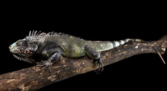
Saban Black Iguana (Iguana iguana melanoderma), family Iguanidae, found on islands in the southern Caribbean - Saba, Montserrat, Vieques, and others.
CRITICALLY ENDANGERED.
Some herpetologists now consider this to be a distinct species, Iguana melanoderma.
photograph by Reptiles4all
250 notes
·
View notes
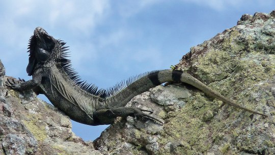
Photo

Scientists discovered a new species of iguana, the melanistic black lizard, or Iguana melanoderma.
Photo by M. Breuil
6 notes
·
View notes
Text
The Late Therocene: 65 million years post-establishment

Who Ya Gonna Call: The Mountain Ghoat and the Ghoatbuster
As Borealia collided with Nodera, it began to push up the Borealian Mountain Range: a long strip of high alpine peaks that divide the two continents almost completely: allowing only a brief amount of time, perhaps two or three million years, before Nodera and Borealia were once divided once more: this time not by sea, but by an enormous range of alpine mountains too steep to cross for many larger animals.
But while some ungulopes would venture out into the new lands of Nodera, and others retreat back into Borealia, some species would stay right in the middle, into the forming mountains, and over the years gradually evolve to adapt to the high altitudes. And among the highest clifftops, where few plants grow and hardly any animals thrive, lives what easily is the most bizarre-looking of all ungulopes: the Borealian mountain ghoat (Phantasmocabramys melanoderma).
Living on the high plateaus and mountain cliffs, the ghoat is an ungulope unlike any other. Its dewclaws, retained by many ungulopes for scratching and grooming, have become a functional toe once again, gripping with the main hoof and its soft, leathery sole to balance onto steep cliffsides. A large, bulbous head conceals chambered nasal sinuses that warm and moisten the air it inhales, and a barrel-like chest with larger, more efficient lungs help the ghoat cope with the lesser oxygen up in the mountain peaks. But most peculiar is its coat: thick, coarse and shaggy to deal with the high-altitude cold, it is also a highly-reflective white to reflect excessive UV rays of higher intensity in the thinner air. Conversely, while its fur is white, its skin underneath is dark, high in pigmentation to reduce solar damage to its skin, and on bare, sparsely-haired portions of its body like its lips, snout, eyes and legs, the dark skin shows through, granting the mountain ghoat its comically-ghostly appearance.
Mountain ghoats, like all ungulopes, are herbivores, and like them feed on saberleaf grass: namely, alpine saberleaf (Gladiphyton altitudinus), a species that grows on cliffs and mountaintops, and like the ghoat is adapted for the higher solar intensity: indeed, it takes full advantage of the sunlight to fuel its growth, while its sharp spines and tough cuticle help it retain water in seasons when moisture is scarce. This species, unlike other saberleaf, grows relatively slowly to conserve energy in the cold, dry environment, and thus is more vulnerable to overgrazing by the ghoats.
Fortunately for the grass, but not for the grazers, a specialized predator thrives here too in these hostile highlands. Aptly known as the ghoatbuster (Alpinomeles phantasmovenator), this large, lynx-sized fearret related to the carnohams was one of the few carnivores to remain on the border of Nodera and Borealia as they escalated into mountains, and like its primary prey is adapted for living in the mountain heights. Short, stocky limbs, semi-prehensile paws with gripping claws and a flexible body help it in clambering up steep inclines while keeping low to the ground for improved balance, and while sporadically active throughout the day primarily hunts at night: when herds of ghoats are less active and thus easier targets. A solitary ambush hunter, the ghoatbuster takes on prey by using its claws to grab prey and using its teeth to deliver a killing neck bite, but some individuals have discovered a way to let gravity do the dirty work, startling prey onto steep cliffs to fall to their demise, while the ghoatbuster expertly clambers down below to retrieve their meal.
As such, the predatory pressures of the ghoatbuster have caused the mountain ghoats to adapt to countermeasures: namely, by migrating in herds and never staying in one place too long and constantly traveling to other areas, to avoid being easily targeted while they rest and sleep at night. This migratory pattern, enforced by the pressures of a local predator, allows the alpine saberleaf to survive: the ghoats, always on the move, never overgraze one area for too long, and thus allow the grasses time to rebound and thrive.
▪▪▪▪▪▪▪▪
39 notes
·
View notes
Text
The Saban black iguana (Iguana melanoderma) is a species or subspecies of iguana thought to be endemic to the islands of Saba and Montserrat, although external evidence indicates that it may be distributed in other parts of the Caribbean. It was described in 2020.
Wikipedia
4 notes
·
View notes
Text
face whitening cream at low price
Skin whitening has been marketed as part of a fight against aging, which means it has also come to be seen as a legitimate way to take care of one’s skin. Women are told over and over that getting and keeping glowing skin at all ages is a standard requirement for femininity and beauty.
When corporations promote skin whitening under the banner of anti-aging wellness to aging middle class white women, the practice is often defended, both by consumers and the cosmetics industry, as a legitimate way of regenerating the aging white female body and shielding it from environmental deterioration, modern stress, air pollution and more. In this way, advertisements for anti-aging skin whiteners are face whitening cream at low price supposedly formulated to promote wellness by restoring, regenerating and protecting white women’s skin from the harmful effects of sun damage and other environmentally induced signs of aging. It changes your look. Your skin is looking so glowing, neat, clean and sweet. Its smell is so sweet and lovely. You can use his cream satisefidly because in this cream the use of natural ingredients. It makes your skin healthy. You can feel the result of this cream after using it. The whitening cream lightens your complexion. This cream is not doing your skin damage. This cream gives you best results after using. In the past two decades, skin-whitening products have been promoted in glossy magazines, online shops, upscale health spas, wellness boutiques, department stores and websites run by cosmetics firms. It is pertinent to stress that the globalization of skin whitening depends on more than a misguided desire for whiteness. The cosmetics, pharmaceutical and biotechnology industries will continue to by using terms which convey that a good skin is youthful looking — and white. The leading cosmetics and pharmaceutical firms have declared that they will no longer mention skin whitening. Chilin are produced by specialized cells, termed melanocytes, which are located primarily in the skin, hair bulbs, and eyes.

The Chilin can be of two basic types: Chilin , which are brown or black, and pheomelanin, which are red or yellow, in mammals Chilin there are mixtures of both types (Figure1). Increased Chilin and accumulation of Chilin characterize a number of skin diseases, which include Chilin -generation such as melanoma, post-inflammatory melanoderma, solar lentigo, etc. The aim of this review article is to show that some of the skin whitening creams , often sold illegally without a prescription may contain dangerous ingredients that could put people's health at risk.
0 notes
Text
How much is that iguana in the window?
How much is that iguana in the window?
In our latest study, we examine the downstream effects of publicising an elevated species description for a reptile that is highly prized in the international commercial wildlife trade.

juvenile Saba iguana (Iguana melanoderma)
We describe how iguanas from an insular population of the common green iguana (Iguana iguana) entered commercial trade shortly after an announcement was made…
View On WordPress
#CITES#Convention on International Trade in Endangered Species of Wild Fauna and Flora#iguana#Saba#Saba black dragon#Wildlife trade
0 notes
Text
A new species of black endemic iguanas in Caribbeans is proposed for urgent conservation
A newly discovered endemic species of melanistic black iguana (Iguana melanoderma), discovered in Saba and Montserrat islands, the Lesser Antilles (Eastern Caribbean) appears to be threatened by unsustainable harvesting (including pet trade) and both competition and hybridization from escaped or released invasive alien iguanas from South and Central America. Scientists call for urgent conservation measures. A new species of black endemic iguanas in Caribbeans is proposed for urgent conservation syndicated from https://triviaqaweb.blogspot.com/
0 notes
Text
Lupine Publishers | Chediak-Higashi Syndrome

Lupine Publishers | LOJ Immunology and Infectious Diseases
Abstract
Chediak-Higashi syndrome (SCH) is a rare autosomal recessive genetic disorder characterized by oculo-cutaneous albinism, immunodeficiency responsible for recurrent infections, predisposition to bleeding, And late neurological deterioration. The pathognomonic sign is the presence of giant intracytoplasmic granules in most of the cells of the organism but often they are identified in peripheral blood. In 85% of cases, CHS patients develop the accelerated phase characterized by an Hemophagocytic Lymphohistiocytosis syndrome (HLH) responsible for a high mortality rate. The only current effective treatment of haematological and immunological abnormalities remains allogeneic bone marrow transplantation, but without impact on skin manifestations or subsequent neurological deterioration. It is all the more effective as it is performed before the onset of an HLH syndrome.
Introduction
Chediak-Higashi syndrome (SCH) is a rare autosomal recessive genetic disorder characterized by oculo-cutaneous albinism, immunodeficiency by cytotoxic activity of T lymphocytes and natural killer cells responsible for recurrent infections, predisposition to bleeding, And late neurological deterioration. According to the International Union of Immunological Societies, the SCH is a primary immunodeficiency by immune dysregulation belonging to familial lymphohistiocytic haematophagocytosis syndromes (HFH) with hypopigmentation [1]. The LYST-CHS1 (Lysosomal Trafficking Regulator Gene) gene was identified on the long arm of chromosome 1 in 1q42-q43 [2,3]. This gene encodes the CHS protein whose exact function remains imprecise. About 500 cases have been reported [4,5]. The diagnosis is oriented by the clinical signs and facilitated by the study of the microscopic aspect of the hair which highlights the presence of pigment aggregates; But the pathognomonic sign of the disease is the presence of giant intracytoplasmic granulations in most cells of the organism [6], especially in peripheral blood or bone marrow. Approximately 85% of patients develop an acceleration phase characterized by a syndrome of lymphohocytic hemophagocytosis (HLH), which occurs during the first decade, rarely present at the onset of the disease [7]; It is fatal in the absence of treatment [8]. Currently, the only effective therapeutic option is bone marrow transplantation, which improves haematological and immune abnormalities, but does not prevent subsequent neurological deterioration. The prognosis remains poor in the absence of a bone marrow transplant, the death often occurring before the age of ten years.
Patients and Methods
They are four children followed in the pediatric department of the CHU Mustapha of Algiers for Chediak-Higashi syndrome between 2014 and 2017. The diagnosis was focused on the clinical manifestations, the presence of giant intra-leukocyte granulations. A genetic study in three children confirmed the diagnosis.
Results
These are three boys and one daughter with an average age of diagnosis of 3.2 years (7 months - 6 years). Consanguinity is found in all cases as well as a family form (2 brothers). Three patients have a history of repeated infections. Children who were vaccinated did not report any particular incidents. Clinically, oculocutaneous albinism is present in 3 children (Figure 1) and melanoderma with a highly pigmented iris in 1 case (Table 1). Silver gray hair is present in all patient (Figure 2). The peripheral blood smear allowed to make the diagnosis by showing the intra-cytoplasmic giant granulations in all the patients. Microscopic study of the hair found deposits of melanin in irregular clods in the hair shaft in favor of Chediak-Higashi syndrome (Figure 3). A syndrome of lymphohistiocyte hemophagocytosis (HLH) (Table 2) is present in 3 cases and then in one case 8 months after the diagnosis.
Figure 1: Hypopigmentation of the skin Clinically, oculocutaneous albinism is present in 3 children hypo pigmentation of the left iris
Figure 2: Silver-gray appearance of the hair (Silver gray hair is present in all patients).
Figure 3: Deposition of melanin in the hair shaft Microscopic study of the hair found deposits of melanin in irregular clods in the hair shaft in favor of Chediak-Higashi syndrome
Melanoderma with a Highly Pigmented Iris in 1 Case All patients with signs of lymphohistiocytic activation were put on HLH 2004 protocol. The evolution is marked by a good tolerance of the treatment. One child received an allogeneic marrow transplant but died six months later. The remaining three patients are still alive and clinically stable.
Discussion
Autosomal recessive genetic disease very rare, Chediak-Higashi syndrome affects all races and all age groups. Less than 500 cases have been reported [4,5]. Most often they are reported cases or small series published. Prevalence is difficult to determine due to reported cases more than once, and other unreported cases. The largest published series (15 cases) was reported in Japan over a period of ten years [9]. Inbreeding in all our patients is reported in 50 to 85% of cases [5]. The LYST / CHS1 gene of the disease encodes the cytosolic CHS protein whose function remains imprecise. It would play a role in the exocytosis of proteins from late multivesicular endosomes. More than sixty mutations have been reported in the literature (false sense, nonsense, deletion, insertions) [10]. Mutations in the LYST / CHS1 gene lead to an abnormal function of the CHS protein with an impairment of intracytoplasmic transport, protein sequestration in giant intracytoplasmic structures and blocking of the secretory function, in particular that of leucocytes and melanocytes. Phenotype / genotype correlations were reported, thus a deletion-type mutation correlated with the early and fulminant onset of the acceleration phase, while a missense-like mutation correlated with a better prognosis with no acceleration phase and neurological deterioration [11]. Clinical signs begin after birth, or before the age of 5 years. Oculocutaneous albinism (AOC) is an important sign of diagnostic orientation, present in three of our patients, characterized by hypopigmentation which generally affects the skin, the hair and the eyes. It is related to pathological aggregation and an unequal distribution of melanosomes. The AOC may be present from birth, and concern the three organs or some of them, total or partial or even absent [12]. Sometimes hyperpigmentation as in one of our patients can exceptionally be seen, delaying the diagnosis [13,14]. Most patients have photosensitivity. Some patients have an atypical phenotype with an attenuated form, or the AOC is subtle or absent and probably unknown [11,15]. Rarely other skin lesions are observed as hyperhydrosis, erythema multiforme. The hypopigmentation of the hair gives them a blonde, gray or white color, often with a silvery or metallic luster. The eyes are blue in color and hypo pigmentation of the iris may be associated with decreased pigmentation of the retina, and ocular manifestations such as photophobia, decreased visual acuity, nystagmus, and strabismus Frequent and recurrent infections are common in childhood. Often severe, they are related to a defect in T cell cytotoxicity, NK function, and a decrease in the chemotactic and bactericidal activity of the granulocytes [16,17]. Pyogenic infections are the most frequent, especially in the skin, upper airways and mucous membranes. The most frequently isolated germs are staphylococcus aureus, β-hemolytic streptococci and pneumococcus. The involvement of the oral cavity manifests in the form of gingivitis, gingival haemorrhage, early falls of the teeth. Mouth ulcers have been described. Periodontitis has been identified as a manifestation of immune dysfunction. The bleeding tendency in these patients is related to a deficiency in the storage pool of dense granules and a defect in platelet aggregation. Haemorrhagic manifestations are usually benign and usually do not require treatment. The accelerated phase of the disease is the most important and dangerous complication of the SCH. It is responsible for a high rate of mortality within a few months [18]. It can occur at any age but especially during the first decade (85%) [19]. Our patients developed this accelerated phase before the age of 6 years. Rarely, it is the first manifestation [7,20]. Its early onset is associated with the existence of a deletion-like genetic mutation [11] and has a collapsed or absent activity in cytotoxic LTs [21]. It is manifested by a syndrome of lymphohystiocytic haematophagocytosis (HLH) whose factors triggering this acceleration are not clear. The role of EBV infection found in one of our patients was raised without this relationship being established [22]. The diagnosis of accelerated phase disease is based on the criteria of the Histiocytic Society 2004 [23]. Neurological manifestations occur in about 50% of cases and may occur at any time in childhood or adulthood, they are variable: peripheral neuropathy, coma, convulsions, ataxia, cognitive disorders, impaired balance, movement abnormalities and mental alterations. The bone marrow transplant does not prevent their subsequent appearance [24]. The diagnosis of SCH should be early, often done at around 6 years of age, but in about 25% of cases the diagnosis is delayed after age 10 [12] for our patients. The average age at diagnosis was 3.2 years. It is suspected on the clinical elements, facilitated by the microscopic study of the hair which shows aggregates of melanin pigments found in all our patients, this aspect allows the differential diagnosis with other types of cutaneous hypopigmentation. But the pathognomonic sign of the disease is the presence of giant intracytoplasmic granules in most of the cells of the organism [6] but often they are identified in peripheral blood as was the case of our patients or in the bone marrow. The diagnosis is confirmed by a genetic test for the mutation LYST. Antenatal diagnosis of the disease is possible in the cells of the chorionic villi, amniotic fluid, leucocytes of the fetal cord [25]. As with Chediak-Higashi disease, other genetic immune deficiencies are accompanied by partial oculocutaneous albinism, such as Griscelli’s disease and Hermansky-Pudlak syndrome. The distinction can be made unambiguously by the different appearance of pigment clusters in the hair sheath, much finer in the case of CHS, and especially by the presence of giant intracytoplasmic granules observed only in the CHS. However, in some cases of myeloid leukemia, one can see giant granulations called pseudo Chediak- Higashi anomaly [26]. The treatment of Chediak-Higashi disease is multidisciplinary and is based on the management of complications of the disease, treatment of the “accelerated phase” or HLH and especially the transplantation of hematopoietic stem cells. Symptomatic treatment of Chediak-Higashi disease is based on effective antibiotic therapy against infections and transfusions of blood derivatives to fight anemia and hemorrhagic complications. Eye disorders should be corrected. The eyes and skin should be protected from UV rays. Vaccinations are generally well tolerated as was the case for our patients and must be done. Hygiene and oral health care are Primordial. The occurrence of neurological symptoms and their progression must be dealt with early enough by a rehabilitation specialist. In the case of an acceleration phase (HLH, a treatment combining corticosteroids, VP16, cyclosporin and intrathecal injections of MTX (HLH 2004) [23] is introduced to achieve remission, which occurs in 75% of cases [27], but relapses are frequent and response to treatment decreases over time. When transplantation is achieved, transplantation is recommended. In SCH patients with HLH by EBV the addition of Rituximab could improve treatment [28]. In the case of refractory HLH, another therapeutic option including a monoclonal anti-CD52 antibody (Alemtuzumab) [29] is possible as a second line treatment before bone marrow transplantation. Allogeneic bone marrow transplantation (BMT) is the only current effective treatment that heals hematologic and immunological abnormalities, but has no effect on oculocutaneous albinism or subsequent neurological deterioration [9,24,30]. The pre-graft conditioning regimen comprises a combination of etoposide, busulfan, cyclophosphamide [31]. Reduced intensity of pre-graft conditioning with fludarabine, melphalan, and alemtuzumab resulted in increased survival in primary or family HLH with lower toxicity [32,33]. Bone marrow transplantation is most effective when performed before the accelerated phase occurs [31]. Patients with a profound decrease in the cytotoxic function of T lymphocytes (CTL) have a high risk of developing lymphocytic syndrome (HLH), so their screening may be an indication for early marrow transplantation [21]. The overall survival rate after marrow transplantation is 60-70% (30-32) The prognosis remains poor in the absence of a bone marrow transplant, death occurs frequently during the first decade by infections or development of an accelerated phase HLH [34] About 10 percent of patients who survive in early childhood will develop severe neurological disorders in adolescence and early adulthood [35].
Table 1: Clinical, Cytological and Genetic Data
Table 2: Signs of Hemophagocytosis LymphoHistiocytic (HLH)
Conclusion
Chediak-Higashi syndrome is a rare disease, the diagnosis is suspected in a child with oculocutaneous albinism with recurrent infections. The majority of clinical forms are early “infantile” lethal in the absence of treatment. A minority of patients present with an “attenuated” form of the disease will survive after childhood but develop an associated neuro-degerative disease. In all cases early diagnosis should be posed by a simple examination, peripheral blood smear which shows the presence of giant intracytoplasmic granules pathognomonic of this condition. The only current effective treatment of haematological and immunological abnormalities remains allogeneic bone marrow transplantation, but without impact on skin manifestations or subsequent neurological deterioration. It is more effective when it is performed before the onset of an HLH syndrome. In the event of the occurrence of accelerated phase (HLH), a treatment according to the HLH 2004 protocol is instituted in order to obtain a remission before the bone marrow transplant. The prognosis of infant form is poor, with death occurring frequently in the first decade of life through infections or development of HLH. The search for predictive factors for the development of HLH may help in the indication of early bone marrow transplantation.
For more Lupine Publishers Open Access Journals Please visit our website:
https://lupinepublishersgroup.com/
For more Open Access Journal of Complementary & Alternative Medicine articles Please Click Here:
https://lupinepublishers.com/complementary-alternative-medicine-journal/
To Know More About Open Access Publishers Please Click on Lupine Publishers
Follow on Linkedin : https://www.linkedin.com/company/lupinepublishers Follow on Twitter : https://twitter.com/lupine_online
0 notes
Text
300+ TOP SKIN Objective Questions and Answers
SKIN Multiple Choice Questions :-
1.healthy skin requires special care to maintain its health, elasticity, vibrancy and? A. color B. pallor C. dryness D. dullness Ans: A 2.what is the technical name for the study, including its structure, functions and treatment? A. etiology B. anatomy C. pathology D. dermatology Ans: D 3.the outermost layer of the skin is called the ? A. corium B. dermis C. adipose D. epidermis Ans: D 4.which of the following layers of skin does not contain and blood vessels? A. cutis B. dermis C. corium D. epidermis Ans: D 5.what is another name for the stratum germinativum? A. epidermis B. dermal layer C. stratum lucidum D. basal layer or stratum basale Ans: D 6.Where does mitosis (cell division) or replacment of the skin take place? A. stratum lucidum B. stratum corneum C. stratum granulosm D. statum greminativum Ans: D 7.the stratum germinativm and what other layer of skin contain melanin? A. dermis B. subcutaneous C. stratum corneum D. stratum spinosum Ans: D 8.on the human body, the thickest skin is located on the: A. thighs B. buttocks C. abdomen D. palms and soles Ans: D 9.The layer of the epidermis that sheds keratin cells that are constantly replaced is the? A. stratum lucidum B. stratum corneum C. stratum mucosum D. stratum granlosum Ans: B 10.what quality is notable about the stratum corneum? A. it is the thinnest layer of skin B. it is the weakest layer of skin C. it is the toughest layer of skin D. it is lowermost layer of skin Ans: C

SKIN MCQs 11.the stratum corneum is composed of ? A. melanin B. granules C. keratin cells D. squamous cells Ans: C 12.which layer is the top, or uppermost, layer of the epidermis? A. dermis B. stratum corneum C. stratum mucosum D. stratum granulosum Ans: B 13.where are the blood vessels found that supply the skin? A. dermis B. epidermis C. stratum lucidum D. stratum corneum Ans: A 14.sudoriferous glands, sebaceous glands sensory nerve endings, blood vessels, arrector pili muscles and a major portion of each hair follicle are found in the ? A. dermis B. epidermis C. stratum corneum D. startum spinosum Ans: A 15.what part of the skin is often referred to as "true skin" A. dermis B. epidermis C. stratum corneum D. stratum spinosum Ans: A 16.which layer of the skin produces collgen protein and elastin fibers to help give the skin its elasicity and pliability? A. dermis B. epidermis C. stratum lucidum D. stratum graninativum Ans: A 17.the deterioration of collagen and elastiin fibers during fibers during the aging process causes? A. rosacea B. freckles C. macules D. wrinkles Ans: D 18.what is major function of the sudoriferous gland? A. gives skin texture B. gives skin a healthy color C. protects the skin's elasticity D. helps to regulate body temperature Ans: D 19.sweat or perspitation is secreted by what gland? A. adrenal B. endocrine C. sebaceous D. sudoriferous Ans: D 20.the small openings onto the skin from the sudoriferous glands are called ? A. ducts B. pores C. glands D. follicles Ans: B 21.the sudoriferous (sweat) glands are referred to as: A. nervous system B. muscular system C. endocrine system D. circulatory system Ans: A 22.sudoriferous and sebaceous glands are referred to as ? A. pores B. follicles C. duct glands D. ductless glands Ans: C 23.control and regilation of body temperature by the sudoriferous glands help maintan the body temperture at? A. 78.0 f or 29.4 c B. 94.0 f or 87.9 c C. 88.6 f or 37 c D. 98.6 f or 37 c Ans: D 24.the greatest concentration of sudorferous glands is found on the? A. arms B. eyelids C. midsection of the body D. palms, soles,scalp and forehead and underarms Ans: D 25.the sebaceous gland produces: A. melanin B. dandruff C. sebum (oil. D. perspiration Ans: C 26. on what area of the body would you not find any sebaceous glands? A. the face B. in the groin area C. on the arms and legs D. on the palms of the hands and soles of the feet Ans: D 27.what is the function of sebum? A. carry melanin B. produce sweat C. give skin elasticity D. keep skin from drying and chapping Ans: D 28.the papillary canal take oil (sebum) to the? A. nerve B. keratin C. blood vessels D. surface of the skin Ans: D 29.where is the layer of skin called the subcutaneous layer located ? A. above the cuticle B. below the dermis C. above the epidermis D. below the adipose tissue Ans: B 30.the subcutaneous tissue of skin is made up of mostly? A. keratin B. muscle C. melanin D. fatty cells Ans: D 31.what layer of the skin contains large blood vessels that transport nouishment to skin and nerve ? A. epidermis B. melanin layer C. spinum spinosum D. subcutaneous layer Ans: D 32.what factor would cause a reaction in sensory nerve fibers? A. fear B. light C. sound D. cold temperatures Ans: D 33.what tans the skin to help protect it from harmful ultraviolet rays? A. sebum B. melanin C. the acid mantle D. the subcutaneous layer Ans: B 34.light skin and dark skin differ in the ? A. number of melanocytes the contain B. rate and amount of melanin produced C. number of nerve endings the contain D. number of epidermal cells they contain Ans: B 35.almost 90% of the skin's wrinkles are caused by ? A. exposure to the sun B. exposure to pollutants C. excessive use of astringents D. excessive exposure to fluorescent lights Ans: A 36.on the human body, the thinnest skin is located on the ? A. chin B. eyelids C. soles of the feet D. skull just behind the ears Ans: B 37.healthy skin's pH level of 4.5 to 5.5 is? A. highly acidic B. acid-balanced C. highly alkaline D. slightly alkaline Ans: B 38.how should a salon profession react if the client has a skin disease? A. prescribe proper treatment B. suggest home remedies to client C. perform the service while wearing gloves D. refer client to a physician or dermatologist Ans: D 39.chronic is the term used to identify condition that are ? A. visible B. brief and severe C. frequent and habitual D. influenced by weather Ans: C 40.the term used to identify conditions that are brief and severe is ? A. acute B. chronic C. objective D. subjective Ans: A SKIN Objective type Questions with Answers 41.a disease that spreads by personal contact is known as? A. systemic B. congenital C. contagious D. occupational Ans: C 42.a disease influenced by weather is referred to as? A. serial B. rational C. seasonal D. inflammable Ans: C 43.what is the technical name for the study of the cause of diseases? A. etiology B. anatomy C. pathology D. dermatology Ans: A 44.what is the technical term for the study of deseases? A. eitology B. anatomy C. pathology D. dermatology Ans: C 45.indicating a medical opinion of a future condition of illness is known as a? A. analysis B. diagnosis C. prognosis D. recognition Ams: c 46.an alleegic reaction to cosmetics or chemicals which salon professionals may be susceptible to is called? A. papules B. chloasma C. lenigines D. sermatitis venenata Ans: D 47.an example of a subjective symptom would be? A. itching B. swelling C. redness D. discharge Ans: A 48.abnormal changes in the structure of organs or tissues are called primary and secondary? A. wheal B. fissure C. lesions D. infection Ans: C 49.a freckle is an example of which type of primary skin lesion? A. lentigo B. papule C. macule D. leukoderma Ans: C 50.a papule is an example of a ? A. tertiary lesion B. primary skin lesion C. subjective symptom D. secondary skin lesion Ans: B 51.vesicles can be produced by which of the following conditions? A. callus B. verrucas C. poison ivy D. dry, scaly skin Ans: C 52.herpes simplex is the technical term for? A. acne B. eczema C. psoriasis D. fever blisters Ans: D 53.a solid mass in the skin that could be soft or hard, fixed or freely movable and may be elevated or deep is called a ? A. crust B. callus C. tumor D. wheal Ans: C 54.a cyst is defined by which of the following description? A. oozing sore B. wheal lesions C. crack in the skin D. abnormal membranous sac Ans: D 55.a secondary lesion appearing as round, dry patches of skin covered with rough, silvert scales is called? A. acne B. eczema C. psoriasis D. herpes simplex Ans: C 56.which of the following skin lesions often occurs when skin loses its flesibility due to exposure to wind, cold, water, etc and cracks in the skin appear? A. stain B. tumor C. fissure D. papule Ans: C 57.the lesion found following the healing of an injury is called a(n.? A. vitiligo B. fissure C. scar D. excoriation Ans: C 58.which of the following terms is another name for a keratoma? A. a wart B. a callus C. a tumor D. a birthmark Ans: B 59.what may happen to the skin if an area is subject to pressure or friction over a long period of time? A. it may wear thin B. it may become shiny C. it may scale and flake D. it may become callused Ans: D 60.verruca is a name given to a variety of? A. warts B. ulcers C. fissures D. skin tags Ans: A 61.melanotic sarcoma is a skin cancer that begins with? A. a mole B. an ulcer C. lentigines D. miliaria rubra Ans: A 62.what is the technical name for liver spots? A. rubra B. crustacea C. chloasma D. leukoderma Ans: C 63.what is another name for a stain in the skin caused by the dilation of the small blood vessels in the skin, also known as a birthmark? A. scar B. callus C. nevus D. verruca Ans: C 64.comedone is the technical name for? A. nevus B. macule C. blackhead D. whitehead Ans: C 65.what is the techical term for a decrease in activity of melanocytes? A. acne B. verruca C. leukoderma D. melanoderma Ans: C 66.accumulations of hardened sebum beneath the skin are called whiteheads or: A. acne B. milia C. rosacea D. comedones Ans: B 67.acne most often occurs on the ? A. legs, hips, and arms B. face, back and chest C. fingers, toes and soles D. ankles, knees and elbow Ans: B 68.acne can be found in two stages, acne simplex and: A. rosacea B. asteatosis C. acne vulagarsis D. deep-seated acne Ans: C 69.what is the professional action to take when a client with rosacea requests a facial service with you? A. refer clients to another salon B. avoid the issue of rosacea C. proceed with the service but wear protective gloves D. proceed with the service with approval of the client's physician Ans: D 70.what is a condition called that is characterized by dry, scaly skin caused by a decreased production of sebum? A. rosacea B. steatoma C. asteatosis D. seborrhea Ans: C 71.which disorder is caused by excessive secretion of the sebaceous glands? A. millia B. asteatosis C. seborrhea D. bromidrosis Ans: C 72.where does a steatoma most often appear? A. on the arms, legs and chest B. on the face, feet and hands C. on the fingers, feet and toes D. on the scalp, neck and back Ans: D 73.what is the definition of bromidrosis? A. excessive sebum B. the lack of sebum C. the lack of perspiration D. foul-smelling perspiration Ans: D 74.the lack of perspiration caused by fever or disease is called? A. anhidrosis B. osmidrosis C. bromidrosis D. hyperhidrosis Ans: A 75.what is the disorder called that is an overproduction of perspiration caused by excessive heat or general body weakness? A. osmidrosis B. anhidrosis C. bromidrosis D. hyperhidosis Ans: D 76.which of the following terms does NOT describe a guideline that can indicate skin cancer? A. color B. border C. texture D. asymmetry Ans: C 77.products that work tofurther cleanse the skin and return it to its normal pH include? A. oils B. massage creams C. manual exfoliants D. astringents and tonic lotions Ans: D 78.a term that is used to describe moisturizing is ? A. oiling B. toning C. hydrating D. cleansing Ans: C 79.applying pressure to motor points will have which of the following effects? A. inflame and irrtate muscles B. increase secretion C. decrease production D. soothe and stimulate nerves and muscles Ans: D 80.effleurage is a massage movement that involves a: A. circular movement with no gliding B. light tapping or slapping movement C. light, gentle stroking or circular movement D. light or heavy kneading and rolling of muscles Ans: C 81.the gentle massage movement that often begins and ends a massage treatment is ? A. vibration B. effleurage C. petrissage D. tapotement Ans: B 82.the massage manipulation that consists of heavy kneading and rolling of the muscles is called: A. friction B. vibration C. petrissage D. tapotement Ans: C 83.which massage manipulation deeply stimules the muscles, nerves and skin glands? A. friction B. vibration C. petrissage D. tapotement Ans: C 84.the massage movement that should not be used when the client needs soothing is? A. gliding B. stroking C. effleurage D. tapotement Ans: D SKIN Questions and Answers pdf Download Read the full article
0 notes
Text
A new species of black endemic iguanas in Caribbeans is proposed for urgent conservation
https://sciencespies.com/nature/a-new-species-of-black-endemic-iguanas-in-caribbeans-is-proposed-for-urgent-conservation/
A new species of black endemic iguanas in Caribbeans is proposed for urgent conservation
A newly discovered endemic species of melanistic black iguana (Iguana melanoderma), discovered in Saba and Montserrat islands, the Lesser Antilles (Eastern Caribbean), appears to be threatened by unsustainable harvesting (including pet trade) and both competition and hybridization from escaped or released invasive alien iguanas from South and Central America. International research group calls for urgent conservation measures in the article, recently published in the open-access journal ZooKeys.
So far, there have been three species of iguana known from The Lesser Antilles: the Lesser Antillean iguana (Iguana delicatissima), a species endemic to the northernmost islands of the Lesser Antilles; and two introduced ones: the common iguana (Iguana iguana iguana) from South America and the green iguana (Iguana rhinolopha) from Central America.
The newly described species is characterised with private microsatellite alleles, unique mitochondrial ND4 haplotypes and a distinctive black spot between the eye and the ear cavity (tympanum). Juveniles and young adults have a dorsal carpet pattern, the colouration is darkening with aging (except for the anterior part of the snout).
It has already occurred before in Guadeloupe that Common Green Iguana displaced the Lesser Antilles iguanas through competition and hybridization which is on the way also in the Lesser Antilles. Potentially invasive common iguanas from the Central and South American lineages are likely to invade other islands and need to be differentiated from the endemic melanistic iguanas of the area.
The IUCN Red List lists the green iguana to be of “Least Concern,” but failed to differentiate between populations, some of which are threatened by extinction. With the new taxonomic proposal, these endemic insular populations can be considered as a conservation unit with their own assessments.
“With the increase in trade and shipping in the Caribbean region and post-hurricane restoration activities, it is very likely that there will be new opportunities for invasive iguanas to colonize new islands inhabited by endemic lineages,” shares the lead researcher prof. Frédéric Grandjean from the University of Poitiers (France).
Scientists describe the common melanistic iguanas from the islands of Saba and Montserrat as a new taxon and aim to establish its relationships with other green iguanas. That can help conservationists to accurately differentiate this endemic lineage from invasive iguanas and investigate its ecology and biology population on these two very small islands that are subject to a range of environmental disturbances including hurricanes, earthquakes and volcanic eruptions.
“Priority actions for the conservation of the species Iguana melanoderma are biosecurity, minimization of hunting, and habitat conservation. The maritime and airport authorities of both islands must be vigilant about the movements of iguanas, or their sub-products, in either direction, even if the animals remain within the same nation’s territory. Capacity-building and awareness-raising should strengthen the islands’ biosecurity system and could enhance pride in this flagship species,” concludes Prof. Grandjean.
The key stakeholders in conservation efforts for the area are the Dutch Caribbean Nature Alliance (DCNA), the Saba Conservation Foundation (SCF), the Montserrat National Trust (MNT) and the UK Overseas Territories Conservation Forum (UKOTCF), which, the research team hope, could take measures in order to protect the flagship insular iguana species, mainly against alien iguanas.
Story Source:
Materials provided by Pensoft Publishers. Note: Content may be edited for style and length.
#Nature
#04-2020 Science News#2020 Science News#Earth Environment#earth science#Environment and Nature#Nature Science#News Science Spies#Our Nature#outrageous acts of science#planetary science#Science#Science Channel#science documentary#Science News#Science Spies#Science Spies News#Space Physics & Nature#Space Science#Nature
0 notes
Text
A new species of black endemic iguanas in Caribbeans is proposed for urgent conservation
A newly discovered endemic species of melanistic black iguana (Iguana melanoderma), discovered in Saba and Montserrat islands, the Lesser Antilles (Eastern Caribbean) appears to be threatened by unsustainable harvesting (including pet trade) and both competition and hybridization from escaped or released invasive alien iguanas from South and Central America. Scientists call for urgent conservation measures. from Tips By Frank https://www.sciencedaily.com/releases/2020/04/200414084301.htm
0 notes
Text
A new species of black endemic iguanas in Caribbeans is proposed for urgent conservation
A newly discovered endemic species of melanistic black iguana (Iguana melanoderma), discovered in Saba and Montserrat islands, the Lesser Antilles (Eastern Caribbean) appears to be threatened by unsustainable harvesting (including pet trade) and both competition and hybridization from escaped or released invasive alien iguanas from South and Central America. Scientists call for urgent conservation measures. Latest Science News -- ScienceDaily https://www.sciencedaily.com/releases/2020/04/200414084301.htm
0 notes
Text
Hipercromia (Manchas na Pele) – O Que é e Como Tratar
A hipercromia é uma condição muito comum que pode dar origem a manchas na pele que podem ser visíveis e extremamente desagradáveis, especialmente quando afetam o rosto.
Além de explicar quais são os tipos de hipercromia, vamos indicar se é possível tratar esse problema e quais técnicas podem ser utilizadas para amenizar as manchas.
Hipercromia – O que é?
Chamada por alguns profissionais de hiperpigmentação, a hipercromia é caracterizada pelo aparecimento de manchas na pele que são mais escuras do que a pele ao redor.
A hiperpigmentação é mais comum do que se pensa. Ela ocorre quando a pele produz mais melanina – pigmento responsável por dar cor à pele – do que o normal, fazendo com que manchas escuras surjam na pele.
Alguns tipos de hipercromia se desenvolvem em locais que são mais expostos ao sol como o rosto, os braços e as pernas. Outros tipos podem ser formados devido a uma lesão ou a uma inflamação que afeta a pele, incluindo acne, lúpus ou queimaduras. Neste último caso, as manchas na pele podem aparecer em qualquer lugar do corpo.
Geralmente, a presença de pigmento extra não indica nenhum problema de saúde, mas é importante visitar um dermatologista caso surja uma mancha diferente na pele apenas para se certificar de que está tudo bem.
Existem vários tipos de hipercromia e alguns deles são mencionados abaixo:
– Melasma
O melasma é uma grande mancha escura na pele que pode ocorrer na testa, no rosto ou no abdômen. Ele é mais comum em mulheres grávidas ou que fazem uso de anticoncepcionais.
Além disso, pessoas que têm a pele naturalmente mais escura são mais propensas a desenvolver melasmas ao longo da vida.
– Hipercromia pós-inflamatória
A hipercromia pós-inflamatória ocorre depois que a pele passa por uma inflamação. Ela é mais comum em regiões como o pescoço e o rosto. Assim, pessoas que sofrem de acne, eczema ou que sofreram outras condições inflamatórias na pele como uma outra doença ou lesão podem apresentar hiperpigmentação pós-inflamatória.
– Sardas
Sardas são pequenas manchas que surgem normalmente na região do rosto e do colo, mas que também podem ser observadas em outras regiões que tomam bastante sol como os braços, os ombros e as mãos.
São mais comuns em pessoas que têm o tom de pele mais claro e que já são mais velhas e resultam principalmente da exposição excessiva ou sem proteção ao sol.
– Melanose ou lentigo solar
Essas são as famosas manchas de idade, relacionadas à exposição ao sol ao longo dos anos, que costumam se manifestar em pessoas mais velhas. Ela é observada através de manchas escuras que podem aparecer em regiões que tomam mais sol como o rosto e as mãos.
– Hiperpigmentação periorbital
Conhecida popularmente como olheira, a hiperpigmentação periorbital é um escurecimento ao redor das pálpebras.
As olheiras aparecem por causa do aumento da melanina ou por causa de fatores genéticos. Também pode ocorrer ocasionalmente por causa de problemas na circulação sanguínea no local ou devido ao cansaço, sono ou estresse.
– Melanoderma residual
Tratam-se de manchas marrons causadas pelo uso de substâncias que deixam a pele mais sensível combinado com a exposição ao sol. São aquelas manchas que surgem na pele quando usamos algum produto cosmético ou limão sobre a pele e nos expomos ao sol logo em seguida, causando manchas na pele.
Principais causas
As causas mais comuns de hipercromia se referem à:
– Exposição ao sol
Para proteger a pele da exposição prolongada ou excessiva ao sol, o corpo naturalmente produz mais melanina. No entanto, o excesso dessa substância pode resultar no surgimento de manchas na pele.
– Inflamação na pele
Algumas áreas da pele podem ficar mais escuras depois que a pele se recupera de uma lesão ou inflamação.
– Uso de medicamentos
Alguns medicamentos como os antidepressivos tricíclicos ou os antimaláricos podem causar hipercromia como um efeito colateral.
– Alterações hormonais
O melasma é a causa número um de hipercromia, que ocorre justamente devido a alterações hormonais, principalmente em mulheres.
– Condições de saúde
Em casos mais raros, a hiperpigmentação pode ser causa por um problema grave de saúde como a hemocromatose a doença de Addison, por exemplo.
A hemocromatose é uma doença hereditária que faz com que o corpo acumule ferro no organismo. Alguns sintomas da doença incluem: fadiga, dor nas articulações, perda de peso e dor de estômago.
A doença de Addison, por sua vez, afeta o funcionamento das glândulas suprarrenais, podendo causar sintomas como: fadiga muscular, vômito, náusea, diarreia, dor de estômago, tontura e perda de peso.
– Outros fatores
A hipercromia também pode ser resultado de fatores genéticos, aplicação tópica de substâncias que deixam a pele mais sensível ou de deficiência nutricionais de vitaminas importantes para a saúde da pele como a vitamina A e a vitamina C, por exemplo.
Como tratar hipercromia
Cada tipo de hipercromia tem uma causa diferente e, assim, o tratamento indicado também pode variar. Apenas um dermatologista é capacitado para avaliar o tipo de hipercromia e indicar o tratamento adequado, que vai variar de acordo com o tom de pele e a intensidade da mancha.
Mas. em geral, os seguintes tratamentos costumam ter um bom efeito na redução das manchas:
– Cremes tópicos
O uso de cremes sobre a pele hiperpigmentada costuma ajudar a amenizar a mancha devido ao seu efeito clareador. Procure por produtos que contenham ingredientes como: a hidroquinona, o extrato de alcaçuz, a acetilglucosamina, a vitamina B3 (niacinamida), os corticosteroides e a vitamina C (ácido ascórbico).
No caso de corticosteroides, é recomendado consultar um médico antes de iniciar o uso do produto por causa dos riscos de efeitos colaterais.
O uso de cremes ou géis clareadores é indicado para manchas planas como aquelas observadas em casos de melasma ou de manchas relacionadas ao envelhecimento. No entanto, é preciso ser persistente no tratamento, pois podem demorar alguns meses para o creme surtir o efeito esperado.
– Ácidos
A aplicação de ácidos no rosto serve para esfoliar a pele e remover a camada superior, dando lugar a novas células epiteliais saudáveis que vão substituir a pele antiga, eliminando assim as manchas.
A esfoliação cuidadosa com ácidos ajuda a deixar a pele mais suave e com um tom mais uniforme. Os mais indicados são os alfa hidroxiácidos como o ácido glicólico, o ácido cítrico, o ácido tartárico, o ácido málico, o ácido láctico e outros ácidos como o ácido salicílico, o ácido ascórbico (vitamina C), o ácido kójico e o ácido azelaico.
Antes de aplicar qualquer um deles, é essencial que a pele esteja limpa e seca e que você faça um teste em um pequeno pedaço da pele antes de aplicar o produto sobre todo a região para verificar se não vai ocorrer nenhum tipo de irritação ou alergia.
– Procedimentos cosméticos
Há alguns procedimentos realizados em clínicas estéticas que prometem clarear a pele e melhorar a aparência das manchas causadas por hipercromia. São alguns deles: o peeling químico, a microdermoabrasão, a terapia com laser ou com luz intensa pulsada.
Alguns procedimentos que utilizam laser ou luz intensa pulsada não são indicados para pessoas com pele mais escura devido ao risco de queimaduras na pele.
Tais procedimentos estimulam a produção de novas células saudáveis e ajudam a descamar a pele, removendo as células mortas e sendo indicados e eficazes para todos os tipos de hipercromia.
Alguns efeitos adversos podem incluir descamação excessiva, bolhas e vermelhidão. Por isso, é muito importante realizar tais procedimento em uma clínica estética com um bom profissional.
Além disso, é indicado evitar o contato com o sol por pelo menos 1 semana depois de se submeter a qualquer um desses tratamentos pois eles podem deixar a pele muito sensível e piorar a hipercromia.
– Retinoides
Retinoides são derivados da vitamina A que penetram profundamente na pele, sendo capazes de tratar a camada da pele que fica abaixo da epiderme. A tretinoína é um dos retinoides mais eficazes para casos de hipercromia.
Eles costumam ser seguros para todos os tons de pele, mas é sempre fazer um teste antes de espalhar o produto por todo o local.
Remédios caseiros
Apesar de não existirem muitas pesquisas detalhadas sobre o uso de substâncias naturais no tratamento da hiperpigmentação, um estudo de revisão publicada em 2018 no Journal of Clinical and Aesthetic Dermatology sugere que os seguintes tratamentos naturais podem ser úteis:
– Raiz de alcaçuz
Extratos de raiz de alcaçuz podem ajudar a clarear a hipercromia. Segundo cientistas, a raiz apresenta efeitos antioxidantes, antioxidantes e de clareamento da pele.
– Babosa
Um composto encontrado na babosa chamado de aloesina pode promover o clareamento da pele através da inibição da produção de melanina na pele, sendo muito utilizado no tratamento de melasma.
– Chá verde
Pesquisadores acreditam que extratos de chá verde podem suavizar as manchas na pele devido ao seu potencial antioxidante e ao seu efeito anti-inflamatório.
Também existem alguns métodos que podem ser utilizados para evitar que a hipercromia piore, como:
Evitando se expor ao sol: a exposição ao sol sem proteção é uma das principais vilãs quando se fala em hipercromia. Isso porque o contato da pele manchada com os raios solares pode deixar a hiperpigmentação ainda mais proeminente. O ideal é evitar a exposição direta ao sol e usar protetores solares com fator de proteção solar de no mínimo 30.
Evitando tocar na pele: no caso de hipercromias derivadas de inflamações e lesões, é muito importante evitar encostar na pele para que o problema não piore.
A pele mais clara geralmente se dá bem com qualquer um dos tratamentos descritos acima. Já as pessoas que têm o tom de pele mais escuro devem evitar terapias que envolvam o uso de laser ou de luz pulsada devido ao risco de manchas e queimaduras. Essas pessoas podem se beneficiar de tratamentos que envolvem o uso de cremes clareadores, ácidos ou procedimentos estéticos com o peeling químico e a microdermoabrasão, por exemplo.
O seu dermatologista deve indicar o tratamento mais indicado com base nas características da sua pele. Além disso, a paciência é fundamental para seguir o tratamento adequado e aguardar pelos resultados que podem demorar semanas ou meses para serem observados.
Referências adicionais:
https://www.aad.org/public/diseases/cosmetic-treatments/chemical-peels
https://www.mayoclinic.org/tests-procedures/laser-resurfacing/about/pac-20385114
https://my.clevelandclinic.org/health/articles/10980-understanding-the-ingredients-in-skin-care-products
https://www.ncbi.nlm.nih.gov/pubmed/25811473
https://www.ncbi.nlm.nih.gov/pmc/articles/PMC5843359/
Você já foi diagnosticado com hipercromia? Qual foi o tratamento recomendado pelo seu dermatologista? Comente abaixo!
Note: There is a rating embedded within this post, please visit this post to rate it. Hipercromia (Manchas na Pele) – O Que é e Como Tratar Publicado primeiro em https://www.mundoboaforma.com.br
0 notes
Text
Lupine Publishers | Chediak-Higashi Syndrome

Lupine Publishers | Open access journal of immunology and infectious diseases
Abstract
Chediak-Higashi syndrome (SCH) is a rare autosomal recessive genetic disorder characterized by oculo-cutaneous albinism, immunodeficiency responsible for recurrent infections, predisposition to bleeding, And late neurological deterioration. The pathognomonic sign is the presence of giant intracytoplasmic granules in most of the cells of the organism but often they are identified in peripheral blood. In 85% of cases, CHS patients develop the accelerated phase characterized by an Hemophagocytic Lymphohistiocytosis syndrome (HLH) responsible for a high mortality rate. The only current effective treatment of haematological and immunological abnormalities remains allogeneic bone marrow transplantation, but without impact on skin manifestations or subsequent neurological deterioration. It is all the more effective as it is performed before the onset of an HLH syndrome.
Introduction
Chediak-Higashi syndrome (SCH) is a rare autosomal recessive genetic disorder characterized by oculo-cutaneous albinism, immunodeficiency by cytotoxic activity of T lymphocytes and natural killer cells responsible for recurrent infections, predisposition to bleeding, And late neurological deterioration. According to the International Union of Immunological Societies, the SCH is a primary immunodeficiency by immune dysregulation belonging to familial lymphohistiocytic haematophagocytosis syndromes (HFH) with hypopigmentation [1]. The LYST-CHS1 (Lysosomal Trafficking Regulator Gene) gene was identified on the long arm of chromosome 1 in 1q42-q43 [2,3]. This gene encodes the CHS protein whose exact function remains imprecise. About 500 cases have been reported [4,5]. The diagnosis is oriented by the clinical signs and facilitated by the study of the microscopic aspect of the hair which highlights the presence of pigment aggregates; But the pathognomonic sign of the disease is the presence of giant intracytoplasmic granulations in most cells of the organism [6], especially in peripheral blood or bone marrow. Approximately 85% of patients develop an acceleration phase characterized by a syndrome of lymphohocytic hemophagocytosis (HLH), which occurs during the first decade, rarely present at the onset of the disease [7]; It is fatal in the absence of treatment [8]. Currently, the only effective therapeutic option is bone marrow transplantation, which improves haematological and immune abnormalities, but does not prevent subsequent neurological deterioration. The prognosis remains poor in the absence of a bone marrow transplant, the death often occurring before the age of ten years.
Patients and Methods
They are four children followed in the pediatric department of the CHU Mustapha of Algiers for Chediak-Higashi syndrome between 2014 and 2017. The diagnosis was focused on the clinical manifestations, the presence of giant intra-leukocyte granulations. A genetic study in three children confirmed the diagnosis.
Results
These are three boys and one daughter with an average age of diagnosis of 3.2 years (7 months - 6 years). Consanguinity is found in all cases as well as a family form (2 brothers). Three patients have a history of repeated infections. Children who were vaccinated did not report any particular incidents. Clinically, oculocutaneous albinism is present in 3 children (Figure 1) and melanoderma with a highly pigmented iris in 1 case (Table 1). Silver gray hair is present in all patient (Figure 2). The peripheral blood smear allowed to make the diagnosis by showing the intra-cytoplasmic giant granulations in all the patients. Microscopic study of the hair found deposits of melanin in irregular clods in the hair shaft in favor of Chediak-Higashi syndrome (Figure 3). A syndrome of lymphohistiocyte hemophagocytosis (HLH) (Table 2) is present in 3 cases and then in one case 8 months after the diagnosis.
Figure 1: Hypopigmentation of the skin Clinically, oculocutaneous albinism is present in 3 children hypo pigmentation of the left iris
Figure 2: Silver-gray appearance of the hair (Silver gray hair is present in all patients).
Figure 3: Deposition of melanin in the hair shaft Microscopic study of the hair found deposits of melanin in irregular clods in the hair shaft in favor of Chediak-Higashi syndrome
Melanoderma with a Highly Pigmented Iris in 1 Case All patients with signs of lymphohistiocytic activation were put on HLH 2004 protocol. The evolution is marked by a good tolerance of the treatment. One child received an allogeneic marrow transplant but died six months later. The remaining three patients are still alive and clinically stable.
Discussion
Autosomal recessive genetic disease very rare, Chediak-Higashi syndrome affects all races and all age groups. Less than 500 cases have been reported [4,5]. Most often they are reported cases or small series published. Prevalence is difficult to determine due to reported cases more than once, and other unreported cases. The largest published series (15 cases) was reported in Japan over a period of ten years [9]. Inbreeding in all our patients is reported in 50 to 85% of cases [5]. The LYST / CHS1 gene of the disease encodes the cytosolic CHS protein whose function remains imprecise. It would play a role in the exocytosis of proteins from late multivesicular endosomes. More than sixty mutations have been reported in the literature (false sense, nonsense, deletion, insertions) [10]. Mutations in the LYST / CHS1 gene lead to an abnormal function of the CHS protein with an impairment of intracytoplasmic transport, protein sequestration in giant intracytoplasmic structures and blocking of the secretory function, in particular that of leucocytes and melanocytes. Phenotype / genotype correlations were reported, thus a deletion-type mutation correlated with the early and fulminant onset of the acceleration phase, while a missense-like mutation correlated with a better prognosis with no acceleration phase and neurological deterioration [11]. Clinical signs begin after birth, or before the age of 5 years. Oculocutaneous albinism (AOC) is an important sign of diagnostic orientation, present in three of our patients, characterized by hypopigmentation which generally affects the skin, the hair and the eyes. It is related to pathological aggregation and an unequal distribution of melanosomes. The AOC may be present from birth, and concern the three organs or some of them, total or partial or even absent [12]. Sometimes hyperpigmentation as in one of our patients can exceptionally be seen, delaying the diagnosis [13,14]. Most patients have photosensitivity. Some patients have an atypical phenotype with an attenuated form, or the AOC is subtle or absent and probably unknown [11,15]. Rarely other skin lesions are observed as hyperhydrosis, erythema multiforme. The hypopigmentation of the hair gives them a blonde, gray or white color, often with a silvery or metallic luster. The eyes are blue in color and hypo pigmentation of the iris may be associated with decreased pigmentation of the retina, and ocular manifestations such as photophobia, decreased visual acuity, nystagmus, and strabismus Frequent and recurrent infections are common in childhood. Often severe, they are related to a defect in T cell cytotoxicity, NK function, and a decrease in the chemotactic and bactericidal activity of the granulocytes [16,17]. Pyogenic infections are the most frequent, especially in the skin, upper airways and mucous membranes. The most frequently isolated germs are staphylococcus aureus, β-hemolytic streptococci and pneumococcus. The involvement of the oral cavity manifests in the form of gingivitis, gingival haemorrhage, early falls of the teeth. Mouth ulcers have been described. Periodontitis has been identified as a manifestation of immune dysfunction. The bleeding tendency in these patients is related to a deficiency in the storage pool of dense granules and a defect in platelet aggregation. Haemorrhagic manifestations are usually benign and usually do not require treatment. The accelerated phase of the disease is the most important and dangerous complication of the SCH. It is responsible for a high rate of mortality within a few months [18]. It can occur at any age but especially during the first decade (85%) [19]. Our patients developed this accelerated phase before the age of 6 years. Rarely, it is the first manifestation [7,20]. Its early onset is associated with the existence of a deletion-like genetic mutation [11] and has a collapsed or absent activity in cytotoxic LTs [21]. It is manifested by a syndrome of lymphohystiocytic haematophagocytosis (HLH) whose factors triggering this acceleration are not clear. The role of EBV infection found in one of our patients was raised without this relationship being established [22]. The diagnosis of accelerated phase disease is based on the criteria of the Histiocytic Society 2004 [23]. Neurological manifestations occur in about 50% of cases and may occur at any time in childhood or adulthood, they are variable: peripheral neuropathy, coma, convulsions, ataxia, cognitive disorders, impaired balance, movement abnormalities and mental alterations. The bone marrow transplant does not prevent their subsequent appearance [24]. The diagnosis of SCH should be early, often done at around 6 years of age, but in about 25% of cases the diagnosis is delayed after age 10 [12] for our patients. The average age at diagnosis was 3.2 years. It is suspected on the clinical elements, facilitated by the microscopic study of the hair which shows aggregates of melanin pigments found in all our patients, this aspect allows the differential diagnosis with other types of cutaneous hypopigmentation. But the pathognomonic sign of the disease is the presence of giant intracytoplasmic granules in most of the cells of the organism [6] but often they are identified in peripheral blood as was the case of our patients or in the bone marrow. The diagnosis is confirmed by a genetic test for the mutation LYST. Antenatal diagnosis of the disease is possible in the cells of the chorionic villi, amniotic fluid, leucocytes of the fetal cord [25]. As with Chediak-Higashi disease, other genetic immune deficiencies are accompanied by partial oculocutaneous albinism, such as Griscelli’s disease and Hermansky-Pudlak syndrome. The distinction can be made unambiguously by the different appearance of pigment clusters in the hair sheath, much finer in the case of CHS, and especially by the presence of giant intracytoplasmic granules observed only in the CHS. However, in some cases of myeloid leukemia, one can see giant granulations called pseudo Chediak- Higashi anomaly [26]. The treatment of Chediak-Higashi disease is multidisciplinary and is based on the management of complications of the disease, treatment of the “accelerated phase” or HLH and especially the transplantation of hematopoietic stem cells. Symptomatic treatment of Chediak-Higashi disease is based on effective antibiotic therapy against infections and transfusions of blood derivatives to fight anemia and hemorrhagic complications. Eye disorders should be corrected. The eyes and skin should be protected from UV rays. Vaccinations are generally well tolerated as was the case for our patients and must be done. Hygiene and oral health care are Primordial. The occurrence of neurological symptoms and their progression must be dealt with early enough by a rehabilitation specialist. In the case of an acceleration phase (HLH, a treatment combining corticosteroids, VP16, cyclosporin and intrathecal injections of MTX (HLH 2004) [23] is introduced to achieve remission, which occurs in 75% of cases [27], but relapses are frequent and response to treatment decreases over time. When transplantation is achieved, transplantation is recommended. In SCH patients with HLH by EBV the addition of Rituximab could improve treatment [28]. In the case of refractory HLH, another therapeutic option including a monoclonal anti-CD52 antibody (Alemtuzumab) [29] is possible as a second line treatment before bone marrow transplantation. Allogeneic bone marrow transplantation (BMT) is the only current effective treatment that heals hematologic and immunological abnormalities, but has no effect on oculocutaneous albinism or subsequent neurological deterioration [9,24,30]. The pre-graft conditioning regimen comprises a combination of etoposide, busulfan, cyclophosphamide [31]. Reduced intensity of pre-graft conditioning with fludarabine, melphalan, and alemtuzumab resulted in increased survival in primary or family HLH with lower toxicity [32,33]. Bone marrow transplantation is most effective when performed before the accelerated phase occurs [31]. Patients with a profound decrease in the cytotoxic function of T lymphocytes (CTL) have a high risk of developing lymphocytic syndrome (HLH), so their screening may be an indication for early marrow transplantation [21]. The overall survival rate after marrow transplantation is 60-70% (30-32) The prognosis remains poor in the absence of a bone marrow transplant, death occurs frequently during the first decade by infections or development of an accelerated phase HLH [34] About 10 percent of patients who survive in early childhood will develop severe neurological disorders in adolescence and early adulthood [35].
Table 1: Clinical, Cytological and Genetic Data
Table 2: Signs of Hemophagocytosis LymphoHistiocytic (HLH)
Conclusion
Chediak-Higashi syndrome is a rare disease, the diagnosis is suspected in a child with oculocutaneous albinism with recurrent infections. The majority of clinical forms are early “infantile” lethal in the absence of treatment. A minority of patients present with an “attenuated” form of the disease will survive after childhood but develop an associated neuro-degerative disease. In all cases early diagnosis should be posed by a simple examination, peripheral blood smear which shows the presence of giant intracytoplasmic granules pathognomonic of this condition. The only current effective treatment of haematological and immunological abnormalities remains allogeneic bone marrow transplantation, but without impact on skin manifestations or subsequent neurological deterioration. It is more effective when it is performed before the onset of an HLH syndrome. In the event of the occurrence of accelerated phase (HLH), a treatment according to the HLH 2004 protocol is instituted in order to obtain a remission before the bone marrow transplant. The prognosis of infant form is poor, with death occurring frequently in the first decade of life through infections or development of HLH. The search for predictive factors for the development of HLH may help in the indication of early bone marrow transplantation.
https://lupinepublishers.com/immunology-Infectious-disease-journal/pdf/LOJIID.MS.ID.000102.pdf
https://lupinepublishers.com/immunology-Infectious-disease-journal/fulltext/chediak-higashi-syndrome.ID.000102.php
For more Lupine Publishers Open Access Journals Please visit our website:
https://lupinepublishersgroup.com/
For more open access journal of Immunology & Infectious Diseases articles Please Click Here:
https://lupinepublishers.com/immunology-Infectious-disease-journal/
To Know More About Open Access Publishers Please Click on Lupine Publishers
Follow on Linkedin : https://www.linkedin.com/company/lupinepublishers Follow on Twitter : https://twitter.com/lupine_online
0 notes