
easyphysiologeasy-blog
2 posts
An open-source physiology website for students!
Don't wanna be here? Send us removal request.
Statistics
We looked inside some of the posts by easyphysiologeasy-blog and here's what we found interesting.
Average Info
Notes Per Post
0
Likes Per Post
0
Reblog Per Post
0
Reply Per Post
0
Time Between Posts
2 minutes
Number of Posts By Type
Text
2
Last Seen Tumblr Blogs


Fun Fact
69% of Tumblr users are millennials.
Text
Clinical Perspective - Multiple Sclerosis and Myelination
Multiple sclerosis is a disease that causes the immune system to attack its own myelin. This can have obvious effects such as laziness, fatigue, loss of motor control and neurophysical effects.
As myelin is degraded, the resistance to leakage channels decrease which can allow potassium to diffuse out, decreasing the strength of the signal and at the same time, slowing down the speed of the signal due to the leakage of current out of the axon. It also causes a spread of VGSC from within the nodes of ranvier as myelin sheaths are missing. The absence of saltatory propagation can slow down action potentials.
This terrible condition simply demonstrates that myelination is a vital part in our normal body function, and without it, we wouldn’t be able to do our everyday activities.
0 notes
Text
Clinical Perspective – Strychnine Pharmacodynamics and Inhibitory Synapses
Strychnine is a neurotoxin which exerts its effects through interaction at inhibitory glycine receptors (GlyRs). Such receptors are ionotropic Cl- channels, hence cause depolarisation in synapsing neurons.
Upon drug absorption, strychnine acts as an antagonist of these receptors primarily within spinal cord motor neurons. Reduced ability for hyperpolarization of such neurons means fewer excitatory postsynaptic potentials (EPSPs) are necessary to reach threshold potentials at the axon hillock and elicit an action potential. Motor neurons are then more easily activated, causing the observed symptoms of strychnine poisoning.
Such unchecked activation causes severe muscle spasms and tetanic contraction, leading to eventual asphyxiation.
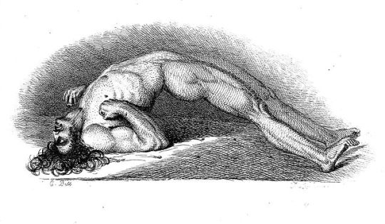
Opisthotonus – spasm of spinal column muscles such as this is evident in strychnine poisoning. Painting by neurologist Sir Charles Bell, 1809.
References
Curtis DR, Duggan AW, Johnston GAR. The Specificity of Strychnine as a Glycine Antagonist in the Mammalian Spinal Cord. Experimental Brain Research 1971; 12(5): 547 - 565.
0 notes