
Statistics
We looked inside some of the posts by optometrycase-blog and here's what we found interesting.
Average Info
Notes Per Post
6
Likes Per Post
4
Reblog Per Post
2
Reply Per Post
0
Time Between Posts
2 days
Number of Posts By Type
Photo
5
Last Seen Tumblr Blogs


Fun Fact
Post activity is at the highest at 4:00 pm EDT; notes peak at 10:00 pm EDT.
Photo
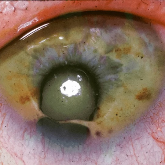
Coloboma. A coloboma (from the Greek koloboma, meaning defect) is a hole in one of the structures of the eye, such as the iris, retina, choroid, or optic disc. The hole is present from birth (except for one case, where it developed within the first few months of the child's life) and can be caused when a gap called the choroid fissure, which is present during early stages of prenatal development, fails to close up completely before a child is born. The classical description in medical literature is of a key-hole shaped defect. A coloboma can occur in one eye (unilateral) or both eyes (bilateral). Most cases of coloboma affect only the iris. The level of visible impairment of those with a coloboma can range from having no vision problems to being able to see only light or dark, depending on severity and the position of the coloboma (or colobomata of more than one is present). It affects less than one in every 10,000 births. The effects a coloboma has on the vision can be mild or more severe depending on the size and location of the gap. If, for example, only a small part of the iris is missing, vision may be normal, whereas if a large part of the retina or optic nerve is missing, vision may be poor and a large part of the visual field may be missing. This is more likely to cause problems with mobility if the lower visual field is absent. Other conditions can be associated with a coloboma. Sometimes, the eye may be reduced in size, a condition called microphthalmia. Glaucoma, nystagmus, scotoma, or strabismus may also occur. #coloboma #iris #eyedoctor #eyeexam #eyecare #optometria #optometrist #optometry #optometrystudent #ophthalmologist #ophthalmology #medicalschool #medicaldoctor #medicalstudent #oculista #eyedisease https://www.instagram.com/p/BsBYmGuA-IK/?utm_source=ig_tumblr_share&igshid=f6d1etju4pdf
#coloboma#iris#eyedoctor#eyeexam#eyecare#optometria#optometrist#optometry#optometrystudent#ophthalmologist#ophthalmology#medicalschool#medicaldoctor#medicalstudent#oculista#eyedisease
0 notes
Photo

Contact lens. // Application of a 20.2mm scleral lens on a transplanted cornea. #contactlenses #scleralenses #contactologia #contactlens #optometry #optometryschool #optometrystudent #optometrylife #fluroscent #transplant #corneatransplant #ophthalmology #ophthalmologist #ophthalmic #ocular #eyedoctor #eyelid #eyelicious #cornea #pupil #slitlamp #eye #medicalschool #meded #medicalstudent #surgery #rehabilitation #lens #implantsurgery https://www.instagram.com/p/BipCgmGFrMw/?utm_source=ig_tumblr_share&igshid=wbula1rinnc2
#contactlenses#scleralenses#contactologia#contactlens#optometry#optometryschool#optometrystudent#optometrylife#fluroscent#transplant#corneatransplant#ophthalmology#ophthalmologist#ophthalmic#ocular#eyedoctor#eyelid#eyelicious#cornea#pupil#slitlamp#eye#medicalschool#meded#medicalstudent#surgery#rehabilitation#lens#implantsurgery
2 notes
·
View notes
Photo
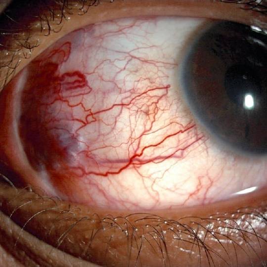
Elevated Episcleral Venous Pressure. Episcleral venous pressure is an important factor in the regulation of IOP. Normal episcleral venous pressure is 8–10 mm Hg, but it can be raised by a variety of clinical entities that either obstruct venous outflow or involve arteriovenous malformations. Patients may note a chronic red eye without discomfort or allergic symptoms. Occasionally, a distant history of significant head trauma may suggest the cause of a carotid-cavernous sinus or dural fistula. However, most cases are idiopathic, often without angiographic abnormalities, and may be familial. Clinically, patients with increased episcleral venous pressure present with tortuous, dilated episcleral veins. These vascular changes may be unilateral or bilateral depending on the location of the vascular anomaly. The anterior segment appears normal in most of these patients, except for elevated IOP and often the gonioscopic finding of blood in Schlemm’s canal. Rarely, signs of ocular ischemia or venous stasis may be present. Sudden, severe carotid-cavernous fistulas may be accompanied by proptosis and other orbital or neurologic signs. These cases may require neuroradiologic intervention. #eyedoctor #eyeexam #eyecare #optometria #optometrist #optometry #optometrystudent #ophthalmologist #ophthalmology #medicalschool #medicalstudent #medicaldoctor #orthoptics #optician https://www.instagram.com/p/Brnnh5zAM9p/?utm_source=ig_tumblr_share&igshid=e0hdlimq7kf6
#eyedoctor#eyeexam#eyecare#optometria#optometrist#optometry#optometrystudent#ophthalmologist#ophthalmology#medicalschool#medicalstudent#medicaldoctor#orthoptics#optician
0 notes
Photo
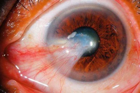
Pterygium. //A pterygium is a pinkish, triangular tissue growth on the cornea of the eye. It typically starts on the cornea near the nose. It may slowly grow but rarely grows so large that the pupil is covered. • • • • • • • • • • • • • • • #pterygium #eyedisease #tissue #cornea #eye #nose #eyesurgery #surgery #aestheticsurgery #uvrays #ophthalmology #ophthalmologist #meded #medicalschool #eyedoctor #pupil #iris #slitlamp #vessel #pathology #pathologist #patologia #optometria #optometrist #eyeexam #optometryschool #eyelicious #eyedrops #genetics https://www.instagram.com/p/Biezx4agiUd/?utm_source=ig_tumblr_share&igshid=5pyb5kth6ok
#pterygium#eyedisease#tissue#cornea#eye#nose#eyesurgery#surgery#aestheticsurgery#uvrays#ophthalmology#ophthalmologist#meded#medicalschool#eyedoctor#pupil#iris#slitlamp#vessel#pathology#pathologist#patologia#optometria#optometrist#eyeexam#optometryschool#eyelicious#eyedrops#genetics
2 notes
·
View notes
Photo

Elevated Episcleral Venous Pressure (EVP). Elevated episcleral venous pressure (EVP) is a clinical finding which may be observed in a variety of primary conditions. It can also be idiopathic, although this is a diagnosis of exclusion. In either case, elevated EVP may be associated with elevated intraocular pressure (IOP) and glaucoma. Idiopathic elevated EVP leading to secondary open angle glaucoma is also known as Radius-Maumenee syndrome in German literature. Average EVP ranges from 8-10 mmHg, although it can transiently elevate with downward displacement of the head. Persistently elevated EVP is a known cause of open-angle glaucoma as it can lead to obstruction of the aqueous drainage into the orbital venous system. If not caught early it can lead to an insidious onset of glaucoma and subsequent vision loss. Idiopathic elevated EVP was first described in 1968 by Thomas Minas and Steven Podos in a case report of a family with two members found to have the condition after ruling out primary entities known to cause secondary elevated EVP. Any history of head trauma is a risk factor for developing a carotid cavernous sinus, dural fistula or other arteriovenous anomaly which can lead to the development of elevated EVP. Patients with elevated EVP may be entirely unaware of their condition or the underlying cause. They generally present without typical glaucomatous signs or symptoms early in their disease. Patients may endorse a distant history of craniofacial trauma that might suggest the cause of a carotid cavernous sinus, dural fistula or any other arteriovenous anomaly. It is important to review the patient’s medical history, particularly for conditions such as amyloidosis, hyperthyroidism, congestive heart failure, hypercoagulable states, vasculitis, superior vena cava syndrome, Sturge–Weber syndrome, or other arteriovenous anomalies which may suggest an underlying cause for the elevated EVP. #eyedoctor #eyedisease #eyeexam #optometrystudent #optometrist #optometry #optometryschool #ophthalmologist #ophthalmology #medicalschool #medicalcondition #medicalstudent #medicaldoctor #eyepressure #glaucoma #humaneye https://www.instagram.com/p/BrLPo3Tl26w/?utm_source=ig_tumblr_share&igshid=1e28g0if7mz73
#eyedoctor#eyedisease#eyeexam#optometrystudent#optometrist#optometry#optometryschool#ophthalmologist#ophthalmology#medicalschool#medicalcondition#medicalstudent#medicaldoctor#eyepressure#glaucoma#humaneye
2 notes
·
View notes