#archenteron
Photo
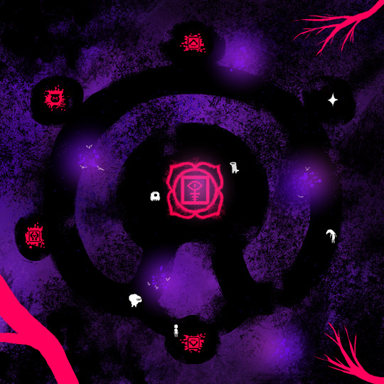
God's Reject (color style test)
color test for Archenteron's map, one ofthe earliest areas in the game.
having a rough time settling on a color scheme.... my initial thought was going for that 80s style of black backgrounds with bright sprites juxtaposed. this, basically: https://www.youtube.com/watch?v=NmhYq5RPJm0
i adore the simplicity of 80s games, it's not even a nostalgia thing for me. i didn't get into gaming until the 00s. i just genuinely like the style, i like simplicity and minimalism in general.
but going full on 1-bit black & white doesn't jive with me. i want a few more colors. purple is my favorite color, so i might just go with that because... why not?
My Socials/Galleries - See new art early on Patreon! - Leave a tip via Ko-Fi!
Posted using PostyBirb
1 note
·
View note
Note
this is a 100% genuine question: I’ve been studying the anatomy of dreamkin, mainly the ones that appear to be more human, and I need to know when their anal canal ends and where their inner void (for lack of a better term) begins. this would also apply to their throats; if I looked into a dreamkin’s throat would it appear as human anatomy or something eldritch?
dreamkin are protostomes, meaning that the initial invagination of the gastrula forms the mouth and primitive gut. in organic life, this archenteron would eventually tunnel through to form the anus. most dreamkin do not require a complex enough digestive system to necessitate an "end" at all—they are biologically and physically immortal, and consumption has no material benefit. most cannot consume anything not of the dreaming, but they will eat dreamfood regularly, which is quickly and efficiently reabsorbed into the dreaming itself at the dead-end of the digestive tract.
the corinthian is an interesting exception to this, not in the least because of his two distinct digestive tracts. the first is typical, beginning at his primary mouth and ending at the cecum. the second connects both his eyes at a medial trachea that loops around his "brain" and descends the body cavity to his anus. despite having this secondary "complete" digestive system, he still cannot derive meaningful nutrition from non-dreamfood. he may consume eyes in dreams and in the waking and understand, vicariously, the life he has taken, but he finds no satiation in it, only pleasure. i have, perhaps, underestimated how hungry he is.
45 notes
·
View notes
Text
Summarization of the Processes involved in Animal Embryonic Development
The first aspect needed to do is the union of sperm cells and egg cells to make a fertilized egg or zygote. After fertilization, there are a series and rapid mitotic divisions called Cleavage. In this process, the zygote divides many times without separating or changing in mass. It looks like an "atis" or sugar apple because of the smaller cells. These smaller cells are called blastomeres and the one large cell is called a blastula. This part also started the implantation in the uterus, where the zygote attaches to the uterus lining. Then, the cells (smaller cells) will rearrange into multi-layers (gastrula), it also observed the movement of the cell in forming the layers (invagination). This process will be called Gastrulation, which leads to an opening (blastopore) and then filled with archenteron. Later on, the opening becomes an anus. The three germ layers are the endoderm (inner part), ectoderm (outer part), and mesoderm (middle part) Now, the three germ layers execute the assigned body parts/systems on them, that direct on the formation of organs, and these process is called Organogenesis. These germ layers differentiate into tissues to form the organs such as; ectoderm for epithelial (skin), nervous system, etc. mesoderm for muscular, skeletal, heart, etc. endoderm for the respiratory system, glands, etc. The embryo will adopt a support membrane for embryo growth and nutrition. And the development will continue in the embryo until it becomes a fetus.
0 notes
Text
The cuckoo is one of the most unique creatures on the planet, but how it develops and hatches is even more interesting. As with most birds, the cuckoo goes through embryonic development, but the process is a bit different. Instead of hatching from an egg directly, they rely on an interesting process to make it happen. This process, known as blastoporic, is where a cuckoo's embryonic development starts and ends.
In the process of blastoporic, a mouth-like opening appears on the embryonic cuckoo's surface. This opening, now known as the archenteron, is where the embryo begins to invaginate. After the completion of the invagination process, the embryo is ready to hatch.
What's remarkable however, is how cuckoos don't require any parental care during the hatching process. Their blastoporic technique does it all for them in a relatively short period of time. Once the opening takes place, the embryo can slowly start hatching and eventually make it's way out of the eggshell.
This unique process also explains why cuckoo birds are great for stacking several eggs in one nest. With other birds, such as chickens, the eggs require constant parental care to help them hatch. However, since cuckoo eggs use the blastoporic method, the eggs hatch almost entirely on their own. That makes them well suited to stacking due to their self-sufficient nature.
In the end, the blastoporic method used by cuckoos is one of the most unique and efficient ways of developing and hatching an egg. It's why they can be stacked up Boston style and why they don't require parental attention when hatching. It's no wonder why this unique bird knows how to make it in the wild.
0 notes
Text
Blastopore in humans

Blastopore in humans skin#
Gastrulation in Gallus gallus (Domestic Chicken).
Blastopore in humans skin#
In the chick embryo, the cells of the ectoderm go on to form the skin and neural tissue, endoderm cells line the respiratory and gastrointestinal tracts, and the kidneys, circulatory system and skeleton are made from the mesoderm cells. Later, the cells of Hensen’s node regress, paving the way for the formation of the central nervous system. The cells making up Hensen’s node at the end of the primitive streak elongate across the blastula. One of the unique features of chick gastrulation is the cellular rearrangement that occurs at the posterior end of the blastula and forms the primitive streak, a thickening of the tissue. The lip is the point where the cells begin to turn and migrate inward, forming the blastopore. Another interesting aspect of frog gastrulation is that the blastopore forms a “lip” exactly 180 degrees opposite from where the sperm entered the egg. Also different, is that the cells of the blastula in the frog form the ectoderm or endoderm while the mesoderm is made from the yolk cells inside. One of the main differences is that the blastula is not hollow but is filled with yolk cells. Gastrulation in the frog is similar to the sea urchin, but it’s more complicated. In sea urchins, all three primary germ layers originate from the same outside layer of cells. The anus forms at the spot where the invagination started on the surface. The archenteron elongates and eventually fuses with the epithelial cells on the surface to form the mouth. Then, the archenteron (primitive gut) forms as the blastocyst invaginates creating the blastopore. In the first step of gastrulation, primary mesenchyme cells use chemical cues inside the blastula to migrate to the inside of the sphere where they will eventually fuse and form the larval skeleton made of spicules formed from calcium carbonate. Gastrulation in sea urchins is used as a starting point for understanding the process, as it is less complicated or more “simple” compared to other species, and the process only takes about 9 hours. Frogs, chickens, and sea urchins are 3 species most studied by developmental biologists and comparative embryologists.įigure 1: The image above shows the process of transformation from a single-celled zygote to a gastrula.įigure 2: The image above shows how gastrulation changes the number of cell layers from one to three. Each species has its own uniqueness when it comes to the process of gastrulation, but there are similarities that span the entire animal kingdom. The layers are called the primary germ layers the endoderm, ectoderm, and mesoderm (Figure 2). This is a critical point in development because it is when the embryo transforms itself from a hollow sphere made from a single layer of cells into a multi-layered structure. It does this by folding itself inward as shown in Figure 1. Experimental removal of the node in avian embryos results in complete absence of the notochord and a failure of neurulation. Gastrulation is a phase in the embryonic development of animals where the blastula reorganizes itself into a gastrula. The primitive node has been recorded in all stage 7 human embryos it produces axial cell populations, the prechordal plate, notochord, embryonic endoderm and the medial halves of the somites.
0 notes
Text
I can’t find my og post but something something Boethiah being one of the earliest daedra to form, paralleling the early stages of embryonic development being the archenteron, the primitive gut, with hunger and the need for consumption and survival from consumption being a requirement for life
14 notes
·
View notes
Photo

Stages of gastrulation in C. subdepressus under light microscope at 100X magnification depicting A) blastula showing animal pole positioned left and thickened vegetal pole positioned to the right B) Ingression of primary mesenchyme cells moving to the left from the vegetal pole C)Invagination of the vegetal plate to form archenteron D) Elongation of archenteron. Scale bar = 30 μm.
20 notes
·
View notes
Note
whats the most interesting part of ur advanced biology
Hi tumblr user lilacborrower,
All bilaterian animals can be put into two categories: in the early embryonic stage called gastrula the embryo goes from being spherical to forming a dent called the blastopore. The blastopore will get deeper and is what will eventually become the gut of the animal. In protostomes the original dent becomes the mouth, while for deuterostomes it becomes the butt. At some point the gut (at this point it’s called the archenteron) will grow deeper and create an opening on the other side (mouth), but that doesn’t happen immediately, so the deuterostomes are at one point just a bottomless anus.
You can take one fat guess which one of the two we are Sara
5 notes
·
View notes
Photo

It has been 1 year And 7 months since I last went to a concert with @malmosymfoniorkester and I am soooo excited! Good luck to @enricoscaccaglia who gets his piece "Archenteron" premiered this evening! . . . #contemporary #classicalmusicians #concert #liveperformance #livemusik #livemusic #liveconcert #MSO #malmösymfoniorkester #orchestral #orchestra #funtimes #finally https://www.instagram.com/p/CUvCAVzMM-5/?utm_medium=tumblr
#contemporary#classicalmusicians#concert#liveperformance#livemusik#livemusic#liveconcert#mso#malmösymfoniorkester#orchestral#orchestra#funtimes#finally
0 notes
Photo

DIFFERENCES BETWEEN THE MERRIAM-WEBSTER PRINT AND ONLINE DICTIONARIES
In 2017, the Scripps National Spelling Bee transitioned from the print version of the Merriam-Webster Third New International dictionary (which is also the CD ROM version) to the online Unabridged dictionary (unabridged.merriam-webster.com).
This blog post will discuss the differences between these two versions of the dictionaries so you are familiar with the changes.
Pronunciation of Words
There are some slight differences in pronunciation between the two versions of the dictionary. Some examples are:
WordPrint/CD DictionaryOnline Dictionary
mattockmad.ǝk, -atǝkˈmatək, -atək
tareta(a)](ǝ)r, ‘te], ]ǝˈter
flairfla(a)](ǝ)r, ‘fle], ]ǝˈfler
squareskwa(a)](ǝ)r, -we], ]ǝˈskwer
aerolithologya(ǝ)(͵)rōli’thälǝjē¦er-ə-li-¦thä-lə-jē
airbusa(a)(ǝ)r͵bǝsˈer-ˌbəs
revertrə̇’vǝr]t, rē’-, -vӛ], -vǝi], usu ]t+Vri-ˈvərt
redressrə̇’dres, rē’-ri-ˈdres
steersti(ǝ)r, -iǝˈstir
boardbō(ǝ)rd, -ȯ(ǝ)rd, -ōǝd, -ȯ(ǝ)dˈbȯrd
forthfō(ǝ)rth, ‘fȯ(ǝ)rth, ‘fōǝth, ‘fȯ(ǝ)thˈfȯrth
coarsekō(ǝ)rs, -ȯ(ǝ)rs, -ōǝs, -ȯ(ǝ)sˈkȯrs
pandapandǝ, ‘paan-ˈpan-də
forthfō(ǝ)rth, ‘fȯ(ǝ)rth, ‘fōǝth, ‘fȯ(ǝ)thˈfȯrth
answeran(t)sǝ(r), ‘aan-, ‘ain-, ‘ån-ˈan(t)-sər
Note: While the print dictionary has the diacritical symbol /d./, the online dictionary has replace this with /t/.
Some words have different alternative pronunciations between the two dictionaries. Here are some of these words:
WordPrint/CD DictionaryOnline Dictionary
jacanajakənəjə-ˈkä-nə, ˌzhä-sə-ˈnäⁿ
chelatorkē͵lātǝ(r)ˈkē-ˌlā-tər; kē-ˈlā-tər also chē-
sleazyslēzē, -ziˈslē-zē also ˈslā-
archenteron(‘)ärk.’entǝ͵rän(ˈ)är-ˈken-tə-ˌrän, -rən
raptorerap͵tō(ǝ)rˈrap-tər, -ˌtȯr
Aristotelian¦arə̇stǝ¦tēlyǝn, ¦a͵ris-, -lēǝn also ¦er- or ǝ¦ris- or a¦ris-¦a-rə-stə-¦tēl-yən
ctenophoretenǝ͵fō(ǝ)rˈte-nə-ˌfȯr
omȯmˈōm, ˈȯm
flotsamflätsǝm -lōt-ˈflät-səm
luxelǝksˈlu̇ks, ˈləks, ˈlüks
leopoldvilleˈlē-ə-ˌpōld-ˌvilˈlē-ə-ˌpōld-ˌvil, ˈlā-
crevallekrǝ’valē, -lǝ, -lākri-ˈva-lē
aminophyllineǝ͵mēnō’fi͵lēn, |amǝ͵nō’-; ͵amǝ’näfǝ͵-ˌa-mə-ˈnä-fə-lən
leipziglīpsi]g, -sē]ˈlīp-sig, -sik
Spelling
Some words are spelled differently between both dictionaries, such as:
Print/CD DictionaryOnline Dictionary
amiziliaamazilia
bogshabogshah
vilnyusvilnius
New Online Words
The online dictionary has a lot of new words that the print dictionary did not have. Scripps has asked a lot of these new online words in their bees, such as xylorimba, turducken, mizuna, and Beringia. In fact, a few of these new words were asked at the National Spelling Bee in 2017, such as arribada, clafouti, Bruneian, and bucatini.
SpellPundit is one of the only study resources that has completely incorporated all the spelling/pronunciation changes in the online dictionary. Our New Online Words Module has with over 4000 words for spellers to learn these words and test themselves.
https://www.spellpundit.com
#Spelling#Spelling Bee Scripps national#spelling bee#spelling preparation#spellingtips#vocabulary tips#octochamps spelling bee champions#spelling words#spelling word lists#spelling module#Free module#vocabulary module#Latin/Greek roots module#homonyms module#language patters#SpellPundit learning mode
0 notes
Text
300+ TOP EMBRYOLOGY Interview Questions and Answers
EMBRYOLOGY Interview Questions for freshers experienced :-
1. What is the cell division process directly related to the embryonic growth?
The embryonic growth depends directly on mitosis. Through this type of cell division, the zygote divides itself giving birth to a series of cells that by mitosis too compose differentiated tissues and organs until the formation of a complete individual.
2. What is the function of the vitellus in the vertebrate egg? How are these eggs classified according to the amount of vitellus within them?
Vitellus (yolk) is the nutritive material that accumulates in the cytoplasm of the egg (zygote) with the function of nourishing the embryo. According to the amount of vitellus in them, the vertebrate eggs are classified as oligolecithal (little yolk), centrolecithal, or heterolecithal (more yolk diffusely distributed) and telolecithal (more yolk concentrated in one end of the egg).
3. What are the animal pole and the vegetal pole of the vertebrate egg?
The animal pole of a telolecithal egg is the portion of the egg with little vitellus, it is opposite to the vegetal pole that is the region where the yolk is concentrated.
Image Diversity: animal and vegetal poles
4. What are the four initial stages of the embryonic development?
The four initial stages of the embryonic development are the morula stage, the blastula stage, the gastrula stage, and the neurula stage.
5. What is the cell division during the first stage of the embryonic development called? How is this stage characterized?
The cell division in the first stage of the embryonic developments is called cleavage, or segmentation. In this stage, mitosis occurs from the zygote forming the new embryo.
6. What are the cells produced in the first stage of the embryonic development called?
The cells that resulted from the cleavage (the first stage of the embryonic development) are called blastomeres. In this stage the embryo is called morula (similar to a “morus”, mulberry).
Image Diversity: morula
7. After the morula stage what is the next stage? What is the morphological feature that defines that stage?
After passing the morula stage in which the embryo was a compact mass of cells, the next stage is the blastula stage. In the blastula stage, the compactness is lost and an internal cavity filled with fluid appears inside, the blastocele.
Image Diversity: blastula
8. After the blastula stage what is the following stage of the embryonic development? How is the passage from blastula to the next stage called?
The blastula turns into gastrula in a process known as gastrulation.
Image Diversity: gastrula
9. What is gastrulation? How during gastrulation are the first two germ layers formed? Which are these germ layers?
Gastrulation is the process through which a portion of the blastula wall undergoes invagination inside the blastocele forming a tube called archenteron (primitive intestine). The cells of the inner side of the tube form the endoderm (germ layer) and the cells of the outer side form the ectoderm (another germ layer). It is the beginning of the tissue differentiation in the embryonic development.
10. What are the archenteron and the blastopore? What is the stage of the embryonic development in which these structures are formed? What are the destinations of the archenteron and of the blastopore?
Archenteron is the tube formed during gastrulation by means of invagination of the blastula wall inside the blatocele. It is the origin of the gastrointestinal tract. Blastopore is the opening of the archenteron to the exterior. The blastopore gives birth to one of the extremities of the digestive tube: the mouth in protostome beings, or the anus in deuterostome beings.

EMBRYOLOGY Interview Questions
11. How is the mesoderm (third germ layer) of triploblastic animals formed?
The mesoderm appears from differentiation of endodermal cells that cover the dorsal region of the archenteron.
12. What are the three types of germ layers that form tissues and organs in animals?
The three germ layers are the ectoderm, the mesoderm, and the endoderm.
Image Diversity: germ layers
13. How are animals classified according to the germ layers present in their embryonic development?
Cnidarians are diploblastic, i.e., they present only endoderm and ectoderm. With the exception of poriferans, all remaining animals are triploblastic. Poriferans do not present differentiated tissue organization and so they do not classify regarding germ layers (although sometimes they are mentioned as diploblastic).
14. How does the embryo turn from gastrula into neurula? How is the neural tube formed? What is the embryonic origin of the nervous system in vertebrates?
The neurula stage is characterized by the appearing of the neural tube along the dorsal region of the embryo. The growing of mesoderm in that region induces the differentiation of ectodermal cells just above. These cells then differentiate forming the neural tube. Therefore, the origin of the nervous system is the ectoderm (the same germ layer that gives birth to the skin).
Image Diversity: neurula
15. What is notochord? How is this structure formed?
Notochord is a rodlike structure that forms the supporting axis of the embryo and gives birth to the vertebral column in vertebrates. It is formed by differentiation of mesodermal cells.
16. What is coelom? To which structures do coeloms give birth? Are all animals coelomate?
Coeloms are cavities delimited by mesoderm. Coeloms originate the cavities where the internal organs of the body are located, like the pericardial cavity, the peritoneal cavity, and the pleural cavity. Besides coelomate animals, there are acoelomate animals, like platyhelminthes, and pseudocoelomate animals, like nematodes.
Image Diversity: coelom
17. What is the germ layer from which the coeloms originate?
The coeloms are originated from mesoderm.
18. What are pleura, pericardium, and peritoneum?
Pleura are the membrane that covers the lungs and the inner wall of the chest; pericardium is the membrane that covers the heart; peritoneum is the membrane that covers most organs of the gastrointestinal tract and part of the abdominal cavity. All these membranes delimit coeloms (internal cavities).
Image Diversity: pleura pericardium peritoneum
19. After the neurula stage and from its ventral portion to the dorsal how can the morphology of the embryo be described?
In a schematic longitudinal section of the embryo after the neurula stage, the outermost layer of cells is the ectoderm. In the ventral region comes the archenteron tube formed of endodermal cells. In both sides of the embryo, coeloms delimited by mesoderm are present. In the central region above the archenteron and in the middle of the coeloms there is the notochord. In the dorsal region just above the notochord lies the neural tube.
20. What are somites?
Somites are differentiated portions of mesodermal tissue longitudinally distributed along the embryo. The somites originate the muscle tissue and portions of the connective tissues.
Image Diversity: somites
21. What are histogenesis and organogenesis?
Histogenesis is the process of tissue formation in the embryonic development. Organogenesis is the process of organ formation. Before histogenesis and organogenesis the primitive embryonic structures have been already formed: germ layers, neural tube, notochord, coeloms, somites.
24. What are extra embryonic membranes?
Extra embryonic membranes are membranous structures that appear paralleling the embryo and play important roles in the embryonic development. They form from the embryo but do not become part of the individual organism after its birth.
25. What are the extra embryonic membranes present in vertebrates?
The extra embryonic membranes that may be present in vertebrates are the yolk sac, the amnion, the chorion, the allantois and the placenta.
26. Are the extra embryonic membranes the same in all vertebrates?
The presence of each extra embryonic membrane varies according to the vertebrate class.
In fishes and amphibians, only the yolk sac is present. In reptiles and aves besides the yolk sac, there are also the amnion, the chorion and the allantois. In placental mammals besides all these membranes, the placenta is present too.
27. How is the yolk sac formed? What is the function of the yolk sac?
The yolk sac is formed from the covering of the vitellus by some cells originated from the primitive gut.
The yolk sac stores vitellus, the main nourishment source of nonplacental embryos.
Image Diversity: yolk sac
28. Which is the extra embryonic membrane whose function is to store nitrogen wastes of the embryo? Is this function present in placental mammalian embryos?
The allantois is the extra embryonic membrane whose function is to store excretes of the embryo.
In placental mammals, the allantois is present but it does not exert that function since the embryonic wastes are collected by the mother’s body through the placenta.
Image Diversity: allantois
29. Why can the allantois be considered an adaptation to terrestrial life?
The allantois is an adaptation to dry land because in embryos of oviparous terrestrial beings, like reptiles and birds, the metabolic residuals cannot be immediately excreted to the aquatic surrounds (as fishes and amphibian larvae do). It was necessary then the appearing of a structure capable of storing the embryonic excretes until hatching.
30. What is the difference between amnion and chorion?
Amnion is the membrane that covers the embryo. Chorion is the membrane that covers the amnion, the yolk sac, and the allantois. The space delimited by the chorion and the amnion is called amniotic cavity and it is filled with aminiotic fluid. The amniotic cavity has the functions of preventing desiccation of the embryo and of protecting it against mechanical shocks.
31. Why can the amnion also be considered an adaptation to terrestrial life?
The amnion is also an adaptation to dry land since one of its functions is to prevent desiccation of the embryo.
32. What is the chorioallantois membrane present in the embryonic development of reptiles and birds? How does this membrane participate in the energetic metabolism of the embryo?
The chorioallantois membrane is formed by juxtaposition of some regions of the chorion and the allantois. Since it is porous, the chorioallantois membrane allows the passage of gases between the embryo and the exterior thus making aerobic cellular respiration possible.
33. In which type of animals does the placenta exist? What is its main function?
True placenta is present in placental mammals.
The placenta is formed from the chorion of the embryo and from the mother’s endometrium. Its main function is to allow the exchange of substances between the fetus and the mother’s body.
Image Diversity: placenta placental mammals
34. What are the main substances transferred from the mother to the fetus through the placenta and from the fetus to the mother?
From the mother to the fetus the main transferred substances through the placenta are water, oxygen, nutrients, and antibodies. From the fetus to the mother, metabolic wastes including urea (nitrogen waste), and carbon dioxide are transferred.
35. Is there exchange of cells between the mother and the fetus through the placenta?
Under normal conditions, there is no passage of cells across the placenta during gestation. The placenta has a smooth mucosa separating the richly vascularized region in contact with the mother’s endometrium from the umbilical cord in contact with the fetal blood. This barrier is known as placental barrier. Although permeable to some substances (selective permeability), the placental barrier forbids the passage of cells.
Image Diversity: umbilical cord placental barrier
36. What are the endocrine functions of the placenta?
The placenta has endocrine function since it secretes the hormones progesterone and estrogens that maintain the endometrium (internal covering of the uterus) and prevent menses during pregnancy. The placenta also secretes other important hormones for pregnancy regulation.
37. What is the function of the umbilical cord?
The umbilical cord is a set of blood vessels that connect the fetus with the placenta. In the fetus, one extremity of the cord inserts into the center of the abdominal wall (the later scar of this insertion is the umbilicus).
38. What are the three types of germ layers that form tissues and organs in animals?
The three germ layers are the ectoderm, the mesoderm, and the endoderm.
39. How is the mesoderm (third germ layer) of triploblastic animals formed?
The mesoderm appears from differentiation of endodermal cells that cover the dorsal region of the archenteron.
40. What are the archenteron and the blastopore? What is the stage of the embryonic development in which these structures are formed? What are the destinations of the archenteron and of the blastopore?
Archenteron is the tube formed during gastrulation by means of invagination of the blastula wall inside the blatocele. It is the origin of the gastrointestinal tract. Blastopore is the opening of the archenteron to the exterior. The blastopore gives birth to one of the extremities of the digestive tube: the mouth in protostome beings, or the anus in deuterostome beings.
41. What is gastrulation? How during gastrulation are the first two germ layers formed? Which are these germ layers?
Gastrulation is the process through which a portion of the blastula wall undergoes invagination inside the blastocele forming a tube called archenteron (primitive intestine). The cells of the inner side of the tube form the endoderm (germ layer) and the cells of the outer side form the ectoderm (another germ layer). It is the beginning of the tissue differentiation in the embryonic development.
42. After the blastula stage what is the following stage of the embryonic development? How is the passage from blastula to the next stage called?
The blastula turns into gastrula in a process known as gastrulation.
43. After the morula stage what is the next stage? What is the morphological feature that defines that stage?
After passing the morula stage in which the embryo was a compact mass of cells, the next stage is the blastula stage. In the blastula stage, the compactness is lost and an internal cavity filled with fluid appears inside, the blastocele.
44. What are the cells produced in the first stage of the embryonic development called?
The cells that resulted from the cleavage (the first stage of the embryonic development) are called blastomeres. In this stage the embryo is called morula (similar to a “morus”, mulberry).
45. What is the cell division during the first stage of the embryonic development called? How is this stage characterized?
The cell division in the first stage of the embryonic developments is called cleavage, or segmentation. In this stage, mitosis occurs from the zygote forming the new embryo.
46. What are the four initial stages of the embryonic development?
The four initial stages of the embryonic development are the morula stage, the blastula stage, the gastrula stage, and the neurula stage.
47. What are the animal pole and the vegetal pole of the vertebrate egg?
The animal pole of a telolecithal egg is the portion of the egg with little vitellus, it is opposite to the vegetal pole that is the region where the yolk is concentrated.
48. What is the function of the vitellus in the vertebrate egg? How are these eggs classified according to the amount of vitellus within them?
Vitellus (yolk) is the nutritive material that accumulates in the cytoplasm of the egg (zygote) with the function of nourishing the embryo. According to the amount of vitellus in them, the vertebrate eggs are classified as oligolecithal (little yolk), centrolecithal, or heterolecithal (more yolk diffusely distributed) and telolecithal (more yolk concentrated in one end of the egg).
49. What is the cell division process directly related to the embryonic growth?
The embryonic growth depends directly on mitosis. Through this type of cell division, the zygote divides itself giving birth to a series of cells that by mitosis too compose differentiated tissues and organs until the formation of a complete individual.
50. How are animals classified according to the germ layers present in their embryonic development?
Cnidarians are diploblastic, i.e., they present only endoderm and ectoderm. With the exception of poriferans, all remaining animals are triploblastic. Poriferans do not present differentiated tissue organization and so they do not classify regarding germ layers (although sometimes they are mentioned as diploblastic).
51. How does the embryo turn from gastrula into neurula? How is the neural tube formed? What is the embryonic origin of the nervous system in vertebrates?
The neurula stage is characterized by the appearing of the neural tube along the dorsal region of the embryo. The growing of mesoderm in that region induces the differentiation of ectodermal cells just above. These cells then differentiate forming the neural tube. Therefore, the origin of the nervous system is the ectoderm (the same germ layer that gives birth to the skin).
52. What is notochord? How is this structure formed?
Notochord is a rodlike structure that forms the supporting axis of the embryo and gives birth to the vertebral column in vertebrates. It is formed by differentiation of mesodermal cells.
53. What is coelom? To which structures do coeloms give birth? Are all animals coelomate?
Coeloms are cavities delimited by mesoderm. Coeloms originate the cavities where the internal organs of the body are located, like the pericardial cavity, the peritoneal cavity, and the pleural cavity. Besides coelomate animals, there are acoelomate animals, like platyhelminthes, and pseudocoelomate animals, like nematodes.
54. What is the germ layer from which the coeloms originate?
The coeloms are originated from mesoderm.
55. What are pleura, pericardium, and peritoneum?
Pleura are the membrane that covers the lungs and the inner wall of the chest; pericardium is the membrane that covers the heart; peritoneum is the membrane that covers most organs of the gastrointestinal tract and part of the abdominal cavity. All these membranes delimit coeloms (internal cavities).
56. After the neurula stage and from its ventral portion to the dorsal how can the morphology of the embryo be described?
In a schematic longitudinal section of the embryo after the neurula stage, the outermost layer of cells is the ectoderm. In the ventral region comes the archenteron tube formed of endodermal cells. In both sides of the embryo, coeloms delimited by mesoderm are present. In the central region above the archenteron and in the middle of the coeloms there is the notochord. In the dorsal region just above the notochord lies the neural tube.
57. What are somites?
Somites are differentiated portions of mesodermal tissue longitudinally distributed along the embryo. The somites originate the muscle tissue and portions of the connective tissues.
58. What are histogenesis and organogenesis?
Histogenesis is the process of tissue formation in the embryonic development. Organogenesis is the process of organ formation. Before histogenesis and organogenesis the primitive embryonic structures have been already formed: germ layers, neural tube, notochord, coeloms, somites.
EMBRYOLOGY Questions and Answers pdf Download
Read the full article
0 notes
Text
creepycreepyspacewizard replied to your post: “Arch-something is an important term” good job...
arch-nemesis!! its not a note its a warning aaaaaaahhhhh
ladydomini replied to your post: “Arch-something is an important term” good job...
Were you being taught by an arch-wizard?
guys stop making me laugh I’m in a library! (ok nvm the people sitting around me keep chatting annoyingly, they can handle the occasional muffled giggle.)
For the record, the word was “archenteron”, aka the rudimentary alimentary cavity of an embryo at the gastrula stage
Which also sounds kinda funny tbh.
#creepycreepyspacewizard#ladydomini#definition taken straight from google#and no I have no idea what that means#but I can point it out on a cross-section of an embryo of the appropriate stage#hopefully
1 note
·
View note
Text
Alyssa Baguss Loves a Challenge and so do I

"Vitamin D: A Pep Rally For Spring," 2018
“Hope locates itself in the premises that we don’t know what will happen and that in the spaciousness of uncertainty is room to act.”
—Solnit, Rebecca. "Grounds For Hope."
What actions do you take when you want to build up a community, improve the place where you live, or make meaningful connections with the world around you? What do you do when it’s 20 degrees below Fahrenheit and winter feels like it will never ever end? If you are Alyssa Baguss, you throw a party or more specifically a pep rally to usher in spring and warm things up. Alyssa’s "Vitamin D: A Pep Rally For Spring," 2018 was a part of “Illuminate South Loop” a three-day creative placemaking festival created by the City of Bloomington, Artistry, and Northern Lights.mnin the lead up to the Super Bowl. It featured cheerleaders and a drum corps and was a simple act of defiant joy.
Our class of Nomads met Alyssa Baguss at Silverwood, an art-focused public park in the Three Rivers District of Hennepin County, Minnesota that she helps run. We were there as part of a class with artist Amanda Lovelee called Art and Place Reconsidered. Both women are part of a tight-knit group of socially engaged artists in the Twin Cities. Alyssa and pretty much everyone else we met in Minnesota, mentioned how deeply cold the area can be and I realized seasonal depression in a place with this much winter must be hard to contend with, the immense amount of joy and humor in her work relates directly to the realities of living in this place.
In another of her workd titled HB2U:MIA, Alyssa created a voluminous spatial installation out of a multicolored parachute (a giant version of the I and many others I played with in elementary school). Taking a theme, in this case, Minneapolis Institute of Arts’s100th birthday and running with it, she accompanied this spatial experience with birthday cake sculptures ready for decorating. Alyssa’s practice is play-based and impulse-driven. She makes space for creative experiences that are for her enjoyment and the community around her.

Silverwood Park, Hennepin Co. Mn

Bethel University. Archenteron, Silverwood Park, Hennepin Co. Mn
In her other life at Silverwood, Alyssa Baguss is the Artist and Arts Programming Coordinator. The park is a relatively new suburban open space focused on encouraging an understanding of nature through art and art-making. The trails have poetry stops and contemporary sculpture nestled into wooded areas, bird habitats, and along the water. There is an artist-in-residency program for writers and an onsite gallery. As programming coordinator, she manages a team of full and part-time staff members to create events and classes, works with artists, and the broader community to build meaningful connections around the environment.

Oceanic,2018
Folded photo wallpaper mural
While we were in Minneapolis, I also got a chance to see some of her installation work at Minneapolis College of Art and Design. These drawings were created as part of a set of new works for the Jerome Fellowship. They are large and beautiful digital depictions of place and space folded like origami, so they move into three dimensions. The works depict the outside world, vast expanses of water and earth, but they feel made only for interior space. Oceanic, 2018 was the first piece I saw when I walked into the gallery, and it floats like an ice-burg reflecting in the speckled highly polished floor. These remind me of rainy-day activities, plans for future adventure, or sets for imagined ones.
I’m impressed with the balance she’s able to maintain between her individual practice, where one participates with one’s community but is personal in nature, and Parks or institutional work, which is service driven and collective in nature. Each piece informing and influencing the other, time made for each.
I live on the east coast of the USA in a pretty rural (relative to things here) town in Southern New Jersey called Mays Landing. There’s a lot of creative placemaking that goes on in other parts of the state and especially in nearby Philadelphia, where between organizations like Philadelphia Contemporary, Mural Arts Philadelphia, and the city government there seems to be a new event, park, or festival every day of the week with a high focus on contemporary public art. I grew up in New Jersey but, I’m pretty new to Mays Landing, and I’m still getting to know this place. I’ve met a lot of people who participate in committees like Friends of the Park, and the historical society and they run some pretty wonderful programming but turn out to things is still really low. The town itself has about four big community events a year and based on anecdotes from friends and family the turn out for those is going down too.
One thing I took away from this trip was that everyone we met manages to make things happen and a huge part of that is the community of artists and educators and how well connected they are to each other. There seemed to only be two-degrees of separation between everyone we met with, and that’s if they didn’t already know each other.
Another is that I’ve had vague ideas of how to start creating programs and things here before we went to Minneapolis but, after this trip and spending time at Silverwood, I think there’s a need to look deeper into my interest in the parks here and my understanding of nature. As part of Amanda Lovelee’s class, we listened to an interview with Robin Wall Kimmerer where she said: “…attention is that doorway to gratitude, the doorway to wonder, the doorway to reciprocity.”It’s about the way children look and through their wonder can build relationships with the world. It’s something I try to keep alive in my work when I research and paint but not something I’ve thought to include in my collaborative projects, not something I thought to share.
More: Video about Alyssa Baguss
0 notes
Video
youtube
Learn how to pronounce Mesenteron in English --- MESENTERON Pronunciation of Mesenteron: /(ˌ)me-ˈzen-tə-ˌrän, ˌmē-, -ˈsen-, -rən/ noun Definition of Mesenteron: the part of the alimentary canal that is developed from the archenteron and is lined with hypoblast ★ http://Learn2Pronounce.com ★ How to pronounce Mesenteron | English pronunciation: https://youtu.be/yKwAEDQA0ck
#how to pronounce How to pronounce Mesenteron | English pronunciation#pronunciation of How to pronoun
0 notes
Photo

The morula is 32 cells big, and keeps growing into a spherical blastula. The blastula is where the gastrulation happens -- the invagination and forming the archenteron. As the archenteron proceeds, is called a blastopore. Coelom development happens during this gastrulation. The coelom is formed from outside in for the protostome; the coelom is formed from the inside out in deuterostomes likes us.
Notably, we experience standard radial cleavage of the cells (so as to make nice normal little spheres) while the protostomes experience spiral cleavage, so they development at an angle.
0 notes
Text
New Post has been published on Biology Dictionary
New Post has been published on https://biologydictionary.net/mesoderm/
Mesoderm
Mesoderm Definition
The mesoderm is a germ layer present in animal embryos that will give rise to specialized tissue types. The mesoderm is one of three germ layers found in triploblastic organisms; it is found between the ectoderm and endoderm. All bilaterally-symmetrical animals are triploblasts, whereas some simpler animals such as cnidaria and ctenophores (jellyfish and comb jellies) have only two germ layers and are diploblasts. The mesoderm plays an important role in animal development. It goes on to form many central structures including the skeletal system, muscular system, and the notochord.
Mesoderm Formation
The tissue layers, or germ layers, form during gastrulation. Early in development the egg consists of a darker animal region and a yolky vegetal region. Future mesoderm cells form from animal region cells at the boundary of these two regions as an equatorial band. Unlike the other two germ layers whose fate is determined by maternal factors in the egg, future mesodermal cells form in response to signals from the future endoderm cells in the vegetal region.
In the early blastula, the future mesoderm cells are found on the surface. During gastrulation the cells will rearrange until the mesoderm (and endoderm) are on the inside of the embryo, and the ectoderm is on the outside surface. This occurs by invagination of the mesoderm and endoderm cells; they migrate to the interior while the ectoderm spreads to cover the exterior.
Mesoderm Function
The mesoderm is responsible for the formation of a number of critical structures and organs within the developing embryo including the skeletal system, the muscular system, the excretory system, the circulatory system, the lymphatic system, and the reproductive system. It also gives rise to connective tissues, the dermis of the skin, the lining of the coelom, the adrenal cortex, and many of the internal organs. The mesoderm is generally separated into a number of regions between the dorsal and ventral sides. The dorsal mesoderm will form the notochord, the central region will form the heart and muscles, and the most ventral region will form the blood and associated organs (e.g., the kidney).
Towards the end of gastrulation, the dorsal mesoderm forms a rod-like notochord that runs along the embryo from head to tail below where the nervous system will form, just above the archenteron (the primitive gut). The notochord then sends a signal to the dorsal ectoderm to form the neural tube that will go on to form the central nervous system. The notochord acts as an anchor around which the vertebrae will form. The notochord itself is transient and will ultimately be incorporated as the inner portion of the vertebral disks.
On either side of the notochord lie paired segments of mesoderm that are arranged successively. These somites are formed from lateral-ventral mesoderm. They will ultimately give rise to muscles, cartilage, the vertebral column, and the dermis of the skin. What structures are formed from each of the somite pairs is specified by Hox genes and signals from adjacent cells.
Some of the mesoderm found along the notochord will form mesenchyme cells that will then relocate to new locations. This lateral plate mesoderm will form internal organs including the kidney and the heart. The kidney is formed from mesenchymal cells that have formed tubules. These tubules combine and each develops a glomerulus which acts in filtration. The heart is formed as a single tubule made up of two epithelial layers of mesenchyme: the endocardium and the myocardium. The tube develops into the two heart chambers, the atrium and the ventricle, which then divide further in mammals and birds to form the four-chamber heart.
In many cases the mesoderm does not develop properly in the embryo. This can result in a number of conditions including heart defects, skeletal abnormalities, or death of the embryo. An example of a mesoderm abnormality is the mutation of the mammalian T gene that gives rise to the brachyury protein. The homozygous form of this mutation is lethal as the mesoderm does not form properly and the skeleton does not develop in the posterior. Heterozygotes for the T gene mutation generally lack some of the sacral vertebrae and have a short tail.
Quiz
1. What organisms do not have a mesoderm?
A. insects
B. fish
C. corals
D. amphibians
Answer to Question #1
C is correct. The mesoderm is found in triploblastic organisms. These include all bilaterally-symmetrical animals. Simpler animals such as cnidarians are diploblastic. Corals are cnidarians, and as such are diploblasts.
2. Which of the following organs is derived from mesoderm?
A. pancreas
B. kidney
C. liver
D. gall bladder
Answer to Question #2
B is correct. Many of the internal organs are formed from the mesoderm, most notably the heart and the kidney. The liver, pancreas, and gall bladder are all associated with the digestive system, and are all derived from the endoderm.
3. What structure is formed from the mesoderm?
A. notochord
B. neural crest
C. neural tube
D. neural plate
Answer to Question #3
A is correct. The mesoderm forms a rod-like structure called the notochord. The notochord then signals the ectoderm to form the neural plate which folds in to form the neural tube.
References
Campbell, N. A., & Reece, J. B. (2005).Biology, 7th. ed. Chs. 32 and 47. San Francisco, CA: Benjamin Cummings. ISBN: 0-8053-7171-0.
Jessell, T., Lawrence, P., Meyerowitz, E., Robertson, E., & Smith, J. (2005).Principles of Development, 3rd. ed. Chs. 1 to 4. New York, NY: Oxford University Press. ISBN: 0-19-927537-8.
0 notes
Last Seen Blogs
truthaboutthebeatlesgirls
Truth About the Beatles' Girls

plexusthings
estascy seller

gabrisart
Gabris Art
chiaochen1028
chiao chen

simsccola
SimsCCola