#symptoms of Navel Displacement
Text
What is Navel Displacement : Symptoms & Treatment in Ayurveda
Navel displacement, also known as Nabhi Chakra Veda or Nabhi Displacement, is a condition where the navel or umbilicus moves from its normal position. In Ayurveda, it is believed that the displacement of the navel can cause an imbalance in the body's energy, leading to various health problems.
Symptoms of navel displacement may include pain or discomfort in the abdomen, indigestion, constipation, bloating, nausea, vomiting, back pain, fatigue, and menstrual problems in women.
According to Ayurveda, the treatment for navel displacement involves restoring the balance of energy in the body through various techniques. One of the primary techniques is Navel Displacement Correction Therapy or Nabhi Chikitsa. This therapy involves massaging the area around the navel with warm oil and then applying a small amount of pressure to the navel. The therapist may also use specific Ayurvedic herbs and oils to help restore the balance of energy in the body.
Other Ayurvedic treatments for navel displacement may include yoga, meditation, dietary changes, and the use of Ayurvedic medicines. It is also essential to maintain good posture and avoid heavy lifting or strenuous activities that may worsen the condition.
In Ayurveda, it is believed that maintaining a healthy balance of energy in the body is crucial for overall well-being. Therefore, it is important to seek treatment for navel displacement if you are experiencing symptoms, to help restore balance and promote optimal health.
image source https://ayurhealthcare.com.au/wp-content/uploads/2021/11/Navel-Displacement.jpg
How do you know if your navel is displaced?
Navel displacement, also known as Nabhi Chakra Veda, can be diagnosed through a physical examination by a healthcare provider or an Ayurvedic practitioner. The practitioner will check the position of the navel and observe any signs of swelling or discomfort in the surrounding area.
However, there are some signs and symptoms that may indicate navel displacement. These include:
Pain or discomfort in the abdomen, especially around the navel area.
Digestive problems such as bloating, constipation, or gas.
Back pain or discomfort.
Menstrual problems in women, such as irregular periods or painful periods.
Fatigue or weakness.
Nausea or vomiting.
A visible shift or bulge in the navel area.
If you are experiencing any of these symptoms, it is essential to seek medical attention to determine the underlying cause and receive appropriate treatment. An Ayurvedic practitioner can also help you restore balance to the body's energy and promote overall well-being.
What are the Causes Of Navel Displacement
In Ayurveda, navel displacement is believed to be caused by an imbalance in the body's energy, or doshas, which can be triggered by various factors. Some of the common causes of navel displacement are:
Poor Posture: Poor posture can cause undue pressure on the abdomen and lead to navel displacement.
Injury: Injury to the abdomen or navel area can cause the navel to move from its normal position.
Heavy Lifting: Lifting heavy objects can also lead to navel displacement due to the pressure it puts on the abdomen.
Digestive Problems: Digestive problems such as constipation, bloating, or gas can cause the navel to shift from its normal position.
Menstrual Problems: In women, menstrual problems such as irregular periods or painful periods can lead to navel displacement.
Emotional Stress: Emotional stress or anxiety can also affect the body's energy and lead to navel displacement.
Obesity: Obesity can put pressure on the abdomen and lead to navel displacement.
It is essential to address the underlying cause of navel displacement to prevent it from occurring or recurring. Ayurvedic treatments aim to restore balance to the body's energy and promote overall health and well-being.
#Navel Displacement#Navel Displacement Treatments#Ayurvedic Treatment For Navel Displacement#symptoms of Navel Displacement#Navel Displacement symptoms
4 notes
·
View notes
Text
A Comprehensive Guide to Navel Displacement Medicine
Navel displacement, a condition where the navel or belly button shifts from its normal position, can be a source of discomfort and concern for many individuals. In this comprehensive guide, we will explore the causes, symptoms, and available treatments for navel displacement. By delving into the world of navel displacement medicine, we aim to provide valuable insights and information to those seeking a deeper understanding of this condition.
What is Navel Displacement?
Navel displacement, also known as umbilical displacement or belly button displacement, occurs when the navel shifts from its central position. This displacement can be caused by a variety of factors, including pregnancy, obesity, abdominal surgery, or trauma to the abdominal area.
Symptoms and Diagnosis
Individuals experiencing navel displacement may notice symptoms such as pain, discomfort, or a visible change in the appearance of their belly button. In some cases, a protrusion or bulge may be observed. Proper diagnosis of navel displacement involves a physical examination by a healthcare professional, who may also use imaging studies such as ultrasound to assess the extent of the displacement.
Navel Displacement Medicine: Treatment Options
Non-Surgical Approaches
Lifestyle modifications: Adopting a healthy lifestyle that includes regular exercise and maintaining a healthy weight can alleviate symptoms associated with navel displacement.
Physical therapy: Targeted exercises prescribed by a healthcare professional can help strengthen the abdominal muscles and support the navel, reducing the likelihood of displacement.
Medical Interventions
Pain management: Over-the-counter pain relievers may be recommended to manage discomfort associated with navel displacement.
Supportive devices: In some cases, the use of supportive devices, such as abdominal binders, may be suggested to provide additional support to the abdominal area.
Surgical Options
Hernia repair: If navel displacement is associated with a hernia, surgical intervention may be necessary to correct the underlying issue and reposition the navel.
Laparoscopic procedures: Minimally invasive techniques may be employed to surgically address navel displacement, minimizing recovery time and scarring.
Prevention Strategies
While not all instances of navel displacement can be prevented, adopting certain measures can reduce the risk of developing this condition. These measures include maintaining a healthy weight, practicing proper lifting techniques, and addressing any underlying medical conditions that may contribute to abdominal strain.
Conclusion:
In conclusion, understanding navel displacement and the role of navel displacement medicine is crucial for individuals affected by this condition. By recognizing the symptoms, exploring treatment options, and adopting preventive measures, individuals can take proactive steps towards managing and alleviating the discomfort associated with navel displacement. Remember, consulting with a healthcare professional is essential for personalized guidance and a tailored treatment plan.
0 notes
Text
Hernia Surgery Benefits, Risks, and What to Expect
What Is Hernia Surgery?
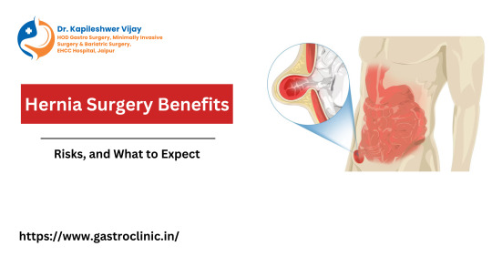
Hernia surgery, also known as herniorrhaphy, is a medical procedure performed to repair a hernia, a condition where an organ or fatty tissue protrudes through a weak spot in the surrounding muscle or connective tissue. Hernias can occur in various areas of the body, but they are most commonly found in the abdominal region, groin, and upper thigh. The surgery involves returning the displaced tissue to its proper position and reinforcing the weakened area with sutures or a mesh patch to prevent recurrence. There are different types of hernia surgeries, including open surgery, where an incision is made directly over the hernia, and laparoscopic surgery, which involves smaller incisions and the use of a camera and specialized instruments for repair. The choice of surgical approach depends on the type and location of the hernia, as well as the patient's overall health and surgeon's recommendation. Hernia surgery is typically safe and effective, providing relief from the discomfort and potential complications associated with untreated hernias. Patients often experience a significant improvement in their quality of life after undergoing this procedure.
Hernia Surgery Benefits and Risks
Hernia surgery, also known as herniorrhaphy or hernioplasty, offers both significant benefits and potential risks to patients. The primary advantage of undergoing hernia surgery is the resolution of the hernia itself. Hernias occur when an organ or tissue pushes through a weakened spot in the surrounding muscle or connective tissue, causing pain and discomfort. Surgery aims to repair this structural defect, relieving pain and preventing complications such as strangulation, a life-threatening condition that occurs when blood supply to the herniated organ is compromised. On the flip side, like any surgical procedure, hernia surgery is not without its risks. Common risks include infection, bleeding, and post-operative pain. It's essential for patients to consult with their healthcare providers to weigh the potential benefits against the associated risks and make an informed decision about whether hernia surgery is the right course of action for their specific case.
Conditions We Treat with Hernia Surgery
Hernia surgery is a medical procedure employed to address a variety of conditions related to hernias, which occur when an organ or fatty tissue protrudes through a weak spot or tear in the surrounding muscle or connective tissue. This surgical intervention is primarily used to treat inguinal hernias, which are the most common type, affecting the groin area. Inguinal hernias often require surgical repair to alleviate pain and discomfort. Additionally, umbilical hernias, which occur around the navel, and incisional hernias, arising from a prior surgical incision, are also treated with hernia surgery. Ventral hernias, located in the abdominal wall, and femoral hernias, typically occurring lower down in the groin region, can also be addressed through this procedure. The aim of hernia surgery is to reinforce the weakened area and reduce the risk of hernia recurrence, ultimately improving the patient's quality of life and alleviating associated symptoms.
During hernia surgery
To start the surgery, you'll receive some form of sedation, so you don't feel any pain during your hernia surgery.
To repair your hernia, your surgeon will:
Push the bulging tissue or organ back where it belongs.
Repair the weak spot or opening in your muscle.
Use surgical mesh to strengthen and cover the hernia defect in some cases.
There are two methods for hernia repair: open and minimally invasive (laparoscopic or robotic) hernia surgery.
Which one is best for you will depend on your hernia's type and size and risk factors from other health issues.
With both options, there's a chance the hernia can come back.
Open hernia surgery
Larger hernias often require open surgery.
Your surgeon makes one long cut in your groin or abdomen to see and repair your hernia.
You'll know ahead of time if a surgical mesh is needed. The mesh keeps a hernia from forming again in the same spot.
Minimally invasive hernia surgery
Your surgeon will make several small cuts in your abdomen and insert:
A tiny camera inside a hollow tube through one cut to see your hernia.
Special tools through other tubes and cuts to repair your hernia.
Hernia surgery recovery
Each person is unique.
How long it takes you to recover after hernia surgery will vary based on factors such as:
The size of your hernia.
The type of hernia surgery you had. Most people recover faster after minimally invasive surgery than they do after open hernia surgery.
Your age and overall health.
Your surgeon will give you specific post-op instructions and advise you when you can resume strenuous exercise or activity.
You will likely be able to resume light activity in the days after the operation.
Your surgeon will also tell you when they want to see you again to make sure you're healing properly. Be sure to make a follow-up appointment within that time frame.
Meet Dr. Kapileshwer Vijay For Hernia Surgery in Jaipur
If you're in need of expert hernia surgery in Jaipur, look no further than Dr. Kapileshwer Vijay. With a wealth of experience and a reputation for excellence in the field of hernia treatment, Dr. Kapileshwer Vijay is the dedicated and skilled gastro surgeon in jaipur you can trust. His commitment to providing top-notch medical care, along with his compassionate approach to patient care, sets him apart as a leading specialist in the region. Whether you're seeking treatment for inguinal, umbilical, or any other type of hernia, Dr. Kapileshwer Vijay is here to provide you with the highest standard of care, ensuring your comfort and a swift road to recovery. Don't compromise on your health; choose the expertise of Dr. Kapileshwer Vijay for your hernia surgery needs in Jaipur.
0 notes
Link
0 notes
Text
Navel Displacement
Navel displacement, also known as Solar Plexus, is directly connected to the nervous system, stomach, and muscles. In this, the navel shifts from the center mostly to the upward or downward direction and can cause discomfort. Among other reasons to cause navel displacement, weak digestion is the most common one. We’ll discuss navel displacement symptoms and their treatments later in this post.
Navel displacement is more common in females and can lead to severe menstrual pain and bleeding. However it can be treated at home with natural remedies, but first consulting a doctor is a must.
https://healthstrives.com/navel-displacement-guide/
0 notes
Photo

Displacement and Prolapsus Introduction:- There are two types of troubles occur in the case of physically weak women- first, moving the uterus from its location that is called the displacement of the uterus and second, coming the uterus to the vagina or coming out from the vagina that is called prolepses. Both these disease is called as ‘Removing the navel’. In this disease, the patient feels pain in the place of her uterus; feels difficulty while excretion and urination; coming leucorrhea; bleeding or lack of blood, sterility, etc. Treatment of this disease by following drugs: 1. Alo- This drug is very useful in the condition of displacement and prolepses with loose motions after delivery. She feels her uterus as heavy that makes her as unable to stand for long time or she does not able to walk for long distance too. Mostly, such type of heaviness is felt in her anus. Thus, Alo 3 should be given to her in these symptoms. It provides benefit. 2. Sepia- The patient feels as if her genitals are coming out through her vagina. Thus, Sepia 12 or 30 should be given in such symptoms. 3. Phaxinus-Americana- In symptoms like- enlarging the uterus, the uterus becomes unable to gets her original location after delivery, prolepses, uterus tumors, she feels heaviness in the lower section of her stomach, etc., taking 10-15 drops of its juice thrice a day with lukewarm water is beneficial. 4. Alumen- It is called alum. After delivery, if woman feels heaviness in the lower section of her stomach due to bleeding in large volume and in the case of prolepses, taking Alumen 30 or 200 is very useful. 5. Rota- In female, if she has the complaint of prolepses due to apply even little force while excretion or before or after delivery and in male, if he has the complaint of prolepses-ani, Rota 6 is very effective in both the cases. 6. Podophylum- If a woman has suffered from prolepses of the uterus after delivery or occurring such types of troubles by lifting heavy weight during pregnancy, etc., Podophylum 6 or its juice should be taken. 7. Heloniyas- If the patient feels heaviness of her uterus with pain in it; persisting pain in her waist after delivery that is mostly. (at Babykuning) https://www.instagram.com/p/B92cgaZBL3L/?igshid=epty90bocc37
0 notes
Text
Sacroiliac Joint Sprain and Dysfunction
There are two Sacroiliac (SI) joints. They connect both sides of the sacrum to the iliac part of the pelvic bones, forming the ring of the pelvis. The sacrum is usually formed by five vertebrae fused together (though it can be four or six). The SI joints are big and very strong and are synovial (fluid filled) joints. They are held together by thick and strong ligaments. They are subjected to a small rocking movement in opposite directions to each other when walking. They are subjected to twisting forces when the spine twists. They are under compressive forces when standing or walking, and extreme forces when landing after a jump. The SI joints are ‘shock absorbers’, and transfer forces from the axial skeleton to the legs, and from the legs to the axial skeleton. In women, a large movement of the SI joints are needed to facilitate childbirth. No single muscle crosses the joints and moves the joints as a ‘prime mover’; instead, the joints are moved as a result of complex musculoskeletal movements such as walking.
What is Sacroiliac Joint Pain?
Sacroiliac pain is often described as pain that is focused in the lower portion of the back and the buttocks. It may radiate out to one hip. In some cases it may travel around to the front on one side, in the groin area. Some males may feel pain in the testicles as well. Pain may radiate down the back of one leg, to either the knee or the ankle, but will rarely be felt in the foot. The pain may be similar to sciatica. The pain may increase by walking or weight bearing on the affected side. Sneezing, coughing, rolling over in bed and bending may increase the pain. Stiffness in the lower back may be experienced as well. The sacroiliac joint can become strained and inflamed, with or without a subluxation (this is a partial dislocation). The bones are slightly displaced, which stretches the ligaments that hold it together and causes them to become inflamed. Subluxation of an SI joint may be caused by bending and lifting to one side, or landing heavily on one leg. Pain can come from the joint surfaces or from the synovial membrane or fibrous capsule of the joint or from the ligaments that hold the joint together. These are rich in nerve endings, and therefore any inflammation or injury to this area results in pain. A subluxed SI joint may give the appearance of a short or long leg on that side, depending on the direction that the SI joint is subluxed. The resultant tilt of the base of the sacrum them puts a twist and side-bending into the lower lumbar spine, and can cause dysfunction and symptoms in this region and sometimes above. A feeling that the leg, or hip, is rotated may accompany this condition, and the toes of one foot may turn out more than the other. One hip bone may appear higher than the other, and there may be asymmetry in the waist.
SI joint sprain may be accompanied by piriformis syndrome, though piriformis syndrome may occur without a SI sprain and be misdiagnosed as a SI sprain. Pain may radiate into one leg, either because there is pressure on some of the nerves to that leg, as some of them passes through the piriformis muscle (which crosses the SI joints); or because of a stretch of the hamstring or adductor muscles due to a shift of their origins caused by the ischial and pubic bones having moved by a subluxation of the SI joint. The hormones of pregnancy soften the sacroiliac ligaments to allow extra movement of the sacrum, to enable the baby to pass through the pelvis at childbirth. Unfortunately this also means that pregnant women are more susceptible to sacroiliac joint strains. Also it is possible for one or both joints to become subluxed and ‘jammed’, which can lead to an obstructed delivery and an emergency caesarean if it is not corrected. Fortunately strained sacroiliac joints in pregnancy are easily treated, though it is important to see an osteopath who specializes in pregnancy, as not all osteopathic colleges train their students in the most suitable techniques. Unfortunately many pregnant women with sacroiliac joint strains are told that it is ‘normal’ and they have to live with it. Sacroiliac Joint Pain may be the result of inflammatory disease processes such as ankylosing spondylitis, which can eventually lead to fusion of the SI joints. Fractures can be caused by acute trauma, such as a car accident, causing severe pain.
Conventional treatments include injections and surgery. Manipulation is best from an osteopath, who will make a complete bio-mechanical analysis of the joints and muscles of the spine and pelvic girdle; and treat where appropriate using both manipulation of the relevant joints and soft tissue work on the muscles.Osteopaths have always believed the sacroiliac joints move and can become strained, and have always treated sacroiliac joint strains and dysfunction. Until recently, medical doctors and physiotherapists didn’t believe that sacroiliac joints could move, let alone become strained. This is because of the teachings of a famous British orthopaedic surgeon called James Cyriax, who died in 1985 at the age of 80. He always maintained that SI joints couldn’t move or become strained, and that osteopaths were fraudulent for claiming that they could, which delayed the recognition of osteopaths. He taught crude and rough manipulation techniques to doctors and physiotherapists, ignoring underlying causes. Since his passing, it has become widely accepted that sacroiliac joints can move and become strained, and osteopaths are now widely recognized and regulated by statute. Unfortunately his methods are still widely practiced by doctors and physiotherapists.
Exercises
If you are looking to do exercises for sacroiliac joint pain then your first focus should be to stabilize the pelvis including the sacrum in proper alignment. DO NOT do SI joint exercises in poor pelvic alignment, you will only stabilize the faulty alignment and cause more problems! See your osteopath first!
1) Learn neutral spine:Neutral Spine is the healthiest and most stable position for the spine and pelvis taking in to account the natural curvature of the spine.
Standing: Back up against a wall with your buttocks and shoulder blades leaning into the wall. Notice whether your lower back is against the wall or if there is an excessive arch there. The latter is more common.To achieve neutral keep the buttocks and shoulders against the wall and then draw the middle part of your back into the wall. You should feel the abdominal muscles engage and/or the ribs drawing in.
Lying: Lying on a mat with your knees bent and feet hip width apart, arms at your side. Begin by releasing your tailbone down creating an arc in the lower back, move up into the mid back and draw it down without flattening the spine. The shoulder blades are down and heavy and the back of the neck is long, do this by drawing your chin down towards your chest leaving the size of a fist space there.
Sitting: When sitting in a chair press your bottom right up against the back of the chair then stack the rest of the spine over it. Your collarbone is over your hip bones and your breastbone is right above the pubic bone. Navel drawn in gently.Proper posture is the best way to reduce tension from sitting at work all day, on computers, driving, etc.
2) Pelvic Stabilization Exercises for Sacroiliac Joint Pain:
Wall squats Position: Standing in neutral against the wall with your feet the length of your thighs away from the wall.Action: Bend your knees no lower than a 90 degree angle keeping your weight in the heels evenly for both feet. Kneecaps should line up with the second toe in each foot. Repeat for 8-12 repetitions. Do 2-3 sets every other day. Cues: Place hands on hip bones and make sure they stay level as you bend and lift, also keep the buttocks, shoulder blades, and mid part of the back against the wall throughout the exercise.
Pelvic clocks Position: Lying on the floor with neutral spine and knees bent.Action: Imagine your pelvis as a clock. 12 o'clock is at your navel, 6 is at your pubic or tailbone, 3 and 9 are the hip bones. Now imagine there is water in that clock or bowl and you are going to empty from 12 o'clock around clockwise and then counterclockwise feeling each number on the clock working. Cues: Keep the knees still you are just mobilizing the pelvis.
Diaphragmatic Breathing Position: Lying in neutral spine. Action: Without changing the position of your spine inhale deeply through the nose filling up or expanding into the ribs and upper back, then exhale through your mouth expelling the air again without changing the spine. On the exhale feel all the air leave your body feeling the muscles tighten around the waist as your abdomen flattens.
3) Strengthening Exercises for Sacroiliac Joint Pain: Once you have a stable and aligned pelvis you can begin mobilizing exercises to continue strengthening.
Leg Circles. Position: Lying on the floor with one leg extended along the mat and the other at a 90 degree angle to the floor and a neutral spine.Action: Keeping the pelvis still circle the thigh (leg) in the hip socket 6 times each direction. Switch legs. Cues: Focus on keeping the torso and leg on the mat very still as you freely circle the leg in the air.
Bridges. Position: Lying in neutral with knees bent arms at your side.Action: Inhale to prepare and exhale as you press into your heels lifting the pelvis up in neutral until weight is between shoulder blades not in the neck. Inhale hold then exhale to bring the tailbone and ribs down all at one time. Cues: Focus on the navel drawn in to lift the pubic bone up to the ceiling. Weight even in the feet. Rotation and side bending exercises can be added as you are symptom free in neutral spine.
4) Stretching Exercises for Sacroiliac Joint Dysfunction.
The main objective of exercises for sciatic pain from sacroiliac joint dysfunction is to restore the range of motion in this joint which can be limited if the joint is inflamed. Performing range of motion exercises directed at the SI joint can often restore normal movement and alleviate the irritation of the sciatic nerve. Three helpful exercises are:
Single knee to chest stretch. Pull one knee up to the chest at a time, gently pumping the knee three to four times at the top of the range of motion. Do 10 repetitions for each leg.
Press-up. From the prone position, press up on the hands while the pelvis remains in contact with the floor. Keep the lower back and buttocks relaxed for a gentle stretch. Hold the press- up position initially for five seconds, and gradually work up to 30 seconds per repetition. Aim to complete 10 repetitions.
Lumbar rotation—non-weight bearing. Starting by lying on the back with both knees bent, keep the feet flat on the floor while rocking the knees from side to side. The thighs should rub together and the knees will not move very far. The lower spine should remain fairly still. Rock the knees for 30 seconds.
0 notes
Text
Because it's important enough to share
Endometriosis
Endometriosis (en-doe-me-tree-O-sis) is an often painful disorder in which tissue that normally lines the inside of your uterus — the endometrium — grows outside your uterus. Endometriosis most commonly involves your ovaries, fallopian tubes and the tissue lining your pelvis. Rarely, endometrial tissue may spread beyond pelvic organs.
With endometriosis, displaced endometrial tissue continues to act as it normally would — it thickens, breaks down and bleeds with each menstrual cycle. Because this displaced tissue has no way to exit your body, it becomes trapped. When endometriosis involves the ovaries, cysts called endometriomas may form. Surrounding tissue can become irritated, eventually developing scar tissue and adhesions — abnormal bands of fibrous tissue that can cause pelvic tissues and organs to stick to each other.
Endometriosis can cause pain — sometimes severe — especially during your period. Fertility problems also may develop. Fortunately, effective treatments are available.
Symptoms
The primary symptom of endometriosis is pelvic pain, often associated with your menstrual period. Although many women experience cramping during their menstrual period, women with endometriosis typically describe menstrual pain that's far worse than usual. They also tend to report that the pain increases over time.
Common signs and symptoms of endometriosis may include:
• Painful periods (dysmenorrhea). Pelvic pain and cramping may begin before your period and extend several days into your period. You may also have lower back and abdominal pain.
• Pain with intercourse. Pain during or after sex is common with endometriosis.
• Pain with bowel movements or urination. You're most likely to experience these symptoms during your period.
• Excessive bleeding. You may experience occasional heavy periods (menorrhagia) or bleeding between periods (menometrorrhagia).
• Infertility. Endometriosis is first diagnosed in some women who are seeking treatment for infertility.
• Other symptoms. You may also experience fatigue, diarrhea, constipation, bloating or nausea, especially during menstrual periods.
The severity of your pain isn't necessarily a reliable indicator of the extent of the condition. Some women with mild endometriosis have intense pain, while others with advanced endometriosis may have little pain or even no pain at all.
Endometriosis is sometimes mistaken for other conditions that can cause pelvic pain, such as pelvic inflammatory disease (PID) or ovarian cysts. It may be confused with irritable bowel syndrome (IBS), a condition that causes bouts of diarrhea, constipation and abdominal cramping. IBS can accompany endometriosis, which can complicate the diagnosis.
When to see a doctor
See your doctor if you have signs and symptoms that may indicate endometriosis.
Endometriosis can be a challenging condition to manage. An early diagnosis, a multidisciplinary medical team and an understanding of your diagnosis may result in better management of your symptoms.
Causes
Although the exact cause of endometriosis is not certain, possible explanations include:
• Retrograde menstruation. In retrograde menstruation, menstrual blood containing endometrial cells flows back through the fallopian tubes and into the pelvic cavity instead of out of the body. These displaced endometrial cells stick to the pelvic walls and surfaces of pelvic organs, where they grow and continue to thicken and bleed over the course of each menstrual cycle.
• Transformation of peritoneal cells. In what's known as the "induction theory," experts propose that hormones or immune factors promote transformation of peritoneal cells — cells that line the inner side of your abdomen — into endometrial cells.
• Embryonic cell transformation. Hormones such as estrogen may transform embryonic cells — cells in the earliest stages of development — into endometrial cell implants during puberty.
• Surgical scar implantation. After a surgery, such as a hysterectomy or C-section, endometrial cells may attach to a surgical incision.
• Endometrial cells transport. The blood vessels or tissue fluid (lymphatic) system may transport endometrial cells to other parts of the body.
• Immune system disorder. It's possible that a problem with the immune system may make the body unable to recognize and destroy endometrial tissue that's growing outside the uterus.
Risk factors
Several factors place you at greater risk of developing endometriosis, such as:
• Never giving birth
• Starting your period at an early age
• Going through menopause at an older age
• Short menstrual cycles — for instance, less than 27 days
• Having higher levels of estrogen in your body or a greater lifetime exposure to estrogen your body produces
• Low body mass index
• Alcohol consumption
• One or more relatives (mother, aunt or sister) with endometriosis
• Any medical condition that prevents the normal passage of menstrual flow out of the body
• Uterine abnormalities
Endometriosis usually develops several years after the onset of menstruation (menarche). Signs and symptoms of endometriosis end temporarily with pregnancy and end permanently with menopause, unless you're taking estrogen.
Infertility
The main complication of endometriosis is impaired fertility. Approximately one-third to one-half of women with endometriosis have difficulty getting pregnant.
For pregnancy to occur, an egg must be released from an ovary, travel through the neighboring fallopian tube, become fertilized by a sperm cell and attach itself to the uterine wall to begin development. Endometriosis may obstruct the tube and keep the egg and sperm from uniting. But the condition also seems to affect fertility in less-direct ways, such as damage to the sperm or egg.
Even so, many women with mild to moderate endometriosis can still conceive and carry a pregnancy to term. Doctors sometimes advise women with endometriosis not to delay having children because the condition may worsen with time.
Ovarian cancer
Ovarian cancer does occur at higher than expected rates in women with endometriosis. But the overall lifetime risk of ovarian cancer is low to begin with. Some studies suggest that endometriosis increases that risk, but it's still relatively low. Although rare, another type of cancer — endometriosis-associated adenocarcinoma — can develop later in life in women who have had endometriosis.
Transvaginal ultrasound
To diagnose endometriosis and other conditions that can cause pelvic pain, your doctor will ask you to describe your symptoms, including the location of your pain and when it occurs.
Tests to check for physical clues of endometriosis include:
• Pelvic exam. During a pelvic exam, your doctor manually feels (palpates) areas in your pelvis for abnormalities, such as cysts on your reproductive organs or scars behind your uterus. Often it's not possible to feel small areas of endometriosis, unless they've caused a cyst to form.
• Ultrasound. This test uses high-frequency sound waves to create images of the inside of your body. To capture the images, a device called a transducer is either pressed against your abdomen or inserted into your vagina (transvaginal ultrasound). Both types of ultrasound may be done to get the best view of your reproductive organs. Ultrasound imaging won't definitively tell your doctor whether you have endometriosis, but it can identify cysts associated with endometriosis (endometriomas).
• Magnetic resonance imaging (MRI). An MRI is an exam that uses a magnetic field and radio waves to create detailed images of the organs and tissues within your body. For some women, an MRI helps with surgical planning, giving your surgeon detailed information about the location and size of endometrial implants.
• Laparoscopy. In some cases, your doctor may refer you to a surgeon for a procedure (laparoscopy) that allows the surgeon to view inside your abdomen. While you're under general anesthesia, your surgeon makes a tiny incision near your navel and inserts a slender viewing instrument (laparoscope), looking for signs of endometrial tissue outside the uterus.
A laparoscopy can provide information about the location, extent and size of the endometrial implants to help determine the best treatment options. Your surgeon may take a tissue sample (biopsy) for further testing. Often, with proper surgical planning, your surgeon can fully treat endometriosis during the laparoscopy so that you only need one surgery.
Treatment
Treatment for endometriosis usually involves medication or surgery. The approach you and your doctor choose will depend on how severe your signs and symptoms are and whether you hope to become pregnant.
Doctors typically recommend trying conservative treatment approaches first, opting for surgery if initial treatment fails.
Pain medication
Your doctor may recommend that you take an over-the-counter pain reliever, such as the nonsteroidal anti-inflammatory drugs (NSAIDs) ibuprofen (Advil, Motrin IB, others) or naproxen sodium (Aleve, others), to help ease painful menstrual cramps.
If you find that taking the maximum dose of these medications doesn't provide full relief, you may need to try another approach to manage your signs and symptoms.
Hormone therapy
Supplemental hormones are sometimes effective in reducing or eliminating the pain of endometriosis. The rise and fall of hormones during the menstrual cycle causes endometrial implants to thicken, break down and bleed. Hormone medication may slow endometrial tissue growth and prevent new implants of endometrial tissue.
Hormone therapy isn't a permanent fix for endometriosis. You could experience a return of your symptoms after stopping treatment.
Therapies used to treat endometriosis include:
• Hormonal contraceptives. Birth control pills, patches and vaginal rings help control the hormones responsible for the buildup of endometrial tissue each month. Most women have lighter and shorter menstrual flow when they're using a hormonal contraceptive. Using hormonal contraceptives — especially continuous cycle regimens — may reduce or eliminate pain for some women.
• Gonadotropin-releasing hormone (Gn-RH) agonists and antagonists. These drugs block the production of ovarian-stimulating hormones, lowering estrogen levels and preventing menstruation. This causes endometrial tissue to shrink. Because these drugs create an artificial menopause, taking a low dose of estrogen or progestin along with Gn-RH agonists and antagonists may decrease menopausal side effects, such as hot flashes, vaginal dryness and bone loss. Your periods and the ability to get pregnant return when you stop taking the medication.
• Progestin therapy. A variety of progestin therapies, including an intrauterine device (Mirena), contraceptive implant, contraceptive injection (Depo-Provera) or progestin pills, can halt menstrual periods and the growth of endometrial implants, which may relieve endometriosis signs and symptoms.
• Aromatase inhibitors. Aromatase inhibitors are a class of medicines that reduce the amount of estrogen in your body. Your doctor may recommend an aromatase inhibitor along with a progestin or combination hormonal contraceptive to treat endometriosis.
Conservative surgery for endometriosis
If you have endometriosis and are trying to become pregnant, surgery to remove the endometriosis implants while preserving your uterus and ovaries (conservative surgery) may increase your chances of success. If you have severe pain from endometriosis, you may also benefit from surgery — however, endometriosis and pain may return.
Your doctor may do this procedure laparoscopically or, less commonly, through traditional abdominal surgery in more-extensive cases. Even in severe cases of endometriosis, most women can be treated with laparoscopic surgery.
In laparoscopic surgery, your surgeon inserts a slender viewing instrument (laparoscope) through a small incision near your navel and inserts instruments to remove endometrial tissue through another small incision. After surgery, your doctor may recommend taking hormone medication to help improve your pain.
Fertility treatment
Women with endometriosis can have trouble conceiving. If you're having difficulty getting pregnant, your doctor may recommend fertility treatment supervised by a fertility specialist. Fertility treatment ranges from stimulating your ovaries to make more eggs to in vitro fertilization. Which treatment is right for you depends on your personal situation.
Hysterectomy with removal of the ovaries
Surgery to remove the uterus (hysterectomy) and ovaries (oophorectomy) was once considered the most effective treatment for endometriosis. But endometriosis experts are moving away from this approach, instead focusing on the careful and thorough removal of all endometriosis tissue.
0 notes
Photo

Naval Displacement Symptoms and Ayurvedic Treatment
. Mustard Oil
2. Aniseed and Jaggery
3. Curd
4. Mint
5. Ayurvedic Medicines
Ayurveda has always regarded navel or the belly button a crucial part of the human body. It is located in the median position and is the central place for the fetus to receive its nutrition. However, under strenuous physical pressure, the belly button can also move from its position and lead to several disturbances in the body. This condition is called as navel displacement or “dharan hilna.” Navel displacement treatment can be conducted with Ayurvedic remedies, since conventional medicine does recognise this disorder.
0 notes
Last Seen Blogs

holdonlonger
Beneath an Empire

thebeautiful-journey
The Beautiful Journey

rightwinged
Politics.

gujratexpert
Untitled

nydisability
Daniel Berger