
Don't wanna be here? Send us removal request.
Statistics
We looked inside some of the posts by glintlab and here's what we found interesting.
Average Info
Notes Per Post
2
Likes Per Post
2
Reblog Per Post
0
Reply Per Post
0
Time Between Posts
2 days
Number of Posts By Type
Text
6
Last Seen Tumblr Blogs


Fun Fact
US Tumblr user growth rate is estimated to slow down to 4.1%.
Text
Transforming Drug Development with Digital Pathology and In Situ Hybridization
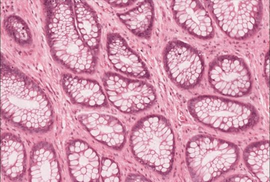
The field of drug development is experiencing significant breakthroughs, thanks to innovative techniques like digital pathology and in situ hybridization. These technologies are reshaping how researchers conduct studies, analyze data, and develop new therapies. Here's an in-depth look at how in situ hybridization and digital pathology services are revolutionizing pathology services for drug research and enhancing drug development research services.
In Situ Hybridization: Precision in Genetic Analysis
In situ hybridization (ISH) is a powerful technique used to detect and localize specific nucleic acid sequences within tissue sections. This method provides precise spatial information about gene expression, making it invaluable for understanding disease mechanisms and identifying potential therapeutic targets. By using labeled probes that bind to specific DNA or RNA sequences, ISH allows researchers to visualize the presence and distribution of these sequences in the context of the tissue's histology.
In drug development, ISH is used to validate biomarkers, study gene expression patterns, and assess the molecular effects of new therapies. The technique’s ability to provide detailed insights into the genetic landscape of tissues aids in the identification of disease pathways and the development of targeted treatments. By combining ISH with other molecular techniques, researchers can gain a comprehensive understanding of disease biology, leading to more effective drug discovery and development.
Digital Pathology Services: Enhancing Research Efficiency
Digital pathology services are revolutionizing the way pathology is conducted, offering a myriad of benefits for drug research. By converting traditional glass slides into high-resolution digital images, digital pathology allows for easier storage, sharing, and analysis of pathology data. This technology enables pathologists and researchers to view and analyze images remotely, facilitating collaboration and speeding up the diagnostic process.
In drug research, digital pathology is crucial for managing large volumes of pathology data, ensuring consistency and accuracy in data interpretation. Advanced image analysis software can automate the quantification of histological features, reducing human error and increasing efficiency. This level of precision and efficiency is essential for large-scale drug studies, where rapid and accurate data analysis can significantly impact the pace of research and development.
Pathology Services for Drug Research: Integrating Innovation
Pathology services for drug research are becoming increasingly sophisticated, integrating technologies like digital pathology and ISH to provide comprehensive solutions for drug development. These services include tissue sample analysis, biomarker validation, and the assessment of drug effects on tissues at the molecular level. By leveraging cutting-edge techniques, pathology services can offer more detailed and accurate insights into the efficacy and safety of new drugs.
Innovative pathology services support every stage of drug development, from early discovery through preclinical studies and clinical trials. They provide the critical data needed to understand how drugs interact with biological systems, helping researchers refine and optimize therapeutic candidates. This integration of advanced pathology techniques into drug research workflows accelerates the development of new therapies, ultimately bringing effective treatments to patients faster.
Drug Development Research Services: Driving New Therapies
Drug development research services encompass a wide range of activities designed to support the discovery and development of new therapies. These services include target identification, lead optimization, preclinical testing, and clinical trial support. By utilizing advanced techniques like digital pathology and ISH, research services can offer more precise and efficient solutions for drug development challenges.
High-quality drug development research services provide the expertise and resources needed to navigate the complex landscape of drug discovery. They help identify promising drug candidates, evaluate their potential, and ensure their safety and efficacy through rigorous testing. By partnering with specialized research services, pharmaceutical companies can enhance their drug development processes, reduce time-to-market, and increase the likelihood of successful outcomes.
Digital pathology and in situ hybridization are transforming the landscape of drug development, offering powerful tools for genetic analysis and data management. These technologies enhance the precision, efficiency, and collaboration essential for modern drug research. By integrating these advanced techniques into pathology services and drug development research, the pharmaceutical industry can achieve significant advancements in discovering and developing new therapies, ultimately improving patient care and outcomes.
1 note
·
View note
Text

Glint Lab provides expert histopathology and digital pathology services tailored to small and midsize biopharma companies engaged in research and preclinical phases of drug discovery. Our state-of-the-art facilities and experienced team ensure precise, reliable results for your pathology needs. From tissue processing and staining to advanced image analysis, we offer comprehensive solutions to accelerate your research.
0 notes
Text
The Crucial Role of Histochemical and Immunohistochemical Staining in Biomedical Research
Histochemical and immunohistochemical staining are indispensable techniques in biomedical research and diagnostic pathology. These methods enable scientists and clinicians to visualize and analyze the complex structures and functions within tissues, contributing significantly to our understanding of various diseases and conditions. This article delves into the importance of histochemical staining, immunohistochemical stains, and immunohistochemistry services, highlighting their applications and benefits.
Histochemical Staining
Histochemical staining involves the use of chemical reactions to identify and visualize specific components within tissue sections. This technique is essential for studying the structure, composition, and function of tissues at the microscopic level.
Histochemical stains can highlight various cellular components, such as carbohydrates, lipids, proteins, and nucleic acids. For instance, Hematoxylin and Eosin (H&E) staining is a widely used histochemical technique that provides detailed information about tissue morphology. Hematoxylin stains cell nuclei blue, while eosin stains the cytoplasm and extracellular matrix pink, creating a clear contrast that helps in identifying cellular structures and abnormalities.
Immunohistochemical Stains
Immunohistochemical (IHC) staining uses antibodies to detect specific antigens in tissue sections. This method combines histological and immunological techniques, making it possible to pinpoint the presence and location of proteins, pathogens, and other molecules within tissues.
The primary advantage of IHC staining is its specificity. By using antibodies that bind to particular antigens, researchers can identify and visualize the distribution and abundance of these targets within the tissue. This is especially valuable in cancer research, where IHC can detect tumor markers, helping to classify different types of cancer and guide treatment decisions.
Immunohistochemistry Staining
Immunohistochemistry (IHC) staining is a broader term that encompasses various techniques and applications of using antibodies for tissue analysis. IHC staining is crucial in both research and clinical settings, offering insights into the molecular mechanisms underlying diseases and aiding in the diagnosis and prognosis of various conditions.
IHC staining involves several steps: tissue preparation, antigen retrieval, antibody incubation, and visualization. Each step requires meticulous attention to detail to ensure accurate and reliable results. The choice of antibodies, detection systems, and visualization techniques can vary depending on the specific research or diagnostic question.
Immunohistochemistry Services
Professional immunohistochemistry services provide comprehensive support for researchers and clinicians who require high-quality IHC staining. These services often include antibody selection, tissue processing, staining, and analysis, ensuring that every aspect of the IHC process is expertly managed.
In San Diego, numerous laboratories and biotech companies offer specialized immunohistochemistry services, leveraging advanced technologies and expertise to deliver precise and reproducible results. These services are invaluable for researchers studying complex diseases, drug development, and biomarker discovery.
Histochemical and immunohistochemical staining are pivotal techniques in the field of biomedical research and pathology. They provide critical insights into the structure and function of tissues, enabling the detection and analysis of specific molecules within complex biological systems. With the availability of advanced immunohistochemistry services, researchers and clinicians can access high-quality staining and analysis, driving forward our understanding of diseases and improving diagnostic and therapeutic strategies. Whether through detailed histochemical staining or highly specific immunohistochemical stains, these techniques continue to be at the forefront of scientific discovery and medical innovation.
0 notes
Text

Glint Lab provides expert histopathology and digital pathology services tailored to small and midsize biopharma companies engaged in research and preclinical phases of drug discovery. Our state-of-the-art facilities and experienced team ensure precise, reliable results for your pathology needs. From tissue processing and staining to advanced image analysis, we offer comprehensive solutions to accelerate your research.
0 notes
Text
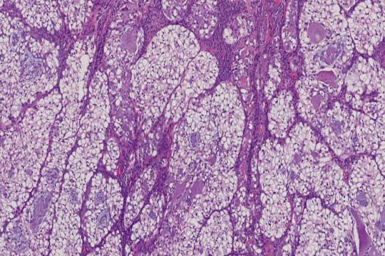
Exploring Advanced Techniques in Drug Discovery and Research
In the realm of drug discovery and biomedical research, advanced techniques and specialized services are crucial for obtaining accurate and reliable data. Keywords like Non GLP Toxicology, Spatial Biology FFPE, Drug Discovery Histology, and Animal Tissue Histopathology represent cutting-edge methodologies that drive innovation and improve outcomes in research. This article delves into these key areas, highlighting their significance and applications in modern scientific investigations.
Non GLP Toxicology: Flexibility in Early-Stage Research
Non GLP Toxicology refers to toxicology studies conducted without the stringent compliance to Good Laboratory Practice (GLP) regulations. These studies are typically performed during the early stages of drug development to quickly assess the safety profile of new compounds. Non GLP toxicology offers greater flexibility and faster turnaround times, allowing researchers to make informed decisions about which compounds to advance into more rigorous GLP-compliant testing. This approach is cost-effective and efficient, providing valuable insights into the potential toxic effects of new drug candidates.
Spatial Biology FFPE: Unlocking Cellular Interactions
Spatial Biology FFPE (Formalin-Fixed, Paraffin-Embedded) is a technique that allows researchers to analyze the spatial organization of cells and molecules within preserved tissue samples. By applying spatial biology methods to FFPE samples, scientists can investigate the complex interactions and heterogeneity within tissues at a high resolution. This is particularly useful in oncology, where understanding the tumor microenvironment is critical for developing targeted therapies. Spatial biology FFPE combines the robustness of traditional histopathology with advanced imaging and molecular profiling techniques, providing a comprehensive view of cellular dynamics.
Drug Discovery Histology: Visualizing Cellular Changes
Drug Discovery Histology involves the microscopic examination of tissues to study the effects of new drug candidates. Histological analysis is essential for understanding the mechanisms of action, efficacy, and safety of potential therapies. By staining and examining tissue sections, researchers can identify pathological changes, assess tissue damage, and evaluate therapeutic effects. This detailed visualization of cellular changes helps in the identification of promising drug candidates and the optimization of treatment regimens. Drug discovery histology is a cornerstone of preclinical research, providing critical data to guide further development.
Animal Tissue Histopathology: Insights from Preclinical Models
Animal Tissue Histopathology plays a vital role in preclinical research by providing insights into the effects of drugs on various organs and tissues in animal models. Histopathological analysis of animal tissues helps researchers understand the safety and efficacy of new therapies before they are tested in humans. This process involves detailed examination of tissue samples to detect any adverse effects, tissue damage, or pathological changes induced by the drug. Animal tissue histopathology is crucial for ensuring that potential therapies are safe and effective, thereby reducing the risk of failure in clinical trials.
Integrating Advanced Techniques in Research
Integrating techniques such as non GLP toxicology, spatial biology FFPE, drug discovery histology, and animal tissue histopathology into research workflows enhances the quality and depth of scientific investigations. These methodologies provide comprehensive data that supports the development of safer and more effective therapies. By leveraging advanced histological and toxicological analyses, researchers can make more informed decisions, accelerate drug development, and ultimately improve patient outcomes.
The fields of Non GLP Toxicology, Spatial Biology FFPE, Drug Discovery Histology, and Animal Tissue Histopathology represent essential components of modern biomedical research. Each technique offers unique advantages and insights that contribute to the overall success of drug discovery and development. By embracing these advanced methodologies, researchers can enhance their understanding of disease mechanisms, improve the safety and efficacy of new therapies, and drive innovation in the pursuit of better healthcare solutions.
Explore the potential of these advanced research techniques and discover how they can elevate your scientific investigations. Embrace the future of drug discovery with cutting-edge methodologies that provide deeper insights and more reliable data.
0 notes
Text

Located in San Diego, Glint Lab is a leading Contract Research Organization (CRO) specializing in histopathology and Digital Pathology services tailored for small to midsize biopharmaceutical companies. Beyond traditional lab services, we are a dedicated team of experienced histologists and scientists committed to advancing preclinical and early drug discovery.
#digital pathology services#Pathology services for drug research#Drug development research services#Pathology CRO San Diego
1 note
·
View note