
A blog for medical microscopy!Unless otherwise indicated, all images are hematoxylin - eosin stained.
Don't wanna be here? Send us removal request.
Statistics
We looked inside some of the posts by picturesofhistology and here's what we found interesting.
Average Info
Notes Per Post
218
Likes Per Post
151
Reblog Per Post
62
Reply Per Post
5
Time Between Posts
7 days
Number of Posts By Type
Text
17
Last Seen Tumblr Blogs


Fun Fact
In 2020, 44% of users from Denmark used Tumblr daily.
Text

Something rare nowadays in pathology: a whole mount slide! We only really ever do them for prostate and for eye globes nowadays. It’s expensive and difficult to cut, but the end effect is of one full slice of the organ. Super cool! This is prostate, with all of the prostatic carcinoma outlined in blue dots for volume measurement purposes. The small slit in the center is the urethra.
3 notes
·
View notes
Text


An amazing find in an ovary - this egg is actually undergoing meiosis! You can see the individual separated chromosomes if you look carefully.
4 notes
·
View notes
Text

Teratoma! Here we have cartilage, salivary gland, sebaceous glands, and ciliated respiratory epithelium all in one field of view.
5 notes
·
View notes
Text

Products of conception are always sad to look at; fetal autopsies absolutely wrecked me in residency. But this fetal kidney is so perfectly kidney shaped that I had to share. You can see the few glomeruli that have started to form, with the rim of dark, immature cells around the outside that are brewing up more. And below, the actual slide with green dots around the little kidney for a sense of scale.

#pathology#histology#products of conception#fetal tissue#abortion trigger warning#kidney#fetal kidney
7 notes
·
View notes
Text

Chondroblastic osteosarcoma
8 notes
·
View notes
Text

The craziest, biggest mitotic figure I’ve ever seen in my life
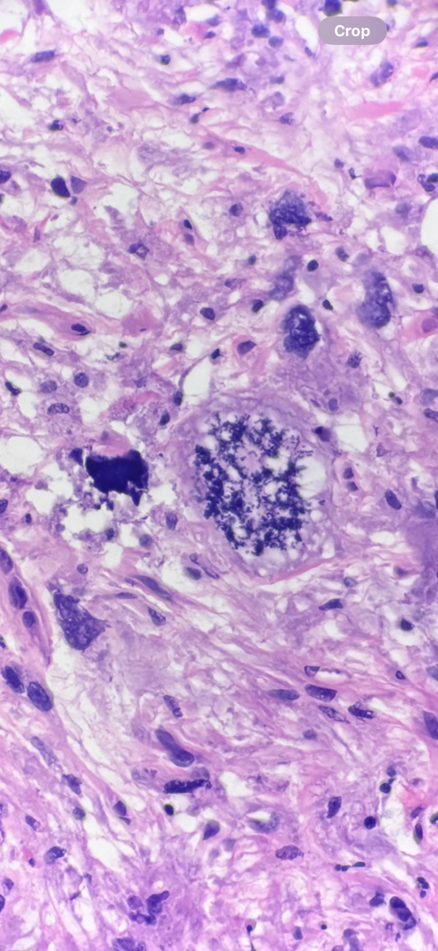
#histology#pathology#sarcoma#undifferentiated pleomorphic sarcoma#parapharyngeal mass after radiation
4 notes
·
View notes
Text


An example of perineural invasion - this is from the goblet cell carcinoma as well, but zoomed in on an area where the cancer has invaded and is tracking along a nerve.
Perineural invasion is often one way that tumors, especially squamous cell carcinomas and prostate cancers, can travel locally (not a way to metastasize). It’s one way in which cancer may evade being entirely removed by surgery, because the tracking is only visible microscopically.
0 notes
Text


Goblet cell tumor from likely appendiceal primary, found in the small bowel
The mucicarmine stain is so pretty and highlights mucin in pink!
For comparison, a normal area of the small bowel:


1 note
·
View note
Text


Molluscum contagiosum, caused by a pox virus
1 note
·
View note
Text

Metastatic colorectal carcinoma in a lymph node
2 notes
·
View notes
Text


Mucormycosis, a fungal infection - you can see the branching at bizarre angles and the lack of septation, both of which are characteristic of Mucor specifically
PAS stain for fungus pops them out in purple so prettily!
Picture credit to one of the residents in my program, who sent them to me
3 notes
·
View notes
Text

A pacinian corpuscle, the organ used to sense pressure in the skin! Found this one in a breast specimen.
2 notes
·
View notes
Text


Juvenile hamartomatous polyp from the GI tract - note the treelike branching architecture, classic for this entity!
4 notes
·
View notes
Text

Image credit to my friend Ryan: a promyelocyte from a patient with promyelocytic leukemia - they classically have butterfly shaped nuclei. This one was packing some junk in its trunk!
1 note
·
View note
Text

Image credit to Catherine from my program! Immature/fetal ovarian tissue, with all of those big cells being eggs.
6 notes
·
View notes
Text

High grade breast cancer with mitotic figures literally EVERYWHERE
49 notes
·
View notes