#C-Kit Mutation Detection Kit
Explore tagged Tumblr posts
Visit Tumblr Blog
Explore Tumblr blogs with no restrictions, modern design and the best experience.
Last Seen Tumblr Blogs


Fun Fact
Tumblr has 16.74 million mobile monthly users in the US.
Text
Enhance Molecular Diagnostics with Advanced C-Kit and CMV Detection Kits
In molecular diagnostics, precision and reliability define the value of any testing solution. Laboratories across the globe are shifting towards highly specialized tools that offer targeted mutation detection and accurate quantification of viral loads. Among these tools, the C-Kit PCR Kit, C-Kit Mutation Detection Kit, and CMV Detection Kit are setting new benchmarks for diagnostic accuracy and…

View On WordPress
0 notes
Text
🔬 GIST Mutation Detection Kit Market: Trends & Insights for 2025 and Beyond
The global GIST (Gastrointestinal Stromal Tumor) Mutation Detection Kit Market is steadily advancing in the precision medicine space.
📈 Market Snapshot:
2024: Valued at $0.39 billion
2025 Forecast: Set to rise to $0.41 billion
2033 Outlook: Expected to reach $0.64 billion
CAGR (2025–2033): 📊 5.8%
✨ These kits are essential for identifying key mutations in PDGFRA and C-KIT, aiding in targeted therapies and research development.
🔍 Segment Insights:
By Type: PDGFRA & C-KIT
By Application:
Medical
Research
Others
🌍 Key Market Players:
U.S.: EntroGen, Bio-Rad, Thermo Fisher Scientific, Medaysis
China: AmoyDx, MCLAB, Beyotime Biotechnology, Genetron Health, Jiangsu MicroDiag, HELIXGEN
Europe: LCM Genect (Italy), NimaGen (Netherlands), ASBACH (Germany), Diogene (Switzerland)
Asia-Pacific: 3B BlackBio Biotech India, Panagene (South Korea)
Canada: Geneseeq
🔗 As demand for molecular diagnostics grows, expect increased innovation, accessibility, and global collaboration.
#Biotech#MolecularDiagnostics#GIST#HealthcareInnovation#Genomics#PrecisionMedicine#MarketTrends#TumblrScience
0 notes
Text
From DNA to Diagnosis: What Happens Inside a Genetic Testing Lab?
Genetic testing may seem like a black box from the outside — you provide a sample, and weeks later, receive a detailed report filled with technical information. But behind the scenes, there’s a meticulous, high-precision process that transforms your DNA into actionable medical insights. Let’s take a closer look at what actually happens inside a modern genetic testing laboratory.

Step 1: Sample Collection and Preparation
It all begins with a simple biological sample — typically blood, saliva, or tissue. Once the sample reaches the lab, it is labeled, logged, and stored under controlled conditions to maintain integrity.
The first laboratory step is DNA extraction, where technicians isolate DNA from the sample using specialized chemical processes or kits. The result is a purified form of DNA ready for further analysis.
Step 2: DNA Quantification and Quality Check
Before testing begins, the extracted DNA is evaluated for quality and quantity. Using tools like spectrophotometers and fluorometers, technicians assess whether the sample has sufficient, high-quality DNA. Only high-integrity samples proceed to the next stage.
Step 3: Genetic Analysis — Sequencing or Targeted Testing
Depending on the clinical question, labs perform different types of genetic analysis:
Targeted Mutation Testing: Looks for known mutations (e.g., BRCA1/BRCA2 for breast cancer)
Panel Testing: Analyzes multiple genes linked to a specific disease (e.g., cardiomyopathy panel).
Whole Exome Sequencing (WES) or Whole Genome Sequencing (WGS): Offers a broad look across the entire coding region or full genome to detect rare or unknown genetic variants.
This is usually performed using Next Generation Sequencing (NGS) — a highly sensitive, high-throughput technology that reads millions of DNA fragments simultaneously.
Step 4: Bioinformatics — Interpreting the Code
The sequencing process generates massive amounts of raw data — essentially long strings of A’s, T’s, G’s, and C’s. Bioinformatics specialists use sophisticated software and algorithms to:
Align DNA sequences to a reference genome
Identify genetic variations
Filter out harmless or common variants
Highlight potentially disease-causing mutations
This digital layer is crucial in turning raw DNA sequences into meaningful insights.
Step 5: Clinical Interpretation and Reporting
Once potentially significant variants are identified, clinical geneticists or molecular pathologists interpret them in the context of medical literature, databases, and the patient’s clinical history. They determine whether a mutation is:
Pathogenic (disease-causing)
Likely pathogenic
Variant of uncertain significance (VUS)
Benign or likely benign
A final report is then generated — clear, clinically relevant, and actionable — and shared with the ordering physician or genetic counselor.
Step 6: Quality Control and Accreditation
Throughout every step, quality control is paramount. Labs like Greenarray Genomics Research and Solutions Pvt. Ltd. follow stringent protocols and use certified equipment to ensure accuracy, reproducibility, and compliance with regulatory standards.
Under the guidance of Dr. Sanjay Gupte, our lab in Pune combines advanced instrumentation with expert oversight to maintain the highest standards in genetic diagnostics.
The Journey of Discovery
From a simple swab to sophisticated sequencing, genetic testing is a fusion of biology, technology, and clinical science. Each step — from DNA extraction to diagnosis — plays a vital role in uncovering the genetic factors behind health and disease.
At Greenarray Genomics, we don’t just test genes — we decode possibilities, empower clinicians, and help patients take informed steps toward better health.
0 notes
Text
🔬🧬 Inside the Genome: GIST Mutation Detection Kit Market Trends 🧬🔬
The GIST Mutation Detection Kit Market is on the rise — driven by rapid advancements in medical diagnostics, cancer research, and the growing demand for targeted therapies. Whether in labs, hospitals, or biotech startups, these kits are shaping how we understand and treat gastrointestinal stromal tumors (GISTs).
🧪 What’s Fueling the Market Boom?
Increased awareness of PDGFRA & C-KIT mutations 🧬
Rising demand for personalized medicine in oncology
Accelerated R&D in molecular diagnostics
Global focus on early cancer detection
🌍 Global Scope, Local Impact: 📍 Regions to Watch:
North America (leading in clinical adoption)
Europe (expanding R&D investment)
Asia-Pacific (rapid healthcare infrastructure growth)
RoW (untapped potential & emerging access)
⚗️ Segment Snapshot: Types:
PDGFRA & C-KIT Detection Kits Applications:
Medical Diagnostics 🏥
Scientific Research 🔬
Others (academic, pharma, etc.)
📊 Market Outlook Includes: ✔️ Growth forecasts through 2033 ✔️ Revenue breakdown by segment & region ✔️ Strategic moves by key players ✔️ Distribution & pricing patterns ✔️ Competitive landscape analysis
📌 Curious about the details? 👉 Click here for the full report + sample
#BiotechTumblr#MolecularDiagnostics#PrecisionMedicine#GISTDetection#Genomics#CancerResearch#HealthcareTrends#BioInnovation#GISTMutation#TumorMarkers#MedTech#OncologyInsights#ClinicalDiagnostics
0 notes
Text
GIST Mutation Detection Kit Market 2031; Region Wise Analysis of Top Players in by its Types and Application
The global "GIST Mutation Detection Kit Market Market" report indicates a consistent and robust growth trend in recent times, projecting a positive trajectory expected to persist until 2031. A significant trend observed in the GIST Mutation Detection Kit Market market is the rising consumer inclination towards environmentally sustainable and eco-friendly products. Furthermore, a notable advancement in this market is the increasing incorporation of technology to elevate both product quality and efficiency. Cutting-edge technologies like artificial intelligence, machine learning, and block chain are actively employed to develop innovative products that outperform traditional options in terms of effectiveness and efficiency. The GIST Mutation Detection Kit Market Market Research Report for 2024 highlights emerging trends, growth opportunities, and potential scenarios envisioned up to the year 2031.
By delving into the latest trends, the report keeps businesses abreast of the dynamic market environment, helping them identify emerging opportunities and navigate potential challenges. The meticulous analysis covers various aspects, offering valuable insights into the historical performance of the market and presenting the current (CAGR) status.
Get a Sample Copy of the Report at: https://www.proficientmarketinsights.com/enquiry/request-sample-pdf/1838
Who are the biggest GIST Mutation Detection Kit Market manufacturers worldwide?
EntroGen (U.S.)
Bio-Rad (U.S.)
AmoyDx (China)
MCLAB (China)
Thermo Fisher Scientific (U.S.)
LCM Genect (Italy)
NimaGen (Netherlands)
Beyotime Biotechnology (China)
Genetron Health (China)
Jiangsu MicroDiag Biomedicine Technology (China)
HELIXGEN (Guangzhou) (China)
Geneseeq (Canada)
ASBACH MEDICAL PRODUCTS GMBH (AMP) (Germany)
Diogene (Swizerland)
3B BlackBio Biotech India (India)
Panagene (South Korea)
Medaysis (U.S.)
The GIST Mutation Detection Kit Market Market is described briefly as follows:
The global GIST Mutation Detection Kit Market Size was USD 409.4 million in 2024 and the market is projected to touch USD 574.2 million by 2031, exhibiting a CAGR of 5.80% during the forecast period.
SWOT Analysis of GIST Mutation Detection Kit Market Market:
A SWOT analysis involves evaluating the strengths, weaknesses, opportunities, and threats of a particular market or business. In the case of the keyword market, we'll be looking at the factors that can impact the industry's performance.
Pestle Analysis of GIST Mutation Detection Kit Market Market:
To better comprehend the market environment, a five-force analysis is performed, which takes into account the bargaining power of the customer, the supplier, the threat of substitutes, the threat of new entrants, and the threat of competition.
Get a Sample Copy of GIST Mutation Detection Kit Market Market Report
What are the Types in GIST Mutation Detection Kit Market Market?
PDGFRA
C-KIT
What are Applications in GIST Mutation Detection Kit Market Market?
Medical
Research
Others
Inquire or Share Your Questions If Any before the Purchasing This Report: https://www.proficientmarketinsights.com/enquiry/queries/1838
Geographical Segmentation:
Geographically, this report is segmented into several key regions, with sales, revenue, market share, and GIST Mutation Detection Kit Market market growth rate in these regions, from 2017 to 2028, covering
North America (United States, Canada and Mexico)
Europe (Germany, UK, France, Italy, Russia and Turkey etc.)
Asia-Pacific (China, Japan, Korea, India, Australia, Indonesia, Thailand, Philippines, Malaysia, and Vietnam)
South America (Brazil etc.)
Middle East and Africa (Egypt and GCC Countries)
Some of the key questions answered in this report:
Who are the worldwide key Players of the GIST Mutation Detection Kit Market Industry?
How the opposition goes in what was in store connected with GIST Mutation Detection Kit Market?
Which is the most driving country in the GIST Mutation Detection Kit Market industry?
What are the GIST Mutation Detection Kit Market market valuable open doors and dangers looked by the manufactures in the worldwide GIST Mutation Detection Kit Market Industry?
Which application/end-client or item type might look for gradual development possibilities? What is the portion of the overall industry of each kind and application?
What centered approach and imperatives are holding the GIST Mutation Detection Kit Market market?
What are the various deals, promoting, and dissemination diverts in the worldwide business?
What are the key market patterns influencing the development of the GIST Mutation Detection Kit Market market?
Financial effect on the GIST Mutation Detection Kit Market business and improvement pattern of the GIST Mutation Detection Kit Market business?
Purchase this Report (Price 3200 USD for A Single-User License) at: https://www.proficientmarketinsights.com/purchase/1838
TOC of Global GIST Mutation Detection Kit Market Market Research Report 2023
1 GIST Mutation Detection Kit Market Market Overview
2 Market Competition by Manufacturers
3 GIST Mutation Detection Kit Market Production by Region
4 GIST Mutation Detection Kit Market Consumption by Region
5 Segment by Type
6 Segment by Application
7 Key Companies Profiled
8 Industry Chain and Sales Channels Analysis
9 GIST Mutation Detection Kit Market Market Dynamics
10 Research Finding and Conclusion
11 Methodology and Data Source
0 notes
Text
Genomics Market Expected To Witness High Growth Driven By Advancements In Technology And Rising Demand For Personalized Medicine

A) Market Overview:
Genomics is the study of genes and their functions, with a focus on understanding the structure, function, evolution, mapping, and editing of genomes. It plays a crucial role in personalized medicine, drug discovery, and the development of precision therapies. The market mainly consists of products such as instruments & consumables, services, and software used in genomic research and diagnostics.
The global Genomics Market is estimated to be valued at US$ 46.2 Bn in 2023 and is expected to exhibit a CAGR of 12.4% over the forecast period 2023-2028, as highlighted in a new report published by Coherent Market Insights.
B) Market Dynamics:
The global Genomics Market is driven by two major factors. Firstly, advancements in technology have revolutionized genomics research and diagnostics. The development of Next-Generation Sequencing (NGS) technologies and its applications in various genomic studies have significantly contributed to market growth. NGS enables faster and cost-effective sequencing of DNA, RNA, and proteins, thereby facilitating a deeper understanding of diseases at a molecular level.
Secondly, there is a rising demand for personalized medicine, which utilizes genomic information to tailor medical treatments to individual patients. The increasing prevalence of chronic diseases such as cancer and genetic disorders has created a need for more targeted therapies. Genomic testing enables the identification of specific genetic mutations associated with these diseases, allowing for personalized treatment plans.
C) Market Key Trends:
One key trend observed in the Genomics Market is the growing adoption of direct-to-consumer genetic testing. Companies such as 23andMe provide consumers with the opportunity to access their genetic information through at-home testing kits. This trend has gained popularity due to increased consumer awareness and curiosity about their DNA and ancestry.
For example, 23andMe offers ancestry reports, health predisposition reports, and carrier status reports, providing individuals with insights into their genetic background and potential health risks. This trend is expected to continue, as more people are interested in exploring their genetic information for various purposes.
D) SWOT Analysis:
Strength:
1. Advancements in technology - Continued advancements in genomic technologies such as NGS drive the market's growth.
2. Increasing demand for personalized medicine - The rising prevalence of chronic diseases and genetic disorders fuels the adoption of genomic approaches in clinical decision-making.
Weakness:
1. High costs associated with genomic testing - The cost of genomic tests and sequencing technologies may limit their accessibility, hindering market growth.
2. Regulatory challenges - Stringent regulations regarding the use and sharing of genetic data pose challenges for the market expansion.
Opportunity:
1. Growing applications in non-invasive prenatal testing - Genomics has the potential to revolutionize prenatal care by enabling non-invasive detection of fetal genetic abnormalities.
2. Expanding use of genomics in agriculture - The agricultural sector can benefit from genomics-based technologies for crop improvement and disease resistance.
Threats:
1. Ethical and privacy concerns - The collection and utilization of genetic data raise ethical questions and privacy concerns among individuals and regulatory bodies.
2. Lack of awareness and limited infrastructure - Limited awareness about genomics and inadequate infrastructure in certain regions can hinder market growth.
E) Key Takeaways:
- The global genomics market is expected to witness high growth, exhibiting a CAGR of 12.4% over the forecast period, driven by advancements in technology and rising demand for personalized medicine.
- North America is currently the largest market for genomics, owing to the presence of well-established healthcare infrastructure, increased funding for genomics research, and a growing consumer base for direct-to-consumer genetic testing.
- Key players operating in the global genomics market include Thermo Fisher Scientific, Agilent Technologies, Bio-Rad Laboratories, Illumina, Pacific Biosciences, Oxford Nanopore Technologies, 23andMe, Foundation Medicine, Hoffmann-La Roche Ltd., and Myriad Genetics Inc. These players focus on product development, partnerships, and acquisitions to maintain their market position and enhance their product offerings.
#Genomics Market#Coherent Market Insights#Genomics Market Demand#Genomics Market Growth#Genomics Market Sale#Genomics Market Trends#\Genomics Market Analysis
0 notes
Text
Germ Cell Tumors - Causes, Symptoms, and Treatment Options - Dr. Mark Cabelin's Firewa ll
Germ cell tumors are a type of neoplasm that primarily originate from germ cells, which give rise to eggs in females and sperm in males. They can occur anywhere in the body but are most commonly found in the ovaries, testicles, and areas where germ cells usually don't reside, such as the chest, abdomen, or brain. Although germ cell tumors are relatively rare, they represent a significant proportion of cancers in adolescents and young adults.
Despite their potential severity, advancements in urology and oncology have led to effective treatment options, and the prognosis for patients with germ cell tumors has improved significantly over the past few decades. The key to successful treatment often lies in early detection and a comprehensive understanding of the tumor's nature and behavior,

Germ cell tumors arise from germ cells, unique cells involved in reproduction. They are the precursors to both egg and sperm cells. Unusual growth of these cells can lead to tumors, which can be benign (non-cancerous) or malignant (cancerous). Malignant germ cell tumors can be life-threatening if not treated promptly and effectively, Dr. Mark Cabelin.
Germ cell tumors are broadly categorized into two types: seminomas and non-seminomas. Seminomas are usually slower-growing and more responsive to radiation therapy. Non-seminomas are more aggressive and comprise several subtypes, including embryonal carcinoma, yolk sac carcinoma, choriocarcinoma, and teratoma. Each of these subtypes has its unique characteristics and requires specific treatment strategies.
In the field of urology, germ cell tumors, specifically testicular germ cell tumors, are of paramount importance. They constitute the majority of testicular cancers, making their understanding crucial for urologists. These tumors often present unique diagnostic and therapeutic challenges, necessitating a multidisciplinary approach involving urologists, oncologists, radiologists, and pathologists.
Their management extends beyond surgical intervention, encompassing chemotherapy, radiation therapy, and constant surveillance. Understanding germ cell tumors is thus critical for comprehensive patient care in urology. Understanding the causes of germ cell tumors is crucial for prevention and treatment.
While the exact cause is unknown, several factors are believed to contribute to their occurrence. Specific genetic abnormalities have been linked to the development of germ cell tumors. For instance, individuals with disorders of sex development (DSDs), who have unusual stories of sexual anatomy, are at a higher risk.
Additionally, carrying specific mutations in genes, such as the c-KIT gene, has been associated with an increased incidence of these tumors. Exposure to certain environmental factors may also increase the risk of developing germ cell tumors. These include exposure to chemicals, radiation, or certain drugs during pregnancy. Smoking and excessive alcohol use have also been implicated.
Research is ongoing to understand the complex interplay of genetic predisposition and environmental exposure in the development of germ cell tumors. The symptoms of germ cell tumors can vary greatly depending on their location and size. The most common sign of testicular germ cell tumors is a painless lump or swelling in the testes.
Other symptoms may include discomfort or pain in the testicle or scrotum, a feeling of heaviness in the scrotum, or a sudden collection of fluid in the scrotum. In females, ovarian germ cell tumors may cause abdominal pain or swelling. When germ cell tumors occur in other areas of the body, symptoms might include chest pain, cough, shortness of breath, or neurological symptoms if the cancer is in the brain.
It’s important to remember that these symptoms can also be caused by conditions other than germ cell tumors, so it’s essential to consult a healthcare professional for an accurate diagnosis. If left untreated, germ cell tumors can grow and spread to other body parts, leading to severe complications. Potential risks include metastasis, where the cancer spreads to other organs, which can significantly complicate treatment and worsen the prognosis, Dr. Mark Cabelin Urologist.
Germ cell tumors can also cause functional disturbances in the affected organ, such as infertility in men if the cancer is in the testes. In addition, treatments for germ cell tumors, like surgery, chemotherapy, and radiation therapy, can also have side effects and long-term health impacts. Regular follow-ups are necessary to monitor for potential complications and manage them promptly.
Diagnosing germ cell tumors usually involves a combination of physical examination, medical history review, and diagnostic tests. The first step often involves a thorough physical examination where the physician may check for lumps or abnormal growth. A detailed discussion about the patient's health history can also provide valuable insights, as certain genetic diseases or previous cancer treatments can increase the risk of germ cell tumors.
Imaging tests play a vital role in diagnosing and staging germ cell tumors. These can include ultrasound, often the first test for suspected testicular or ovarian tumors. It can determine whether a lump is solid (likely cancer) or filled with fluid (probably a cyst).
Computed Tomography (CT) scans, and Magnetic Resonance Imaging (MRI) can help detect tumors in other body parts and determine if cancer has spread. Positron Emission Tomography (PET) scans may also detect metastasis or evaluate the response to treatment. Pathological tests are essential for confirming the diagnosis and understanding the type and stage of the germ cell tumor.
These tests often involve a biopsy, where a tissue sample is removed from the tumor and examined under a microscope. A biopsy can reveal the presence of cancer cells and provide insights into the tumor's aggressiveness. Other pathological tests, such as serum tumor marker tests, measure the levels of certain substances in the blood that may increase when a germ cell tumor is present.
Pathological diagnosis is crucial in urology for the management of germ cell tumors. Treating germ cell tumors is typically a multidisciplinary effort involving urologists, oncologists, and other healthcare professionals.
Treatment decisions are based on factors such as the type and stage of the tumor, the patient's overall health, and personal preferences. Surgery is often the first line of treatment for germ cell tumors. The goal is to remove the cancer entirely.
In males, this may involve a radical inguinal orchiectomy, the surgical removal of the entire testicle. In females, surgery may include the removal of one or both ovaries. In some cases, lymph nodes in the abdomen may also need to be removed.
Radiation therapy uses high-energy rays to kill cancer cells. It's often used in conjunction with surgery, either before to shrink the tumor or after to kill any remaining cancer cells. Radiation therapy can also treat germ cell tumors that have spread to other body parts.
Chemotherapy is a systemic therapy that uses drugs to kill cancer cells throughout the body. It's often used when advanced germ cell tumors have spread to other body parts. Several chemotherapy regimens are used to treat germ cell tumors, and the choice of regimen depends on factors such as the type and stage of the tumor and the patient's overall health. Research is ongoing to develop more effective and less toxic treatments for germ cell tumors.
These emerging treatments include targeted therapies that aim to attack specific characteristics of cancer cells and immunotherapies that help the body's immune system fight cancer. Clinical trials are exploring the potential of these new treatments, and patients with germ cell tumors may have the opportunity to participate in these trials. Germ cell tumors, although rare, can have significant implications on a patient's health and quality of life.
Early detection and prompt treatment are crucial to improving the prognosis and reducing the risk of complications. Diagnosis involves comprehensive physical examinations, imaging tests, and pathological tests. Treatment is typically multidisciplinary, involving surgery, radiation therapy, and chemotherapy, often in combination.
Interestingly, the field of urology continues to explore new and innovative treatment methods, including targeted therapies and immunotherapies. Regular follow-ups, monitoring, and patient education are also integral to managing this condition effectively. Remember, each patient's journey is unique, and treatment should be personalized based on their specific circumstances and preferences.
Click Here-
https://www.linkedin.com/in/mark-cabelin-a544158
youtube
0 notes
Text
Genes Affecting Progenitor/Stem Cell Function
In the yolk sac, hematopoiesis occurs multifocally. Such distribution of blood islands suggests that hematopoiesis may be initiated through numerous progenitors. These progenitors, of low proliferative potential, contribute to the relatively small pool of circulating embryonic erythrocytes. However in the adult, abundant hematopoiesis is the result of a few self-renewing definitive hematopoietic progenitors (85) with high proliferative potential, as shown by comparisons of yolk sac, fetal liver, and bone marrow in vitro colonies (17). Thus, gene mutations differentially affecting yolk sac and fetal liver hematopoiesis may be involved in proliferative/clonogenic potential of hematopoietic progenitors and should be readily apparent. Alternatively, such genes may be involved in the generation, maintenance, or self-renewal of definitive progenitors/stem cells. Defects in these characteristics of progenitor/stem cells are more difficult to detect since in vivo, single hematopoietic progenitors/stem cells can generate a complete fully differentiated adult blood system (8). Examples of some candidate genes leading to such proposed defects are found in mice mutant for the transcription factors GATA-2 (86), core binding factor (CBF) (87-90), GATA-3 (91,92), myb (93,94) and PD. 1 (95-97), and for the receptor tyrosine kinase c-kit (69,98,99) and its ligand steel factor (SCF) cck8 formula. More subtle examples are found in mice lacking growth factors interleukin-6 (IL-6) (100), or leukemia inhibitory factor (LIF) (101), or the receptor tyrosine kinase Flk-21F1t-3 (102). Brief descriptions of the effects of each mutation are given.
1 note
·
View note
Text
Autoimmune Disease Diagnostics Market Worth $7.53 Billion by 2029
According to a new market research report titled, ‘Autoimmune Disease Diagnostics Market by Product (Reagents, Kits, Assays, Instruments), Test Type (ANA, CRP, ESR, Rheumatoid Factor, Routine Test), Disease Type (Systemic, Localized), End User (Hospital, Diagnostic Lab, Research) – Global Forecast to 2029,’ published by Meticulous Research®, the autoimmune disease diagnostics market is expected to reach $7.53 billion by 2029, at a CAGR of 6.5% from 2022 to 2029.
Download Free Report Sample Now @ https://www.meticulousresearch.com/download-sample-report/cp_id=5405
Autoimmune diseases occur when the immune system attacks healthy cells and tissues. The trigger to these attacks can be genetic mutations, microorganisms, viruses, drugs, and environmental factors. The early diagnosis of these autoimmune diseases is necessary for an effective course of treatment or management. Autoimmune diseases can be systemic, affecting multiple organs or systems, or specific, affecting a single organ. Several tests such as western blot, antinuclear antibody, C-reactive protein (CRP), erythrocyte sedimentation rate (ESR), rheumatoid factor, immunoassays, routine blood tests, immunohistochemistry (IHC), and flow cytometry are used for diagnosing autoimmune diseases.
Increasing Prevalence of Autoimmune Diseases Driving the Growth of Autoimmune Disease Diagnostics market
There are more than 80 autoimmune diseases in which the immune system of a patient attacks its own body. The tests for autoimmune prove crucial for the early detection of these diseases. The early diagnosis of autoimmune diseases is also crucial to reduce the financial burden. For instance, according to the American Autoimmune Related Disease Association, around 50 million people in the U.S. suffer from autoimmune diseases and incur USD 86 billion per year in managing their conditions. Autoimmune disease testing is expected to increase due to the increasing prevalence of these diseases. For instance, according to the Lancet Diabetes & Endocrinology, in 2021, around 8.4 million people were affected with type-1 diabetes (one of the most common autoimmune diseases) globally. This number is estimated to increase to 13.5–17.4 million people by 2040. Additionally, around 4% of the global population suffers from one or more autoimmune diseases.
Speak to our Analysts to Understand the Impact of COVID-19 on Your Business: https://www.meticulousresearch.com/speak-to-analyst/cp_id=5405
The autoimmune disease diagnostics market is segmented based on Product Category (Reagents, Assays, & Kits and Instruments), Test Type (Antinuclear Antibodies (ANA), Western Blot, Immunoassays, C-reactive Protein (CRP), Erythrocyte Sedimentation Rate (ESR)), Rheumatoid Factor, Immunoglobulins, Routine Blood Tests, Immunohistochemistry (IHC), Flow Cytometry, and Other Test Types), Disease Type (Systemic Autoimmune Diseases and Organ-specific Autoimmune Diseases), End Users (Hospitals & Clinics, Diagnostics Laboratories, Academic & Research Institutes), and Geography (North America, Europe, Asia-Pacific, Latin America, and Middle East & Africa). The study also evaluates industry competitors and analyzes the market at the global and regional levels.
Based on the product category, the reagents, kits, and assays segment is slated to register the higher CAGR during the forecast period. The frequent use of reagents for diagnosing diseases and growing awareness and testing capabilities are the major factors driving the growth of this segment.
Based on test type, the antinuclear antibody test segment is slated to register the fastest growth rate during the forecast period. The antibody responsible for autoimmune diseases are formed in the blood much earlier than the onset of the actual disease in an individual; this enables the early diagnosis of the disease, which helps to reduce morbidity and mortality due to the disease. The antinuclear antibodies test is simple and has higher accuracy than other tests. Therefore, the increasing prevalence of autoimmune diseases, the need for early disease diagnosis, and the ease of use and low turnaround time of ANA test drive the segment’s growth.
Quick Buy – Autoimmune Disease Diagnostics Market- Global Opportunity Analysis And Industry Forecast (2022-2029), Research Report: https://www.meticulousresearch.com/Checkout/84306372
Based on disease type, the systemic segment is slated to register the highest CAGR during the forecast period. Systemic autoimmune diseases affect multiple organs or systems. The rising prevalence of systemic autoimmune diseases has led companies operating in the autoimmune disease diagnostics market to launch tests for diagnosing systemic autoimmune diseases. For instance, in June 2022, Thermo Fisher Scientific Inc. (U.S.) launched EliA RNA Pol III and EliA Rib-P tests cleared by the U.S. FDA. These tests are used to diagnose Systemic Sclerosis (SSS) and Systemic Lupus Erythematosus (SLE). Thus, the increasing number of companies launching tests for systemic autoimmune diseases drives this segment’s growth.
Based on end user, in 2022, the hospitals & clinics segment is expected to account for the largest share of the market. The large market share of this segment is attributed to the high adoption rate of testing technologies by hospitals and the rising expansion of existing testing capabilities to provide cost- and time-efficient tests.
Based on geography, in 2022, North America is expected to account for the largest share of the autoimmune disease diagnostics market, followed by Europe and Asia-Pacific. North America’s major market share is attributed to factors such as high healthcare expenditure, high prevalence of autoimmune diseases, increased awareness among patients regarding autoimmune diseases, and increased funding and investments towards R&D of autoimmune diseases.
The report includes an extensive assessment of product portfolios, geography, and key growth strategies adopted by leading market participants between 2020 and 2022.
Some of the key players operating in the autoimmune disease diagnostics market are Thermo Fisher Scientific Inc. (U.S.), Bio-Rad Laboratories, Inc. (U.S.), Beckman Coulter, Inc. (U.S.), Abbott Laboratories (U.S.), Myriad Genetics, Inc. (U.S.), EUROIMMUN Medizinische Labordiagnostika AG (Germany), F. Hoffmann-La Roche Ltd (Switzerland), Siemens Healthineers AG (Germany), Grifols, S.A. (Spain), and Creative Diagnostics (U.S.).
To gain more insights into the market with a detailed table of content and figures, click here: https://www.meticulousresearch.com/product/autoimmune-disease-diagnostics-market-5405
Scope of the Report
Autoimmune Disease Diagnostics Market, by Product Type
Reagents, Assays, and Kits
Instruments
Autoimmune Disease Diagnostics market, by Test Type
Antinuclear Antibodies (ANA) Test
Rheumatoid Factor Test
Immunoglobulins Test
Routine Blood Tests
C-reactive Protein (CRP) Test
Erythrocyte Sedimentation Rate (ESR) Test
Other Test Types
Note: Other test types include Anti-double stranded DNA, Extractable nuclear antigen, Anti-signal recognition particle tests, Ferritin, and Anti-cyclic Citrullinated Peptide (Anti-CCP) Antibodies.
Autoimmune Disease Diagnostics Market, by Disease Type
Systemic Autoimmune Diseases
Organ-specific Autoimmune Diseases
Autoimmune Disease Diagnostics market, by End User
Hospitals & Clinics
Diagnostic Laboratories
Academic & Research Institutes
Autoimmune Disease Diagnostics Market, by Geography
North America
U.S.
Canada
Europe
Germany
France
U.K.
Italy
Spain
Rest of Europe (RoE)
Asia-Pacific (APAC)
Japan
China
India
Rest of APAC (RoAPAC)
Latin America (LATAM)
Brazil
Mexico
Rest of Latin America (RoLATAM)
Middle East & Africa
Download Free Report Sample Now @ https://www.meticulousresearch.com/download-sample-report/cp_id=5405
0 notes
Text
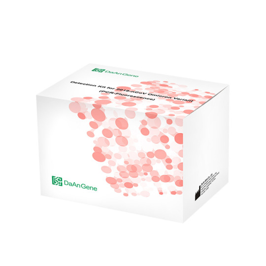
nCoV Omicron Variant Detection Kit (PCR-Fluorescence)
This omicron variant detection kit is used for the in vitro qualitative detection of S gene mutations K417N, E484A, and P681H in 2019-nCoV Omicron Variant testing in the oropharyngeal swabs, nasal swabs, and saliva specimens from suspected pneumonia cases infected by novel coronavirus, clustered cases and others needing test for omicron variant. Features of COVID-19 Omicron Variant Detection Kit Targets: 3 mutation sites & 1 conserved gene Endogenous internal control UDG enzyme to control contamination in PCR Specification of COVID-19 Omicron Variant Detection Kit
Target S gene mutations K417N, E484A, and P681H Specimen Type Oropharyngeal swabs, nasal swabs and saliva Storage and shelf life Stored at -20±5°C, and the shelf life is 12 months Certificate CE Test Principle Real-time fluorescent PCR Applicable Instruments ABI 7500, Bio-Rad CFX96
Order Information of COVID-19 Omicron Variant Detection Kit Description Cat. No. Package 2019-nCoV Omicron Variant Detection Kit (PCR-Fluorescence) DA1220 Large package, 24 tests/kit 2019-nCoV Omicron Variant Detection Kit (PCR-Fluorescence) DA1221 Large package, 48 tests/kit
0 notes
Text
Enhance Molecular Diagnostics with Advanced C-Kit and CMV Detection Kits

In molecular diagnostics, precision and reliability define the value of any testing solution. Laboratories across the globe are shifting towards highly specialized tools that offer targeted mutation detection and accurate quantification of viral loads. Among these tools, the C-Kit PCR Kit, C-Kit Mutation Detection Kit, and CMV Detection Kit are setting new benchmarks for diagnostic accuracy and clinical relevance.
The C-Kit PCR Kit is designed for researchers and clinicians working in oncology, particularly those focusing on gastrointestinal stromal tumors (GISTs), mast cell diseases, and other malignancies where C-Kit mutations play a central role. This kit enables precise detection of mutations in the C-Kit gene, especially in exon 9 and exon 11, which are known to affect treatment decisions significantly. By using real-time PCR technology, the C-Kit PCR Kit ensures both sensitivity and specificity, critical for early diagnosis and personalized therapy plans.
The C-Kit Mutation Detection Kit holds closely aligned applications and significance in the diagnostic landscape. This diagnostic tool supports the detection of clinically significant mutations that impact patient prognosis and therapeutic strategies. Unlike conventional sequencing methods, this kit provides a faster turnaround, allowing healthcare providers to initiate targeted treatments without delay. The C-Kit Mutation Detection Kit is especially beneficial in settings where quick, yet accurate, results are essential for life-saving decisions.
For clinical laboratories that deal with transplant patients and immunocompromised individuals, monitoring viral infections like cytomegalovirus (CMV) is a high priority. The CMV Detection Kit offers a robust solution for quantifying CMV DNA in clinical samples, aiding in timely diagnosis and monitoring of infection progression or treatment efficacy. Utilizes real-time PCR, kit delivers high results with excellent sensitivity and reproducibility. The CMV Detection Kit is particularly valuable in managing patients at risk of CMV reactivation, ensuring that clinicians can act promptly and effectively.
One of the key advantages of the C-Kit PCR Kit and C-Kit Mutation Detection Kit is their user-friendly protocol, which reduces hands-on time and minimizes the risk of contamination. These kits are optimized for use with various sample types, providing flexibility for different clinical scenarios. The integration of internal controls in these kits ensures the reliability of every test run, boosting confidence in the results obtained.
Similarly, the CMV Detection Kit includes rigorous internal quality controls to ensure the accuracy of every test. Its ability to detect even low levels of viral DNA makes it indispensable in critical care settings. The kit’s compatibility with widely used PCR instruments further enhances its adaptability in diverse laboratory environments.
Incorporating these diagnostic kits into routine clinical workflows can significantly improve disease detection, risk stratification, and treatment monitoring. The streamlined processes and precise analytics offered by the C-Kit Mutation Detection Kit and C-Kit PCR Kit contribute to more informed clinical decision-making. Meanwhile, the CMV Detection Kit plays a crucial role in virology labs, safeguarding patient outcomes through early and accurate viral monitoring.
All three kits are products of extensive research and development aimed at simplifying complex diagnostic challenges. Their performance is backed by scientific validation and real-world usage, making them reliable choices for institutions that demand accuracy and efficiency.
To explore more about the C-Kit PCR Kit and C-Kit Mutation Detection Kit, visit 3B BlackBio’s Oncology Solutions. For detailed information on the CMV Detection Kit, refer to their virology diagnostics section at CMV Quantification Kit.
By choosing advanced molecular tools like these, laboratories and clinics can ensure they are delivering the highest standards of diagnostic care—quickly, accurately, and with maximum clinical relevance.
0 notes
Text
Covid-19 Variants - Our Proactive Health Plans
No question - this has been a year from...
However, with every miserable thing that happens in life - is an opportunity to learn.
I will always ask (gently and diplomatically!) someone with a chronic illness, "how did you know?" Looking back, were there any signs, that maybe we sloughed off as stress, age, or something else?
How do these covid variants happen?
Let's first have a crash course on virology. Viruses are probably the best example of our concept of " to infinity and beyond". They are the most abundant form of "life" on earth, and probably elsewhere! Exponential growth is an understatement. Literally, trillions to the power of another few trillion.
A virus has a shell, capsid, which protects its viral material, or genome, safe. Some particularly resilient viruses such as Hep C, coronavirus, herpes, HIV have an extra protective coating referred to as envelopes.
Viral envelopes hate soap, soap is their kryptonite, which is why washing with soap is paramount to destroy the virus.
Every time the coronavirus transmits between people, it picks up microscopic changes to its genetic code. The result is coronavirus variants, Researchers are observing patterns as to how the virus is mutating. Covid-19 seems to be the lead track star of viruses - redefining how fast a virus can mutate. The bottom line is, we do not know how many variants exist now, or, potentially how many there could be in the future.
Remaining vigilant about preventing transmission is no less vital now - it is even more important now to help halt the potential for mutations.
Hand washing with soap, masks, distancing, covid-19 testing, abiding by lockdowns... are all vital to harnessing this virus from further mutations. The vaccine is NOT a get out of jail for free card!!!
Hopefully, our vaccines will be able to keep up with these mutations. We know that every year (less actually) our flu vaccines need to be updated. The coronavirus vaccines will demand constant updates - more than any software we have ever complained about updating!
Evolution in action... survival of the fittest: In most viruses and disease-causing bacteria, the use of treatments and vaccines challenges them to evolve ways of escaping them so they can continue to thrive... These viruses certainly appear to have an evolutionary advantage.
What can we do to proactively strengthen us against these multiple coronaviruses?
Yes, we ALL must abide by the Lyophilized SARS-COV-2 detection kit rules to prevent transmission. But how can we strengthen ourselves, that if we come into contact with these viruses, we minimize our risk of a deadly reaction?
I have many articles on here about the role of chronic inflammation in every one of our chronic illnesses from heart disease, diabetes, vascular disease, arthritis, depression, cancers... Chronic inflammation is the ultimate underlying cause of a compromised ability for our body to shake off any nasties that hit us. AKA - defences are down.
In patients with a severe reaction to the virus, the lungs are damaged, and patients need supplemental oxygen. The risk of mortality these cases is over 40%.
March 2021 Journal of Immunity: Donna Farber, PhD, professor of microbiology & immunology, Chief of Surgical Sciences Columbia University: "Even though individuals are getting vaccinated, severe COVID-19 remains a significant risk for certain individuals and we need to find ways to treat people who develop severe disease."
"People refer to patients experiencing a cytokine storm in the blood, but what we're seeing in the lungs is on another level. The immune cells in the lung went into overdrive releasing these cytokines."
Very complicated science. Bottom line is - lowering our levels of chronic inflammation, is probably our best defense against a drastic reaction to the virus. This explains why, people who already have underlying chronic conditions are more vulnerable.
Please read the many articles I have here on chronic inflammation, and how we can reduce our risk, to not just covid, but all of our chronic diseases. The research continues to support this necessity. My goal in chronic care management, is to bring reducing chronic inflammation, to the very top of our health "to-do" list. All of our other "healthy lifestyle" habits, fall under the umbrella of reducing inflammation. However, please be careful you are not "paving the road to hell with good intentions". In other words, get the true science on lowering inflammation, and not the pop science.
In Health.
see more : https://www.biotephy.com/
1 note
·
View note
Text
Lupine Publishers | Effects of Kav001 (A Kavain Analogue) on LPS-Induced Cytokines and Neutrophil Infiltration Lupine Publishers | LOJ Pharmacology & Clinical Research

Abstract
Background: TNF-α, a cytokine, is known to be involved in LPS-induced inflammation. Our recent data indicate that treatment of Kavain in mouse or human cells significantly suppresses TNF-α production in response to LPS. In order to completely identify one Kavain analogue (Kav001) reported in our previous data, we further analyzed its biological function in this paper.
Methods and Results: Commercial Kavain or Kav001 as inhibitor was used. ELISA was performed for detection of TNF-α, IL-1β, IL-6, IL-4, or Caspase 1 after treatment of THP-1 cells with Kav001. Wild type mice (C57BL/6) were used for neutrophil infiltration assay. A kinase/protein array was performed to analyze if Kav001 affects any kinases/protein in response to LPS. Conclusions: It is clear that Kav001 not only inhibits TNF-α, but also IL-1β, IL-6, caspase 1 and neutrophil infiltration in response to LPS compared to control.
Introduction
Kavain is known as a treatment for certain diseases [1-3]. Amar’s group recently reported that, in vivo, Kavain mediated alveolar bone loss and inflammatory infiltrate in a mnouse model [4]. Kavain is found to affect TNF-α production through regulation of NF-kB [5] and to be inhibited by ERK2 dephosphorylation in vitro or in vivo [6]. Our previous data indicate that Kavain inhibits P. gingivalis/LPS-induced inflammation/inflammatory disease [7,8]. Recently, we found that Kavain grants small amounts of toxicity to mammalian cells, most likely due to its insoluble chemical structure with hydrophobicity. To address this issue, we designed and synthesized compounds with the basic chemical structure of Kavain with a different number of hydrophilic bonds. One soluble compound (named Kav001) from the Kavain analogue was selected and further confirmed to have a stronger biological function than Kavain by in vitro/in vivo experiments such as CAIA mouse models or endotoxic shock [9]. In this paper, we further found that Kav001 not only inhibits TNF-α, but also IL-1β, IL-6, caspase 1 and neutrophil infiltration. However, this phenomenon cannot be observed when macrophages are treated with LPS plus Kavain. We believe that Kav001 may mediate a novel link between Kav001 and LPS-induced inflammation and may be used as a key inhibitor to LPS-induced inflammation/inflammatory disease.
Materials and Methods
Animals and cells
Wild type mice (8-10 week-old C57BL/6, Jackson Labs) were maintained under strict specific pathogen-free (SPF) conditions. All protocols were approved by the Boston University Institutional Animal Care and Use Committee and were performed in compliance with the relevant animal care and use laws and institutional guidelines. THP-1 cells (TIB-202, ATCC) were cultured in a RPMI- 1640 media (Cat#: 11875–093, Life Technologies, NY) with 10% FBS at 37 °C in a 5% CO2 atmosphere.
Compounds
Commercial Kavain (Cat#59780, Sigma‐Aldrich Co., Natick, MA) and custom synthesized analogue Kav001 (Lot#KB180125, KareBay Biochem, Inc.) were obtained. The analysis of each compound shows that ≥95% purity. The stock concentration of each compound was prepared with 10μg/μl. The compound was diluted to 100μg/ml in 25% DMSO for Kavain or in water for Kav001 just before the experiment.
Elisa
The treated media from THP-1 cells were subjected to ELISA for TNF-α detection with the kit (Cat#: ab181421, Abcam), IL-1β detection with the kit (Cat#: ab100562, Abcam), IL-6 detection with the kit (Cat#: KHC0061, Life technologies), IL-4 detection with the kit (Cat#: BMS225-2, Thermo Fisher), or Caspase 1 (Cat#: ab219633, Abcam). ELISA immunoreactivity was measured by a reader (Model 680, Bio-Rad). The data were graphed.
Neutrophil Infiltration Assay
Mice (n=3) were untreated or treated by i.p. injection of E.coli LPS (1mg/kg body weight) and/or oral gavage of Kav001 (30mg/ kg) for 24hrs. Skin tissues from each group were taken and fixed in 4% paraformaldehyde, then embedded in O.C.T. compound [10,11]. Skin sections (5μm) were made and stained with H&E (hematoxylin and eosin). Results were analyzed and imaged.
Protein Array
1 × 10⁵ THP-1 cells were treated with 100μg/ml of E. coli LPS (Sigma) or plus Kav001 (0.1mg/ml) for 16 hrs at 37 °C. The medium from each test was centrifuged at 1,500 × g and then treated with a specific kit for human protein cytokine array (RayBiotech, Norcross, GA) based on the manufacturer’s instructions. The data were analyzed using a Verrado Imaging System (Bio-Rad) and graphed.
Statistical Analysis
All tests were done in triplicate and statistical analyses were done with the SAS software package. All data were normally distributed. For multiple mean comparisons, we conducted ANOVA analysis by the Student’s t-test for single mean comparison. P values less than 0.05 were considered significant.
Results
In order to examine whether Kav001 also inhibits other cytokines besides TNF-α, we made a relevant experiment via ELISA. As shown in Figure 1A-1C, co-treatment of LPS plus Kav001 (A-C, #4) significantly reduces LPS-induced TNF-α, IL-1β, or IL-6 compared to the positive controls (A-C, #2). This phenomenon for detection of TNF-α was also observed while co-treatment of LPS plus Kavain but was not detectable for IL-1β or IL-6 production (1B&C, #3) As shown in Figure 1D,1B. Additionally, neither Kavain nor Kav001 was found to affect IL-4. Because our preliminary data indicate that Kav001 affects some cytokines in response to LPS, we were therefore interested in whether Kav001 is involved in other stimuli-mediated cytokines. As shown in Figure 1E & 1G, Kav001 strongly reduced all three stimuli-induced cytokine productions, TNF-α (E, No.4), IL-1β (F, No.4), and IL-6 (G, No.4) compared to their positive control (No.2 of E, F, or G). This phenomenon for reduction of IL-1β (F, No.3) or IL-6 (G, No.3) was not observed with co-treatment of stimuli plus Kavain. Since it is known that caspase 1 expression is activated with TNF-α, IL-1 beta, or IL-6 [12-14], we wanted to further test whether Kav001 inhibits caspase 1 caused by TNF-α, IL-1 beta, or IL-6. As shown in Figure 1F & 1H, Kav001 (H, No.6) reduced LPS-induced caspase 1 compared to the positive control (H, No.2).
As a control, Kavain did not reduce LPS-induced caspase 1, but instead increased its value (H, No.5). In order to analyze whether the treatment of Kav001 affects any kinases/protein upstream of caspase 1 in response to LPS, a kinase/protein array was performed. As shown in Figure 2B & 2C Kav001 significantly reduced LPSinduced kinases such as Akt (Protein kinase B) at 17%, ATM (Ataxia telangiectasia mutated) at 13%, caspase 3 at 11.5%, caspase 7 at 8.6%, CHK1 (Checkpoint kinase 1) at 26.4%, CHK2 (Checkpoint kinase 2) at 15.3%, and ERK1/2 at 12.7%/14.8%, but does not affect p27, p53, or TAK1 (Transforming growth factor beta-activated kinase 1 (Figure 2A & 2D). As it is known that subcutaneous injection of LPS in mice induces neutrophil infiltration of tissues [15,16], we were therefore interested in whether Kav001 treatment is able to suppress LPS-induced neutrophil infiltration. As shown in Figure 3A & 3D, indeed, subcutaneous treatment of 30mg/kg of Kav001 significantly reduced LPS-induced Neutrophil infiltration of tissues in mouse (DI,II, III) compared to the positive control (CI,II, III).
Figure 2:Kinase/Protein array. (A) The stamped identification number on the array. (B) THP-1 cells were untreated or treated with LPS alone or LPS+ Kav001 over night. The conditioned medium from each test group was assessed by protein assay and quantified by a Versace Imaging System. (C) Analysis of the intensity on the array filter. The cell intensity treated by LPS was assigned to 100% as base value and compared to the others. There ratio was calculated in this table.
Discussion
Kavain was shown to affect TNF-α transcriptional regulation [6- 8]. Compared with Kavain, Kav001 not only inhibits LPS-induced TNF-α, , but also IL-1β, IL-6, caspase 1 or Neutrophil infiltration. DMSO is known to be widely used as reagent solvent but sometimes causes a little toxicity to mammalian cells [17]. It is known that Kavain is dissolved in the higher concentration of DMSO (≥25%) while Kav001 is completely dissolved in water. This water-soluble property may be the reason why Kav001 prevent DMSO-mediated toxicity to cells with a longer period of efficacy. Our most recent data support this potential hypothesis [9]. Additionally, neutrophils are the innate immune response and thus, neutrophilic infiltration of tissues is involved in many inflammatory diseases [18,19]. In order to inhibit the neutrophilic infiltration from tissue injury (I/R injury, brain injury, ethanol feeding-induced liver injury), several inhibitors such as MgIG (Magnesium isoglycyrrhizinate), Clenbuterol, Phillyrin, or PAI-1 (Plasminogen activator inhibitor-1) have been widely studied [20,21]. Researcher groups have also suggested that these inhibitors suppress infiltration of neutrophils via regulation of factors such as MyD88 or p38 MAPK [22,23]. However, these studies of certain inhibitors leave several questions, such as whether there is a common key point between their pathways. Our data here also show that the treatment of Kav001 significantly reduces LPS-induced neutrophilic infiltration. Thus, Kav001 may be used for suppression of neutrophil infiltration induced by other stimuli or injuries. Since TNF-α, IL-1β, or IL-6 was found here to be inhibited by Kav001, we assume that Kav001 may have treatment for inflammatory diseases caused by these cytokines (TNF-α, IL-1β, or IL-6). We will try to address this hypothesis for future research.
https://lupinepublishers.com/pharmacology-clinical-research-journal/pdf/LOJPCR.MS.ID.000122.pdf
https://lupinepublishers.com/pharmacology-clinical-research-journal/fulltext/effects-of-kav001-(a-kavain-analogue)-on-lps-induced-cytokines-and-neutrophil-infiltration.ID.000122.php
0 notes
Photo

MCQ of the day- 19 september
6. The drug which acts at A in the above image is
(A) Bedaquiniline
(B) Delamanid
(C) PAS
(D) Pyrazinamide
Answer: Option (B) is the correct answer
Reference: Guidelines for use of delamanid in the treatment of drug resistant TB 2018; Pg 2
Option (B): Delamanid is a nitroimidazooxazole compound which inhibits mycolic acid synthesis
Option (A): Bedaquiniline is a diarylquinoline which inhibits ATP synthase
Option (C): PAS – inhibits synthesis of DNA precursor.
Option (D): Pyrazinamide – disrupts plasma membrane
7. A 49yr old aquarium worker presented to the OPD with multiple nodules and few non healing ulcers in right forearm. He gives a history of cleaning aquarium tanks regularly. Biopsy from the nodule showed acid fast bacilli. The probable etiological agent is
(A) Mycobacterium leprae
(B) Nocardia
(C) Mycobacterium marinum
(D) Mycobacterium avium intercellulare complex.
Answer: Option (C) is the correct answer
Reference: Apurba Sastry Essentials of Medical Microbiology 2nd edition pg no: 294
Option (C): The occupational exposure is suggestive of M.marinum which causes fish tank granuloma or swimming pool granuloma. It presents as tender nodules, papules and ulcers
Option (A): Mycobacterium leprae causes leprosy with hypopigmented anaesthetic patches in early lesions
Option (B): Nocardia causes Pulmonary nocardiosis, Disseminated nocardiosis and actinomycetoma.
Option (D): MAC complex is usually associated with pulmonary lesion and lymphadenitis.
8. Match the above
(A) a- ii, b-v, c-i, d-iii
(B) a-v, b-iv, c-iii, d-i
(C) a- ii, b- iv, c-i, d-v
(D) a- iii, b-v, c-ii, d-i
Answer: Option (C) is the correct answer
Reference: Apurba Sastry Essentials of Medical Microbiology 2ndedition pg no: 295
Option (C): As per Runyons classification a) M.marinum – photochromogens; b) M.gordonae - scotochromogens; c) M.abscessus – rapid grower; d) M.ulcerans – Non photochromogens
9. Which of the following statement is true regarding GeneXpert used in TB diagnosis
(A) Can detect rifampicin resistance only
(B) Can detect isoniazid resistance only
(C) Can detect both rifampicin and isoniazid resistance
(D) does not detect drug resistance
Answer: Option (A) is the correct answer
Reference: Apurba Sastry Essentials of Medical Microbiology 2nd edition pg no: 288
Option (A): GeneXpert simultaneously detects: (i) MTB complex DNA and (ii)
rifampicin resistance (mutations of the rpoBgene). It uses five probes targeting various sequences of rpoBgene
10. This instrument is based on
(A) RT PCR
(B) Automated liquid culture system
(C) CBNAAT
(D) Line probe assay
Answer: Option (B) is the correct answer
Reference: Apurba Sastry Essentials of Medical Microbiology 2nd edition pg no: 288
Option (B): This is BACTEC MGIT(Mycobacteria growth indicator tube) which is an automated liquid culture system to diagnose TB
Option (A): Nested PCR targeting IS6110 gene was the most common molecular test used earlier. Other gene targets in PCR are MPT64 gene, 65 KDa and 38 KDa genes.
Option (C): Cartridge based PCR used in TB diagnosis is CBNAAT – Genexpert.
Option (D): Line probe assay involves probe-based detection of amplified DNA in the specimen. The commercial kits available to perform LPA are GenoType and INNO-LiPA
For daily updates follow us on:
Website: https://koncptnext.in/
Youtube: https://www.youtube.com/results?search_query=koncptnext
Twitter: https://twitter.com/KoncptNext
Instagram:https://www.instagram.com/koncptnext_exit/
Linked in:https://www.linkedin.com/in/koncpt-next-8257661b3/
Facebook: https://www.facebook.com/koncptnextexit
0 notes
Text
Quantification of MicroRNAs for the Diagnostic Screening of Colon Cancer in Human Stool by Absolute Digital(d)PCR*| Lupine Publishers

Abstract
There is currently no validated micro(mi)RNA diagnostic stool test to screen for colon cancer (CC) on the market because of the complexity of fecal density, vulnerability of stool to daily changes, and the presence of three sources of miRNAs in stool (cell-free from fecal homogenates, exsosomal miRNAs from fecal exosomes, and fecal colonocytes). By employing earlier on a microarray miRNA experiment, using Affymetrix GeneChip miRNA 2.0 Arrays, on immunocaptured and enriched stool colonocytes of 15 subjects [three healthy controls and twelve colon cancer patients [three TNM stage 0-1 (e.g., polyps 1 cm, villous or tubvillous, or with high grade dysplasia), three stage 2, three stage 3, and three stage 4] in triplicates, this allowed for selection of a smaller panel of 14 preferentially expressed mature miRNAs associated with colon cancer (12 Up-Regulated, miR-19a, miR-20a, miR-21, miR-31, miR-34a, miR-96, miR-106a, miR-133a, miR-135b, miR-206, miR-224 and miR-302; and 2 Down-Regulated, miR-143 and miR-145). Then carrying out an absolute quantitative digital PCR on these 15 stool samples from TNM stages 0-4 on total small RNA extracted by immunocapture, followed by RT that employed a Custom TaqMan® miRNA Reverse Transcription (RT) Kit and TaqMan RT Primer Pool, and absolute quantification of miRNAs, in copies/μl, measured using a chip-based Absolute QuantStudio 3D Digital PCR analysis, allowed for validating the microarray results. To ensure that human and not bacterial small total RNA was chosen, coextraction protocols with E. coli K1 strain RS18 was carried out, followed by comparing Agilent electrophoretic patterns with human and bacterial electrophoretic patterns, and also random samples were sequenced using mRNA/miRNA sequencing, to ensure that human and not bacterial mRNA was chosen.
Introduction
Quantitative dPCR miRNA data presented in herein, show that the quantitative changes in the expression of a few mature miRNA genes in stool, which are associated with right and left colon cancer, would provide for a more convenient, sensitive and specific diagnostic screening molecular markers, more useful than markers currently available on the market, such as the low-sensitivity (<15%) fecal occult blood test(FOBT); result in better compliance; and is more economical than the invasive and expensive colon cancer colonoscopy exam, resulting in a higher probability of curing that cancer, if detected at the early TNM stages, and which becomes incurable and deadly if not diagnosed before metastasis.
Advantages of Using a MiRNA Diagnostic Colon Cancer Screening Test
The expression of individual genes may be altered by mutations in the DNA, or by a change in their regulation at the RNA or protein levels [1]. Epigenetic silencing is an important mechanism that contributes to gene inactivation in colorectal cancer (CRC) [2]. Analysis of promoter methylation of hypermethylated in cancer 1 (HIC1) gene in human stool showed it to be highly specific (98%) for both colon adenoma and carcinoma [3], but the sensitivity was quite low (31% for adenoma & 42% for all cancer), suggesting that an epigenetic marker only is not adequate for an accurate diagnostic screening, but a combination of genetic and epigenetic markers would be required to reliably identify CRC at an early disease stage [4]. Working with the stable DNA has been relatively easy compared to working with the fragile RNA molecule [1].A study by scientists at Exact Sciences Corp, Marlborough, MA, which markets a mutation-based DNA test “Cologuard”, assessed a newer version of a fecal DNA test for CRC screening using a vimentin methylation marker and another mutation DY marker plus nondegraded DNA in a limited sample of 44 CRC patients and 122 normal controls [5].
It cited a sensitivity of 88% and a specificity of 82%, only for advanced cancer, but not for the early adenoma stage. Besides, DNA mutation tests are not cost-effective, as screening for multiple mutations is expensive because these demanding mutation tests are not automated and are labor intensive. In addition, mutation detection in oncogenes and suppressor genes suffers from: a) the detection of mutations in these genes in fewer than half of large adenomas and carcinomas, b) the detection of gene mutations in non-neoplastic tissues, c) mutations found only in a portion of the tumor, and d) mutations often produce changes in the expression of many other genes [6,7]. Protein-based methods are currently not suited for screening and early diagnosis, either because proteins are not specific to one tumor or tissue type (e.g., CEA), their susceptibility to proteases, current lack of means to amplify proteins, no function is known for more than 75% of predicted proteins of multicellular organisms, there is not always a direct correlation between protein abundance and activity, and most importantly because detection of these markers exfoliately often signifies the presence of an advanced tumor stage. The dynamic range of protein expression in minimally-invasive body fluids (e.g., blood) is as large as 1010. Moreover, mRNA levels do not necessarily correlate with protein expressions.
Protein microarray studies revealed that protein expression vastly exceeds RNA levels, and only post translationally modified proteins are involved in signal transduction pathways leading to tumorigenesis. There is no well-documented protein test that has been shown in clinical trials to be a sensitive and a specific indicator of colon neoplasia, especially in early stages [8]. A serum proteomic study employing liquid chromatography (LC)-mass spectrometry (MS) carried out in a non-biased fashion failed to differentiate between individuals with large adenoma ( 1 cm) and normal individuals [9]. Compared to nucleic acids, proteomic research is a newer discipline; therefore, it will take considerable time to identify and validate proteins suitable for use as clinical markers, and resolve issues of bias and validations [10].On the other hand, a transcriptomic mRNA approach, has been shown to detect both adenomas and colon carcinomas with high sensitivity and specificity in preliminary studies [1], but no randomized, standardized, blinded prospective clinical studies have been carried out to validate the superiority of the mRNA approach.
A study indicated that a combination of a transcriptomic mRNA and miRNA expression signatures improves biomolecular classification of CRC [11]. Furthermore, not only does miRNAs regulate mRNA, but they also regulate protein expression. Two studies have shown that a single miRNA act as a rheostat to fine tune the expression of hundreds of proteins [12,13]. Hence, for CRC screening, miRNA markers are much more comprehensive and preferable to a DNA-, epigenetic-, mRNA- or a protein-based marker [14-18]. An added advantage for the use of the stable, nondegradable miRNAs by PCR expression, by chip-based methods, is its being automatable, making them much more economical and more easily acceptable by laboratory personnel performing these assays [4]. The discovery of small non-coding protein sequences,17-27 nucleotides long RNAs (microRNAs), has opened new opportunities for developing a non-invasive screening test for early diagnosis of many cancers. The latest miRbase release 22 on, March 12, 2018 [http://ww.mirbase.org] indicates the total number of miRNAs labeled “high confidence” has increased by 168, to 1996, than in the previous release [19].
MiRNA functions seem regulate development [20], apoptosis [21], and specific miRNAs are essential in oncogenesis [22,23], effective in classifying solid [24-26] and liquid tumors [27,28], and could serve as oncogenes or suppressor genes [29]. MiRNA genes are frequently found at fragile sites, as well as minimal regions of loss of heterozygosity, or amplification of common break-point regions [30], implying their involvement in carcinogenesis. MiRNAs have potential to serve as biomarkers for cancer diagnosis, prognosis and/or response to therapy [31,32]. Profiles of miRNA expression differ between normal and tumor tissues (33,34), suggesting that their expression profiles cluster similar tumor types together more accurately than expression profiles of protein-coding mRNA genes [33,34]. A study that examined global expression of 735 miRNAs in 315 samples of normal colonic mucosa, tubulovillus adenomas, adenocarcinomas proficient in DNA mismatch repair (pMMR), and defective in DNA mismatch repair (dMMR) representing sporadic and inherited CRC stages I-IV suggest involvement of common biologic pathways in pMMR and dMMR tumors in spite of the presence of numerous molecular differences between them, including differences at the miRNA level; indicating the need to pay attention to mismatch DNA repair (MMR) [34] .
Unlike screening for large numbers of messenger (m)RNA [1], a modest number of miRNAs is used to differentiate cancer from normal [35], and unlike mRNA, miRNAs in stool remain largely intact and stable for detection [36], therefore, leading to conclude that miRNA molecules are better markers to use for developing a reliable noninvasive diagnostic marker screen for colon cancer [14-18], since: a) the presence of the bacterium Escherichia coli does not hinder detection of miRNA by a sensitive technique such as dPCR [36], and b) the miRNA expression patterns are the same in primary tumor, or diseased tissue, as in stool samples [35-37]. The gold standard to which the miRNA test is compared to, has been “colonoscopy”, obtained from patients’ medical records [38]. However, because the low sensitivity guaiac FOBT is still the most commonly used screen in annual checkups (www.cancer.org) [39], this test should also be included for comparison with the proposed dPCR diagnostic miRNA screening approach in human stool.
Advantages of Stool Over Other Testing Media
Stool testing has several advantages over other colon cancer screening methods a s it is truly noninvasive and requires no unpleasant cathartic preparation, formal health care visits, or time away from work or routine activities [40-43]. Unlike sigmoidoscopy, it reflects the full length of the colorectum and samples can be taken in a way that represents the right and left side of the colon. It is also believed that colonocytes are released continuously and abundantly into the fecal stream, contrary to blood that is released intermittently as in guaiac fecal occult blood test (FOBT) [39]; therefore, this natural enrichment phenomenon partially obviates the need to use a laboratory-enrichment technique to enrich for tumorigenic colonocytes, as for example when blood is used for screen testing [44]. Furthermore, because testing can be performed on mail-in-specimens, geographic access stool screening is essentially unimpeded [4]. The American Cancer Society (ACS) has recognized stool-based molecular testing as a promising screening technology for CRC (www.cancer.org).
Isolation of colonocytes from stool, and comparing the Agilent electrophoretic (18S and 28S) patterns to those obtained from total RNA extracted from whole stool [45-47], and differential lysis of colonocytes by RT lysis buffer (Qiagen), could be construed as a validation that the electrophoretic pattern observed in stool (18S and 28S) is truly due to the presence of human colonocytes, and not due to stool contamination with Escherichia coli (16S and 23S). Taking into account that some exsosomal RNA will be released from purified colonocytes into stool, attempts must be made to correct for exsosomal RNA effect [48].
MiRNA dPCR Study Design
To test miRNAs as reliable, quantitative, sensitive and specific diagnostic biomarkers, for early non-invasive screening of colon cancer, using absolute dPCR test, preliminary work must be validated in a study using a nested case control epidemiology design and employing a prospective specimen collection, retrospective blind evaluation (Probe) of control subjects and test colon cancer patients, as specifically delineated by the National Cancer Institute’s Early Detection Research Network http://edrn.nci.nih.gov for cancer biomarker discovery studies. Selection of 14 miRNAs, 12 of them showed increased expression and 2 showed decreased expression for analysis of absolute miRNAs expression by a chipbased digital (d) PCR test is presented in Tables 1 & 2, and Figure 1. Absolute Quantitative Digital PCR Approach Digital PCR is a new approach to miRNAs quantification that offers an alternate method to qPCR for absolute quantification, by partitioning a sample of DNA or cDNA into many individual, parallel PCR reactions; some of these reactions contain the target molecule (positive), while others do not (negative).
Figure 1: Diagram illustrating QuantStudioTM 3D Digital PCR System Chip; ChipCase Lid (1); Digital PCR 20K 10 mm2 nanofluidic v2 chip (2), which contains 20,000 reaction wells; QuantStudioTM 3D Digital PCR Chip Case (3); Chip ID (4); Fill port (5); and Reaction wells, the 20,000 physical holes that suspend individual PCR reactions.
Table 1: Characteristics of Fourteen Up- or Down-Regulated MicroRNAs in Human Stool.
Table 2: Absolute Quantification of Up-/Down- Regulated miRNAs in Stool by QuantStudioTM 3D Chip-Based Digital PCR.
A single molecule can be amplified a million-fold or more. During amplification, TaqMan chemistry with dye-labeled probes is used to detect sequence-specific targets. When no target sequence is present, no signal accumulates. Following PCR analysis, the fraction of negative reactions is used to generate an absolute count of the number of target molecules in the sample, without the need for standards or endogenous controls. In conventional qPCR, the signal from wild-type sequences dominates and obscures the signal from rare sequences [49-51]. By minimizing the effect of competition between targets, dPCR overcomes the difficulties inherent to amplifying rare sequences and allows for sensitive & precise absolute quantification of the selected miRNAs. Applied Biosystem QuantStudio™ 3D instrument used in this research study, only performs the imaging and primary analysis of the digital chips. The chips themselves must be cycled offline on a Dual Flat Block GeneAmp® 9700 PCR System. or the ProFlex™ 2x Flat PCR System. The QuantStudio™ 3D Digital PCR System can read the digital chip in less than 1 minute, following thermal cycling [45].
It allows for one sample per chip; although, duplexing allows for analsis of two targets per chip. Sample prep for digital PCR is no different than for real-time qPCR, when using the QuantStudio™ 3D Digital PCR System. To figure out the concentration of cDNA stock from results, if one includes all of the necessary dilution factors into the AnalysisSuite™ software, the software will give the copies/��L in the stock. There are 2 dilutions that one needs to take into account:
a) The first is the dilution of the sample in the reaction,. and
b) The second is the dilution of the stock that one makes before adding it to the digital PCR reaction.
For example, if one wants to add 1 μL of a sample that has been diluted 1:10 from the stock. Thus, if one adds 1 μL of his/her sample to a 16 μL (final volume) reaction, the dilution factor of the sample is 1:16 or 1/16 = 0.0625. Since the stock has also been diluted 1:10 (0.1), one also need to factor this in. The final dilution factor to enter into the software is 0.0625 x 0.1 = 0.00625 (1:160). One can use either annotation to indicate the dilution factor in the AnalysisSuite™ software. If one enters that value into the “Dilution” column, the software will give the copies/μL in the starting material (stock).
The Poisson Plus algorithm for projects that contain QuantStudio™ 3D Chips with target, quantities >2000 copies/μL. The Poisson Plus algorithm corrects for well-to-well load volume variation, on a per Chip basis. This becomes important at higher target concentrations. There is also an option to export the Chip data as XML on the Export tab-thousands of discrete subunits prior to amplification by PCR, each ideally containing either zero or one (or at most, a few) template molecules [50]. Each partition behaves as an individual PCR reactions –as with real-time PCR-fluorescent FAM probes [or others, as VIC fluorescence]. Samples containing amplified products are considered positive (1, fluorescent), and those without product –with little or no fluorescence (i.e., are negative, 0). The ratio of positives to negatives in each sample is the basis of amplification. Unlike real-time qPCR, dPCR does not rely on the number of amplification cycles to determine the initial amount of template nucleic acid in each sample, but it relies on Poisson Statistics to determine the absolute template quantity.
The unique sample partitioning step of dPCR, coupled with Poisson Statistics allows for higher precision than both traditional end point PCR. and qPCR methods; thereby allowing for analysis of rare miRNA targets quantitatively and accuratley [47,48]. The use of a nanofluidic chip, shown below, provides a convenient and straight forward mechanism to run thousands of PCR reactions in parallel. Each well is loaded with a mixture of sample, master mix, and Applied Biosystems TaqMan Assay reagents, and individually analyzed to detect the presence (positive) or absence (negative) of an endpoint signal. To account for wells that may have received more than one molecule of the target sequence, a correction factor is applied using the Poisson model. It features a filter set that is optimized for the FAM™, VIC®, and ROX™ dyes, available from Life Technologies [46]. The chips themselves must be cycled offline on a Dual Flat Block GeneAmp® 9700 PCR System. or the ProFlex™ 2x Flat PCR System. The QuantStudio™ 3D Digital PCR System can read the digital chip in less than 1 minute, following thermal cycling [45].
It allows for one sample per chip; although, duplexing allows for analsis of two targets per chip. Sample prep for digital PCR is no different than for real-time PCR, when using the Quant Studio™ 3D Digital PCR System. To figure out the concentration of cDNA stock from results, if one includes all of the necessary dilution factors into the AnalysisSuite™ software, the software will give the copies/sL in the stock. There are 2 dilutions that one needs to take into account:
a) The first is the dilution of the sample in the reaction,. and
b) The second is the dilution of the stock that one makes before adding it to the digital PCR reaction.
For example, if one wants to add 1 μL of a sample that has been diluted 1:10 from the stock. Thus, if one adds 1 μL of his/her sample to a 16 μL (final volume) reaction, the dilution factor of the sample is 1:16 or 1/16 = 0.0625. Since the stock has also been diluted 1:10 (0.1), one also need to factor this in. The final dilution factor to enter into the software is 0.0625 x 0.1 = 0.00625 (1:160). One can use either annotation to indicate the dilution factor in the AnalysisSuite™ software.
If one enters that value into the “Dilution” column, the software will give the copies/μL in the starting material (stock). The Poisson Plus algorithm for projects that contain Quant Studio™ 3D Chips with target, quantities >2000 copies/μL. The Poisson Plus algorithm corrects for well-to-well load volume variation, on a per Chip basis. This becomes important at higher target concentrations. There is also an option to export the Chip data as XML on the Export tab-thousands of discrete subunits prior to amplification by PCR, each ideally containing either zero or one (or at most, a few) template molecules [47]. Each partition behaves as an individual PCR reactions –as with real-time PCR—fluorescent FAM probes [or others, as VIC fluorescence]. Samples containing amplified products are considered positive (1, fluorescent), and those without product –with little or no fluorescence (i.e., are negative, 0). The ratio of positives to negatives in each sample is the basis of amplification. Unlike real-time qPCR, dPCR does not rely on the number of amplification cycles to determine the initial amount of template nucleic acid in each sample, but it relies on Poisson Statistics to determine the absolute template quantity.
The unique sample partitioning step of dPCR, coupled with Poisson Statistics allows for higher precision than both traditional and qPCR methods; thereby allowing for analysis of rare miRNA targets quantitatively and accuratley [47]. The use of a nanofluidic chip, shown below, provides a convenient and straight forward mechanism to run thousands of PCR reactions in parallel. Each well is loaded with a mixture of sample, master mix, and Applied Biosystems TaqMan Assay reagents, and individually analyzed to detect the presence (positive) or absence (negative) of an endpoint signal. To account for wells that may have received more than one molecule of the target sequence, a correction factor is applied using the Poisson model. It features a filter set that is optimized for the FAM™, VIC®, and ROX™ dyes, available from Life Technologies [46]. A workflow of the dPCR procedure by the QuantStudioTM 3D Digital PCR System is presented in (Figure 2) Workflow of a digital miRNAs PCR for colon cancer profiling in human colon tissue or stool samples.
Figure 2: Workflow of a digital miRNAs PCR for colon cancer profiling in human colon tissue or stool samples.
Digital PCR, however, has Several Tips to Follow:
i. A rough estimate of the concentration of miRNAs of interest has to be first carried out, in order to make appropriate dilutions, so that not too many partitions will get multiple copies that prevent accurate calculation of the copy number of miRNAs of interest;
ii. Non-template controls and a RT negative control must be set up for each miRNA, when using a “primer pool method” for retro-transcription;
iii. A chip-based dPCR method requires less pipetting steps, which reduces potential PCR contamination compared to another type of dPCR marketed by Bio-Rad Laboratories, thus called “Bio-Rad’s droplet digital PCR”, which requires multiple pipette transfers that potentially increase the risk of contamination [47], and
iv. Quant StudioTM 3D chip has 20,000 fixed reaction wells, whereas Bio-Rad’s droplet PCR relies upon the generation of droplets;
a step that could be extremely variable, as reported by Miotto et al (11,48) Absolute dPCR data tabulated in Tables 1 & 2, and presented graphically in Figure 1 below, which show 14 preferentially expressed mature miRNAs associated with colon cancer (12 Up-Regulated, miR-19a, miR-20a, miR-21, miR-31, miR- 34a, miR-96, miR-106a, miR-133a, miR-135b, miR-206, miR-224 and miR-302; and 2 Down-Regulated, miR-143 and miR-145) in stool samples from healthy controls, and stages 0-1 to 4 individuals with colon cancer . Standard deviations (sd) obtained from the one-way ANOVA, using the 5 level factor Type (normal, stage01, stage2, stage3, stage4) were calculated. The adjusted R-squared values representing the proportion of variation explained by Type are also reported. Type was statistically significant for every gene; all p-values were less than 0.000001 (no adjustments for multiple comparisons). These data are tabulated in Table 3 and shown graphically in Figure 1. For each gene on the graph in Figure 1, the min and max have been shown, in order to make the presentation clearer. At top left is high exxpression Value of 9985, which is the maximum value for that gene, at the bottom one finds the value for the minimum the colors range from dark blue (control) to orange (stage 4).
Table 3: Representation of SDs and R2 for miRNAs tested by absolute digital PCR.
The groups are also distinguished by line type: control (solid), stage 0-1 (long dash), stage 2 (dash), stage 3 (dot), stage 4 (dash nd dot). The figure is a parallel coordinate plot made in R, using the package MASS. For statistical analysis. Innovation and Clininical Significance of the dPCR-miRNA Diagnostic Stool Screening Approach Innovation lies in the collective use of many methods, such as: immunoparamagnetic beads to capture colonocytes from the harsh, noninvasive stool environment, whose extracted fragile total small RNA is stabilized shortly after stool excretion by commercial kits so it does not ever fragment, followed by standardized analytical quantitative miRNA dPCR-chip profiling in stool samples, which are neither labor intensive, nor require extensive sample preparation, to develop a panel of few stable miRNAs for absolute quantitative diagnostic screening of early sporadic colon cancer (stage 0-1), cheaper, with higher sensitivity and specificity than any other colon cancer screening test on the market . Isolation of colonocytes from stool samples is needed to provide a quantitative estimate of how our proposed miRNA method performs. Although we may miss exosome RNA, a parallel test could also be carried out on miRNAs obtained from stool samples to compare the extent of loss when colonocytes are only used, and an appropriate correction for exsosomal loss can be made [48]. Figure 3 Absolute Quantification of Up- or Down-Regulated miRNAs in Human Stool by Quant Studio TM 3D Digital PCR Chip System.
Figure 3: Absolute Quantification of Up- or Down-Regulated miRNAs in Human Stool by QuantStudioTM 3D Digital PCR Chip System.
Acknowledgments
We express our thanks to participating volunteers who provided stool samples for the study; Dr. Paul W. Vos of the Department of Biostatistics, East Carolina University for Statistical Analysis, and Dr. Clark D. Jeffries at Renaissance Computing Institute, University of North Carolina at Chapel Hill for his insight on bioinformatics’ analysis.
To know more about our Journal of Surgery click on https://lupinepublishers.com/surgery-case-studies-journal/
To know more about Lupine Publishers click on https://lupinepublishers.us/
To know more about Open access publishers click on Lupine Publishers
0 notes
Text
Autoimmune Disease Diagnostics Market Worth $7.53 Billion by 2029
According to a new market research report titled, ‘Autoimmune Disease Diagnostics Market by Product (Reagents, Kits, Assays, Instruments), Test Type (ANA, CRP, ESR, Rheumatoid Factor, Routine Test), Disease Type (Systemic, Localized), End User (Hospital, Diagnostic Lab, Research) - Global Forecast to 2029,’ published by Meticulous Research®, the autoimmune disease diagnostics market is expected to reach $7.53 billion by 2029, at a CAGR of 6.5% from 2022 to 2029.
Download Free Report Sample Now @ https://www.meticulousresearch.com/download-sample-report/cp_id=5405 Autoimmune diseases occur when the immune system attacks healthy cells and tissues. The trigger to these attacks can be genetic mutations, microorganisms, viruses, drugs, and environmental factors. The early diagnosis of these autoimmune diseases is necessary for an effective course of treatment or management. Autoimmune diseases can be systemic, affecting multiple organs or systems, or specific, affecting a single organ. Several tests such as western blot, antinuclear antibody, C-reactive protein (CRP), erythrocyte sedimentation rate (ESR), rheumatoid factor, immunoassays, routine blood tests, immunohistochemistry (IHC), and flow cytometry are used for diagnosing autoimmune diseases.
Increasing Prevalence of Autoimmune Diseases Driving the Growth of Autoimmune Disease Diagnostics market
There are more than 80 autoimmune diseases in which the immune system of a patient attacks its own body. The tests for autoimmune prove crucial for the early detection of these diseases. The early diagnosis of autoimmune diseases is also crucial to reduce the financial burden. For instance, according to the American Autoimmune Related Disease Association, around 50 million people in the U.S. suffer from autoimmune diseases and incur USD 86 billion per year in managing their conditions. Autoimmune disease testing is expected to increase due to the increasing prevalence of these diseases. For instance, according to the Lancet Diabetes & Endocrinology, in 2021, around 8.4 million people were affected with type-1 diabetes (one of the most common autoimmune diseases) globally. This number is estimated to increase to 13.5–17.4 million people by 2040. Additionally, around 4% of the global population suffers from one or more autoimmune diseases.
Speak to our Analysts to Understand the Impact of COVID-19 on Your Business: https://www.meticulousresearch.com/speak-to-analyst/cp_id=5405
The autoimmune disease diagnostics market is segmented based on Product Category (Reagents, Assays, & Kits and Instruments), Test Type (Antinuclear Antibodies (ANA), Western Blot, Immunoassays, C-reactive Protein (CRP), Erythrocyte Sedimentation Rate (ESR)), Rheumatoid Factor, Immunoglobulins, Routine Blood Tests, Immunohistochemistry (IHC), Flow Cytometry, and Other Test Types), Disease Type (Systemic Autoimmune Diseases and Organ-specific Autoimmune Diseases), End Users (Hospitals & Clinics, Diagnostics Laboratories, Academic & Research Institutes), and Geography (North America, Europe, Asia-Pacific, Latin America, and Middle East & Africa). The study also evaluates industry competitors and analyzes the market at the global and regional levels.
Based on the product category, the reagents, kits, and assays segment is slated to register the higher CAGR during the forecast period. The frequent use of reagents for diagnosing diseases and growing awareness and testing capabilities are the major factors driving the growth of this segment.
Based on test type, the antinuclear antibody test segment is slated to register the fastest growth rate during the forecast period. The antibody responsible for autoimmune diseases are formed in the blood much earlier than the onset of the actual disease in an individual; this enables the early diagnosis of the disease, which helps to reduce morbidity and mortality due to the disease. The antinuclear antibodies test is simple and has higher accuracy than other tests. Therefore, the increasing prevalence of autoimmune diseases, the need for early disease diagnosis, and the ease of use and low turnaround time of ANA test drive the segment's growth.
Quick Buy – Autoimmune Disease Diagnostics Market- Global Opportunity Analysis And Industry Forecast (2022-2029), Research Report: https://www.meticulousresearch.com/Checkout/84306372
Based on disease type, the systemic segment is slated to register the highest CAGR during the forecast period. Systemic autoimmune diseases affect multiple organs or systems. The rising prevalence of systemic autoimmune diseases has led companies operating in the autoimmune disease diagnostics market to launch tests for diagnosing systemic autoimmune diseases. For instance, in June 2022, Thermo Fisher Scientific Inc. (U.S.) launched EliA RNA Pol III and EliA Rib-P tests cleared by the U.S. FDA. These tests are used to diagnose Systemic Sclerosis (SSS) and Systemic Lupus Erythematosus (SLE). Thus, the increasing number of companies launching tests for systemic autoimmune diseases drives this segment's growth.
Based on end user, in 2022, the hospitals & clinics segment is expected to account for the largest share of the market. The large market share of this segment is attributed to the high adoption rate of testing technologies by hospitals and the rising expansion of existing testing capabilities to provide cost- and time-efficient tests.
Based on geography, in 2022, North America is expected to account for the largest share of the autoimmune disease diagnostics market, followed by Europe and Asia-Pacific. North America’s major market share is attributed to factors such as high healthcare expenditure, high prevalence of autoimmune diseases, increased awareness among patients regarding autoimmune diseases, and increased funding and investments towards R&D of autoimmune diseases.
The report includes an extensive assessment of product portfolios, geography, and key growth strategies adopted by leading market participants between 2020 and 2022.
Some of the key players operating in the autoimmune disease diagnostics market are Thermo Fisher Scientific Inc. (U.S.), Bio-Rad Laboratories, Inc. (U.S.), Beckman Coulter, Inc. (U.S.), Abbott Laboratories (U.S.), Myriad Genetics, Inc. (U.S.), EUROIMMUN Medizinische Labordiagnostika AG (Germany), F. Hoffmann-La Roche Ltd (Switzerland), Siemens Healthineers AG (Germany), Grifols, S.A. (Spain), and Creative Diagnostics (U.S.).
To gain more insights into the market with a detailed table of content and figures, click here: https://www.meticulousresearch.com/product/autoimmune-disease-diagnostics-market-5405
0 notes