#density gradient centrifugation principle
Explore tagged Tumblr posts
Visit Tumblr Blog
Explore Tumblr blogs with no restrictions, modern design and the best experience.
Last Seen Tumblr Blogs


Fun Fact
Tumblr Inc. has $15.1M in annual revenue.
Text
Advancing Cell Separation: Comprehensive Solutions for Leukocyte Enrichment and PBMC Isolation

This article explores the advanced cell separation technologies, highlighting the ready-to-use density gradient media for efficient isolation and enrichment of leukocytes and PBMCs from blood samples, which are crucial for immunological and clinical research.
Cell separation technology is a cornerstone of immunological and clinical research, enabling scientists to isolate specific cell populations from complex biological samples. pluriSelect offers a range of ready-to-use density gradient media designed for the precise and efficient isolation of various blood cell populations. These media facilitate single-step density gradient centrifugation, providing high yields and purity for downstream applications such as antibody cell separation, particle filtration, and cell culture.
Density Gradient Centrifugation: A Key Technique
Density gradient centrifugation is an essential method in cell separation technology. This technique separates cells based on their density by layering blood or other sample materials over a density gradient medium and subjecting them to centrifugal force. The cells stratify into distinct layers, allowing researchers to extract target cell populations with high purity.
PBMC-Spin®: Precision in PBMC Isolation
PBMC-Spin® is a density gradient medium with a density of 1.077 g/ml, optimized for isolating peripheral blood mononuclear cells (PBMCs), including lymphocytes and monocytes, from fresh samples. This ready-to-use medium ensures efficient separation within 12 hours of sample collection. PBMC-Spin® is ideal for obtaining PBMCs for various research applications, including immunological studies and cell culture.
Leuko Spin: Enriching All Leukocytes
Leuko Spin is another innovative product designed for the enrichment of all leukocytes from fresh human peripheral blood or buffy coat. This sterile, ready-to-use medium allows for the isolation of mononuclear and polymorphonuclear cells through single-step density gradient centrifugation. When combined with PBMC-Spin® in a double gradient centrifugation process, Leuko Spin enables the simultaneous isolation and separation of granulocytes and PBMCs, providing high yield and purity. The enriched cells are suitable for a range of downstream applications, such as magnetic activated cell sorting (MACS) and stem cell research.
PBMC24+ Spin: Effective Isolation from Older Samples
For researchers working with blood samples older than 12 to 24 hours, PBMC24+ Spin is the recommended choice. This medium minimizes contamination with granulocytes and erythrocytes that typically increase over time. PBMC24+ Spin ensures high purity and yield, even 24 hours post-sampling, making it an excellent solution for isolating PBMCs from older samples. These cells can be used in various downstream applications, including MACS and stem cell research.
PLT Spin: Optimized for Platelets
PLT Spin Medium is specifically formulated for the isolation of platelets from fresh human peripheral blood. This ready-to-use, sterile medium facilitates high-yield platelet separation through single-step centrifugation, making it invaluable for research applications requiring pure platelet populations.
Monocyte Spin: Gentle Enrichment for Monocytes
Monocyte Spin Medium offers a gentle alternative to bead-mediated monocyte enrichment methods. This ready-to-use, sterile medium is designed for the isolation of monocytes from fresh peripheral blood or buffy coat, achieving purity levels of 60-80%, depending on the sample. Monocyte Spin is particularly suitable for downstream applications such as cell culture and differentiation, where maintaining cell integrity is crucial.
Customizable Density Solutions: HDSM and DDM Spin Medium Set
For researchers requiring tailored density gradients, pluriSelect provides the High Density Spin Medium (HDSM) and Density Diluent Medium (DDM) set. This set allows for the preparation of customized density working solutions for the enrichment of cells, bacteria, and other particles. By mixing HDSM with DDM, users can achieve densities between 1.0 and 1.1 g/ml, accommodating a wide range of sample types and isolation requirements.
Applications and Benefits
The enriched cell populations obtained using pluriSelect’s density gradient media are suitable for various applications, including:
Immunological Research: Isolating specific immune cell populations to study immune responses.
Stem Cell Research: Enriching stem cells for differentiation and proliferation studies.
MACS: Using magnetic beads to further purify specific cell types.
Cell Culture: Culturing isolated cells for in vitro studies.
In conclusion, pluriSelect’s comprehensive range of density gradient media supports efficient and precise cell separation, facilitating advancements in immunological and clinical research. These products enable researchers to achieve high yields and purity in their cell separation procedures, contributing to the development of targeted therapies and a deeper understanding of cellular biology.
#strainer cascade#best Lab Cell Strainers#Antibody Cell Separation#particle separation techniques#in line strainer#density gradient centrifugation principle#cell separation technology#particle filtration#cascade straining#cell enrichment techniques#stainless steel basket strainer#70 um cell strainer#40 um cell strainer#cell strainer 100 um
0 notes
Text
Korea Centrifugation Market Future Trends to Look Out | Bis Research
Centrifugation techniques used in Korea or developed by Korean scientists, would involve the separation of substances using centrifugal force, a method commonly used in various fields such as biology, chemistry, and medicine.
Centrifugation involves spinning a mixture at high speeds, causing the components to separate based on their density, size, and shape. This process finds applications in purifying biological samples, separating components of blood, isolating cellular organelles, and various other laboratory procedures.
The Korea centrifugation market was valued at $46.1 million in 2023 and is expected to reach $80.6 million by 2033, growing at a CAGR of 5.75% between 2023 and 2033
Korea Centrifugation Market Overview
Centrifugation is a fundamental laboratory technique used for separating components of heterogeneous mixtures based on their density, size, and shape differences. It involves spinning samples at high speeds, generating centrifugal forces that cause particles to move radially outward or inward according to their mass and buoyant density.
Centrifugation exploits the principle of sedimentation, where denser particles sediment faster under centrifugal force.
Types of Centrifugation
Differential Centrifugation
Density Gradient Centrifugation
Ultracentrifugation
Korea Centrifugation Market Drivers
Biotechnology and Life Sciences Research
Clinical Diagnostics and Healthcare
Industrial Applications
Technological Innovations
Korea's centrifugation market is driven by factors such as research and innovation, healthcare demand, industrial applications, environmental considerations, and global competitiveness, shaping the dynamics of the industry and driving technological advancements and market growth.
Have an understanding of our report Click Here !
Market Segmentation
Based on Product
Based on Model Type
Based on Application
Key Market Players
Agilent Technologies, Inc.
Andreas Hettich GmbH & Co. KG
ARI Medical Technology Co., Ltd.
Biolab Scientific
Bioneer Corporation
And many others
Download our sample page to have a better understanding click here !
Types of Korea Centrifugation Market
Ultracentrifugation
Differential Centrifugation
Density Gradient Centrifugation
Preparative Centrifugation
Analytical Centrifugation
Recent Developments in the Korea Centrifugation Market
• In April 2023, Eppendorf, a prominent life science company, unveiled the Centrifuge 5427 R, marking the introduction of Eppendorf's inaugural microcentrifuge equipped with hydrocarbon cooling. This innovation contributes to fostering a more sustainable laboratory environment. Through this advancement, users had the capability to conduct a diverse range of molecular and cell biology applications using a refrigerated device that incorporated a natural cooling agent boasting a nearly zero Global Warming Potential (GWP). The primary goal was to safeguard both samples and the planet.
•In February 2022, Beckman Coulter Life Sciences introduced its most versatile 3-liter benchtop centrifuge to date, the Allegra V-15R refrigerated centrifuge. It featured 10 rotor configurations and 50 programmable runs, along with a comprehensive array of adapters, allowing for a wide range of workflows and applications. These capabilities extended from cell and blood separation to high-throughput screening.
Visit our LifeSciences and Biopharma page click here !
Key Question Answers
Q What is the regulatory framework in the Korea centrifugation market?
Q What are the key trends and the impact of COVID-19 on the Korea centrifugation market?
Q What are the major market drivers, challenges, and opportunities in the Korea centrifugation market?
Q How is each product segment of the Korea centrifugation market expected to grow during the forecast period 2023-2033, and what is the anticipated revenue generated by each of the segments by the end of 2033?
Conclusion
In conclusion, the centrifugation market in Korea presents a dynamic landscape shaped by various factors driving demand, innovation, and growth. With a robust scientific research community, thriving industrial sectors, and a commitment to technological advancement, Korea stands as a key player in the global centrifugation market.
0 notes
Text
Unlocking The Power of Lymphoprep: A Comprehensive Guide to Mobile Separation
Lymphoprep can be a beneficial Software used in mobile biology laboratories throughout the world for isolating specific cell populations from advanced mixtures. In this post, we will delve into what Lymphoprep is, how it works, and its purposes in study and medical configurations.
Knowing Lymphoprep Lymphoprep is usually a sterile, iso-osmotic Remedy employed for density gradient centrifugation. It is actually composed of a mixture of polysaccharides and inorganic salts that kind a colloidal suspension with an outlined density. Any time a sample containing different cell forms is layered in addition to Lymphoprep and centrifuged, the cells different based on their buoyant densities, enabling for that isolation of precise mobile populations.
How Lymphoprep Is effective The principle behind Lymphoprep's functionality lies in the fact that various cell kinds have varying densities. When a sample is layered on to a gradient of Lymphoprep solution and centrifuged, cells with higher densities sink to The underside of your tube, while cells with lower densities remain suspended at better degrees during the gradient. This process results in the separation of various mobile populations alongside the gradient, enabling for your isolation of particular mobile forms.
Apps of Lymphoprep Lymphoprep finds vast-ranging applications in both investigate and medical settings:
Mobile Isolation: Lymphoprep is often used to isolate specific mobile populations, including lymphocytes, monocytes, and stem cells, from entire blood, bone marrow, or other tissue samples.

Immunology Exploration: Researchers use Lymphoprep to review immune mobile populations and their features in various disorder states, such as autoimmune Diseases, infectious ailments, and most cancers.
Stem Cell Analysis: Lymphoprep facilitates the isolation of stem cells from bone marrow or peripheral blood, enabling scientists to study their differentiation likely and therapeutic purposes.
Transplantation Medicine: In medical options, Lymphoprep is used to organize mobile suspensions for stem mobile transplantation processes, for instance bone marrow transplants and hematopoietic stem mobile transplantation.
Strategies for Prosperous Mobile Separation with Lymphoprep To achieve ideal effects when making use of Lymphoprep for mobile separation, take into consideration the following suggestions:
Enhance Centrifugation Parameters: Change the velocity and period of centrifugation in accordance with the specific cell kinds and sample volumes getting processed To maximise cell recovery and purity.
Use Clean Samples: Course of action samples straight away after collection to make sure cell viability and reduce the risk of cell activation or degradation.
Layer Samples Cautiously: Steer clear of disturbing the gradient when layering the sample on top of Lymphoprep to prevent mixing of mobile populations and be certain exact separation.
Accomplish Sterile Approaches: Retain sterility all over the cell separation method to minimize the potential risk of contamination and maintain cell integrity.
Conclusion Lymphoprep is a flexible Device that plays an important purpose in cell separation and isolation methods in both investigate and medical laboratories. By knowing how Lymphoprep functions and following finest procedures for its use, researchers and clinicians can proficiently isolate distinct cell populations for downstream Investigation, advancing our knowledge of mobile biology and facilitating the development of novel therapeutic procedures.
Get more info. here: NatTrol
0 notes
Text
Principles and Types of Bench Top Centrifuges

Benchtop centrifuges are used in laboratories to separate and filter molecular mixtures in liquid media based on their density gradient. Centrifugation is widely used in biochemistry laboratories for investigating and isolating cells, subcellular fractions, molecular complexes, and biological macromolecules such as proteins, DNA, and RNA. Centrifuges are high-speed machines that use vacuum, gravitational acceleration, and centrifugal force to separate molecules from liquid mixtures while avoiding scorching the samples. In 1924, Nobel Laureate Theodor Svedberg invented the first analytical centrifuge for sediment monitoring. Later, in the 1940s, Claude and his colleagues refined the centrifugation method, which became the foundation of biomedical and biological research over the next few decades. Small-capacity benchtop centrifuges are becoming an essential tool for routine biomedical research.
The particles are evenly disseminated in a medium prior to centrifugation. The denser particles in the medium sink to the bottom during centrifugation, while the lighter particles rise. The top liquid fraction obtained following centrifugation is referred to as “supernatant.” The part that sinks to the bottom is referred to as “pellet.” The supernatant and the pellet have an interaction. The fraction of particles left in the pellet after centrifugation is referred to as particle recovery. This recovery is affected by particle density and size.
What exactly is a bench top centrifuge?
A benchtop centrifuge is a tiny, laboratory-grade centrifuge intended for use on a laboratory bench or countertop. Centrifugal force is commonly employed to separate substances in a liquid or solid mixture. This type of centrifuge is extensively used in medical, scientific, and industrial contexts to isolate cells or cell components, separate blood components, purify proteins and nucleic acids, and prepare samples for analysis.
Benchtop centrifuges are available in a variety of sizes and configurations, and can be powered by electricity or a hand crank. Depending on the model, they may also have different features and capabilities, such as the ability to adjust the rotor speed, the capacity to hold different types of tubes or other containers, and the ability to run for a specific amount of time or until a certain number of revolutions have been completed. Some tabletop centrifuges are intended to be used in conjunction with a cooling system, while others can be operated at room temperature.
Benchtop Centrifuge Types
Benchtop centrifuges are classified into numerous categories, including:
Microcentrifuges: These are small, lightweight centrifuges designed to handle small amounts of liquid, typically 0.2 to 2 mL. They are often employed for cell separation, DNA and RNA isolation, and sample preparation for analysis.
Mini centrifuges: Mini centrifuges take up even less area than ordinary tabletop centrifuges. They have an eight-tube maximum processing capacity and a maximum speed of 6000rpm. These centrifuges are great for laboratories with limited space, however they may not be appropriate for laboratories with high production.
Plate Centrifuges: Plate centrifuges are widely used in PCR laboratories. These centrifuges make certain that all reagents are placed to the bottom of the wells for accurate concentrations and results. A maximum horizontal spin speed of 400xg is possible with plate centrifuges. To avoid spillage, these benchtop centrifuges use a distinctive “wing-out rotor design.”
Refrigerated Centrifuges: Temperature-sensitive samples require cooled centrifuges since even minor temperature changes can destroy them. These seem nearly identical to their non-refrigerated cousins. They do, however, allow temperature adjustment between -10°C and 40°C.
Tabletop centrifuges: These are larger, heavier-duty centrifuges designed for use with larger amounts of liquid, typically 10 to 100 mL. They are frequently utilized in the separation of blood components, the purification of proteins, and the isolation of cell components.
High-speed centrifuges: These are powerful centrifuges with high-speed rotors that can achieve extremely high centrifugal forces. They are typically employed to separate particles that are denser or heavier than the surrounding liquid in a combination.
Refrigerated centrifuges: Refrigerated centrifuges are those that have a cooling system that keeps the samples at a consistent, low temperature during the centrifugation process. They are widely employed for separating biological samples that are temperature sensitive or for working with samples that require low-temperature storage.
Centrifuges with fixed-angle rotors: These have a fixed-angle rotor, which implies that the tubes or containers being spun are held at a fixed angle relative to the axis of rotation. They are frequently employed for particle separation based on size or density.
Swinging bucket rotor centrifuges: These centrifuges have a swinging bucket rotor, which allows the tubes or containers being spun to vary angle relative to the axis of rotation while the rotor rotates. They are frequently employed for particle separation based on size or density.
Different types of centrifuge rotors
In a centrifuge, numerous types of rotors can be employed, including:
Fixed-angle rotors: Set-angle rotors are intended to hold tubes or containers at a set angle relative to the axis of rotation. They are frequently employed for particle separation based on size or density.
Swinging bucket rotors: Swinging bucket rotors are designed to hold tubes or containers in a swinging bucket, allowing them to alter angle relative to the axis of rotation as the rotor spins. They are frequently employed for particle separation based on size or density.
Vertical rotors: Vertical rotors are intended to hold tubes or containers vertically, with the axis of rotation passing through the center of the tubes. They’re frequently employed to separate cells or cell components.
Horizontal rotors: Horizontal rotors are intended to hold tubes or containers horizontally, with the axis of rotation running perpendicular to the tubes. They are frequently used to separate blood components or to purify proteins.
Zonal rotors: These rotors are meant to support vertical tubes or containers, with the axis of rotation passing through the center of the tubes. They are separated into zones, each with its own centrifugal force. They’re frequently employed to separate cells or cell components based on size or density.
Benchtop Centrifuge Principle
The gravitational force ‘g’ (g = 9.81ms-2) exerted by the Earth’s gravitational field causes substances to separate based on their density. The sedimentation rate increases when these samples are accelerated in a centrifugal field (G > 9.81ms-2). The relative gravitational field is frequently represented as a multiple of gravitational acceleration. When employing benchtop centrifuges, underlying factors must be addressed.
The more dense biomolecules sediment faster in a centripetal field.
The larger the mass of a molecule, the faster it settles in the centripetal field.
The biological structure moves slowly via a more dense buffer system.
The particle’s velocity decreases as the coefficient of friction increases.
Particles settle faster under higher centrifugal forces.
When the density of a biomolecule matches that of the surrounding medium, its sedimentation rate becomes zero.
A biological medium’s frictional force in a viscous medium acts in the opposite direction of sedimentation. It is equal to the product of the particle’s velocity and frictional coefficient. As previously stated, the centrifugal field is related to the Earth’s gravitational field. At a given radius and speed, the relative centrifugal field (RCF) is the ratio of centrifugal force to standard gravitational acceleration.
This Blog Originally Posted Here:
https://ibusinessday.com/principles-and-types-of-bench-top-centrifuges/
0 notes
Text
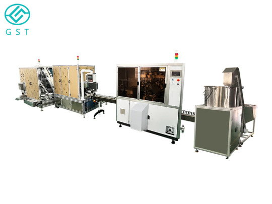
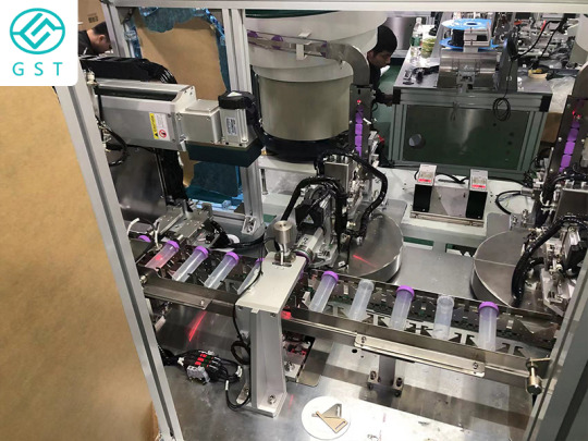
The most commonly used centrifuge tube automatic production line solution in the laboratory
Centrifuge tube automation production line solution manufacturers know that the most inconspicuous consumable in the laboratory - centrifuge tube, is also the most commonly used consumable. In biological sciences, especially in the field of biochemistry and molecular biology research, centrifuge tubes are widely used, and every biochemistry and molecular biology laboratory is inseparable from centrifuge tubes. Every biochemistry laboratory will prepare various types of centrifuges. The working principle of the centrifuge tube is to rotate the solution at a high speed to generate centrifugal force, so that the suspended tiny particles settle at a certain speed, so as to achieve the purpose of separation from the solution.
1. Centrifuge tubes are classified according to material
Plastic centrifuge tubes: Plastic centrifuge tubes include polyethylene (PE), polycarbonate (PP), polypropylene (PP), etc. The advantages are transparent or translucent, and its hardness is small. The gradient can be taken out by puncture method. The disadvantage is that It is easy to deform, has poor corrosion resistance to organic solvents, and has a short service life. It is generally recommended to choose polypropylene centrifuge tubes.
Glass centrifuge tubes: Glass centrifuge tubes cannot withstand large centrifugal forces, and they need rubber pads when in use, and the airtightness is not good enough, so they are generally not used in high-speed centrifuges.
Steel centrifuge tube: Steel centrifuge tube has many advantages, such as high strength, no deformation, frost resistance, and corrosion resistance, so it has the widest range of use, but its surface is non-transparent, making it difficult to observe the separation. Although the corrosion resistance of steel centrifuge tubes is very good, it should also avoid contact with strong corrosive chemicals when using them.
2. Classified by capacity
Large-capacity centrifuge tubes (250mL, 500mL), also known as centrifuge bottles, are the centrifugal choice for large-capacity cell harvesting, pelleting and protein purification;
Ordinary centrifuge tubes (15mL, 50mL), the most common centrifuge tubes in laboratories;
Microcentrifuge tubes (0.6mL, 1.5mL, 2mL,) are often used in conjunction with microcentrifuges for the separation of micro reagents, and are often used in micromanipulation experiments in molecular biology.
3. Classified by bottom shape
Round bottom centrifuge tube: when collecting density gradients, you can choose a round bottom; the area of the round bottom is large, and it can withstand greater centrifugal force than the pointed bottom;
Conical bottom centrifuge tube: When the sample is small, when collecting the precipitate, use the conical bottom for better separation, and the upper layer is easier to absorb with a straw;
Flat-bottomed centrifuge tube: The purpose is the same as that of the conical-bottomed centrifuge tube, but the centrifuge tube can stand upright, which is more convenient to put and take out in the experiment. Other flat-bottomed centrifuge tubes are mostly centrifuge bottles and sample tubes.
4. Classified according to the closing method of the lid
Gland type: commonly used in microcentrifuge tubes;
Screw cap: It is often used for centrifuge tubes or centrifuge bottles with large capacity.
5. Classified by centrifugal speed
Low-speed centrifugation: generally refers to the maximum speed of less than 8000r/min, that is, 8000 rpm;
Normal speed centrifugation: generally refers to the maximum speed less than 15000r/min, that is, 15000 rpm;
High-speed centrifugation: generally refers to the maximum speed greater than 15,000 revolutions but less than 30,000 revolutions per minute.
In the case of such high centrifugal force, centrifuge tubes with poor tolerance will crack or even burst, which will lead to sample loss and unnecessary contamination. Sometimes it will even cause irreversible damage to the centrifuge, and the leaked aerosol will pose a threat to the health and safety of laboratory researchers, which is not conducive to the development and effective conduct of experiments.
0 notes
Text
Biomed Grid | Automatic Generation of Drug Concentration Gradient by a Modified Ink-jet Printer and its Application in Drug Screening
Abstract
Manual preparation of drug concentration gradient is labor-intensive and limits the efficiency of a drug screening to some extent, especially at large scale. In this study, we reported an automatic method to generate drug concentration gradient in 24-well plate. We loaded drug solution in a modified hot ink-jet printer, designed printing pattern in the WORD software, customized the concentration gradient in the WORD software by RGB coding, and finally printed the drug concentration gradient in the 24-well plate. A drug concentration gradient can be automatically generated within 5s in arbitrary pattern. Based on this flexible technology, we developed an ink-jet printer-based drug screening assay. Concentration gradient of 5-fluorouracil, as well as Mcf-7 cells, were printed in the 24-well plate, and the inhibition rate of Mcf-7 cells was measured by MTT method. This was the first time that hot inkjet printing technology was used in drug screening.
Keywords: Bioprinting; Drug screening; Concentration gradient; Ink-jet printer; RGB
Introduction
Preparation of a concentration gradient of drug candidate is a prerequisite to study cell-drug interaction quantitatively. Currently, this step is manually operated in the laboratory, which is labor intensive and annoying. Automation is desirable in practice. Derringer et al. developed an automatic method to generate concentration gradient [1], and Ye et al. [2] used this method in drug screening [2,3], however, this method was only applicable in microfluidic device, not in 96-well or 24-well plate, thus its applications were limited. Previously, we established drug screening device based on biomedical engineering methods [4]. In this study, we developed a method to generate concentration gradient in plate automatically, using normal commercial ink-jet printer and WORD software. We also used the same printer to print the cell suspension, thus we finally developed a drug screening assay based on hot ink-jet printing. Hot ink-jet printing technique is useful for printing cells in biomedical engineering. It works by the principle in which the heating element turns the “biological inks” into bubbles at bottom of the nozzle instantly and the pressure generated makes a certain amount of inks free from the surface tension there and pushed from the nozzle. As an approach for cell printing, hot ink-jet printer has been used in tissue engineering [5], biological microarray construction [6], stem cell research [7] and gene transcription [8]. However, there are few reports in its application in drug screening at cellular level
In this paper, a hot ink-jet printer was custom-modified as an automatic dispensing tool for the engineering of cell arrays and generation of concentration gradient of drugs in a 24-well plate. By means of this apparatus, cell/drug composite arrays can be automatically generated for drug screening. RGB value was used as the output gauge of drug concentrations for the first time. A Mcf-7 cell/5-fluorouracil composite array was engineered to demonstrate the applicability of the hot ink-jet printer in the drug screening.
Experimental
Materials
Hoechst 33342 fluorescence dye and 3-(4,5-dimethylthiazol-2- yl)-2,5-diphenyltetrazolium bromide (MTT) were purchased from Kubolai inc. Beijing. Propidium iodide was obtained from Jingchun technology inc. Shanghai. 5-fluorouracil was purchased from Sigma- Aldrich, United Kingdom. Phosphorate buffer saline (pH7.4) was prepared and sterilized in lab. DMEM cell culture medium and fetal bovine serum from HyClone, New Zealand, was used in this study. Methanol of HPLC grade from local company was used. Ultrapure water was obtained with Millipore water purification system.
Instruments
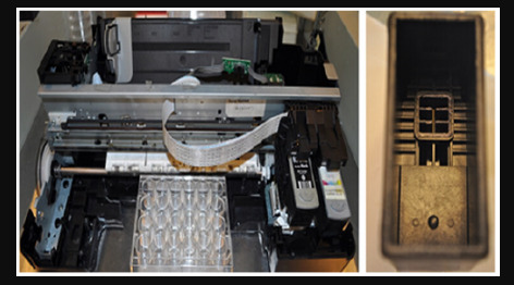
Canon PIXMA ip 1980 ink-jet printer with PG830 ink box from Canon, Japan, was modified in this study. Inverted fluorescence microscope IX71 from Olympus, Japan, was used. Agilent 1200 HPLC was from Agilent, USA. Synergy H1 microplate reader was from BioTek, USA (Figure 1). A Canon PIXMA ip 1980 printer and a Canon PG830 ink box for biological printing after modification. The white plate was a 24-well plate used as “paper”.
Modification of the Printer and the Cartridge
A Canon PIXMA ip 1980 hot-injet printer was modified as follows the front cover, paper support, rear tray, paper thickness lever and paper guide were dissembled sequentially while cartridge holder and paper sensor being kept intact. The printer after modification was shown as in Figure 1 (left part). Before use, the body of the printer was sterilized and kept inside an ultraviolet-lighted laminar hood overnight. A Canon PG830 ink box was modified as follows: after the top cover of the ink box was ripped from the main body of the box, the sponge inside the ink box was discarded and the metal filter at the bottom of the box was dismantled with the ink reservoir exposed, followed by a thorough cleanup. The ink box after modification was shown as in Figure 1 (right part). Prior to use, the box was emerged in 75% ethanol and air dried in a laminar airflow, followed by being exposed with ultraviolet light for at least 30 mins.
Figure 1: A Canon PIXMA Ip 1980 Printer and a Canon PG830 Ink Box.
Printing Concentration Gradient of 5-Fluorouracil in the 24-Well Plate
10ml 8mg/ml 5-fluorouracil solution was precisely obtained when 0.08g 5-fluorouracil was dissolved in PBS solution in a 10 ml volumetric flask and filtered through a 0.22 μm diameter membrane to remove possible bacteria. This working solution was loaded in the ink box. The printing pattern was designed in the WORD software with designated RGB values. Clicked the “print” button in the WORD software and the concentration gradient of 5-fluorouracil was automated created in the 24-well plate within 5s. We collected the drug solution printed in each well and analyzed their concentrations with HPLC. In a typical setup, a SB-C18 HPLC column (4.6mm diameter, 250mm long) was used and the sample was detected by ultraviolet absorption with a single wavelength of 265 nm. The temperature of the column was set at 35℃. The volume of each sample was 10 μl and the mobile phase was made of methanol and water (5:95) with a flow rate of 1.0ml/min.
Preparation and Printing of Cell Suspension
Mcf-7 cells of the third generation with a high viability was chosen as the main raw material for preparation of the cell suspension. Prior to being loaded into the ink box, a Mcf-7 cell suspension were prepared by pancreatic digestion, centrifuging, collection, counting and re-suspending. The Mcf-7 cell suspensions were prepared at four different concentrations, 3×10⁶, 6×10⁶, 9×10⁶ and 1.2×10⁷/ml. Following the pattern of a round spot with the diameter of 5 mm, the cell suspension with different densities were printed into a 24-well plate with the modified printer as above. Observation was made with an inverted microscope equipped with a X4 zoom objective.
Counting of Printed Cell Numbers and Viability Analysis
400 μl 10% NCBS culture media was added into the plate loaded with the printed Mcf-7 cells to keep their viability. And 10μl 0.4mg/ml Hoechst 33342 solution was added into the plate at 37℃ for 35mins in the dark, followed by the addition of 10 μl 0.2mg/ ml PI solution for another 10 mins. Then the cell suspension in the plate were collected and centrifuged. The cells from the bottom of centrifuge tube were collected and washed with PBS solution for 3 times. Then the cells washed were re-suspended in 1 ml cell culture medium to form a new cell suspension. 20μl new cell suspension was pipetted out and added dropwise onto a cell counter. Counting of cell number and observation of cells were conducted with an inverted fluorescence microscope with a X 10 zoom objective.
Procedure of Drug Screening
Mcf-7 cells with a high viability was selected for construction of cell/drug arrays. The Mcf-7 cell suspension were prepared by pancreatic digestion, centrifuging, collection, counting, and resuspension with a concentration of 9×10⁶/ml. The Mcf-7 cell suspension was loaded into sterilized a PG830 ink box and printed into 5-mm-diameter round spots in a 24-well plate to form a Mcf-7 cell array. 400μl 10% NCBS culture media was immediately loaded into the plate. After being cultured at 37℃ under 5% CO2 for 24hrs, 8 mg/ml 5-fluorouracil solution were printed into the plate which were already loaded with Mcf-7 cells, with different RGB codes. As a result, cell/drug composite arrays (n=3) were formed. After being at 37℃, 5% CO2 for another 48 hrs, 40μl 5mg/ml MTT solution were loaded into the plate and incubated at 37℃ for 4hrs. Then the supernatant was discarded and 500 μl DMSO was added. The plate was shaken for 15mins to achieve DMSO thoroughly dissolved. Then 200 μl supernatant was collected and transferred into a 96-well plate and the adsorption under 490 nm wavelength was recorded.
Results
Printed Concentration Gradient of 5-Fluorouracil
The printing pattern was designed in WORD software, as shown in Figure 2. Each round spot, corresponding to a well in 24-well plate in location, was 5mm in diameter. These round spots varied in RGB values. We printed drugs following the pattern in Figure 2 and measured the concentration of drug in each well. We plotted the drug concentration in each well, as shown in Figure 3. This technology has five advantages compared to manual method
a) It is automatic;
b) We can obtain arbitrary concentration gradient by customizing the RGB values (Figure 3 A-D);
c) The RGB code value was linearly related to the concentration of printed 5-fluorouracil. This can be expressed as Y = -0.7691X+53.975 with R2 being equal to 0.9878 (Figure 3A);
d) It is fast, and a concentration gradient can be generated within 5s;
e) THE hot inkjet printer was cheaper than a micropipette which is used for manual preparation. (Figure 2)
Figure 2: The Printing Pattern Designed in The WORD Software.
Figure 3: Different Types of Drug Concentration Gradient Printed in the 24-Well Plate.
The printing pattern designed in the WORD software. Each row was designed for generation a specific drug concentration gradient. In the first row, the RGB values from left to right were (20,20,20), (30,30,30), (40,40,40), (50,50,50), (60,60,60), (70,70,70). In the second row, the RGB values from left to right were (70,70,70), (20,20,20), (70,70,70), (70,70,70), (20,20,20), (70,70,70). In the third row, the RGB values from left to right were (20,20,20), (20,20,20), (50,50,50), (50,50,50), (70,70,70), (70,70,70). In the fourth row, the RGB values from left to right were (70,70,70), (70,70,70), (50,50,50), (50,50,50), (20,20,20), (20,20,20). (Figure 3) On the top of each plot was the printing pattern designed in the WORD software, and the number below was RGB value. (Figure 4) (from left to right: 3×10⁶/ml, 6×10⁶ /ml,9×106 /ml), scale bar: 500 μm.
Printing of Mcf-7 Cell Suspension
Figure 4: The Bright Field Images of Printed Round Spots of Mcf-7 Cells with Varied Cell Concentration.
Figure 4 shows the printed pattern of Mcf-7 cells, with different concentration of Mcf-7 suspension loaded in the printer. When the concentrations were 3×10⁶ /ml and 6×10⁶ /ml, the printed round spots were incompletely constructed with sparse distributions of cells. It might be caused by the low concentration of cell suspension. When the concentration was 1.2×10⁷/ml, few inks were injected from the nozzle and the printed round spot was hard to be identified. It might be attributed to the high concentration of the cell suspension which jammed the nozzle. However, when the concentration was 9×10⁶/ml, the printed round spot was full of well-distributed cells. It showed that the cell suspension with a concentration of 9×10⁶/ml is the best-performed one and should be adopted in the experiment.
After counting, the average number of cells in each round spot in the cell array was 3×10⁴. After printing, the viability of the cells was the main gauge which reflects the validity of the cell printing process. Hot ink-jet printing was suspected to cause heat damage or mechanical damage on cells [8]. Hoechst33342 is a membrane permissive dye and emits blue fluorescence under 350 nm excitation after combining with DNA in cells. And PI is only capable of penetrating into dead cells. After PI combines with DNA in cells and emits red fluorescence under 543 nm excitation. Therefore, we can investigate the viability of cells when coupling Hoechst33342 and PI dyes after cell printing. As seen in Figure 5, the number of dead cells which emit red fluorescence is negligible and it supports that the negative effect of our cell printing technique on the viability of printed cells can be ignored (Figure 5).
Figure 5: Microscopic observations of printed cells dyed by Hoechst33342(left) and PI (right), under excitations of 350 nm and 543 nm wavelengths, respectively. Scale bar: 500 μm.
Drug Screening Assay Based on Printing
Table 1: Absorbance by MTT and relative inhibition rate (n = 3).
The concentration of the printed 5-fluorouracil can be pretuned with the RGB code setup from the WORD software. A Mcf-7 cell/5-fluorouracil composite array was formed after incubation for 48 hrs. And the inhibition rate of the experimental group at varied concentrations and the control group were tested with MTT method. And the statistics of their optical absorbance at different RGB values and the relative inhibition rates were listed in Table 1. It can be seen from Table 1 that within the range we investigated, the biological viability of the cells in the composite array increased as the RGB value ramped up. In our experimental setup in which RGB code value was kept between 20 and 70, the concentration of the printed 5-fluorouracil increased as the RGB value dropped. In another word, a low RGB value yielded a cell/drug composite array with a relatively high concentration and dose of 5-fluorouracil. Therefore, as the RGB value was lowered, a stronger interaction between Mcf-7 and 5-fluorouracil was achieved, and a higher inhibition phenomenon occurred. This suggested that the Mcf-7 cell/5-fluorouracil composite array established by our method can be used for the inhibition study of 5-fluorouracil at different concentrations and it possessed the potential of evaluating in vitro cellular viability.
Discussion
Cell printing technique has demonstrated remarkable advantages as following :
a) High precision, cells can be precisely localized and distributed in three dimensions [9];
b) Rapid speed, tissue samples with biological viability can be constructed in a short time;
c) High automation grade, it has the possibility to be industrialized at a large scale [10];
d) Simple to be established and operated, it is straightforward to be modified or upgraded [11].
In our experiment, RGB tuning was used for controlling the output doses of 5-fluorouracil and the hot inkjet technique was adopted to build cell/drug composite arrays. And the viability of the cells in the composite arrays were evaluated. Inspired by the model from Boland ‘s group [11,12], a Canon hot inkjet printer and an ink box were successfully modified into a biological printer. And a Mcf- 7 cell suspension with a concentration of 9×10⁶ cells/ml showed the best printing performance in cell distribution and density on the array. The printed cell array demonstrated patterns with the most complete morphology. By testing the concentration of drug solutions with HPLC, we were able to identify the concentration of 5-fluorouracil on the printed matrix produced from different RGB code value. The results show that high-definition Mcf-7 cell arrays can be constructed with our modified printer and ink box. And moreover, the concentration of 5-fluorouracil arrays can be tuned by changing the RGB color code value. The viability of cells by printing is the major indicator for the feasibility of the printing operation. We used Hoechst33342 and PI to dye the printed cells and applied excitations of 350 nm and 543 nm, respectively. The results showed that hot inkjet printing has little damage on the Mcf-7 cells, and they are in consistency with the literatures [13,14]. Also, we used MTT method to identify the inhibition rate of 5-fluorouracil on Mcf- 7 in the cell/drug array. When RGB code value was 30, the dose of the 5-fluorouracil was 30.09±2.69 μg/ml with a relative inhibition rate of 49.02%. These results supported the feasibility of applying the cell/drug printing method in investigating in vitro cellular viability and it provides new possibility of drug screening based on biological printing.
Conclusion
In this study, we developed an automatic method to generate drug concentration gradient in 24-well plate based on hot inkjet printer. The total time was less than 5s. Arbitrary concentration gradient can be easily generated by RGB coding. The cost of printing equipment was very low. This method is expected to make drug screening experiment more user-friendly.
Read More About this Article: https://biomedgrid.com/fulltext/volume5/automatic-generation-of-drug-concentration-gradient-by-a-modified-ink-jet-printer-and-its-application-in-drug-screening.000876.php
For more about: Journals on Biomedical Science :Biomed Grid | Current Issue
#biomedgrid#american journal of biomedical science & research#Journals on vaccination#Health science Journal of Open access
0 notes
Text
300+ TOP GENETIC CODE & REGULATION Objective Questions and Answers
GENETIC CODE and REGULATION Multiple Choice Questions :-
1. Which of the following techniques was carried out by Nirenberg and Matthaei in 1961 to determine the first codon? A. In vitro synthesis of a polypeptide using UUUUU B. Labeled peptide binding to a ribosome C. Mixed co-polymer mRNA synthesis D. none of the above Answer: A 2. Bacterial protein called catabolic activator protein (CAP) is an example of A. negative control of gene expression B. positive control of gene expression C. second type of positive control of gene expression D. none of the above Answer: C 3. How many different codons are possible? A. 3 B. 20 C. 64 D. An infinite number Answer: C 4. The genetic code is A. universal B. universal except for rare exceptions in mitochondria and some protozoa C. species-specific D. kingdom-specific Answer: B 5. Which of the following has been used as an evidence that primitive life forms lacked both DNA and enzymes? A. RNA can both code genetic information and act as a catalyst B. DNA and enzymes are only present in the most advanced cells C. Advanced cells lack RNA D. All of the above Answer: A 6. Crick demonstrated that the genetic code involved three bases and suggested that the code was degenerated. What experimental technique Crick conducted to suggest genetic code degeneration? A. Gel electrophoresis B. Density gradient centrifugation C. Frameshift mutagenesis D. Restriction digests of the rII gene Answer: C 7. Codon that specify the amino acids often differs in the A. first base B. second base C. third base D. none of these Answer: C 8. In some organelles in eukaryotes, the genetic code for some codons A. differs from that used in prokaryotes B. are same C. are partially same D. none of the above Answer: A 9. The codons which do not specify an amino acid are called A. initiation code B. termination code C. propagation code D. none of these Answer: B 10. The genetic code is degenerated. Which of the following codons represents the principle of degeneracy? A. UAA and UAC B. AUG and AUA C. CAU and CAC D. UUA and UUC Answer: C

GENETIC CODE & REGULATION MCQs GENETIC CODE and REGULATION Objective type Questions with Answers 11. The sequence of one strand of DNA is: 5' ATTGCCA 3', what is the sequence of the other strand? A. 5' TAACGGT 3' B. 5' TGGCAAT 3' C. 5' ATTGCCA 3' D. 5' UAAGCCU3' Answer: B 12. In prokaryotes, AUG encodes A. methionine B. N-formyl methionine C. a stop codon D. alanine Answer: B 13. How many amino acids will be encoded by 5' GAU GGU UGA UGU 3' sequence? A. One B. Two C. Three D. Four Answer: B 14. In protein synthesis in prokaryotes A. the initiating amino acid is N- formyl methionine B. the initiating amino acid is methionine C. the initiating amino acid is phenyl alanine D. none of the above Answer: A 15. AUG codes for methionine act as a A. initiation code B. elongation code C. termination code D. propagation code Answer: A GENETIC CODE and REGULATION Questions and Answers pdf Download Read the full article
0 notes
Photo

H2O>O #Repost @physicsfun with @get_repost ・・・ Bubble Trouble Puzzle: physics brain teaser- the trick here is to move all air bubbles to the center bulb. As with all puzzles featured here on @physicsfun, the solution relies on some fun basic physics principles- can you figure out which ones? Answer below and swipe to reveal solution. ➡️ Follow the link in my profile for info on where to find puzzles like this one and other amazing items featured here on @physicsfun #physics #physicsfun #physicstoy #physicspuzzle #puzzle #brainteaser #density #fluiddynamics #bouyancy #centripetal #centripetalacceleration #centrifugal #rotation #kineticenergy #potentialenergy #science ✏️ Solution: spinning the puzzle about its center makes it into a simple centrifuge, where centripetal acceleration creates a density gradient, and the water, which has much greater density than air, moves away from the center towards the ends of the tube. https://ift.tt/2PnMiWd
0 notes
Text
Why Labs Are Choosing PluriMate: Key Benefits of Non-Disruptive Cell Separation

Laboratories aiming to separate leukocytes and peripheral blood mononuclear cells (PBMCs) with minimal contamination are increasingly turning to tools that leverage the density gradient centrifugation principle. Among these, PluriMate stands out due to its innovative design that simplifies cell separation while maintaining sample purity. PluriMate's unique sponge barrier enhances efficiency, making it a preferred choice for labs conducting regular cell isolation from whole blood or bone marrow.
1. Simplified Sample Preparation Process
One of the key benefits of PluriMate is its ease of use. Traditional cell separation methods often involve careful and time-consuming layering of blood over the separation medium, which requires skill and can increase the risk of cross-contamination. PluriMate eliminates this need entirely. Researchers can pour anticoagulated blood or bone marrow directly into the PluriMate tube without worrying about manual layering. This not only saves valuable time but also reduces the potential for errors, making it an ideal tool for labs looking to streamline their processes.
2. Innovative Particle Separation Techniques
PluriMate utilizes a built-in porous sponge made from high-grade polyurethane that acts as a non-disruptive barrier, facilitating reliable particle separation techniques. During centrifugation, this barrier prevents unwanted cells—like erythrocytes and granulocytes—from mixing with the desired cell population. The density gradient within the PluriMate tube separates leucocytes, lymphocytes, and PBMCs based on their specific densities, allowing them to collect in an interphase above the separation medium. This layer of targeted cells can then be easily harvested without contamination from denser, undesired cells. PluriMate’s particle separation techniques ensure that researchers receive high-quality samples for downstream applications.
3. Reduced Risk of Contamination
Contamination is a significant concern during cell separation, especially in multi-step processes where each step increases exposure to contaminants. PluriMate addresses this with its non-disruptive design that minimizes the need for handling. Once centrifugation is complete, the sponge barrier remains intact, preventing recontamination of the enriched cell fraction as it is harvested. This design is particularly beneficial when processing large batches or working with valuable samples, as it ensures that the isolated cells remain pure and ready for analysis or further applications, such as cell enrichment studies or cell-based assays.
4. Efficient Particle Filtration
Effective particle filtration is essential for isolating high-quality cell populations. PluriMate’s porous sponge acts as a built-in filter, preventing the mixture of the sample material with the separation medium, thereby preserving the integrity of the cells that need to be isolated. By keeping unwanted particles and cells out of the desired layer, PluriMate provides a reliable way to filter and enrich the sample without additional equipment. This feature makes it particularly attractive for labs seeking straightforward solutions for cell separation and filtration, with minimal need for extra tools or manual intervention.
5. Versatile for Various Applications
PluriMate is adaptable to both large and small sample volumes, making it a versatile tool in laboratories with diverse sample processing needs. Whether pre-filled with the separation medium or provided as an unfilled tube, PluriMate offers flexibility for a wide range of cell separation protocols. Additionally, it can be combined with pluriSpin for negative cell separation, further expanding its utility. This versatility allows researchers to conduct multiple applications, including cell enrichment and density-based separations, making it an invaluable addition to modern labs.
Conclusion
For labs prioritizing efficiency, reduced contamination, and reliable separation, PluriMate is an effective solution that operates on the density gradient centrifugation principle. With its innovative sponge barrier, easy handling, and adaptability to various cell separation tasks, PluriMate meets the demands of researchers focused on high-quality results. By simplifying the cell separation process, PluriMate enables laboratories to achieve consistent outcomes while maintaining sample purity, making it a top choice for professionals in the field of particle separation and cell isolation.
0 notes
Text
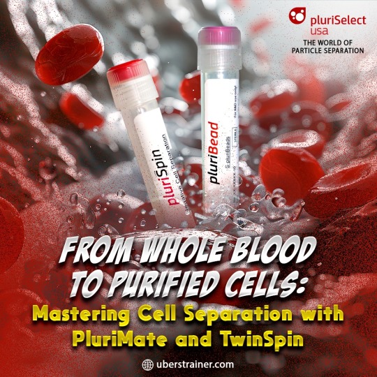
From Whole Blood to Purified Cells: Mastering Cell Separation with PluriMate and TwinSpin
PluriMate and TwinSpin streamline the cell separation process by utilizing the density gradient centrifugation principle to efficiently isolate high-quality cells for research.
visit us :- https://uberstrainer.com/
#antibody cell separation#cell separation technology#particle separation techniques#particle filtration#40 um cell strainer#cell enrichment techniques#uberstrainer#cellsepration
0 notes
Text
Advanced Techniques for Optimizing PBMC Isolation and Enhancing Cell Purity
Due to their significant role in immune response, PBMCs are widely used in research and clinical applications. Isolating PBMCs efficiently and effectively is crucial, and advanced techniques like density gradient centrifugation principle play a pivotal role in this process.
#uberstrainer#antibody cell separation#particle separation techniques#particle filtration#cell separation technology#40 um cell strainer#cell enrichment techniques
0 notes
Text

Empowering Cell Enrichment: Strategies for Maximizing Yield and Purity
The article discusses strategies for maximizing yield and purity in cell enrichment, highlighting Pluriselect-usa's advanced technologies, pluriBead, and pluriSpin, which streamline cell separation processes and enhance the efficiency and accuracy of biomedical research.
Cell enrichment is a critical process in biomedical research, enabling scientists to isolate specific cell types from a mixture for downstream applications. Achieving high yield and purity in cell enrichment is essential for reliable experimental outcomes. Pluriselect-usa, Inc. offers advanced products designed to streamline and enhance the cell enrichment process, ensuring that researchers can obtain the highest quality cells efficiently. In this article, we will explore two innovative cell separation technologies namely pluriBead and pluriSpin, and discuss strategies for maximizing yield and purity in cell enrichment.
pluriBead: Redefining Cell Separation
pluriBead is a unique cell separation technology that eliminates the need for magnetic components, making the process simple and efficient. The pluriBead method involves sieving pluriBeads with bound target cells through a cell strainer. The pluriBeads, along with the target cells, remain on top, while unwanted cells pass through. After detaching the pluriBeads, the target cells are ready for use.
Key Features of pluriBead:
No Sample Preparation Required: pluriBead allows for the use of 200 µl to 45 ml of sample without the need for gradient centrifugation or erythrolysis, simplifying the process significantly.
Versatile Sample Material Compatibility: This technology can handle various sample materials, including whole blood, buffy coat, PBMC, secretion/excretion material, brain homogenate, spleen, and liver.
High Species Range: pluriBead is compatible with samples from a wide range of species, including human, mouse, rat, bovine, canine, and sheep.
Fast and Gentle Isolation: The isolation process can start from as little as five minutes, ensuring a high yield of viable cells while minimizing sample requirements.
Universal Compatibility: pluriBead can be used with any external antibody, making it a flexible tool for diverse research needs.
Simultaneous and Sequential Cell Isolation: pluriBead Cascade allows for the simultaneous separation of two different cell types from one sample, while sequential isolation can target up to six different cell types from a single sample.
pluriSpin: Efficient Negative Cell Isolation
The pluriSpin system is an advanced negative cell separation technology that enables the isolation of untouched, highly purified cells directly from whole blood, buffy coat, or cord blood. This method avoids the use of magnets or columns, reducing the risk of activating or damaging the target cells.
How pluriSpin Works:
To isolate cells using pluriSpin, researchers incubate their sample with the pluriSpin reagent and then perform a standard density gradient centrifugation. pluriSpin binds unwanted cells, which pellet during centrifugation, leaving the untouched, highly purified target cells at the interface between the plasma and the density gradient medium. The purified cells can then be aspirated from this interface into a new tube.
Key Features of pluriSpin:
Fast and Easy Isolation: The pluriSpin protocol is straightforward and can be completed quickly using standard density gradient centrifugation principle.
No Special Training or Equipment Needed: pluriSpin does not require specialized instruments or magnets, only a mixing device such as a rolling mixer or a magnetic stirrer adapter (pluriPlix).
Highly Viable and Functional Cells: The cells isolated using pluriSpin are functional and ready for applications like cell stimulation, cytotoxicity assays, cultivation, differentiation, and high-throughput screening (HTS) that require viable, functional cells with minimal manipulation.
Strategies for Maximizing Yield and Purity
To maximize yield and purity in cell enrichment using pluriBead and pluriSpin, researchers should consider the following strategies:
Optimize Sample Volume: Use the recommended sample volumes to ensure efficient cell separation and minimize loss.
Careful Handling: Gentle handling of samples during the isolation process helps maintain cell viability and functionality.
Follow Protocols Precisely: Adhering to the optimized protocols for pluriBead and pluriSpin ensures consistent and reproducible results.
Regular Calibration: Ensure that all equipment used, such as centrifuges and mixing devices, is regularly calibrated and maintained.
Quality Control: Implement quality control steps, such as flow cytometry, to assess the purity and viability of the isolated cells.
By utilizing pluriBead and pluriSpin, researchers can achieve high yields of pure, viable cells efficiently. These advanced cell separation technologies from Pluriselect-USA empower scientists to conduct their research with confidence, driving scientific discoveries forward.
#antibody cell separation#particle separation techniques#cell separation technology#cell enrichment techniques#cell strainer 40 um#cell strainer 70 um
0 notes
Text
Korea Centrifugation Market Future Trends to Look Out | Bis Research
Centrifugation techniques used in Korea or developed by Korean scientists, would involve the separation of substances using centrifugal force, a method commonly used in various fields such as biology, chemistry, and medicine.
Centrifugation involves spinning a mixture at high speeds, causing the components to separate based on their density, size, and shape. This process finds applications in purifying biological samples, separating components of blood, isolating cellular organelles, and various other laboratory procedures.
Korea Centrifugation Market Overview
Centrifugation is a fundamental laboratory technique used for separating components of heterogeneous mixtures based on their density, size, and shape differences. It involves spinning samples at high speeds, generating centrifugal forces that cause particles to move radially outward or inward according to their mass and buoyant density.
Centrifugation exploits the principle of sedimentation, where denser particles sediment faster under centrifugal force.
Types of Centrifugation
Differential Centrifugation
Density Gradient Centrifugation
Ultracentrifugation
Korea Centrifugation Market Drivers
Biotechnology and Life Sciences Research
Clinical Diagnostics and Healthcare
Industrial Applications
Technological Innovations
Korea's centrifugation market is driven by factors such as research and innovation, healthcare demand, industrial applications, environmental considerations, and global competitiveness, shaping the dynamics of the industry and driving technological advancements and market growth.
Have an understanding of our report Click Here !
Market Segmentation
Based on Product
Based on Model Type
Based on Application
Key Market Players
Agilent Technologies, Inc.
Andreas Hettich GmbH & Co. KG
ARI Medical Technology Co., Ltd.
Biolab Scientific
Bioneer Corporation
And many others
Download our sample page to have a better understanding click here !
Types of Korea Centrifugation Market
Ultracentrifugation
Differential Centrifugation
Density Gradient Centrifugation
Preparative Centrifugation
Analytical Centrifugation
Recent Developments in the Korea Centrifugation Market
• In April 2023, Eppendorf, a prominent life science company, unveiled the Centrifuge 5427 R, marking the introduction of Eppendorf's inaugural microcentrifuge equipped with hydrocarbon cooling. This innovation contributes to fostering a more sustainable laboratory environment. Through this advancement, users had the capability to conduct a diverse range of molecular and cell biology applications using a refrigerated device that incorporated a natural cooling agent boasting a nearly zero Global Warming Potential (GWP). The primary goal was to safeguard both samples and the planet.
•In February 2022, Beckman Coulter Life Sciences introduced its most versatile 3-liter benchtop centrifuge to date, the Allegra V-15R refrigerated centrifuge. It featured 10 rotor configurations and 50 programmable runs, along with a comprehensive array of adapters, allowing for a wide range of workflows and applications. These capabilities extended from cell and blood separation to high-throughput screening.
Visit our LifeSciences and Biopharma page click here !
Key Question Answers
Q What is the regulatory framework in the Korea centrifugation market?
Q What are the key trends and the impact of COVID-19 on the Korea centrifugation market?
Q What are the major market drivers, challenges, and opportunities in the Korea centrifugation market?
Q How is each product segment of the Korea centrifugation market expected to grow during the forecast period 2023-2033, and what is the anticipated revenue generated by each of the segments by the end of 2033?
Conclusion
In conclusion, the centrifugation market in Korea presents a dynamic landscape shaped by various factors driving demand, innovation, and growth. With a robust scientific research community, thriving industrial sectors, and a commitment to technological advancement, Korea stands as a key player in the global centrifugation market.
0 notes