#pdhc
Explore tagged Tumblr posts
Visit Tumblr Blog
Explore Tumblr blogs with no restrictions, modern design and the best experience.
Last Seen Tumblr Blogs


Fun Fact
12.7% of mobile users access Tumblr.
Note
I want your corkscrew hcs!!! just rly anything. It can be fun stuff like his favorite soda or fave movies. Maybe some stuff specific to living in Russia that I'd have no idea was a thing otherwise if u got anything like that! I feel like all my hcs for him will be so lacking because I know so little about living there, but living in Sweden has rly upped my love for learning all the lil mundane differences in daily life from country to country
Okay small and specific shtop hcs leggoo
OUFFF my time has come i guess....finally expirience living in *there* would be useful lmao FIRST of all i wanna mention this one russian song in goin postal playlist
i think its a tribute in shtopors honor. bc like why else rws would put this ru song in there???
ANYWAY.
im not really good at explaining my feelings so im gonna throw a bunch of memes that has STRONG SHTOP VIBES. Memes are the ultimate association language right? im not apologising for klukva (stereotype memes) bc?? original dude is lit basic american trash so errything he knows abt russia is a bunch of stereotypes. makes sense ig
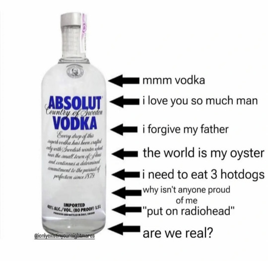

ok now here goes ru memes💥💥💥💥💥


(2nd pic: your pigs grunt violates the laws of the Ural (including shutting the rotten mug up, p.18) and i told my bois about it. I hope, your identity will be established. When crossing the border of Ekaterinburg you will kicked the fuck out with bicycle chains. This is Ural, not Moscow) (3rd pic: [i] hate the rednecks)
Ok NOW ONE SPECIFIC THING. once i mentioned that hes an airbone and here, when the national airbone day comes, its a common joke that the ones who served in it will jump into a fontains like PARATROOPERS WOOHOOOO. yea.
Tbh its really hard to tell where is the diff between your culture (i.e ur everyday life) and others when theres nothing to compare with. BUT. ill try
His concept of personal boundaries is really strange and fucked up compare to others bc here [russia] we r suspicious as hell (imo.) but also like a big family. but the cracked one. Or kinda sorta like that. BUT at the same time he is ready to help everyone who need it and he will not ask any questions. Also he would let live his friends in his small flat if they needed to. It would be really tricky tho. Also shtop is really REALLY LOUD. in every sense of the word
Shtopor knows how to survive in harsh conditions. How to make not-that-bad-edible food out of canned food. Just google navy-styled macaroni! (or Makarony po-flotski) yeass NAVY styled aaand hes the "army one". Out of all dudes at least shtop knows how to COOK. blini, macaroni, syrniki, okroshka, - all of it actually cheap, easy and real tasty. LOOK AT OKROSHKA I MEANNNN. cold soup of raw vegetables. ok.

Shtopor one of those dudes who is a coldfucker. He is actually low-temperatures-proof. (small preface) shtop is an eastern orthodox. AND ANOTHER ONE russkaya zabava. When the baptism of jesus comes we have a tradition to cut out the ICE HOLE in the river AND JUMP INTO IT 👍 or another similar thing when ur hot after the banya you fall into a snowdrift. (banya is like sauna or steam bath. u rarely go to the banya alone.) ((public banyas divides to male and female, commonly yall naked in there, now imagine shtops in public banya and his bi neuron activation haahahah)) (personay i dont really like public banyas cuz its PUBLIC)
Another notable mention i think shtop is actually dgaf what to whatch on tv but he likes to talk with it. That sofa commentators type of ppl u know em. After a really looong day he would mix vodka and beer, sat down in front of tv and gave his VERY IMPORTANT COMMENTS ON LITERALLY EVERY THEME IN THE WORLD in until he fell asleep.
Russian curse words is veeery various, it can contain a lot of definition for one word, but be different on vibes and occasions to use it. U can say an entire sentence without a single cultural word in it. And therefore shtop complains about the lack of swear words in english and often swears in Russian. "--da blyaaaaat' kak zhe zae- -- OH gimme this gun. you started speaking russian which means things are bad." ill brb later with more things but now im kinda tired rn so here u go.
23 notes
·
View notes
Text
Merging Care Management and Homecare - the Big Benefits
By Merging care management and homecare, you will make 4 times the revenue Merging care management and homecare’s big financial payoff of this merger is found in the 2013 Care Management Benchmark Study. Care managers who owned a home care agency made four times the revenue in-home care in 95% and, in all cases, made double their revenue. Merging care management and homecare gives you ultimate…

View On WordPress
#adding geriatric care management#adding geriatric care management to PDHC#adding homecare to care management#aging family#aging life care manager#aging parent care#aging parent crisis#Benefits of merging Care management and homecare#care management profit#care manager#case manager#geriatric care manager#Homecare agency#homecare profit#marketing geriatric care#marketing geriatric care management#merger#merging homecareand care management#nurse advocate#nurse care manager#PR for geriatric care management
0 notes
Note
97 and xisuma

Holding out for the light left on
#97 - Hide Out (The Western Den)
Send me a prompt and a number and I'll do a doodle based on that song from my spotify wrapped!
#thanks for the prompt! i don't think I've ever drawn X except for on pdhc so this was something interesting#and not my best work but oh well#xisuma#xisumavoid#hermitcraft#hermitcraft 9#spark draws#spotify drawings
379 notes
·
View notes
Photo

did that character sheet by @blocky-blaze :)
#this is why my name on pdhc is 'red' :p#minecraft#minesona#mineblr#mine#despite my minecraft skin being a redstone man i am not actually that adept at redstone.#my original skin concept was just a red man but i couldnt get it to look decent and then i remembered redstone blocks exist.-.
328 notes
·
View notes
Text
Projeto Dom Helder Câmara oferece assistência técnica rural para reduzir desigualdade no semiárido
Projeto Dom Helder Câmara oferece assistência técnica rural para reduzir desigualdade no semiárido
Ações do Projeto Dom Helder Câmara (PDHC) têm transformado a vida de 57.486 famílias rurais que vivem no semiárido brasileiro através da oferta de assistência técnica permanente. A iniciativa acontece por meio de parceria entre a Secretaria de Agricultura Familiar e Cooperativismo (SAF) do Ministério da Agricultura, o Fundo Internacional para Desenvolvimento Agrícola (Fida) e a Agência Nacional…
View On WordPress
1 note
·
View note
Text
Dichloroacetic acid and rapamycin synergistically inhibit tumor progression
Mammalian target of rapamycin (mTOR) controls cellular anabolism, and mTOR signaling is hyperactive in most cancer cells. As a result, inhibition of mTOR signaling benefits cancer patients. Rapamycin is a US Food and Drug Administration (FDA)-approved drug, a specific mTOR complex 1 (mTORC1) inhibitor, for the treatment of several different types of cancer. However, rapamycin is reported to inhibit cancer growth rather than induce apoptosis. Pyruvate dehydrogenase complex (PDHc) is the... http://dlvr.it/Sp9jgg
0 notes
Text
Jornal Opinião Goiás - Fida e Mapa estabelecem ações para potencializar resultados do Projeto Dom Helder Câmara
Jornal Opinião Goiás – Fida e Mapa estabelecem ações para potencializar resultados do Projeto Dom Helder Câmara
Por meio de assistência técnica permanente, o projeto busca reduzir os níveis de pobreza e de desigualdades no semiárido Foi realizada nesta quinta-feira (27), em Brasília, a assinatura de um memorando de ações entre o Projeto Dom Helder Câmara (PDHC) e o Fundo Internacional para Desenvolvimento Agrícola (Fida). O evento marca o término da missão de apoio do organismo internacional, realizada de…

View On WordPress
0 notes
Text
Jornais de Goiânia - Comunidade em Água Branca (AL) ganha primeiro viveiro de aclimatação de mudas de palma forrageira
Jornais de Goiânia – Comunidade em Água Branca (AL) ganha primeiro viveiro de aclimatação de mudas de palma forrageira
A comunidade de Alto dos Coelhos, no município de Água Branca (AL), conta agora com o primeiro viveiro polo de aclimatação de mudas de palma forrageira, resultado da parceria entre o Projeto Dom Helder Câmara (PDHC) e a Companhia de Desenvolvimento dos Vales do São Francisco e do Parnaíba (Codevasf). A ação vai atender 660 agricultores de forma direta e, indiretamente, 6.600 agricultores no…

View On WordPress
0 notes
Photo

Jornal JA 7 – Conselho gestor do Projeto Dom Helder Câmara realiza primeira reunião Iniciativa visa combater a pobreza e apoiar o desenvolvimento rural sustentável no semiárido O Conselho Gestor do Projeto Dom Helder Câmara (PDHC) promoveu sua primeira reunião.
0 notes
Note
POSTAL HCs💥
POSTAL HCS LEGGO💥💥
Where do i start. Ofc. P1 u go first. Once i mentioned this hc before but anyway. P1s has a military past. Field medic, if more specific. And due to his military past back then, now the smell of medication, blood and hospitals does a tricky thing to his mind. To his psychosis. Brings back the memories. Other dudes (except stopor) didnt served anywhere. Also p1 is grungepunk nu metal enjoyer. AND his hair ISNT oily or greasy. When the sickness is everywhere you WILL try ro keep yourself as clean as possible. But his house is a fucking mess. Not in a garbage way but in a dusty-abandoned way. And also. He doesnt own a mobile. For delusion reasons. But overall his radio and low-tech skills above average.
Something on p2. Ohohhoh he survived the attempt to shot himself (or he didnt survived?) He ia basically a walking corpse; cold hands, dull stare, sometimes smells bad. But somehow well preserved. No. I know why he is well preserved and why he survived but this is for another topic. He will joking on everything even being one step away from death (again). Bc its way too painful to take his own problems seriously. Ohhh boi this man is FULL OF copium. Doe is his older sister. And in their siblings duo doe is the only one who has A BRAINCELL. His skull is empty. There is a bullet. And u can hear it ringing back and forth
Uuuhggg ooo i forgor everything
12 notes
·
View notes
Photo

Damn right I Support It! Thank you @queenjofficial for your support! Pride 2019 #pride #damnrightisupportit #pridemonth #KreativeMindsPrinting #heattransfervinyl #tshirts #tshirtdesign #customtshirts #custom #houston #HTOWN #onetimeforthebirthdayboy #birthdayboy #birthday #birthdayshirt #supportblackbusiness #customapparel #staymotivated #support #ordercompleted✔️ #orderdelivered✔ Dm for all inquiries and pricing... https://www.instagram.com/p/BzB0Hd-pDhc/?igshid=1x80fkhpe4h6f
#pride#damnrightisupportit#pridemonth#kreativemindsprinting#heattransfervinyl#tshirts#tshirtdesign#customtshirts#custom#houston#htown#onetimeforthebirthdayboy#birthdayboy#birthday#birthdayshirt#supportblackbusiness#customapparel#staymotivated#support#ordercompleted✔️#orderdelivered✔
0 notes
Text
First Case of Ceftriaxone-Resistant Multidrug-Resistant Neisseria gonorrhoeae in Singapore
The SGH laboratory has been doing antimicrobial surveillance testing on regular systematic sample collections of Neisseria gonorrhoeae sent to us from the Department of Sexually Transmitted Infection Control, National Skin Centre since 1992 (at least). These results are submitted to the World Health Organization Gonococcal Antimicrobial Surveillance Programme (WHO GASP). If you are interested, the reports are freely available at the WHO GASP website (https://www.who.int/reproductivehealth/topics/rtis/gonococcal_resistance/en/).
Local isolates of N. gonorrhoeae are often resistant to penicillin, tetracycline, and ciprofloxacin. The key antibiotic is therefore a third generation cephalosporin like ceftriaxone, with azithromycin thrown in for good measure (https://www.dsc-clinic.sg/Healthcare-Professionals/Books%20and%20Publications/Pages/STI-Management-Guideline-6th-Edition-(2013).aspx).
Ceftriaxone has remained a reliable antibiotic for a surprisingly long time but resistance has finally emerged. The first ceftriaxone-resistant N. gonorrhoeae was reported in Japan in 2011 (Ohnishi M, 2011). This strain named H041 had a ceftriaxone MIC of 2 mg/L. Since then, ceftriaxone-resistant N. gonorrhoeae have been reported in an increasing number of countries. Resistance is due to a mosaic penA allele (i.e. the gene is made up of bits of DNA from different genomes, N. gonorrhoeae can do this because they can acquire DNA from their environment by a process called transformation). This results in an altered penicillin-binding protein with reduced affinity to beta-lactam antibiotics.
A particular clone, named after the original strain FC428 isolated in Japan in 2015 has spread internationally (Lee K, 2019). In 2017, the World Health Organization published a list of bacteria for which new antibiotics are urgently needed. Cephalosporin-resistant N. gonorrhoeae were listed as Priority 2: High.
Our lab diagnosed the first ceftriaxone-resistant N. gonorrhoeae to be isolated in Singapore in early 2018.

The isolate was resistant to ceftriaxone with an MIC of 1 mg/L (left) and an annular radius of 5 mm by the CDS method.

Whole genome sequencing was performed and analysis showed that the strain had the same penA allele as the FC428 clone. Furthermore it belonged to ST13871 (allele profile abcZ 126 adk 39 aro E67 fumC 987 gdh 148 pdhC 153 pgm 65) which is a single locus variant of ST1903 (allele profile abcZ 126 adk 39 aroE 67 fumC 157 gdh 148 pdhC 153 pgm 65), the common multi locus sequence type of the FC428 clone. More details can be found in our paper which has just been published (Ko K, 2019).
As many of the cases of ceftriaxone-resistant N. gonorrhoeae were reported to have been acquired in south-east asia, we can only expect this problem to become worse.
Ohnishi M, Saika T, Hoshina S, Iwasaku K, Nakayama S, Watanabe H, Kitawaki J. Ceftriaxone-resistant Neisseria gonorrhoeae, Japan. Emerg Infect Dis. 2011 Jan;17(1):148-9. doi: 10.3201/eid1701.100397. PubMed PMID: 21192886; PubMed Central PMCID: PMC3204624. (free access)
Lee K, Nakayama SI, Osawa K, Yoshida H, Arakawa S, Furubayashi KI, Kameoka H, Shimuta K, Kawahata T, Unemo M, Ohnishi M. Clonal expansion and spread of the ceftriaxone-resistant Neisseria gonorrhoeae strain FC428, identified in Japan in 2015, and closely related isolates. J Antimicrob Chemother. 2019 Apr 19. pii: dkz129. doi: 10.1093/jac/dkz129. [Epub ahead of print] PubMed PMID: 31002306. (no free access)
Ko KKK, Chio MT, Goh SS, Tan AL, Koh TH, Abdul Rahman NB. First Case of Ceftriaxone-Resistant Multidrug-Resistant Neisseria gonorrhoeae in Singapore. Antimicrob Agents Chemother. 2019 Mar 11. pii: AAC.02624-18. doi: 10.1128/AAC.02624-18. [Epub ahead of print] PubMed PMID: 30858209. (free access from May 2020)
0 notes
Text
How Do Ketogenic Diets Work?
Restore Your Health with a Ketogenic Diet
Ketogenic diets are powerful metabolic tools that help your frame switch from burning sugar (carbs) to burning fats. The result of that transfer in gasoline is an improvement in all styles of fitness conditions. In practice, you get to consume actual foods in the form of herbal fat and keto x factor protein (meat, fish, poultry) whilst carbohydrates (sugars and starches) are restricted. On this website, I’ll communicate about how the food plan works, and percentage details on right implementation of the food plan.

How Do Ketogenic Diets Work?
The digestion of carbohydrates (sugar and starch) releases sugar (glucose) into the bloodstream. Greater carb consumption consequences in rising blood sugar and insulin, a pancreatic hormone that manages blood sugar. Going keto replaces nutritional carb with fats and protein. Over time, your cells switch metabolic pathways, and burn saved and dietary fat as a primary power supply instead of sugar. As extra fats are burned, some of it is converted into ketone our bodies. As blood glucose and insulin tiers fall, and ketone stages upward push, your muscular tissues (skeletal and coronary heart), use the fat in the bloodstream as fuel, whilst your mind makes use of the ketones. The end result is greater electricity, clearer wondering and better health. Ketones are beneficial in lots of distinctive ways, and being on this metabolic state of "dietary ketosis" (where blood sugar is low and ketone levels are slight) has some effective consequences in your metabolism. There are powerful studies proof that these metabolic-affecting diets may be used to deal with the subsequent scientific situations:
Cancer: Metabolic Therapy
The cutting-edge body of research on metabolic most cancers treatments the use of a keto weight loss program keeps to develop. Keto or metabolic most cancers remedy is relatively distinctive than the remedy for different illnesses, and is discussed in detail in my eBook Fight Cancer with a Ketogenic Diet, which is primarily based on the metabolic remedy research of Dr. Thomas Seyfried and Dr. Dominic D'Agostino. The primary idea at the back of the usage of a ketogenic weight loss plan to deal with cancer is to starve most cancers cells of the sugar and different fuels they want to survive, and to offer guide and protection for regular electricity tactics in healthful cells. The gain of this remedy protocol is that it's far non-toxic to the body, and in "be patient" instances, it may offer an main fitness guide advantage to the patient. The e book become written for the patient and has all the details and studies in an easy-to-use layout.
This new 0.33 edition of Fight Cancer with a Ketogenic Diet is likewise to be had in paperback shape on Amazon. Ten percentages of the earnings of this eBook go to support Dr. Seyfried and Dr. D'Agostino's research, and I'm grateful with a view to make contributions to their critical paintings.
Diabetes in All Forms
Type 2 Diabetes: The current mainstream diabetes remedy advice is to eat 45-sixty five% of energy from carbohydrate. Since carbohydrate is the high driving force of better blood sugar, this advice is unfavorable to diabetic fitness because it effects in blood sugar spikes and crashes, which in turn causes a more want for medication and insulin. Those high blood sugars additionally bring about the complications of diabetes. In evaluation, a ketogenic eating regimen reduces and in lots of instances, eliminates the want for diabetic medicines and lowers the variety of insulin devices had to control blood sugar. For people with Type 2 diabetes, ketogenic diets get rid of the trigger (carbohydrate intake) and reverse the underlying insulin resistance which causes the disorder. As a end result, long time headaches are reversed or prevented. Learn greater in our Conquer Type 2 Diabetes e-Book or click on the e-book cowl at left.
Type 1 Diabetes: Lowering carb intake and increasing fats consumption is also beneficial for people with Type 1, Type 1.Five diabetes and LADA. The weight loss plan can help lower the number and severity of hypoglycemic episodes, lessen HbA1c ratings and minimize future headaches. Learn greater in The Ketogenic Diet for Type 1 Diabetes e-Book or click on the book cowl at proper. These advantages also are possible for children with Type 1 and their dad and mom need to recognize that they have got alternatives.
Other Health Issues Ketogenic Diets Can Improve
In addition, there's a brilliant deal of technology based totally proof which suggests the subsequent conditions also can be reversed or significantly advanced on a keto weight-reduction plan:
Insulin Resistance, Metabolic Syndrome and Pre Diabetes: those conditions are at once associated with the effects of chronically excessive blood sugar and insulin resulting in insulin resistance (IR). In addition, a related condition which includes fatty liver sickness and PCOS are also as a result of IR. Low carb, excessive fats diets had been used in studies to treat and reverse all of these IR conditions.
Heart ailment: Cardiac danger elements improve when blood sugar and insulin stages are lowered through nutritional modifications. HDL cholesterol goes up on a low carb, high fats food plan and triglycerides fall dramatically. I also talk what reasons coronary heart disease, and it isn't always cholesterol or saturated fats consumption.
Autism: New studies is showing that keto diets are beneficial within the treatment of autism.
Neurological disorders: those encompass ALS, Multiple Sclerosis, and Parkinson's Disease: studies is displaying that ketone bodies exert a protective affect on the fearful gadget and slow the development of nerve associated sicknesses.
Mitochondrial help: Keto diets provide mitochondrial guide and may enhance metabolic energy issues consisting of pyruvate dehydrogenase complicated deficiency (PDHC), glucose transporter type 1 (GLUT1) deficiency, glycogen garage diseases which include McArdle disease, and various mitochondrial myopathies.
Aging:The ketogenic diet reduces inflammation by way of quelling loose radical pastime and down regulating a few inflammatory markers. The cease end result is a slower progression of the human getting old technique.
Acid Reflux/GERD (heartburn): folks who be afflicted by acid reflux disorder or chronic heartburn will discover great remedy after adopting a keto food plan, in particular if all grain and fermentable carb resources (FODMAPS) are eliminated from the food plan. In reality, I agree with that a number of the useful results of a ketogenic food plan are rooted within the removal of pollution in ingredients that the keto weight loss plan limits.
Diet Plans, Benefits and More Information
A traditional ketogenic meal includes a three-5 oz. Of protein, typically cooked in natural fat (as an instance, butter, lard, duck fats, cream, olive oil, red meat tallow, or coconut oil) with the addition of non-starchy or green leafy vegetables which includes lettuce, spinach, summer season squash, or kale.
On a “regular” American weight loss program, carbohydrate intake is high (approximately 40-60% of calories) while fats intake, and particularly saturated fat, is confined. In assessment, carbohydrate consumption on a keto food plan is most effective approximately 2-4% of energy. When carb intake is low, food are scrumptious and satiating. Hunger goes away, and more importantly, this dietary change has some effective and beneficial metabolic effects at the human body, in part because it lowers blood sugar and insulin levels.
The backside line right here is that the ketogenic diet is a effective metabolic tool for treating a extensive range of illnesses. It is not a fad eating regimen, and if it is applied effectively, it corrects metabolic function on the cellular stage. This internet site discusses in element the mechanisms of a ketogenic food regimen, consisting of side consequences, benefits and different facts.
If you're prepared to get started, check out the keto healthy eating plan page, or the low carb meals list. You can study approximately the benefits of the eating regimen, or see a few recipes and meal plans. Or just peruse the navigation bar at the left and click on on what hobbies you.
Podcast Resources
You can also concentrate to 3 podcasts I've performed:
Krisstina Wise of Wealthy Wellthy Life and I discussed Adopting a Ketogenic Diet to Fight Cancer.
Jimmy Moore interviewed me approximately translating the science on metabolic therapy right here.
I additionally did a a laugh interview with Raphael Sirtoli at Break vitamins at the medical makes use of of a ketogenic weight loss plan.
I additionally talked to Brian over at Ketovangelist on the use of a keto diet to deal with various health conditions.
Well Being Journal (WBJ) posted a piece of writing I wrote approximately the fitness results of keto diets of their July/August 2012 trouble. It consists of a detailed review of the advantages of ketogenic diets.
You also can read my hypoglycemia and coffee-carb food regimen myths posted in the 2012 Nov/Dec difficulty of Well Being Journal.
#Keto X Factor#xfactor supplements#x factor supplement#x-factor supplement#x factor diet#Keto Factor
0 notes
Photo

Jornal Opinião Goiás – Conselho gestor do Projeto Dom Helder Câmara realiza primeira reunião Iniciativa visa combater a pobreza e apoiar o desenvolvimento rural sustentável no semiárido O Conselho Gestor do Projeto Dom Helder Câmara (PDHC) promoveu sua primeira reunião.
0 notes
Text
Comparative multi-omics systems analysis reveal the glycolysis / gluconeogenesis signal pathway play an important role in virulence attenuation in fish-derived GBS YM001.
Related Articles Comparative multi-omics systems analysis reveal the glycolysis / gluconeogenesis signal pathway play an important role in virulence attenuation in fish-derived GBS YM001. PLoS One. 2019;14(8):e0221634 Authors: Liu Y, Li L, Huang T, Wang R, Liang W, Yang Q, Lei A, Chen M Abstract Streptococcus agalactiae(GBS) is a seriously threat to the farmed tilapia, and oral vaccination was considered to be the most desirable means which requires deep understanding of virulence mechanism of the fish-derived GBS. Our previous genome study of the fish-derived attenuated strain YM001 showed that there were two large deletions in YM001 compared to its parental virulent strain HN016. In this study, a combined transcriptomic and proteomic analysis was performed on YM001 and HN016 strains, and the important genes were verified by RT-qPCR in bacteria strains and infected-tilapia tissues. Overall, we have shown that a total of 958 genes and 331 proteins were significantly differential expressed between YM001 and HN016. By functional annotation of these DEGs and DEPs, genes that were enriched in pentose phosphate pathway(pgm, ptsG, pgi pfkA, fbaA and FBP3) and pyruvate metabolism pathway(pdhA, pdhB, pdhC and pdhD) were identifed as important candidate genes for leads low growth ability in attenuated strain, which may be an important reasons leading virulence attenuation in the end. The expression levels the candidate genes in pentose phosphate pathway and pyruvate metabolism pathway were significant differential expressed in tilapia' brain and spleen when infected with YM001 and HN016. Our study indicated that the pentose phosphate pathway and pyruvate metabolism pathway that affecting the growth of the strain may be one of the important reasons for the virulence attenuation in HN016. PMID: 31449567 [PubMed - indexed for MEDLINE] http://dlvr.it/RRQw20
0 notes
Text
Potential Role of Mitochondrial Dysfunction in Diabetic Hypertriglyceridemia- Juniper Publishers
Abstract
Type 2 diabetes patients have increased oxidative stress and hypertriglyceridemia. We tested the hypothesis that these two are related and the latter could be the result of peroxide-mediated mitochondrial dysfunction and increased acetate production. Treatment of isolated liver mitochondria or primary hepatocytes with oxidized linoleic acid (LOOH) or hydrogen peroxide (H2O2) resulted in a drastic decrease in activities of pyruvate dehydrogenase (PDHC), aconitase and α-ketoglutarate dehydrogenase (KDHC). In contrast, the incorporation of 14C-acetate into lipids was not affected by peroxides suggesting that fatty acid synthesis was not affected. The livers of Db/db diabetic mice showed reduced enzyme activities as compared to control non-diabetic mice. In vitro reaction of pyruvate in the presence of LOOH or H2O2showed that pyruvate was non-enzymatically converted to acetate together with the release of carbon dioxide (CO2).
These results show that diabetic mice may convert more pyruvate non-enzymatically into acetate in the cytoplasm in the presence of peroxides. In addition, mitochondria in diabetic state may have poor capacity to utilize acetate by TCA cycle to generate energy. Combined with the findings that peroxides did not affect acetate incorporation of acetate into fatty acids, one could expect a net increase in fatty acid triacylglycerol (TG) production.
Abbreviations: EDTA: Ethylene Diamine Tetraacetic Acid; EGTA: Ethylene Glycol-Bis (β-aminoethyl Ether)-N,N,N’,N’-Tetraacetic Acid; FCCP: Carbonyl Cyanide-P-Trifluoromethoxy Phenylhydrazone; HBSS: Hanks Balnced Salt Solution; HNE: Hydroxy Non Enal; KDHC: Α-Ketoglutarate Dehydrogenase; LMB: Leukomethylene Blue Reaction; LOH: Lipid Hydroxide; LOOH: Lipid Peroxide; MSH: Mannitol Sucrose Hepes; NADH: Nicotinamide Adenine Dinucleotide Hydrogenase; NMR: Nuclear Magnetic Resonance; PBS: Phosphate Buffer Saline; PDHC: Pyruvate Dehydrogenase; RNS: Reactive Nitrogen Species; ROS: Reactive Oxygen Species; TBARS: Thiobarbituric Acid Reactive Substances; TCA: Tricarboxylic Acid Cycle
Go to
Introduction
Increased oxidative stress observed in both clinical and experimental diabetes mellitus has been implicated in the etiology of chronic diabetic complications [1-4]. Hyperglycemia leads to an increase in lipid peroxidation in diabetic patients and animals reflecting a rise in reactive oxygen species production [5-8]. Association of diabetic pathology to mitochondrial dysfunction and oxidative stress have been well documented [9-12]. Various studies point to generalized mitochondrial dysfunction in type 2 diabetes patients [13]. For example, mitochondria of type 2 diabetes patients have been shown to possess reduced electron transport chain capacities and reduced citrate synthase activity [14]. Type 2 diabetes patient’s show reduced fatty acid oxidative capacities [15]. Free fatty acid levels are increased together with decreases in fat oxidative capacity in obese, and diabetic patients, and in time this can result in accumulation of fatty acids and acyl glycerols in tissues [13,16,17]. Under both normal and pathological conditions, mitochondria are considered as the major endogenous source of ROS [18-20]. During normal metabolism 1-2% of the electrons that flow into the respiratory chain catalyze the incomplete reduction of O2 generating superoxide anion and hydrogen peroxide. However, under certain pathophysiological conditions the generation of these oxidants dramatically increases, leading to an imbalance between the pro-oxidant and the antioxidant systems. In addition, the high content of polyunsaturated fatty acids in mitochondrial membranes enhances mitochondrial susceptibility to lipid peroxidation, leading to alterations in major enzymes involved in energy production. Liver is heavily dependent on mitochondrial oxidative catabolism for the majority of their ATP requirements.Elevated levels of ROS in liver cells are particularly dangerous because they mediate mitochondrial damage, which in turn can generate further oxidative stress in the cells [21]. Fatty acids are particularly sensitive to ROS/RNS oxidation, resulting in the formation of lipid peroxides, which are cytotoxic and lead to free-radical damage to other lipids, proteins and DNA [13].
Lipid abnormalities are commonly associated with diabetes, especially high risk for hyperlipidemia, most commonly in the form of elevated triacylglycerol levels and decreased highdensity lipoprotein (HDL) levels. The most important pathogenic mechanisms, such as increased oxidative stress and increase free fatty acids and triacylglycerols followed by inactivation of mitochondrial enzymes have been identified in experimental studies [4,22-27]. Although multiple oxidant moieties may participate, there is in vitro and ex vivo evidence to support a role for superoxide anion and H2O2 in the pathogenesis of vascular dysfunction, lipid peroxidation, and formation of glycooxidation products in diabetes [23,26-30]. Various markers of oxidative stress such as increases in oxidized lipoproteins, red cell membrane lipid peroxidation, advanced glycation end products have been documented in blood and tissues of human and experimental diabetic subjects [31-34].
Recent studies shown defects in TCA cycle enzymes are associated with accumulation of very long chain fatty acids and may be accompanied by alterations in the intracellular pool of fatty acid and fatty acyl CoAs, which are known to alter mitochondrial function [35]. Most importantly, mitochondrial alterations in TCA cycle enzymes directly cause defects in electron transport chain (ETC). The pyruvate dehydrogenase complex (PDHC) is a mitochondrial matrix enzyme located exclusively in the mitochondrial matrix that catalyzes the oxidative decarboxylation of pyruvate and represents the sole bridge between anaerobic and aerobic cerebral energy metabolism. α-Ketoglutarate dehydrogenase complex (KGDHC) located in the inner mitochondrial matrices is crucial in the cellular production of reducing equivalents (NADH) and in the maintenance of the mitochondrial redox state [36]. Both pyruvate dehydrogenase [37] and α-Ketoglutarate dehydrogenase [38] are highly susceptible to oxidants inactivation in vitro. Aconitase contains an (4Fe-4S) cluster and is present in two isoforms. The mitochondrial isoform catalyzes the conversion of citrate to isocitrate in the tricarboxylic acid (TCA) cycle, and the cytosolic isoform is involved in iron metabolism [39]. The (4Fe-4S) cluster confers a marked sensitivity to oxidative stress, and the enzyme is inactivated by reactive oxygen species (ROS) [40-42].
Because mitochondria are a site of free radical production and oxidative damage in diabetes [43,44], it is likely that free radical events contribute to declines in mitochondrial function in those subjects. Since mitochondria are the major cellular site involved in fatty acid metabolism, and the main source of reactive oxygen species (ROS), they could play a key role in fat storage and related complications. However, only limited dataexist on the involvement of the mitochondrial compartment in this process.
The present study show increased oxidative stress in diabetic compared to age matched normal mice. Diabetic mice show an inactivation of mitochondrial energy producing enzymes. In addition, these mitochondrial enzymes are very susceptible to inactivation by LOOH (HPODE) or H2O2. There is little doubt that one of the most reactive products of fat oxidation is lipid hydroperoxide. Our in vitro studies show that under oxidative stress pyruvate is converted non-enzymatically into acetate that further converted into free fatty acids and triacylglycerols. The goals of this study were to
Determine the mechanism of increased production of free fatty acids and triglycerides in diabetic subjects and
To identify mitochondrial enzymes inactivated in diabetes under oxidative influence of LOOH or H2O2.
Go to
Materials And Methods
Animals
Male BKS.Cg-m +/+ Leprdb/J (Db/db) mice (Jackson Laboratories) of 7-9 months of age were housed in a temperature controlled room with 12:12hr light cycle and maintained with access to food and water ad libitum. Db/db mice have a spontaneous Leprdb mutation and develop hyperinsulinemia, hyperglycemia and obesity by 1 to 2 months of age. Aged matched, C57BKS/J mice were used as controls (BKS). All procedures were conducted with the approval of the Institutional Animal Care and Use Committee of the Ohio State University, Columbus, OH, USA and in accordance with National Institutes of Health Guidelines for the Care and Use of Laboratory Animals. Blood glucose was checked via tail stick with a freestyle glucose monitor in order to verify the presence of hyperglycemia in the Db/db mice.
Isolation of mitochondria
Liver mitochondria were isolated from male BKS.Cg-m +/+ Leprdb/J (Db/db) or C57BKS/J mice and Sprague-Dawley rats by differential centrifugation and purified by Percoll purification as described [45]. In brief, liver was removed, washed, and homogenized in MSH EDTA (mannitol, 220mm; sucrose, 70mm; HEPES, 5mm; EDTA, 1mm; pH 7.40). The homogenate was centrifuged at 1,000 x g for 10min, and the supernatant was re-centrifuged at 10,000 x g for 10min to obtain crude mitochondrial fraction. The enriched mitochondrial pellet was layered on a Percoll solution (25%) and centrifuged for 30min at 100,000×g. The middle layer was extracted and washed twice in MSH (mannitol, 220mm; sucrose, 70mm; HEPES, 5mm; pH 7.40) and centrifuged for 10 min at 10,000×g. The purified mitochondria pellet was resuspended in 0.5ml MSH and the purity of the mitochondrial preparation was determined. All steps were carried out at 4 °C. Purity of the isolated mitochondria was assessed by measuring cytochrome a using ε605-630nm 12mm-1 cm-1 and by measuring transmembrane potential that is rapidly reversed by the uncoupler FCCP. Only mitochondria with less than 5% impurity were used.
Human HepG2 culture and treatments
Human HepG2 cells (ATCC) were cultured in DMEM containing 10% fetal bovine serum, 2mm glutamine, 100 units/ml penicillin and 100μg/ml streptomycin and 4.5mg/L D-glucose. The cells were cultured in 75cm2 flask s for 5 days at 37 °C under a humidified atmosphere of 95% air and 5% CO2 to about 80% confluence. Cells were treated with 50μM LOOH and 1mm H2O2 for a period of 4 hours. After treatment, cells were washed and supplemented with fresh DMEM and incubated with (13C) Na-Acetate (1mm) for 2 hours. At the end of incubation, the medium was aspirated, cells were washed twice with PBS. For the determination of de novo fatty acid synthesis, cells were scrapped from flasks, homogenized with glass homogenizer and protein content was assayed by Bradford method (1976) [46]. The cells were saponified with aqueous KOH at 37°C for 120min. The total fatty acids were extracted with chloroform. Fatty acid synthesis was determined by LC/MS.
Liver perfusion and culture of primary hepatocytes
Sprague-Dawley rats were anesthetized by intraperitoneal injection of pentobarbital sodium (50mg/kg b.w.). Initially inferior venacava was cannulated and the liver was perfused in situ with an oxygenated Hank’s buffer salt solution (HBSS; pH 7.4) containing penicillin/streptomycin (100U/ml) at the rate of 98ml/min for 10min 37 °C. Liver was further perfused with oxygenated HBSS containing penicillin/streptomycin (100U/ ml), and insulin (1 x 10⁻⁷ M) followed by another perfusion with 0.04 % collagenase type IV (pH 7.4) for 10min. After perfusion the liver was gently removed and minced in HBSS containing CaCl2 (1mm), MgCl2 (1mm), penicillin/streptomycin (100U/ ml), and insulin (1 x 10⁻⁷ M) (pH 7.4). The liver cell suspension was filtered with Falcon cell strainers and centrifuged at 50 x g for 4min.
Rat hepatocytes were cultured in Williams medium supplemented with penicillin/streptomycin (100U/ml) and insulin (1 x 10⁻⁷M) for 1-2 days and treated with LOOH or H2O2 for 4 hours. After treatments hepatocytes were scrapped from wells. Mitochondria were isolated from hepatocytes as described under, broken down by freeze and thaw followed by sonication and centrifugation at 10,000 x g to get clear mitoplasts.
Preparation of mitochondria from cultured hepatocytes
Mitochondria from hepatocytes were isolated as described [47]. Briefly, cells were homogenized in lysis buffer (250mm sucrose, 10mm Tris/HCl (pH 7.4), 1mm EDTA, 10μg/ml leupeptin, 40 komberg international units/ml aprotinin, 10μg/ ml pepstatin A, 0.2mm phenylmethylsulfonyl fluoride) and the homogenate was centrifuged at 1500 × g for 10min at 4 °C, and the supernatant was kept on ice. The pellet was re-homogenized with a further 3ml of isolation buffer (320mm sucrose, 1mm K+-EDTA,10mm Tris-HCl, pH 7.4), and the homogenate was centrifuged at 1500 × g for 5min at 4 °C. The two supernatants were pooled and centrifuged at 1500 × g for 10min at 4 °C. Supernatant was further centrifuged at 17,000 × g for 10min at 4 °C. The pellet, i.e. the mitochondrial fraction, was then resuspended in 200μl of isolation buffer to obtain about 4mg of mitochondrial protein/ ml. These mitochondria, incubated in PBS, were checked for their intactness and coupling by measuring the cytochrome a using ε605-630nm 12mm-1 cm-1 and by measuring transmembrane potential that is rapidly reversed by the uncoupler FCCP.
Broken mitochondria and mitoplasts were prepared by 5 times freezing the intact mitochondria in liquid nitrogen followed by thawing and sonication (3min with 30s interval). Sonicated mitochondria were centrifuged at 25,000 x g for 10min and the supernatant was used for assay of PDHC, aconitase and KGDHC activities.
Preparation of LOOH (HPODE)
Linoleic acid hydroperoxide (HPODE) was prepared by oxidation of linoleic acid by soybean lipoxidase [48]. The isolated hydroperoxide was estimated by U.V. spectrophotometry [49] using the Leukomethylene Blue assay.
Go to
Assays Of Mitochondrial Enzymes
Pyruvate dehydrogenase complex (PGDHC)
PGDHC activity was measured spectrophotometrically using a modified method of Hinman and Blass [50] by following the formation of NADH at 340nm at 37 °C. Isolated mitochondria were resuspended in the assay buffer (50mm phosphate buffer, pH 7.4). The reaction mixture contained the assay buffer, 0.2mm thiamine pyrophosphate, 1mm MgCl2, 2mm NAD+, 0.2mm EGTA, 2.6mm L-cysteine, 0.5mm CaCl2, 0.3mm dithiotreitol, 2mm pyruvate, and mitochondria (60μg/ml). The reaction was initiated by the addition of 0.2mm coenzyme A. Blank samples containing no pyruvate were included in all assays. The activity of the pyruvate dehydrogenase complex was expressed as nanomoles of NADH produced per minute per milligram of mitochondrial protein.
Aconitase
Aconitase activity was assessed by the method of Drapier JC and Hibbs (1976) [51]. The mitochondria (60μg/ml) was resuspended in 100mm Tris•HCl buffer, pH 7.4, containing 1mm MgCl2, 1mm NADP, and 1mm potassium citrate. The reaction was started by adding isocitrate dehydrogenase (2U/ml), carried out at 37°C. Blank samples containing no isocitrate dehydrogenase were included in all assays. The activity of aconitase was expressed as nanomoles of NADPH formed per minute per milligram of protein.
α-Ketoglutarate dehydrogenase complex (KGDHC)
KGDHC activity was measured according to the method of Tretter and Adam-Vizi, (2000) [52] by following the formation of NADH at 340nm at 37 °C. Mitochondrial aliquots (60μg/ml)were added to a 50mm phosphate buffer (pH 7.4) containing 0.2mm thiamine pyrophosphate, 1mm MgCl2, 2mm NAD+, 0.2mm EGTA, 2.6mm L-cysteine, 0.5mm CaCl2, 0.3mm dithiotreitol, and 2mm α-ketoglutarate. The reaction was initiated by the addition of 0.2mm coenzyme A. Blank samples containing no α-ketoglutarate were included in all assays. The activity of α-ketoglutarate dehydrogenase complex was expressed as nanomoles of NADH produced per minute per milligram of mitochondrial protein.
Statistical analysis
Unless otherwise indicated, data are shown as mean±SD. Data were compared by Student’s t-test. For all experiments, p<0.05 denoted statistical significance.
Go to
Results
Body and weight in diabetic mice.
Db/db mice displayed significantly increased body weight as compared to BKS mice (in g: 56.2±1.2 vs. 27.5±0.4, respectively, p<0.05).
Lipid peroxidation
To assess oxidative damage in liver mitochondria, TBARS was tested. Our results showed an increase in TBARS levels in liver mitochondria of diabetic mice compared to normal mice (data not shown).
Decreased activities of PDHC, aconitase and KGDHC in diabetic mice
Next we tested the possibility that increased lipid peroxides and consequently increased oxidative stress may cause alterations in mitochondrial energy producing enzymes, making it potentially difficult to maintain energy production in mitochondria. We measured activities of liver PDHC, aconitase and KGDHC in diabetic and normal mice. Our study show decrease in activities of PDHC, aconitase and KGDHC in liver of diabetic mice compared to normal (Figure 1).
Decreased activities of PDHC, aconitase and KGDHC in rat liver isolated mitochondria and hepatocytes by LOOH and H2O2
In order to verify potential effects of lipid peroxides on mitochondrial energy producing enzymes, we treated isolated rat liver mitochondria with LOOH or H2O2 for 1hr at room temperature followed by 1hr in ice. Treated mitochondria were disrupted by freeze and thaw followed by sonication and clear homogenous liquid of mitoplasts was obtained for assay. As shown in Figure 2 treatment of mitochondria with both LOOH and H2O2 caused drastic inactivation of PDHC, aconitase and KGDHC. We further verified inactivation of these mitochondrial enzymes using cultured primary hepatocytes treated with LOOH or H2O2. After treatment mitochondria were isolated from cells, disrupted by freeze and thaw followed by sonication for determination of enzymes activities. As shown in Figure 2, our results show a similar decrease in PDHC, aconitase and KGDHC activities in mitochondria obtained from hepatocytes.
Increased fatty acid after treatment of LOOH
We anticipate that peroxides, in contrast to mitochondrial enzymes, would not adversely affect fatty acid synthesis in the liver cells. Cells were incubated with 13C -acetate in the presence of hydrogen peroxide for two hours, lipids were extracted and saponified. Tandem MS conditions were optimized with pure palmitic acid (PA). The results presented in Figure 3, shows that 13C-acetate was incorporated very efficiently. The number of 13C-atoms increased up to 4 acetate units in the 2hr incubation and then slowly decreased, indicating that the acetate was efficiently utilized for FA synthesis even in the presence of peroxides.
Loss of pyruvate and LOOH
First, we tested loss of pyruvate in the presence of H2O2. Incubation of 50mm pyruvate with 50mm H2O2 causes disappearance of pyruvate with the lapse of time. We noted a decrease of 37% to 22% with increase in incubation time from 30min to 90min (Figure 4A). The reaction was stoichiometric with 1:1 mole equivalents of reactants. We employed mill molar concentrations of reactants in order to follow spectrophotometric detection.
LOOH contains both peroxide component as well as conjugated diene structure (Figure 4B). When LOOH is reduced to LOH, the peroxide component (as measured by LMB reaction) will be lost while the conjugated diene structure will be unaffected. Incubation of LOOH alone did not result in the loss of either peroxide or conjugated diene content As shown in Figure 4, both the open and closed bars remained at 100 % of the initial levels at the end of incubation. Similar incubations of LOOH with pyridine for 60min at 37 °C resulted in a complete loss of LMB assay activity (P<0.05). The levels of conjugated diene however, remained at original levels. We used acetate instead of pyruvate as control in these studies and there was no reduction in either LMB reactivity or conjugated diene in these incubations (data not shown). We used several other α-keto acids (phenyl pyruvic acid, α-ketoglutarate and dehydroascorbate) with result similar to that obtained using pyridine. Thus α-ketoacids are readily decarboxylated by LOOH or H2O2 to yield acetate. As increased cytoplasmic glucose has been reported to generate increased levels of pyruvate, the results can be interpreted to suggest that increased levels of acetate might be generated when there is a oxidative stress.
Non-enzymatic conversion of pyruvate into acetate
Next we tested non-enzymatic conversion of pyruvate into acetate. In order to demonstrate that acetate was indeed formed in the reaction, we used proton NMR as separation of acetate and pyruvate cannot be achieved by simple means. We incubated 1μmole of LOOH with 5μmoles of pyruvate and the products were analyzed by a Varian 400MHz NMR spectrometer. The 3H-NMR spectrum of sodium pyruvate in deuterated water exhibited a sharp singlet signal at δ 2.309 (Figure 5) showing the presence of methyl group whereas methyl group in sodiumacetate resonated at δ 1.861 (Figure 5). Due to presence of electron withdrawing keto group adjacent to methyl group in sodium pyruvate deshielded the methyl protons and shifted the signal toward downfield. The 3H-NMR spectrum of pyruvate and LOOH reaction mixture in deuterated water (D2O) gave two signals indicating the presence of both the reactant (pyruvate) and the product acetate (the reactant pyruvate was added at 5 times excess). The proton NMR spectrum of LOOH did not yield any of those signals indicating the change in chemical shift value and it must have occurred due to loss of keto group in sodium pyruvate catalyzed by LOOH or pyruvate yielding sodium acetate. Controls that lacked either LOOH or PYR did not show the generation of acetate.
Release of CO2 by decarboxylation of α-keto acids
Carbon dioxide evolved in H2O2 and pyruvate reaction was measured using radioactive calcium carbonate trapping method. Pyruvate was incubated alone, with H2O2 or H2O2 alone in a vial with a cap lined with 45CaCO3 (Figure 6A). Carbon dioxide released in experiment where pyruvate was treated with hydrogen peroxide was overwhelming (100%) compared to pyruvate (8%; p<0.001) or H2O2 alone (16%; p<0.002). Similarly formation of carbon dioxide evolution was higher when pyruvate was treated with LOOH (Figure 6B) (100%) when compared to pyruvate (10%; p<0.005) or LOOH alone (52%). The increased production of CO2 from LOOH alone was perplexing but interesting and reproducible. This suggest a direct peroxide mediated decarboxylation reaction. Such reactions are not unprecedented as it might suggest the formation of a per-acid step.
Go to
Discussion
In this study, we establish for the first time that a mouse model of type II diabetes, Db/db mice exhibits not only oxidative stress in liver but also inactivation of three major mitochondrial enzymes, PDHC, aconitase and KGDHC activities.
ROS production is indicated as one of the potential causes leading to insulin resistance and hepatic disease in diabetic animals [53]. For these reasons, we also assessed the oxidative status of liver mitochondria by measuring TBARS reactive substance. Our results show higher TBARS in diabetic mice that indicate an increased oxidative damage compared to normalmice. Increased ROS production in fatty liver has already been documented [54,55], and has been indicated as one of the causes rendering the liver cell more vulnerable to further injury. The main cellular site of fatty acid oxidation is the mitochondrion. It is possible that excess fat deposition in the liver is partly due to alteration in mitochondrial function. Using the diabetic mice, in the present paper, we determined whether mitochondrial function is altered in the liver compared to age-matched normal mice. Increase in oxidative stress can inactivate mitochondrial enzymes. In an attempt to determine whether diabetic mice show inactivation of mitochondrial enzymes we measured PDH, aconitase and KGDHC. These dehydrogenases were chosen because the activities of these enzymes are required for the synthesis of NADH and because these enzymes have been shown to be highly susceptible to free radical-mediated inactivation. We found that all three mitochondrial enzymes are inactivated in mitochondria isolated from diabetic mice (Figure 2). A direct consequence of inactivation of key mitochondrial enzymes and increased oxidative stress as has been obtained in the present study is that all NADH producing steps in the TCA cycle are inhibited that may be reflected by dramatically reduced TCA cycle flux. These observations show that diabetic mice have lower capacity of mitochondrial NADH production. A low level of mitochondrial NADH may stimulate β-oxidation and TCA cycle flux and decrease triacylglyceride formation [56]. However, a high level of pyruvate that accumulate because of inactivation of PDHC, aconitase and KGDHC could form acetate non-enzymatically that may convert into fatty acids and triacylglycerol. Unutilized pyruvate and inactivation of mitochondrial enzymes may be related to an increase in triacylglycerols in two possible ways. First, as shown in Figure 5 pyruvate non-enzymatically converted to acetate and triacylglycerol and secondly, enzymes that lead to formation of free fatty acids and triacylglycerol are stable in oxidative environments in contrast to PDHC, aconitase and KGDHC that are highly susceptible to oxidative environments. Thus inactivation of mitochondrial enzymes and consequently over accumulation of pyruvate may be an alternative mechanism of triacylglycerol deposition in diabetic mice.
These diabetic mice have been reported to have higher triacylglycerols level. Under the conditions that found in those diabetic mice liver, we anticipate that treating isolated mitochondria or primary hepatocytes with LOOH or H2O2 reflect a combination of oxidative stress and increased lipid peroxides. Our study clearly show that PDH, aconitase and KGDHC are exquisitely sensitive to LOOH or H2O2.
Oxidative stress has been implicated in liver mitochondrial dysfunction in diabetic subjects [57-60]. Oxidative stress is associated with increases in lipid hydroperoxides, lipid peroxidation, and production of lipid peroxidation products such as and 4-hydroxy-2-nonenal (HNE) [61-63], and modification to mitochondrial protein by oxidized lipids [64,65]. HNE is a major product of lipid peroxidation that readily reacts withand inactivates protein [66-69]. Lipid peroxides and H2O2 have been viewed primarily from the perspective of the damage they may impart. It is becoming increasingly apparent, however, that by virtue of the ability to alter protein function [70] they can modify mitochondrial functions. To determine whether observed declines in mitochondrial enzymes could, at least in part, due to increased reactive oxygen species or oxidized lipids, mitochondria were isolated from perfused rat liver and treated with LOOH or H2O2. The inactivation of enzyme was then determined after two hours of incubation. As shown in Figure 2, PDH, aconitase and KGDHC activities are drastically inhibited with LOOH and H2O2. We further verified these results in primary hepatocytes treated with LOOH or H2O2. Our results show similar results in mitochondria isolated with hepatocytes treated with LOOH or H2O2. Based on results presented in Figure 2, we conclude that inactivation of PDH, aconitase and KGDHC could affect supply of NADH to the electron transport chain that controls the rate of NADH-linked mitochondrial respiration. The results of this study indicate that, LOOH and H2O2 interact with PDH, aconitase and KGDHC resulting in enzyme inactivation.
Previous studies showed the susceptibility of aconitase to free radical inactivation [71-75]. Aconitase contains an active site iron-sulfur (4Fe-4S)2+ complex. Using electronic spin resonance, it was determined that when purified mitochondrial aconitase is treated with superoxide the (4Fe-4S)2+ cluster is oxidized to (3Fe-4S)1+, resulting in the release of Fe(II) and H2O2 [76]. Similarly KGDHC is also very susceptible to oxidative species. In vitro studies show that lipid peroxidation and lipid peroxidation products induced inactivation of KGDHC involves modification of essential lipoic acid residues covalently linked to E2 subunits of the enzyme [77]. One consequence of inactivation of KGDHC may be oxidation of glutamate by glutamate dehydrogenase that result in the accumulation of α-ketoglutarate. The conversion of glutamate to α-ketoglutarate by glutamate dehydrogenase is thermodynamically unfavorable (Keq = 1.8 × 10⁻¹³), and a buildup of α-ketoglutarate would be expected to reduce the rate of glutamate utilization. In addition, pyruvate dehydrogenase, an enzyme that shares structural and functional similarities with KGDH and contains covalently bound lipoic acid residues [78], may exhibit a similar response to LOOH or H2O2 inactivation. Together inactivation of these mitochondrial enzymes in diabetic mice as well as in vitro experiments in isolated mitochondria and hepatocytes treated with LOOH or H2O2 suggesting impairment of NADH production.
During the course of present study, we noted an increase of 13C acetate as detected by 13C LC/MS of the hepatocytes treated with LOOH or H2O2. The impact of LOOH or H2O2 on increase in the fatty acid concentration is a direct reflection of its importance as a catastrophic pathway that lead to accumulation of triacylglycerols in the tissue. Under normal conditions, fatty acid synthesis and fatty acid oxidation must always balanced. However, with the inactivation of PDH, aconitase and KGDHC,pyruvate and TCA cycle intermediates may accumulate. Increase in these intermediates may cause allosteric inhibition of TCA cycle activity and fatty acid oxidation [79].
Thus, inactivation of PDHC, aconitase and KGDHC appears to be the primary mechanism by which LOOH or H2O2 may cause an increase in free fatty acids and triacylglycerols in tissues. Because the diabetic mice has more oxidative stress as shown by increased TBARS substances, therefore these mice would be expected to be more sensitive to mitochondrial enzyme inactivation.
It is noteworthy that, under the conditions of our experiments, the magnitude to which PDH, aconitase and KGDH were inactivated was similar, regardless of the concentration of LOOH or H2O2 utilized (data not shown).
Go to
Conclusion
Maintenance of mitochondrial function depends on the ability of mitochondria to sense changes in redox status and respond in a manner commensurate with metabolic requirements. In this study, we have provided clear evidence that overall mitochondrial enzymes are inactivated by the addition of LOOH and H2O2 that promotes formation of fatty acids and triacylglycerols in a switch over mechanism from catabolic pathway to anabolic pathway. Whereas these observations suggest a role of oxidative stress in the liver mitochondrial impairments, this study points out a mechanisms whereby mitochondrial enzyme inactivation leads to formation of free fatty acids and triacylglycerols in diabetic subjects. In addition, experiments designed to test the effects of exogenously added LOOH and H2O2 will further enhance our understanding of the process and its physiological significance. In conclusion, our results indicate that alterations in the mitochondrial enzymes induced by LOOH or H2O2 are associated with the ectopic fat storage in the liver. Although this association cannot distinguish between causes and effects, it is interesting that our results fit with the emerging idea that mitochondrial dysfunction can lead to the development of metabolic diseases, such as obesity, type 2 diabetes mellitus. These results suggest that LOOH or H2O2 production may serve to regulate mitochondrial function.
For more Journals in Juniper Publishers please click on https://juniperpublishers.com/journals.php
For more articles in Journal of Thyroid Research please click on: https://juniperpublishers.com/jetr/index.php
0 notes