#reflexaction
Explore tagged Tumblr posts
Visit Tumblr Blog
Explore Tumblr blogs with no restrictions, modern design and the best experience.
Last Seen Tumblr Blogs


Fun Fact
If you dial 1-866-584-6757, you can leave an audio post for your followers.
Photo
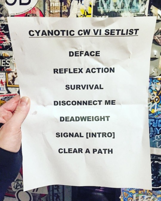
The Cya guys had great times on stage at @coldwavesfestival this past weekend. They did the thing, they did it well, and the #coldwaves crowd had fun @cyanotic #coldwavesfestival #coldwavesvi #setlist #cyanotic #technoir #cyaguys #deface #wedesignthefuture #cyberpunk #postcyberpunk #clearapath #glitchmode #glitchmoderecordings #metrochicago #inmemory #newmusic #musiclove #chicago #industrialmusic #angryrobotmusic #mechanical #angryrobotnoises #ourrolemodelsaremachines #survivalinstincts #aperfectorganism #cybernated #signalthemachines #reflexaction #livemusic #disconnectme (at Metro Chicago)
#inmemory#glitchmoderecordings#cyaguys#ourrolemodelsaremachines#reflexaction#clearapath#technoir#aperfectorganism#coldwaves#angryrobotnoises#cybernated#mechanical#coldwavesfestival#cyanotic#livemusic#chicago#survivalinstincts#cyberpunk#glitchmode#wedesignthefuture#angryrobotmusic#newmusic#industrialmusic#deface#signalthemachines#metrochicago#coldwavesvi#setlist#postcyberpunk#musiclove
3 notes
·
View notes
Photo

PIBE Winter 2016/17 #Issue1 'INSTINCTS' #reflexaction photographed by @viktorflume styling @ursulawangander #pibemagazine #playitbyear #letspibe #instincts #beauty #fashion #art
2 notes
·
View notes
Text
Nervous system , memory and reflex actions

Nervous system
Nervous system The neurons will be the basic units of the nervous system. They assist in conducting the stimuli among the receptor organs - spinal-cord, brain and effector organs. The neurons do the stimulus as electrochemical occurrences. These sequential events involve the migration of ‘Na’ and ‘K’ ions outside and inside the neuronal cells. This phenomenon is recognized as SodiumPotassium pump. This sequence of electro substance events is known as the impulse.

nervous system The junctions of neurons in the nerve pathway are called the synapses. A synapse is created between the bulb-like end structure of the axon called boutons and the cyton or dendrite of the adjacent neuron. At the junction, there's a difference called the synaptic cleft, which is usually about 10 to 20 nm. At this time, transmission of stimulus happens through transmitter chemicals such as acetylcholine. Inside the nervous system, the bundles of parallel axons of the nervous tissue having myelin sheath constitute the white subject. The assortment of neurons having unmyelinated axons from the greyish subject. The axons constitute the white subject of the CNS for nerve tracts. They propagate action potentials. The greyish subject performs integrative functions. The outer surface of the brain (cortex) and the central section of the spinal cord consist of grey matter. Within the mind, collections of grey subject form centers called nuclei.
The brain
There are greater than a thousand million neurons in the adult mind. An estimate shows that the cerebral cortex exclusively has about 102783000 synapses. Thus the mind is a complicated organ. On the structural and useful basis, the mind can be divided into 3 regions. They can be (1). Forebrain, (2). Midbrain, (3). Hindbrain.
Fore Brain (Prosencephalon):-
This region of the mind comprises Diencephalon and the cerebrum. The diencephalon is formed of thalamus and hypothalamus.
Thalamus:-
It's the largest area of the diencephalon. This region includes a cluster of nuclei. A lot of the sensory inputs are conducted to the cerebral cortex through the thalamus. Axons having auditory, aesthetic and other sensory information synapses with specific nuclei of the region. This region may also impact mood and basic body movements credited to strong feelings such as fear or anger.
Hypothalamus :
This region contains small nuclei and nerve tracts. The nuclei called mamillary bodies are involved in olfactory reflexes and psychological replies to odors. The funnel formed infundibulum from the hypothalamus attaches it to the posterior pituitary or neurohypophysis. This region settings the secretions of the pituitary gland. It receives signals from sensory systems and external genitalia. It really is associated with emotional and mood connections. It provides a relaxed sense. Feeling good after a meal, rage and dread are also because of this region. In addition, it coordinates reactions to the sleep-wake cycle with other areas.
Cerebrum :
It is the largest area of the brain. It weighs about 1400g in men and 1200g in females. Bigger brains are usually associated with much larger bodies and not with greater intellect. The grey subject on the outer surface of the cerebrum is the cortex. It forms clusters deep inside the mind called nuclei. The internal area of the brain, among the cortex and the nuclei, have white matter known as-as cerebral medulla.
Cerebral cortex :
The cortex contains several primary sensory areas. These areas include taste area, most important auditory cortex for handling auditory stimuli, aesthetic cortex for perceiving visual images and areas for other cutaneous feelings. The cortical areas next to the principal sensory centers are called the association areas. These areas are involved in the procedure of recognition. For example, the sensory stimulus from the retina of the attention reaches the aesthetic association section of the cortex. Here the aesthetic information is weighed against past encounters. Further, this area has contacts with other areas of the cortex, which affect decisions. Thus aesthetic information is judged many times. This can be one of why two people who see the same event can present slightly different variations of what took place. The primary motor unit section of the cortex controls many voluntary activities, especially the finer electric motor activities of the hands. Muscles such as facial muscles, that contain many electric motor units have increased innervation. They can be represented by a big section of the motor cortex. Anterior to the primary motor area will be the premotor area. It is the staging area in which motor unit functions are structured before these are initiated in the motor cortex. For instance, in case a person decides to lift a palm, the neurons of the premotor area are stimulated first. This area decides the order and the amount to which the muscles must contract. This area is well toned only in primates and especially in humans. Our mental behavior and mood are handled by this area.
The midbrain or Mesencephalon :
The roof of the region contains four nuclei. The nuclei form mounds. They are simply collectively called corpora quadrigemina. It is produced of 2 superior colliculi or mounds and 2 second-rate colliculi or mounds. The superior colliculi get excited about visual reflexes. They control eyeball and head moves. They assist in aesthetic traffic monitoring of moving things. The inferior colliculi get excited about hearing.
The hindbrain or Rhombencephalon :
Hindbrain consists of three parts 1. Cerebellum, 2.Pons, and 3.Medulla oblongata.
Cerebellum :
This region communicates with other regions of the CNS through three large nerve tracts called the cerebellar peduncles. The cerebellum contains the following three parts Parts Control 1. flocculonodular balance and maintenance of muscle build. 2. vermis -anterior part engine coordination and muscle build. 3. vermis- posterior part fine electric motor coordination and Cerebellar dysfunction may cause decreased muscle build, imbalance and insufficient coordination.
Pons :
Pons works as a relays station between the cerebrum and the cerebellum. It also contains sleep center and respiratory centers. These centers along with medulla help control respiratory movements.
Medulla oblongata :
It functions as a conduction pathway for both ascending and descending nerve tracts. The nuclei inside medulla oblongata work as centers of several reflexes mixed up in regulation of heart rate, blood vessels vessel contractions, breathing, swallowing, vomiting, coughing and sneezing.
Memory
The term ‘memory means the ability of the brain to store information. The duration of storage area varies from a couple of seconds or hours to several years.
Types of memory :
1. Sensory memory space:- It means the ability to retain sensory impulses in the sensory regions of the brain for a short interval of time following the genuine sensory experience. This is actually the initial level of storage area process. 2. Primary memory space:- It's the storage area of facts, words, numbers, words or other information. The info in this storage is instantaneously made available to a person do not need to search through his / her mind for this. 3. Secondary memory:- It's the storage in the mind of information that can be recalled at some later time(hours, days, months or years later). That is also called long-term memory, fixed memory or permanent memory.
Physiology of memory:-
Certain anatomical, physical or chemical changes occur in the presynaptic terminals (a part in the neuron) or simply in whole neurons that permanently facilitate the transmitting of impulses at the synapses. All of the synapses (nerves junctions) are thus facilitated in a thought circuit. This circuit can be re-excited by anybody of many diverse indicators at later dates thereby causing storage. The entire facilitated circuit is named a storage area engram or a ram trace. Amnesia:- Amnesia means memory space loss. It is the inability to recall memories from days gone by.
Conditioned reflex
The cerebral cortex adjustments the conditioned reflexes. Additionally, it is called the traditional conditioning, in which a stimulus comes to elicit a reply similar to the response produced by some other stimulus. In his test, Pavlov presented your dog with a sound of a bell (Neutral stimulus) followed by meat (Unconditioned stimulus). This blended stimuli stimulated your dog to salivate (unconditioned response). After many such conditioned pairings, the sound together (Conditioned stimulus) would promote the dog to salivate (Conditioned response - CR). The bell sound, in the beginning, called the neutral stimulus which after lots of trials pairing with the unconditioned stimulus (UCS) viz., the beef part, becomes the conditioned stimulus. Similarly, the salvation of your dog during the original tests of pairing (NS + UCS) is named Unconditioned Response (UR). This unconditioned response, later on, becomes chosen into a conditioned response (CR). It'll be elicited simply by bell sound (CS). The conditioned reflexes are developed in an animal’s life in association with previously established reflexes. This is actually the basis of learning and storage.
Electroencephalography (EEG)
The tissues of the cerebral cortex contain a huge selection of neuronal elements. The mind tissue is characterized by a continuing rhythmic oscillation of electric potential. EEG is a tool to record the electrically powered activity of the mind via electrodes mounted on the head. It exhibits a worldwide web average of all the neuron’s potential. EEG produces information about the mind function in health insurance and disease. It offers data about brain functions during various functional or behavioral activities such as sleeping or waking areas. Additionally, it is found in the diagnosis of brain disease such as tumor, lesions and also in epilepsy.
Right and Still left brain concept
Within the cerebral cortex, the remaining hemisphere is linked to your skin receptors in the right 1 / 2 of the body. In addition, it handles the muscles in the right area of your body. The right hemisphere is linked to the sensory receptors on the eventually left half of your body. Further, it adjustments muscles on the remaining side. Both hemispheres are also linked to the eyes. The sensory nerve tracts of the kept eye are linked to the right hemisphere and the right eyes left hemisphere through the optic chiasma. Thus the kept half views the right 50 percent of the world as the right half recognizes only the remaining 50 percent of the world. The left and right part share information through a group of axons known as the corpus callosum, and also through anterior commissure and hippocampal commissure. Both hemispheres have their own specific functions and the above-mentioned section of functions or labor between your two hemispheres is called ‘lateralization’. The corpus callosum permits the right and left 1 / 2 of the brain to use cooperatively, rather than separately. The anterior commissure works an important role in unifying the emotional replies of two sides of the brain. Damage to the corpus callosum blocks the exchange of information between your two hemispheres. The left brain can be involved with language, number skills, reasoning, spoken language, scientific skills, and right-hand control. The proper brain can be involved with art awareness, imagination, visual functions, emotions, music awareness, 3D forms and eventually left-hand controls. The kept brain is intended for analytical tasks as the right brain is for creating jobs.
Spinal-cord functioning
The spinal-cord remains as a connecting, functional stressed structure in between the brain and sensory / effector organs. The sensory inputs received by sense organs are conducted towards appropriate parts of the brain. Likewise, from the mind motor sensations are sent towards effector buildings. Further, as the mind, the spinal cord can affect the motor unit initiation and cause an impact. This activity is recognized as a reflex action.
Reflex action
Reflex action is the spontaneously involuntary response caused credited to stimulation of receptor organ. E.g. The quick closure of eyes lid when some subject details the eyelashes.; the unexpected withdrawal of hands when the hand details hot pan. A reflex action can be an involuntary process and will not involve the treatment of consciousness. The anatomical basis of reflex action is the reflex arc. It really is a nerve string between receptor organ and effector organ. The reflex arc gets the following route. Sensory organ → sensory or afferent neuron → greyish matter of the spinal-cord → intermediary or relay neuron → efferent or motor neuron → effector organ.
Cerebro Spinal Substance (CSF)
The ventricle of the mind and the central canal of the spinal-cord contain, a fluid just like plasma called cerebrospinal fluid (CSF). CSF is created by several cells called the choroid plexus located inside the four ventricles. In individual, the volume of CSF is 150 ml and the pace of its secretion is 550 ml/day. Functions: 1. CSF cushions the mind against mechanical impact when the head moves. 2. It serves as a protective covering for the CNS and confers buoyancy to the brain. 3. The CSF also offers a reservoir of hormones and diet for the mind and spinal cord. 4. It acts as a mechanical buffer. Remaining inside and outside the CNS, it equalizes the mechanical pressure. When the intracranial pressure will go up the CSF is pressured out. In case the pressure tends to land, more CSF is maintained. Read the full article
0 notes