#MIT Institute for Medical Engineering and Science (IMES)
Explore tagged Tumblr posts
Visit Tumblr Blog
Explore Tumblr blogs with no restrictions, modern design and the best experience.
Last Seen Tumblr Blogs


Fun Fact
Tumblr is available in 18 languages.
Text
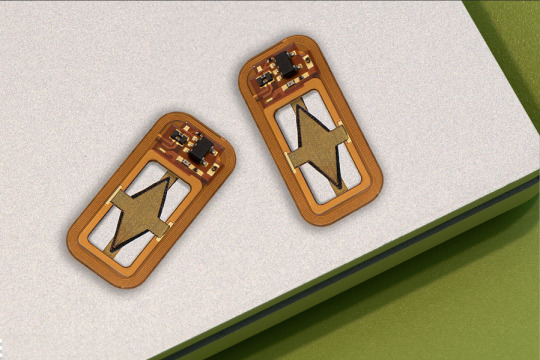
An implantable device could enable injection-free control of diabetes
The device contains encapsulated cells that produce insulin, plus a tiny oxygen-producing factory that keeps the cells healthy.
Anne Trafton | MIT News
One promising approach to treating Type 1 diabetes is implanting pancreatic islet cells that can produce insulin when needed, which can free patients from giving themselves frequent insulin injections. However, one major obstacle to this approach is that once the cells are implanted, they eventually run out of oxygen and stop producing insulin.
To overcome that hurdle, MIT engineers have designed a new implantable device that not only carries hundreds of thousands of insulin-producing islet cells, but also has its own on-board oxygen factory, which generates oxygen by splitting water vapor found in the body.
The researchers showed that when implanted into diabetic mice, this device could keep the mice’s blood glucose levels stable for at least a month. The researchers now hope to create a larger version of the device, about the size of a stick of chewing gum, that could eventually be tested in people with Type 1 diabetes.
“You can think of this as a living medical device that is made from human cells that secrete insulin, along with an electronic life support-system. We’re excited by the progress so far, and we really are optimistic that this technology could end up helping patients,” says Daniel Anderson, a professor in MIT’s Department of Chemical Engineering, a member of MIT’s Koch Institute for Integrative Cancer Research and Institute for Medical Engineering and Science (IMES), and the senior author of the study.
While the researchers’ main focus is on diabetes treatment, they say that this kind of device could also be adapted to treat other diseases that require repeated delivery of therapeutic proteins.
MIT Research Scientist Siddharth Krishnan is the lead author of the paper, which appears today in the Proceedings of the National Academy of Sciences. The research team also includes several other researchers from MIT, including Robert Langer, the David H. Koch Institute Professor at MIT and a member of the Koch Institute, as well as researchers from Boston Children’s Hospital.
Replacing injections
Most patients with Type 1 diabetes have to monitor their blood glucose levels carefully and inject themselves with insulin at least once a day. However, this process doesn’t replicate the body’s natural ability to control blood glucose levels.
“The vast majority of diabetics that are insulin-dependent are injecting themselves with insulin, and doing their very best, but they do not have healthy blood sugar levels,” Anderson says. “If you look at their blood sugar levels, even for people that are very dedicated to being careful, they just can’t match what a living pancreas can do.”
A better alternative would be to transplant cells that produce insulin whenever they detect surges in the patient’s blood glucose levels. Some diabetes patients have received transplanted islet cells from human cadavers, which can achieve long-term control of diabetes; however, these patients have to take immunosuppressive drugs to prevent their body from rejecting the implanted cells.
More recently, researchers have shown similar success with islet cells derived from stem cells, but patients who receive those cells also need to take immunosuppressive drugs.
Another possibility, which could prevent the need for immunosuppressive drugs, is to encapsulate the transplanted cells within a flexible device that protects the cells from the immune system. However, finding a reliable oxygen supply for these encapsulated cells has proven challenging.
Some experimental devices, including one that has been tested in clinical trials, feature an oxygen chamber that can supply the cells, but this chamber needs to be reloaded periodically. Other researchers have developed implants that include chemical reagents that can generate oxygen, but these also run out eventually.
The MIT team took a different approach that could potentially generate oxygen indefinitely, by splitting water. This is done using a proton-exchange membrane — a technology originally deployed to generate hydrogen in fuel cells — located within the device. This membrane can split water vapor (found abundantly in the body) into hydrogen, which diffuses harmlessly away, and oxygen, which goes into a storage chamber that feeds the islet cells through a thin, oxygen-permeable membrane.
A significant advantage of this approach is that it does not require any wires or batteries. Splitting this water vapor requires a small voltage (about 2 volts), which is generated using a phenomenon known as resonant inductive coupling. A tuned magnetic coil located outside the body transmits power to a small, flexible antenna within the device, allowing for wireless power transfer. It does require an external coil, which the researchers anticipate could be worn as a patch on the patient’s skin.
Drugs on demand
After building their device, which is about the size of a U.S. quarter, the researchers tested it in diabetic mice. One group of mice received the device with the oxygen-generating, water-splitting membrane, while the other received a device that contained islet cells without any supplemental oxygen. The devices were implanted just under the skin, in mice with fully functional immune systems.
The researchers found that mice implanted with the oxygen-generating device were able to maintain normal blood glucose levels, comparable to healthy animals. However, mice that received the nonoxygenated device became hyperglycemic (with elevated blood sugar) within about two weeks.
Typically when any kind of medical device is implanted in the body, attack by the immune system leads to a buildup of scar tissue called fibrosis, which can reduce the devices’ effectiveness. This kind of scar tissue did form around the implants used in this study, but the device’s success in controlling blood glucose levels suggests that insulin was still able to diffuse out of the device, and glucose into it.
This approach could also be used to deliver cells that produce other types of therapeutic proteins that need to be given over long periods of time. In this study, the researchers showed that the device could also keep alive cells that produce erythropoietin, a protein that stimulates red blood cell production.
“We’re optimistic that it will be possible to make living medical devices that can reside in the body and produce drugs as needed,” Anderson says. “There are a variety of diseases where patients need to take proteins exogenously, sometimes very frequently. If we can replace the need for infusions every other week with a single implant that can act for a long time, I think that could really help a lot of patients.”
The researchers now plan to adapt the device for testing in larger animals and eventually humans. For human use, they hope to develop an implant that would be about the size of a stick of chewing gum. They also plan to test whether the device can remain in the body for longer periods of time.
“The materials we’ve used are inherently stable and long-lived, so I think that kind of long-term operation is within the realm of possibility, and that’s what we’re working on,” Krishnan says.
“We are very excited about these findings, which we believe could provide a whole new way of someday treating diabetes and possibly other diseases,” Langer adds.
The research was funded by JDRF, the Leona M. and Harry B. Helmsley Charitable Trust, and the National Institute of Biomedical Imaging and Bioengineering at the National Institutes of Health.
Make sure to follow us on Tumblr!
5 notes
·
View notes
Text
MIT engineers design a robotic replica of the heart’s right chamber

MIT engineers design a robotic replica of the heart’s right chamber. The realistic model could aid the development of better heart implants and shed light on understudied heart disorders. Robotic Replica - A ballet of beats A heart’s shelf-life December 08, 2023 - MIT engineers have created a robotic replica of the right ventricle of the heart that mimics the beating and blood-pumping action of a living heart. The robo-ventricle is made up of real heart tissue and synthetic, balloon-like artificial muscles that allow scientists to control the ventricle's contractions while also observing how its natural valves and other intricate structures work. The artificial ventricle can be programmed to simulate both healthy and diseased states. The researchers manipulated the model to simulate right ventricular dysfunction conditions such as pulmonary hypertension and myocardial infarction. The model was also used to test cardiac devices. For example, the researchers implanted a mechanical valve to repair a naturally malfunctioning valve, then observed how the ventricle's pumping changed as a result. They claim that the new robotic right ventricle, or RRV, can be used as a realistic platform for studying right ventricle disorders and testing devices and therapies to treat them. “The right ventricle is particularly susceptible to dysfunction in intensive care unit settings, especially in patients on mechanical ventilation,” says Manisha Singh, a postdoc at MIT’s Institute for Medical Engineering and Science (IMES). “The RRV simulator can be used in the future to study the effects of mechanical ventilation on the right ventricle and to develop strategies to prevent right heart failure in these vulnerable patients.” Singh and her colleagues report details of the new design in an open-access paper appearing today in Nature Cardiovascular Research. Her co-authors include Associate Professor Ellen Roche, who is a core member of IMES and the associate head for research in the Department of Mechanical Engineering at MIT; along with Jean Bonnemain, Caglar Ozturk, Clara Park, Diego Quevedo-Moreno, Meagan Rowlett, and Yiling Fan of MIT; Brian Ayers of Massachusetts General Hospital; Christopher Nguyen of Cleveland Clinic; and Mossab Saeed of Boston Children’s Hospital.
Robotic Replica - A ballet of beats
The right ventricle is one of the heart’s four chambers, along with the left ventricle and the left and right atria. Of the four chambers, the left ventricle is the heavy lifter, as its thick, cone-shaped musculature is built for pumping blood through the entire body. The right ventricle, Roche says, is a “ballerina” in comparison, as it handles a lighter though no-less-crucial load. “The right ventricle pumps deoxygenated blood to the lungs, so it doesn’t have to pump as hard,” Roche notes. “It’s a thinner muscle, with more complex architecture and motion.” This anatomical complexity has made it difficult for clinicians to accurately observe and assess right ventricle function in patients with heart disease. “Conventional tools often fail to capture the intricate mechanics and dynamics of the right ventricle, leading to potential misdiagnoses and inadequate treatment strategies,” Singh says To improve understanding of the lesser-known chamber and speed the development of cardiac devices to treat its dysfunction, the team designed a realistic, functional model of the right ventricle that both captures its anatomical intricacies and reproduces its pumping function. The model includes real heart tissue, which the team chose to incorporate because it retains natural structures that are too complex to reproduce synthetically. “There are thin, tiny chordae and valve leaflets with different material properties that are all moving in concert with the ventricle’s muscle. Trying to cast or print these very delicate structures is quite challenging,” Roche explains
A heart’s shelf-life
In the new study, the team reports explanting a pig’s right ventricle, which they treated to carefully preserve its internal structures. They then fit a silicone wrapping around it, which acted as a soft, synthetic myocardium, or muscular lining. Within this lining, the team embedded several long, balloon-like tubes, which encircled the real heart tissue, in positions that the team determined through computational modeling to be optimal for reproducing the ventricle’s contractions. The researchers connected each tube to a control system, which they then set to inflate and deflate each tube at rates that mimicked the heart’s real rhythm and motion. To test its pumping ability, the team infused the model with a liquid similar in viscosity to blood. This particular liquid was also transparent, allowing the engineers to observe with an internal camera how internal valves and structures responded as the ventricle pumped liquid through. They found that the artificial ventricle’s pumping power and the function of its internal structures were similar to what they previously observed in live, healthy animals, demonstrating that the model can realistically simulate the right ventricle’s action and anatomy. The researchers could also tune the frequency and power of the pumping tubes to mimic various cardiac conditions, such as irregular heartbeats, muscle weakening, and hypertension. “We’re reanimating the heart, in some sense, and in a way that we can study and potentially treat its dysfunction,” Roche says To show that the artificial ventricle can be used to test cardiac devices, the team surgically implanted ring-like medical devices of various sizes to repair the chamber’s tricuspid valve — a leafy, one-way valve that lets blood into the right ventricle. When this valve is leaky, or physically compromised, it can cause right heart failure or atrial fibrillation, and leads to symptoms such as reduced exercise capacity, swelling of the legs and abdomen, and liver enlargement. The researchers surgically manipulated the robo-ventricle’s valve to simulate this condition, then either replaced it by implanting a mechanical valve or repaired it using ring-like devices of different sizes. They observed which device improved the ventricle’s fluid flow as it continued to pump. “With its ability to accurately replicate tricuspid valve dysfunction, the RRV serves as an ideal training ground for surgeons and interventional cardiologists,” Singh says. “They can practice new surgical techniques for repairing or replacing the tricuspid valve on our model before performing them on actual patients.” Currently, the RRV can simulate realistic function over a few months. The team is working to extend that performance and enable the model to run continuously for longer stretches. They are also working with designers of implantable devices to test their prototypes on the artificial ventricle and possibly speed their path to patients. And looking far in the future, Roche plans to pair the RRV with a similar artificial, functional model of the left ventricle, which the group is currently fine-tuning. “We envision pairing this with the left ventricle to make a fully tunable, artificial heart, that could potentially function in people,” Roche says. “We’re quite a while off, but that’s the overarching vision.” This research was supported, in part, by the National Science Foundation. Source: MIT Read the full article
1 note
·
View note
Text
Collin Stultz named co-director and MIT lead of the Harvard-MIT Program in Health Sciences and Technology | MIT News
Collin Stultz named co-director and MIT lead of the Harvard-MIT Program in Health Sciences and Technology | MIT News
Collin M. Stultz, the Nina T. and Robert H. Rubin Professor in Medical Engineering and Science at MIT, has been named co-director of the Harvard-MIT Program in Health Sciences and Technology (HST), and associate director of MIT’s Institute for Medical Engineering and Science (IMES), effective June 1. IMES is HST’s home at MIT. Stultz is a professor of electrical engineering and computer science…

View On WordPress
#Collin Stultz#Emery N. Brown#Harvard-MIT Health Sciences and Technology (HST)#MIT Institute for Medical Engineering and Science (IMES)#MIT Research Laboratory of Electronics
0 notes
Photo

MIT Engineers Create an Inhalable Form of Messenger RNA
Messenger RNA, which can induce cells to produce therapeutic proteins, holds great promise for treating a variety of diseases. The biggest obstacle to this approach so far has been finding safe and efficient ways to deliver mRNA molecules to the target cells.
In an advance that could lead to new treatments for lung disease, MIT researchers have now designed an inhalable form of mRNA. This aerosol could be administered directly to the lungs to help treat diseases such as cystic fibrosis, the researchers say.
“We think the ability to deliver mRNA via inhalation could allow us to treat a range of different diseases of the lung,” says Daniel Anderson, an associate professor in MIT’s Department of Chemical Engineering, a member of MIT’s Koch Institute for Integrative Cancer Research and Institute for Medical Engineering and Science (IMES), and the senior author of the study.
The researchers showed that they could induce lung cells in mice to produce a target protein — in this case, a bioluminescent protein. If the same success rate can be achieved with therapeutic proteins, that could be high enough to treat many lung diseases, the researchers say.
Read more.
53 notes
·
View notes
Link
Artificial intelligence yields new antibiotic
A deep-learning model identifies a powerful new drug that can kill many species of antibiotic-resistant bacteria.
Anne Trafton | MIT News Office
Using a machine-learning algorithm, MIT researchers have identified a powerful new antibiotic compound. In laboratory tests, the drug killed many of the world’s most problematic disease-causing bacteria, including some strains that are resistant to all known antibiotics. It also cleared infections in two different mouse models.
The computer model, which can screen more than a hundred million chemical compounds in a matter of days, is designed to pick out potential antibiotics that kill bacteria using different mechanisms than those of existing drugs.
“We wanted to develop a platform that would allow us to harness the power of artificial intelligence to usher in a new age of antibiotic drug discovery,” says James Collins, the Termeer Professor of Medical Engineering and Science in MIT’s Institute for Medical Engineering and Science (IMES) and Department of Biological Engineering. “Our approach revealed this amazing molecule which is arguably one of the more powerful antibiotics that has been discovered.”
In their new study, the researchers also identified several other promising antibiotic candidates, which they plan to test further. They believe the model could also be used to design new drugs, based on what it has learned about chemical structures that enable drugs to kill bacteria.
“The machine learning model can explore, in silico, large chemical spaces that can be prohibitively expensive for traditional experimental approaches,” says Regina Barzilay, the Delta Electronics Professor of Electrical Engineering and Computer Science in MIT’s Computer Science and Artificial Intelligence Laboratory (CSAIL).
Barzilay and Collins, who are faculty co-leads for MIT’s Abdul Latif Jameel Clinic for Machine Learning in Health (J-Clinic), are the senior authors of the study, which appears today in Cell. The first author of the paper is Jonathan Stokes, a postdoc at MIT and the Broad Institute of MIT and Harvard.
A new pipeline
Over the past few decades, very few new antibiotics have been developed, and most of those newly approved antibiotics are slightly different variants of existing drugs. Current methods for screening new antibiotics are often prohibitively costly, require a significant time investment, and are usually limited to a narrow spectrum of chemical diversity.
“We’re facing a growing crisis around antibiotic resistance, and this situation is being generated by both an increasing number of pathogens becoming resistant to existing antibiotics, and an anemic pipeline in the biotech and pharmaceutical industries for new antibiotics,” Collins says.
To try to find completely novel compounds, he teamed up with Barzilay, Professor Tommi Jaakkola, and their students Kevin Yang, Kyle Swanson, and Wengong Jin, who have previously developed machine-learning computer models that can be trained to analyze the molecular structures of compounds and correlate them with particular traits, such as the ability to kill bacteria.
The idea of using predictive computer models for “in silico” screening is not new, but until now, these models were not sufficiently accurate to transform drug discovery. Previously, molecules were represented as vectors reflecting the presence or absence of certain chemical groups. However, the new neural networks can learn these representations automatically, mapping molecules into continuous vectors which are subsequently used to predict their properties.
In this case, the researchers designed their model to look for chemical features that make molecules effective at killing E. coli. To do so, they trained the model on about 2,500 molecules, including about 1,700 FDA-approved drugs and a set of 800 natural products with diverse structures and a wide range of bioactivities.
Once the model was trained, the researchers tested it on the Broad Institute’s Drug Repurposing Hub, a library of about 6,000 compounds. The model picked out one molecule that was predicted to have strong antibacterial activity and had a chemical structure different from any existing antibiotics. Using a different machine-learning model, the researchers also showed that this molecule would likely have low toxicity to human cells.
This molecule, which the researchers decided to call halicin, after the fictional artificial intelligence system from “2001: A Space Odyssey,” has been previously investigated as possible diabetes drug. The researchers tested it against dozens of bacterial strains isolated from patients and grown in lab dishes, and found that it was able to kill many that are resistant to treatment, including Clostridium difficile, Acinetobacter baumannii, and Mycobacterium tuberculosis. The drug worked against every species that they tested, with the exception of Pseudomonas aeruginosa, a difficult-to-treat lung pathogen.
To test halicin’s effectiveness in living animals, the researchers used it to treat mice infected with A. baumannii, a bacterium that has infected many U.S. soldiers stationed in Iraq and Afghanistan. The strain of A. baumannii that they used is resistant to all known antibiotics, but application of a halicin-containing ointment completely cleared the infections within 24 hours.
Preliminary studies suggest that halicin kills bacteria by disrupting their ability to maintain an electrochemical gradient across their cell membranes. This gradient is necessary, among other functions, to produce ATP (molecules that cells use to store energy), so if the gradient breaks down, the cells die. This type of killing mechanism could be difficult for bacteria to develop resistance to, the researchers say.
“When you’re dealing with a molecule that likely associates with membrane components, a cell can’t necessarily acquire a single mutation or a couple of mutations to change the chemistry of the outer membrane. Mutations like that tend to be far more complex to acquire evolutionarily,” Stokes says.
In this study, the researchers found that E. coli did not develop any resistance to halicin during a 30-day treatment period. In contrast, the bacteria started to develop resistance to the antibiotic ciprofloxacin within one to three days, and after 30 days, the bacteria were about 200 times more resistant to ciprofloxacin than they were at the beginning of the experiment.
The researchers plan to pursue further studies of halicin, working with a pharmaceutical company or nonprofit organization, in hopes of developing it for use in humans.
Optimized molecules
After identifying halicin, the researchers also used their model to screen more than 100 million molecules selected from the ZINC15 database, an online collection of about 1.5 billion chemical compounds. This screen, which took only three days, identified 23 candidates that were structurally dissimilar from existing antibiotics and predicted to be nontoxic to human cells.
In laboratory tests against five species of bacteria, the researchers found that eight of the molecules showed antibacterial activity, and two were particularly powerful. The researchers now plan to test these molecules further, and also to screen more of the ZINC15 database.
The researchers also plan to use their model to design new antibiotics and to optimize existing molecules. For example, they could train the model to add features that would make a particular antibiotic target only certain bacteria, preventing it from killing beneficial bacteria in a patient’s digestive tract.
“This groundbreaking work signifies a paradigm shift in antibiotic discovery and indeed in drug discovery more generally,” says Roy Kishony, a professor of biology and computer science at Technion (the Israel Institute of Technology), who was not involved in the study. “Beyond in silica screens, this approach will allow using deep learning at all stages of antibiotic development, from discovery to improved efficacy and toxicity through drug modifications and medicinal chemistry.”
The research was funded by the Abdul Latif Jameel Clinic for Machine Learning in Health, the Defense Threat Reduction Agency, the Broad Institute, the DARPA Make-It Program, the Canadian Institutes of Health Research, the Canadian Foundation for Innovation, the Canada Research Chairs Program, the Banting Fellowships Program, the Human Frontier Science Program, the Pershing Square Foundation, the Swiss National Science Foundation, a National Institutes of Health Early Investigator Award, the National Science Foundation Graduate Research Fellowship Program, and a gift from Anita and Josh Bekenstein.
Topics: ResearchBiological engineeringElectrical Engineering & Computer Science (eecs)Institute for Medical Engineering and Science (IMES)Computer Science and Artificial Intelligence Laboratory (CSAIL)Broad InstituteSchool of EngineeringBacteriaMicrobesMedicineHealthMachine learningArtificial intelligenceAlgorithmsJ-ClinicNational Institutes of Health (NIH)National Science Foundation (NSF)
0 notes
Text
Covid-19 Diagnostic Based on MIT Technology Might be tested on Patient Samples Soon

As more Covid-19 cases appear in the United States and around the world, the need for fast, easy-to-use diagnostic tests is becoming ever more pressing. A startup company spun out from MIT is now working on a paper-based test that can deliver results in under half an hour, based on technology developed at MIT’s Institute for Medical Engineering and Science (IMES). Cambridge-based E25Bio, which developed the test, is now preparing to submit it to the FDA for emergency use authorization, which would grant temporary approval for using the device on patient samples during public health emergencies.
The technology behind the new E25Bio diagnostic was developed by Lee Gehrke, the Hermann L.F. von Helmholtz Professor at IMES, and other members of his lab, including Irene Bosch, a former IMES research scientist who is now the CTO of E25Bio. The tests consist of strips of paper that are coated with antibodies that bind to a specific viral protein. A second antibody is attached to gold nanoparticles, and the patient’s sample is added to a solution of those particles. The test strip is then dipped in this solution. If the viral protein is present, it attaches to the antibodies on the paper strip as well as the nanoparticle-bound antibodies, and a colored spot appears on the strip within 20 minutes.
Continue:-
The post Covid-19 Diagnostic Based on MIT Technology Might be tested on Patient Samples Soon appeared first on Gadgets, gizmos, and tech from the East.
Covid-19 Diagnostic Based on MIT Technology Might be tested on Patient Samples Soon published first on https://greatestspeakers.tumblr.com/
0 notes
Text
Alex Shalek named director of the Institute for Medical Engineering and Science
New Post has been published on https://thedigitalinsider.com/alex-shalek-named-director-of-the-institute-for-medical-engineering-and-science/
Alex Shalek named director of the Institute for Medical Engineering and Science

Alex K. Shalek, the J. W. Kieckhefer Professor in the MIT Institute for Medical Engineering and Sciences (IMES) and Department of Chemistry, has been named the new director of IMES, effective Aug. 1.
“Professor Shalek’s substantial contributions to the scientific community as a researcher and educator have been exemplary. His extensive network across MIT, Harvard, and Mass General Brigham will be a tremendous asset as director of IMES,” says Anantha Chandrakasan, chief innovation and strategy officer, dean of the School of Engineering, and the Vannevar Bush Professor of Electrical Engineering and Computer Science. “He will undoubtedly be an excellent leader, bringing his innovative approach and collaborative spirit to this new role.”
Shalek is a core member of IMES, a professor of chemistry, and holds several leadership positions, including director of the Health Innovation Hub. He is also an extramural member of MIT’s Koch Institute for Integrative Cancer Research; a member of the Ragon Institute of Mass General, MIT, and Harvard; an institute member of the Broad Institute of MIT and Harvard; an assistant in immunology at Mass General Brigham; and an instructor in health sciences and technology at Harvard Medical School.
The Shalek Lab’s research seeks to uncover how communities of cells work together within human tissues to support health, and how they become dysregulated in disease. By developing and applying innovative experimental and computational technologies, they are shedding light on a wide range of human health conditions.
Shalek and his team use a cross-disciplinary approach that combines genomics, chemical biology, and nanotechnology to develop platforms to profile and control cells and their interactions. Collaborating with researchers across the globe, they apply these tools to study human diseases in great detail. Their goal is to connect what occurs at a cellular level with what medical professionals observe in patients, paving the way for more precise ways to prevent and treat diseases.
Over the course of his career, Shalek’s groundbreaking research has earned him widespread recognition and numerous awards and honors. These include an NIH New Innovator Award, a Beckman Young Investigator Award, a Searle Scholar Award, a Pew-Stewart Scholar Award, an Alfred P. Sloan Research Fellowship in Chemistry, and an Avant-Garde (DP1 Pioneer) Award. Shalek has also been celebrated for his dedication as a faculty member, educator, and mentor. He was awarded the 2019-20 Harold E. Edgerton Faculty Achievement Award at MIT and the 2020 HMS Young Mentor Award.
Shalek received his bachelor’s degree in chemical physics from Columbia University and his master’s and PhD in chemical physics from Harvard University. Prior to joining MIT’s faculty in 2014, he was a postdoc at the Broad Institute.
Shalek succeeds Elazer Edelman, the Edward J. Poitras Professor in Medical Engineering and Science, who has led IMES since April 2018.
“I am grateful to Professor Edelman for his incredible leadership and service to IMES over the past six years,” says Chandrakasan. “His contributions to IMES have been invaluable, and we are thankful for his dedication and vision during his tenure as director.”
#approach#Biology#Broad Institute#Cancer#career#Cells#chemical#chemistry#collaborative#Community#computer#Computer Science#course#Disease#Diseases#Edgerton#engineering#experimental#Faculty#genomics#harvard#Harvard-MIT Health Sciences and Technology#Health#Health sciences and technology#how#human#Human health#immunology#Innovation#Institute for Medical Engineering and Science (IMES)
0 notes
Text
An alternative ultrasound technique not requiring contact with the body to see inside a patient

- By Nuadox Crew -
MIT engineers have conceived an alternative technique to conventional ultrasound which does not require contact with the body to see inside a patient. This new method utilizes an eye- and skin-safe laser system to remotely image the inside of a person. Such could become quite useful to assess patients in hard-to-reach places.
For most people, getting an ultrasound is a relatively easy procedure: As a technician gently presses a probe against a patient’s skin, sound waves generated by the probe travel through the skin, bouncing off muscle, fat, and other soft tissues before reflecting back to the probe, which detects and translates the waves into an image of what lies beneath.
Conventional ultrasound doesn’t expose patients to harmful radiation as X-ray and CT scanners do, and it’s generally noninvasive. But it does require contact with a patient’s body, and as such, may be limiting in situations where clinicians might want to image patients who don’t tolerate the probe well, such as babies, burn victims, or other patients with sensitive skin. Furthermore, ultrasound probe contact induces significant image variability, which is a major challenge in modern ultrasound imaging.
Now, MIT engineers have come up with an alternative to conventional ultrasound that doesn’t require contact with the body to see inside a patient. The new laser ultrasound technique leverages an eye- and skin-safe laser system to remotely image the inside of a person. When trained on a patient’s skin, one laser remotely generates sound waves that bounce through the body. A second laser remotely detects the reflected waves, which researchers then translate into an image similar to conventional ultrasound.
In a paper published today by Nature in the journal Light: Science and Applications, the team reports generating the first laser ultrasound images in humans. The researchers scanned the forearms of several volunteers and observed common tissue features such as muscle, fat, and bone, down to about 6 centimeters below the skin. These images, comparable to conventional ultrasound, were produced using remote lasers focused on a volunteer from half a meter away.

Image: A new ultrasound technique uses lasers to produce images beneath the skin, without making contact with the skin as conventional ultrasound probes do. The new laser ultrasound technique was used to produce an image (left) of a human forearm (above), which was also imaged using conventional ultrasound (right). Credit: MIT.
“We’re at the beginning of what we could do with laser ultrasound,” says Brian W. Anthony, a principal research scientist in MIT’s Department of Mechanical Engineering and Institute for Medical Engineering and Science (IMES), a senior author on the paper. “Imagine we get to a point where we can do everything ultrasound can do now, but at a distance. This gives you a whole new way of seeing organs inside the body and determining properties of deep tissue, without making contact with the patient.”
Early concepts for noncontact laser ultrasound for medical imaging originated from a Lincoln Laboratory program established by Rob Haupt of the Active Optical Systems Group and Chuck Wynn of the Advanced Capabilities and Technologies Group, who are co-authors on the new paper along with Matthew Johnson. From there, the research grew via collaboration with Anthony and his students, Xiang (Shawn) Zhang, who is now an MIT postdoc and is the paper’s first author, and recent doctoral graduate Jonathan Fincke, who is also a co-author. The project combined the Lincoln Laboratory researchers’ expertise in laser and optical systems with the Anthony group's experience with advanced ultrasound systems and medical image reconstruction.
Yelling into a canyon — with a flashlight
In recent years, researchers have explored laser-based methods in ultrasound excitation in a field known as photoacoustics. Instead of directly sending sound waves into the body, the idea is to send in light, in the form of a pulsed laser tuned at a particular wavelength, that penetrates the skin and is absorbed by blood vessels.
The blood vessels rapidly expand and relax — instantly heated by a laser pulse then rapidly cooled by the body back to their original size — only to be struck again by another light pulse. The resulting mechanical vibrations generate sound waves that travel back up, where they can be detected by transducers placed on the skin and translated into a photoacoustic image.
While photoacoustics uses lasers to remotely probe internal structures, the technique still requires a detector in direct contact with the body in order to pick up the sound waves. What’s more, light can only travel a short distance into the skin before fading away. As a result, other researchers have used photoacoustics to image blood vessels just beneath the skin, but not much deeper.
Since sound waves travel further into the body than light, Zhang, Anthony, and their colleagues looked for a way to convert a laser beam’s light into sound waves at the surface of the skin, in order to image deeper in the body.
Based on their research, the team selected 1,550-nanometer lasers, a wavelength which is highly absorbed by water (and is eye- and skin-safe with a large safety margin). As skin is essentially composed of water, the team reasoned that it should efficiently absorb this light, and heat up and expand in response. As it oscillates back to its normal state, the skin itself should produce sound waves that propagate through the body.
The researchers tested this idea with a laser setup, using one pulsed laser set at 1,550 nanometers to generate sound waves, and a second continuous laser, tuned to the same wavelength, to remotely detect reflected sound waves. This second laser is a sensitive motion detector that measures vibrations on the skin surface caused by the sound waves bouncing off muscle, fat, and other tissues. Skin surface motion, generated by the reflected sound waves, causes a change in the laser’s frequency, which can be measured. By mechanically scanning the lasers over the body, scientists can acquire data at different locations and generate an image of the region.
“It’s like we’re constantly yelling into the Grand Canyon while walking along the wall and listening at different locations,” Anthony says. “That then gives you enough data to figure out the geometry of all the things inside that the waves bounced against — and the yelling is done with a flashlight.”
In-home imaging
The researchers first used the new setup to image metal objects embedded in a gelatin mold roughly resembling skin’s water content. They imaged the same gelatin using a commercial ultrasound probe and found both images were encouragingly similar. They moved on to image excised animal tissue — in this case, pig skin — where they found laser ultrasound could distinguish subtler features, such as the boundary between muscle, fat, and bone.
Finally, the team carried out the first laser ultrasound experiments in humans, using a protocol that was approved by the MIT Committee on the Use of Humans as Experimental Subjects. After scanning the forearms of several healthy volunteers, the researchers produced the first fully noncontact laser ultrasound images of a human. The fat, muscle, and tissue boundaries are clearly visible and comparable to images generated using commercial, contact-based ultrasound probes.
The researchers plan to improve their technique, and they are looking for ways to boost the system’s performance to resolve fine features in the tissue. They are also looking to hone the detection laser’s capabilities. Further down the road, they hope to miniaturize the laser setup, so that laser ultrasound might one day be deployed as a portable device.
“I can imagine a scenario where you’re able to do this in the home,” Anthony says. “When I get up in the morning, I can get an image of my thyroid or arteries, and can have in-home physiological imaging inside of my body. You could imagine deploying this in the ambient environment to get an understanding of your internal state.”
Source: MIT
Read Also
New wearable ultrasound patch non-invasively monitors blood pressure in arteries
0 notes
Text
A new computational framework illuminates the hidden ecology of diseased tissues
New Post has been published on https://sunalei.org/news/a-new-computational-framework-illuminates-the-hidden-ecology-of-diseased-tissues/
A new computational framework illuminates the hidden ecology of diseased tissues

To understand what drives disease progression in tissues, scientists need more than just a snapshot of cells in isolation — they need to see where the cells are, how they interact, and how that spatial organization shifts across disease states. A new computational method called MESA (Multiomics and Ecological Spatial Analysis), detailed in a study published in Nature Genetics, is helping researchers study diseased tissues in more meaningful ways.
The work details the results of a collaboration between researchers from MIT, Stanford University, Weill Cornell Medicine, the Ragon Institute of MGH, MIT, and Harvard, and the Broad Institute of MIT and Harvard, and was led by the Stanford team.
MESA brings an ecology-inspired lens to tissue analysis. It offers a pipeline to interpret spatial omics data — the product of cutting-edge technology that captures molecular information along with the location of cells in tissue samples. These data provide a high-resolution map of tissue “neighborhoods,” and MESA helps make sense of the structure of that map.
“By integrating approaches from traditionally distinct disciplines, MESA enables researchers to better appreciate how tissues are locally organized and how that organization changes in different disease contexts, powering new diagnostics and the identification of new targets for preventions and cures,” says Alex K. Shalek, the director of the Institute for Medical Engineering and Science (IMES), the J. W. Kieckhefer Professor in IMES and the Department of Chemistry, and an extramural member of the Koch Institute for Integrative Cancer Research at MIT, as well as an institute member of the Broad Institute and a member of the Ragon Institute.
“In ecology, people study biodiversity across regions — how animal species are distributed and interact,” explains Bokai Zhu, MIT postdoc and author on the study. “We realized we could apply those same ideas to cells in tissues. Instead of rabbits and snakes, we analyze T cells and B cells.”
By treating cell types like ecological species, MESA quantifies “biodiversity” within tissues and tracks how that diversity changes in disease. For example, in liver cancer samples, the method revealed zones where tumor cells consistently co-occurred with macrophages, suggesting these regions may drive unique disease outcomes.
“Our method reads tissues like ecosystems, uncovering cellular ‘hotspots’ that mark early signs of disease or treatment response,” Zhu adds. “This opens new possibilities for precision diagnostics and therapy design.”
MESA also offers another major advantage: It can computationally enrich tissue data without the need for more experiments. Using publicly available single-cell datasets, the tool transfers additional information — such as gene expression profiles — onto existing tissue samples. This approach deepens understanding of how spatial domains function, especially when comparing healthy and diseased tissue.
In tests across multiple datasets and tissue types, MESA uncovered spatial structures and key cell populations that were previously overlooked. It integrates different types of omics data, such as transcriptomics and proteomics, and builds a multilayered view of tissue architecture.
Currently available as a Python package, MESA is designed for academic and translational research. Although spatial omics is still too resource-intensive for routine in-hospital clinical use, the technology is gaining traction among pharmaceutical companies, particularly for drug trials where understanding tissue responses is critical.
“This is just the beginning,” says Zhu. “MESA opens the door to using ecological theory to unravel the spatial complexity of disease — and ultimately, to better predict and treat it.”
0 notes
Text
MIT's implantable device could manage diabetes without injections

MIT's implantable device could manage diabetes without injections. The implantable device contains encapsulated cells that produce insulin, plus a tiny oxygen-producing factory that keeps the cells healthy. MIT's implantable device Replacing injections MIT's implantable device - Drugs on demand One promising approach to treating Type 1 diabetes is implanting pancreatic islet cells that can produce insulin when needed, which can free patients from giving themselves frequent insulin injections. However, one major obstacle to this approach is that once the cells are implanted, they eventually run out of oxygen and stop producing insulin. To overcome that hurdle, MIT engineers have designed a new implantable device that not only carries hundreds of thousands of insulin-producing islet cells, but also has its own on-board oxygen factory, which generates oxygen by splitting water vapor found in the body. The researchers showed that when implanted into diabetic mice, this device could keep the mice’s blood glucose levels stable for at least a month. The researchers now hope to create a larger version of the device, about the size of a stick of chewing gum, that could eventually be tested in people with Type 1 diabetes. “You can think of this as a living medical device that is made from human cells that secrete insulin, along with an electronic life support-system. We’re excited by the progress so far, and we really are optimistic that this technology could end up helping patients,” says Daniel Anderson, a professor in MIT’s Department of Chemical Engineering, a member of MIT’s Koch Institute for Integrative Cancer Research and Institute for Medical Engineering and Science (IMES), and the senior author of the study. says Daniel Anderson, a professor in MIT’s Department of Chemical Engineering, a member of MIT’s Koch Institute for Integrative Cancer Research and Institute for Medical Engineering and Science (IMES), and the senior author of the study.

Pictured is the device submerged in water, generating oxygen (bottom) and hydrogen (top) bubbles without the need for any batteries or wires. Credits: Image: Courtesy of Claudia Liu and Dr. Siddharth Krishnan, MIT/Boston Children’s Hospital
MIT's implantable device Replacing injections
Most patients with Type 1 diabetes have to monitor their blood glucose levels carefully and inject themselves with insulin at least once a day. However, this process doesn’t replicate the body���s natural ability to control blood glucose levels. “The vast majority of diabetics that are insulin-dependent are injecting themselves with insulin, and doing their very best, but they do not have healthy blood sugar levels,” Anderson says. “If you look at their blood sugar levels, even for people that are very dedicated to being careful, they just can’t match what a living pancreas can do.” A better alternative would be to transplant cells that produce insulin whenever they detect surges in the patient’s blood glucose levels. Some diabetes patients have received transplanted islet cells from human cadavers, which can achieve long-term control of diabetes; however, these patients have to take immunosuppressive drugs to prevent their body from rejecting the implanted cells. More recently, researchers have shown similar success with islet cells derived from stem cells, but patients who receive those cells also need to take immunosuppressive drugs. Another possibility, which could prevent the need for immunosuppressive drugs, is to encapsulate the transplanted cells within a flexible device that protects the cells from the immune system.
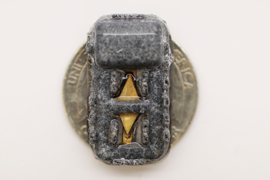
This photo shows the cathode side of fully assembled device, with a United States quarter-dollar coin for scale. Credits: Image: Courtesy of Claudia Liu and Dr. Siddharth Krishnan, MIT/Boston Children’s Hospital However, finding a reliable oxygen supply for these encapsulated cells has proven challenging. Some experimental devices, including one that has been tested in clinical trials, feature an oxygen chamber that can supply the cells, but this chamber needs to be reloaded periodically. Other researchers have developed implants that include chemical reagents that can generate oxygen, but these also run out eventually. The MIT team took a different approach that could potentially generate oxygen indefinitely, by splitting water. This is done using a proton-exchange membrane — a technology originally deployed to generate hydrogen in fuel cells — located within the device. This membrane can split water vapor (found abundantly in the body) into hydrogen, which diffuses harmlessly away, and oxygen, which goes into a storage chamber that feeds the islet cells through a thin, oxygen-permeable membrane. A significant advantage of this approach is that it does not require any wires or batteries. Splitting this water vapor requires a small voltage (about 2 volts), which is generated using a phenomenon known as resonant inductive coupling. A tuned magnetic coil located outside the body transmits power to a small, flexible antenna within the device, allowing for wireless power transfer. It does require an external coil, which the researchers anticipate could be worn as a patch on the patient’s skin.
MIT's implantable device - Drugs on demand
After building their device, which is about the size of a U.S. quarter, the researchers tested it in diabetic mice. One group of mice received the device with the oxygen-generating, water-splitting membrane, while the other received a device that contained islet cells without any supplemental oxygen. The devices were implanted just under the skin, in mice with fully functional immune systems. The researchers found that mice implanted with the oxygen-generating device were able to maintain normal blood glucose levels, comparable to healthy animals. However, mice that received the nonoxygenated device became hyperglycemic (with elevated blood sugar) within about two weeks. Typically when any kind of medical device is implanted in the body, attack by the immune system leads to a buildup of scar tissue called fibrosis, which can reduce the devices’ effectiveness. This kind of scar tissue did form around the implants used in this study, but the device’s success in controlling blood glucose levels suggests that insulin was still able to diffuse out of the device, and glucose into it. This approach could also be used to deliver cells that produce other types of therapeutic proteins that need to be given over long periods of time. In this study, the researchers showed that the device could also keep alive cells that produce erythropoietin, a protein that stimulates red blood cell production. “We’re optimistic that it will be possible to make living medical devices that can reside in the body and produce drugs as needed,” Anderson says. “There are a variety of diseases where patients need to take proteins exogenously, sometimes very frequently. If we can replace the need for infusions every other week with a single implant that can act for a long time, I think that could really help a lot of patients.” The researchers now plan to adapt the device for testing in larger animals and eventually humans. For human use, they hope to develop an implant that would be about the size of a stick of chewing gum. They also plan to test whether the device can remain in the body for longer periods of time. “The materials we’ve used are inherently stable and long-lived, so I think that kind of long-term operation is within the realm of possibility, and that’s what we’re working on,” Krishnan says. “We are very excited about these findings, which we believe could provide a whole new way of someday treating diabetes and possibly other diseases,” Langer adds. The research was funded by JDRF, the Leona M. and Harry B. Helmsley Charitable Trust, and the National Institute of Biomedical Imaging and Bioengineering at the National Institutes of Health. Source: MIT Read the full article
0 notes
Text
Collin Stultz named co-director and MIT lead of the Harvard-MIT Program in Health Sciences and Technology | MIT News
Collin Stultz named co-director and MIT lead of the Harvard-MIT Program in Health Sciences and Technology | MIT News
Collin M. Stultz, the Nina T. and Robert H. Rubin Professor in Medical Engineering and Science at MIT, has been named co-director of the Harvard-MIT Program in Health Sciences and Technology (HST), and associate director of MIT’s Institute for Medical Engineering and Science (IMES), effective June 1. IMES is HST’s home at MIT. Stultz is a professor of electrical engineering and computer science…
View On WordPress
#Collin Stultz#Emery N. Brown#Harvard-MIT Health Sciences and Technology (HST)#MIT Institute for Medical Engineering and Science (IMES)#MIT Research Laboratory of Electronics
0 notes
Text
Researchers produce first laser ultrasound images of humans
For most people, getting an ultrasound is a relatively easy procedure: As a technician gently presses a probe against a patient's skin, sound waves generated by the probe travel through the skin, bouncing off muscle, fat, and other soft tissues before reflecting back to the probe, which detects and translates the waves into an image of what lies beneath.
Conventional ultrasound doesn't expose patients to harmful radiation as X-ray and CT scanners do, and it's generally noninvasive. But it does require contact with a patient's body, and as such, may be limiting in situations where clinicians might want to image patients who don't tolerate the probe well, such as babies, burn victims, or other patients with sensitive skin. Furthermore, ultrasound probe contact induces significant image variability, which is a major challenge in modern ultrasound imaging.
Now, MIT engineers have come up with an alternative to conventional ultrasound that doesn't require contact with the body to see inside a patient. The new laser ultrasound technique leverages an eye- and skin-safe laser system to remotely image the inside of a person. When trained on a patient's skin, one laser remotely generates sound waves that bounce through the body. A second laser remotely detects the reflected waves, which researchers then translate into an image similar to conventional ultrasound.
In a paper published today by Nature in the journal Light: Science and Applications, the team reports generating the first laser ultrasound images in humans. The researchers scanned the forearms of several volunteers and observed common tissue features such as muscle, fat, and bone, down to about 6 centimeters below the skin. These images, comparable to conventional ultrasound, were produced using remote lasers focused on a volunteer from half a meter away.
"We're at the beginning of what we could do with laser ultrasound," says Brian W. Anthony, a principal research scientist in MIT's Department of Mechanical Engineering and Institute for Medical Engineering and Science (IMES), a senior author on the paper. "Imagine we get to a point where we can do everything ultrasound can do now, but at a distance. This gives you a whole new way of seeing organs inside the body and determining properties of deep tissue, without making contact with the patient."
Anthony's co-authors on the paper are lead author and MIT postdoc Xiang (Shawn) Zhang, recent doctoral graduate Jonathan Fincke, along with Charles Wynn, Matthew Johnson, and Robert Haupt of MIT's Lincoln Laboratory.
Yelling into a canyon -- with a flashlight
In recent years, researchers have explored laser-based methods in ultrasound excitation in a field known as photoacoustics. Instead of directly sending sound waves into the body, the idea is to send in light, in the form of a pulsed laser tuned at a particular wavelength, that penetrates the skin and is absorbed by blood vessels.
The blood vessels rapidly expand and relax -- instantly heated by a laser pulse then rapidly cooled by the body back to their original size -- only to be struck again by another light pulse. The resulting mechanical vibrations generate sound waves that travel back up, where they can be detected by transducers placed on the skin and translated into a photoacoustic image.
While photoacoustics uses lasers to remotely probe internal structures, the technique still requires a detector in direct contact with the body in order to pick up the sound waves. What's more, light can only travel a short distance into the skin before fading away. As a result, other researchers have used photoacoustics to image blood vessels just beneath the skin, but not much deeper.
Since sound waves travel further into the body than light, Zhang, Anthony, and their colleagues looked for a way to convert a laser beam's light into sound waves at the surface of the skin, in order to image deeper in the body.
Based on their research, the team selected 1,550-nanometer lasers, a wavelength which is highly absorbed by water (and is eye- and skin-safe with a large safety margin). As skin is essentially composed of water, the team reasoned that it should efficiently absorb this light, and heat up and expand in response. As it oscillates back to its normal state, the skin itself should produce sound waves that propagate through the body.
The researchers tested this idea with a laser setup, using one pulsed laser set at 1,550 nanometers to generate sound waves, and a second continuous laser, tuned to the same wavelength, to remotely detect reflected sound waves. This second laser is a sensitive motion detector that measures vibrations on the skin surface caused by the sound waves bouncing off muscle, fat, and other tissues. Skin surface motion, generated by the reflected sound waves, causes a change in the laser's frequency, which can be measured. By mechanically scanning the lasers over the body, scientists can acquire data at different locations and generate an image of the region.
"It's like we're constantly yelling into the Grand Canyon while walking along the wall and listening at different locations," Anthony says. "That then gives you enough data to figure out the geometry of all the things inside that the waves bounced against -- and the yelling is done with a flashlight."
In-home imaging
The researchers first used the new setup to image metal objects embedded in a gelatin mold roughly resembling skin's water content. They imaged the same gelatin using a commercial ultrasound probe and found both images were encouragingly similar. They moved on to image excised animal tissue -- in this case, pig skin -- where they found laser ultrasound could distinguish subtler features, such as the boundary between muscle, fat, and bone.
Finally, the team carried out the first laser ultrasound experiments in humans, using a protocol that was approved by the MIT Committee on the Use of Humans as Experimental Subjects. After scanning the forearms of several healthy volunteers, the researchers produced the first fully noncontact laser ultrasound images of a human. The fat, muscle, and tissue boundaries are clearly visible and comparable to images generated using commercial, contact-based ultrasound probes.
The researchers plan to improve their technique, and they are looking for ways to boost the system's performance to resolve fine features in the tissue. They are also looking to hone the detection laser's capabilities. Further down the road, they hope to miniaturize the laser setup, so that laser ultrasound might one day be deployed as a portable device.
"I can imagine a scenario where you're able to do this in the home," Anthony says. "When I get up in the morning, I can get an image of my thyroid or arteries, and can have in-home physiological imaging inside of my body. You could imagine deploying this in the ambient environment to get an understanding of your internal state."
This research was supported in part by the MIT Lincoln Laboratory Biomedical Line Program for the United States Air Force and by the U.S. Army Medical Research and Material Command's Military Operational Medicine Research Program.
0 notes
Text
A Short Review on Bio-compatible/Bio-degradable| Photopolymers for Stereolithography Bio-3D Printing - Juniper publishers

Abstract
One of the important procedures in Bio-3D printing is to print/fabricate the scaffold for tissue engineering. A scaffold is a porous biomedical implant, which provides a short-term support to seeded cells in order to direct the formation of new tissues. The scaffold must be non-toxic, biodegradable, biomechanical properties, specific chemical composition and have a precisely defined pore size and geometry. For this reason, it is very important to fabricate scaffolds with high precision. In this review, 3D printing of biomedical scaffolds using photo polymerization process is briefly reviewed. As per requirements of tissue engineering, the choice of best 3d printing method and photopolymer were discussed. Apart from this bio-3D printing application, the bio-compatible photopolymer will be widely used in dental application, like the direct printing of aligner in orthodontics and temporary denture fabrication.
Keywords: Tissue Engineering; Bio-compatible; Photopolymers; Stereolithography
Abbreviations: FDM: Fused Deposition Modelling; SLS: Selective Laser Sintering; 3DP: Three Dimensional Printing; LS: Laser Stereolithography; AM: Additive Manufacturing; DLP: Digital Light Projection; UV: Ultra-Violet;SL: Stereolithography; PCL: Polycaprolactone; PCL-DA: Polycaprolactone- Diacrylate; PGSA: Polyglycerol sebacate Acrylate
Introduction
In biomedical engineering, the tissue engineering is a rapidly growing multidisciplinary research area to reconstruct organs and tissues [1] by using a biodegradable and biocompatible scaffolding structure. As described early, it is very important to fabricate scaffolds with high precision. The scaffold has been fabricated using various 3D printing techniques such as FDM extrusion, SLS, 3DP. In these methods, the smallest printable size is 50-200 |im which is too large to be used for some biomedical implant or certain tissue engineering applications [2].
Stereolithography
LS is presently one ofthe most rapidly growing AM technique widely used in different areas of science and technology, engineering and biomedicine. LS is a polymerization process in which the spatially controlled solidification of resin is achieved using various types of laser radiations. The key benefit of using LS are high spatial resolution (-0.1 mm), fast manufacturing speed, high precision and a large variety of materials [3]. Commercially available LS 3D printers can print parts with an accuracy of 20|im. A recently developed two-photon polymerization LS setup can build micron-sized structures with sub-micron accuracy [4].
DLP is another emerging SL technique in which an array of millions of independently rotatable mirrors is used to project the light in order to polymerize photosensitive materials layer- by-layer under the action of UV or visible light [5]. LS is one of the best choice to fabricate scaffolds with required size and resolution but expensive. In a recent study, Jeng et al. [6] employed projection based SL technique and precisely printed biocompatible porous matrix structures for tissue engineering.
For 3D bio-printing of implants using Vat photo polymerization technology, the biological material need to benon-toxic, biocompatible and biodegradable photopolymer. Therefore, it is not easy to develop photopolymers for scaffolds and medical related applications. That's why they are rarely available commercially. There are biocompatible/ biodegradable polymers available, but mostly are not photo curable and need to be modified to make it photo curable. The photopolymer changes its structural properties when exposed to light, mostly cured by UV light. Photo polymeric materials consists of three main components including monomers (long- chain molecules), Photo initiators (split into radicals after energy input) and additives (UV stabilisators) [7]. A very limited number of photo-curable biocompatible materials are available for scaffold 3d printing. We are capable to describe only a few of them.
Bio-Compatible Photopolymers
PCL is a semi-crystalline polymer of aliphatic polyester group, which is a thermoplastic biodegradable material derived from crude oil by ring opening polymerization. Due to excellent biodegradability, high flexibility and biocompatibility, PCL is widely used in biomedical implants. In 2002, Kweon et al. [8] used PCL-diol with a molecular weight of 2000 for the preparation of PCL-DA through a series of chemical reactions. At Massachusetts Institute of Technology (MIT), another vital material PGSA was prepared in 2007, which is based on a chemical change of PGS with acryl ate moieties. Instead of PGS, PGSA is a rapidly cured material to form polymeric networks at ambient temperature. In a most recent research, Cheng et al. [9] prepared a photo-curable scaffold material by mixing PCL-DA and PGSA and concluded that the new material had improved mechanical properties compared to individuals.
Conclusion
Though several AM techniques are used for bio-3DP applications, Stereolithography setup can print porous biocompatible/biodegradable scaffolds with required size, resolution and surface finish. The bio-compatibility of the photopolymer is one of the most concerns in this VAT process.
PCL and PGSA are photo-curable non-toxic materials with excellent biocompatibility and mechanical properties, however, new materials can be made by combining liquid acrylated polymer precursor with other acrylated molecules for a number of potential biomedical applications. Definitely, there will be more new bio-compatible photopolymer material will be investigated to meet the application of bio 3D Printing and even bio-medical device like dental application.
To Know More About Trends in Technical and Scientific Research click on: https://juniperpublishers.com/ttsr/index.php
To Know More About Open Access Journals Please click on: https://juniperpublishers.com/index.php
0 notes
Text
Artificial intelligence yields new antibiotic never seen before
See on Scoop.it - Design, Comm, Sci and Tech
Using a new machine-learning algorithm, MIT researchers have identified a powerful antibiotic that can kill a wide range of species of pathogenic bacteria, including some that are resistant to all known antibiotics.
The computer model, which can screen more than a hundred million chemical compounds in a matter of days, is designed to pick out potential antibiotics that kill bacteria using different mechanisms than those of existing drugs. "We wanted to develop a platform that would allow us to harness the power of artificial intelligence to usher in a new age of antibiotic drug discovery," says James Collins, the Termeer Professor of Medical Engineering and Science in MIT's Institute for Medical Engineering and Science (IMES) and Department of Biological Engineering. "Our approach revealed this amazing molecule which is arguably one of the more powerful antibiotics that has been discovered."
0 notes
Text
Women in STEM — A celebration of excellence and curiosity
New Post has been published on https://thedigitalinsider.com/women-in-stem-a-celebration-of-excellence-and-curiosity/
Women in STEM — A celebration of excellence and curiosity
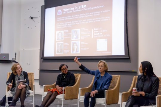
What better way to commemorate Women’s History Month and International Women’s Day than to give three of the world’s most accomplished scientists an opportunity to talk about their careers? On March 7, MindHandHeart invited professors Paula Hammond, Ann Graybiel, and Sangeeta Bhatia to share their career journeys, from the progress they have witnessed to the challenges they have faced as women in STEM. Their conversation was moderated by Mary Fuller, chair of the faculty and professor of literature.
Hammond, an Institute professor with appointments in the Department of Chemical Engineering and the Koch Institute for Integrative Cancer Research, reflected on the strides made by women faculty at MIT, while acknowledging ongoing challenges. “I think that we have advanced a great deal in the last few decades in terms of the numbers of women who are present, although we still have a long way to go,” Hammond noted in her opening. “We’ve seen a remarkable increase over the past couple of decades in our undergraduate population here at MIT, and now we’re beginning to see it in the graduate population, which is really exciting.” Hammond was recently appointed to the role of vice provost for faculty.
Ann Graybiel, also an Institute professor, who has appointments in the Department of Brain and Cognitive Sciences and the McGovern Institute for Brain Research, described growing up in the Deep South. “Girls can’t do science,” she remembers being told in school, and they “can’t do research.” Yet her father, a physician scientist, often took her with him to work and had her assist from a young age, eventually encouraging her directly to pursue a career in science. Graybiel, who first came to MIT in 1973, noted that she continued to face barriers and rejection throughout her career long after leaving the South, but that individual gestures of inspiration, generosity, or simple statements of “You can do it” from her peers helped her power through and continue in her scientific pursuits.
Sangeeta Bhatia, the John and Dorothy Wilson Professor of Health Sciences and Technology and Electrical Engineering and Computer Science, director of the Marble Center for Cancer Nanomedicine at the Koch Institute for Integrative Cancer Research, and a member of the Institute for Medical Engineering and Science, is also the mother of two teenage girls. She shared her perspective on balancing career and family life: “I wanted to pick up my kids from school and I wanted to know their friends. … I had a vision for the life that I wanted.” Setting boundaries at work, she noted, empowered her to achieve both personal and professional goals. Bhatia also described her collaboration with President Emerita Susan Hockfield and MIT Amgen Professor of Biology Emerita Nancy Hopkins to spearhead the Future Founders Initiative, which aims to boost the representation of female faculty members pursuing biotechnology ventures.
A video of the full panel discussion is available on the MindHandHeart YouTube channel.
#Biology#biotechnology#Brain#Brain and cognitive sciences#brain research#Cancer#career#Careers#channel#chemical#Chemical engineering#Collaboration#computer#Computer Science#curiosity#deal#Diversity and Inclusion#Electrical Engineering&Computer Science (eecs)#engineering#Faculty#Full#Future#Health#Health sciences and technology#History#History of MIT#how#Inspiration#Institute for Medical Engineering and Science (IMES)#International Women's Day
0 notes
Text
Researchers identify cells likely targeted by Covid-19 virus
Researchers at MIT; the Ragon Institute of MGH, MIT, and Harvard; and the Broad Institute of MIT and Harvard; along with colleagues from around the world have identified specific types of cells that appear to be targets of the coronavirus that is causing the Covid-19 pandemic.
Using existing data on the RNA found in different types of cells, the researchers were able to search for cells that express the two proteins that help the SARS-CoV-19 virus enter human cells. They found subsets of cells in the lung, the nasal passages, and the intestine that express RNA for both of these proteins much more than other cells.
The researchers hope that their findings will help guide scientists who are working on developing new drug treatments or testing existing drugs that could be repurposed for treating Covid-19.
“Our goal is to get information out to the community and to share data as soon as is humanly possible, so that we can help accelerate ongoing efforts in the scientific and medical communities,” says Alex K. Shalek, the Pfizer-Laubach Career Development Associate Professor of Chemistry, a core member of MIT’s Institute for Medical Engineering and Science (IMES), an extramural member of the Koch Institute for Integrative Cancer Research, an associate member of the Ragon Institute, and an institute member at the Broad Institute.
Shalek and Jose Ordovas-Montanes, a former MIT postdoc who now runs his own lab at Boston Children’s Hospital, are the senior authors of the study, which appears today in Cell. The paper’s lead authors are MIT graduate students Carly Ziegler, Samuel Allon, and Sarah Nyquist; and Ian Mbano, a researcher at the Africa Health Research Institute in Durban, South Africa.
Digging into data
Not long after the SARS-CoV-2 outbreak began, scientists discovered that the viral “spike” protein binds to a receptor on human cells known as angiotensin-converting enzyme 2 (ACE2). Another human protein, an enzyme called TMPRSS2, helps to activate the coronavirus spike protein, to allow for cell entry. The combined binding and activation allows the virus to get into host cells.
“As soon as we realized that the role of these proteins had been biochemically confirmed, we started looking to see where those genes were in our existing datasets,” Ordovas-Montanes says. “We were really in a good position to start to investigate which are the cells that this virus might actually target.”
Shalek’s lab, and many other labs around the world, have performed large-scale studies of tens of thousands of human, nonhuman primate, and mouse cells, in which they use single-cell RNA sequencing technology to determine which genes are turned on in a given cell type. Since last year, Nyquist has been building a database with partners at the Broad Institute to store a huge collection of these datasets in one place, allowing researchers to study potential roles for particular cells in a variety of infectious diseases.
Much of the data came from labs that belong to the Human Cell Atlas project, whose goal is to catalog the distinctive patterns of gene activity for every cell type in the human body. The datasets that the MIT team used for this study included hundreds of cell types from the lungs, nasal passages, and intestine. The researchers chose those organs for the Covid-19 study because previous evidence had indicated that the virus can infect each of them. They then compared their results to cell types from unaffected organs.
“Because we have this incredible repository of information, we were able to begin to look at what would be likely target cells for infection,” Shalek says. “Even though these datasets weren’t designed specifically to study Covid, it’s hopefully given us a jump start on identifying some of the things that might be relevant there.”
In the nasal passages, the researchers found that goblet secretory cells, which produce mucus, express RNAs for both of the proteins that SARS-CoV-2 uses to infect cells. In the lungs, they found the RNAs for these proteins mainly in cells called type II pneumocytes. These cells line the alveoli (air sacs) of the lungs and are responsible for keeping them open.
In the intestine, they found that cells called absorptive enterocytes, which are responsible for the absorption of some nutrients, express the RNAs for these two proteins more than any other intestinal cell type.
“This may not be the full story, but it definitely paints a much more precise picture than where the field stood before,” Ordovas-Montanes says. “Now we can say with some level of confidence that these receptors are expressed on these specific cells in these tissues.”
Fighting infection
In their data, the researchers also saw a surprising phenomenon — expression of the ACE2 gene appeared to be correlated with activation of genes that are known to be turned on by interferon, a protein that the body produces in response to viral infection. To explore this further, the researchers performed new experiments in which they treated cells that line the airway with interferon, and they discovered that the treatment did indeed turn on the ACE2 gene.
Interferon helps to fight off infection by interfering with viral replication and helping to activate immune cells. It also turns on a distinctive set of genes that help cells fight off infection. Previous studies have suggested that ACE2 plays a role in helping lung cells to tolerate damage, but this is the first time that ACE2 has been connected with the interferon response.
The finding suggests that coronaviruses may have evolved to take advantage of host cells’ natural defenses, hijacking some proteins for their own use.
“This isn’t the only example of that,” Ordovas-Montanes says. “There are other examples of coronaviruses and other viruses that actually target interferon-stimulated genes as ways of getting into cells. In a way, it’s the most reliable response of the host.”
Because interferon has so many beneficial effects against viral infection, it is sometimes used to treat infections such as hepatitis B and hepatitis C. The findings of the MIT team suggest that interferon’s potential role in fighting Covid-19 may be complex. On one hand, it can stimulate genes that fight off infection or help cells survive damage, but on the other hand, it may provide extra targets that help the virus infect more cells.
“It’s hard to make any broad conclusions about the role of interferon against this virus. The only way we’ll begin to understand that is through carefully controlled clinical trials,” Shalek says. “What we are trying to do is put information out there, because there are so many rapid clinical responses that people are making. We’re trying to make them aware of things that might be relevant.”
Shalek now hopes to work with collaborators to profile tissue models that incorporate the cells identified in this study. Such models could be used to test existing antiviral drugs and predict how they might affect SARS-CoV-2 infection.
The MIT team and their collaborators have made all the data they used in this study available to other labs who want to use it. Much of the data used in this study was generated in collaboration with researchers around the world, who were very willing to share it, Shalek says.
“There’s been an incredible outpouring of information from the scientific community with a number of different parties interested in contributing to the battle against Covid in any way possible,” he says. “It’s been incredible to see a large number of labs from around the world come together to try and collaboratively tackle this.”
The research was funded by the Searle Scholars Program, the Beckman Young Investigator Program, the Pew-Stewart Scholars Program for Cancer Research, a Sloan Fellowship in Chemistry, the National Institutes of Health, the Aeras Foundation, the Bill and Melinda Gates Foundation, the Richard and Susan Smith Family Foundation, the National Institute of General Medical Sciences, the UMass Center for Clinical and Translational Science Project Pilot Program, and the Office of the Assistant Secretary of Defense for Health Affairs.
Researchers identify cells likely targeted by Covid-19 virus syndicated from https://osmowaterfilters.blogspot.com/
0 notes