#pathological lymph node status
Explore tagged Tumblr posts
Visit Tumblr Blog
Explore Tumblr blogs with no restrictions, modern design and the best experience.
Last Seen Tumblr Blogs


Fun Fact
If you dial 1-866-584-6757, you can leave an audio post for your followers.
Text
youtube
#Breast cancer#postmastectomy radiotherapy#PMRT#overall survival#clinical prognostic stages#neoadjuvant therapy#NAT#ypN0#pathological lymph node status#radiation oncology#local recurrence#distant metastases#disease-free survival#personalized medicine#cancer recurrence#therapeutic benefits#breast cancer treatment#oncological outcomes#clinical oncology#survivorship care.#Youtube
0 notes
Text
AIDS-Related Lymphoma (ARL) Treatment in India: Cost, Procedure, and Outcomes
AIDS-Related Lymphoma (ARL) is a cancer of the lymphatic system that occurs in individuals with HIV/AIDS. It progresses rapidly and needs prompt, specialized treatment. India has emerged as a global destination for ARL treatment due to its affordable pricing, world-class oncology infrastructure, and experienced cancer specialists.
The cost of AIDS-related lymphoma treatment in India ranges from USD 5,000 to USD 15,000, depending on the stage and the treatment protocol required. In comparison, treatment can cost over USD 40,000–60,000 in the US or UK.
What Is AIDS-Related Lymphoma (ARL)?
AIDS-Related Lymphoma is a group of aggressive cancers that develop in people living with HIV. As the immune system weakens, the risk of certain lymphomas—especially non-Hodgkin’s lymphomas (NHL)—increases significantly. ARL commonly affects lymph nodes but can also involve the brain, gastrointestinal tract, or bone marrow.
Types of AIDS-Related Lymphomas
1. Diffuse Large B-Cell Lymphoma (DLBCL)
The most common type of ARL, DLBCL is fast-growing and typically arises in lymph nodes or extranodal areas.
2. Primary Central Nervous System Lymphoma (PCNSL)
A rare ARL that affects the brain or spinal cord, usually seen in patients with very low CD4 counts.
3. Burkitt Lymphoma
An extremely aggressive lymphoma subtype that progresses rapidly but responds well to intensive chemotherapy.
4. Plasmablastic Lymphoma
An uncommon variant, often associated with oral or gastrointestinal lesions in HIV-positive patients.
What Are the Symptoms of ARL?
Symptoms may vary depending on the type and location of the lymphoma, but often include:
Painless swollen lymph nodes
Fever and night sweats
Significant weight loss
Fatigue and weakness
Abdominal pain or fullness
Confusion or neurological symptoms (in PCNSL)
Cough or chest pain (if lymphoma involves lungs)
How Is AIDS-Related Lymphoma Diagnosed?
Diagnosis involves multiple steps to confirm the type, stage, and extent of lymphoma:
Blood tests: To assess immune function (CD4 count, viral load) and organ function.
Biopsy of lymph node/tumor: To confirm cancer cells and determine lymphoma subtype.
Imaging (CT, PET-CT, MRI): To detect spread in the body.
Bone marrow biopsy: To see if cancer has spread to the bone marrow.
Lumbar puncture: If lymphoma is suspected in the central nervous system.
Treatment Protocol for ARL in India
Treatment in India follows globally accepted cancer protocols and is customized for each patient based on their HIV status and general health.
1. Antiretroviral Therapy (ART)
All patients with HIV must continue or initiate ART to boost immune strength during cancer treatment.
2. Chemotherapy
Standard treatment includes aggressive chemotherapy using regimens like:
CHOP (Cyclophosphamide, Doxorubicin, Vincristine, Prednisone)
EPOCH (Etoposide, Prednisone, Vincristine, Cyclophosphamide, Doxorubicin)
Treatment is given over 4–6 cycles, depending on response.
3. Intrathecal Chemotherapy
If the central nervous system is involved, chemotherapy is also administered into the spinal fluid.
4. Radiation Therapy
Used in localized disease or to shrink bulky tumors when needed.
5. Supportive Care
Includes nutritional support, infection prevention, and transfusions when necessary.
Cost of ARL Treatment in India
ServiceEstimated Cost (USD)Consultation and Diagnosis$300 ��� $600Biopsy and Pathology$500 – $1,000PET-CT / MRI / Imaging$400 – $800Chemotherapy (4–6 cycles)$3,000 – $6,000Hospital Stay and Supportive Care$2,000 – $4,000Antiretroviral Therapy (ART)$200 – $500 (monthly)Radiation Therapy (if needed)$1,500 – $2,500Total Treatment Cost$5,000 – $15,000
Cost Comparison with Other Countries
CountryEstimated Cost (USD)India$5,000 – $15,000USA$50,000 – $80,000UK$45,000 – $70,000Thailand$15,000 – $25,000Turkey$18,000 – $30,000
Why Choose India for ARL Treatment?
Affordable, High-Quality Care: World-class cancer treatment at a fraction of Western costs.
Experienced Oncologists: Specialists trained in managing HIV-associated malignancies.
International Accreditation: Many Indian hospitals are JCI or NABH-accredited.
Advanced Infrastructure: Latest PET-CT, bone marrow facilities, and immunotherapy.
No Waiting List: Quick appointment scheduling and treatment initiation.
Services for International Patients
India offers full support for international patients including:
Visa assistance
Free medical opinion before travel
Airport pickup and drop
Translator/interpreter support
Customizable treatment packages
Hotel/hospital accommodation coordination
Success Rate and Prognosis
With early diagnosis and prompt treatment, the outcomes for ARL have improved. Factors that impact prognosis include:
CD4 count and HIV viral load
Type of lymphoma
Stage at diagnosis
Overall health and nutrition
Overall remission rates range from 60% to 80%, especially with integrated HIV and cancer care.
Patient Testimonial
James K., Nigeria "I was diagnosed with AIDS-related lymphoma and couldn’t afford treatment in my country. My brother found an oncology hospital in India. I underwent chemotherapy and supportive care at less than one-fourth the cost in the US. Today, my cancer is in remission, and I’m back on my feet. The doctors saved my life."
0 notes
Text
🔬 Thyroid Cancer Treatment in Dubai – Trusted Experts at the Forefront of Care 🔬
Rising Thyroid Cancer Cases in Dubai: What You Need to Know
Thyroid cancer is a growing health concern in the UAE. Fortunately, it is highly treatable when diagnosed early. With state-of-the-art medical infrastructure and world-class expertise, Thyroid Cancer Dubai services offer comprehensive care for both local residents and international patients seeking expert evaluation and treatment.
As the incidence of thyroid cancer increases, early detection and timely treatment are more critical than ever.
Recognize the Symptoms and Take Action Early
The thyroid gland, located in the lower neck, plays a crucial role in metabolism and hormone regulation. When cancer develops in the gland, it may go unnoticed until symptoms become more apparent.
Symptoms to watch out for:
A lump or swelling in the neck
Difficulty swallowing or breathing
Persistent voice changes
Swollen lymph nodes
Fatigue or unexplained weight changes
If you notice any of these signs, schedule an appointment at the Best Thyroid Center Dubai for a thorough evaluation.
Why Dubai is a Global Leader in Thyroid Cancer Treatment
Dubai has emerged as a medical hub in the Middle East, offering high-quality care backed by advanced technology and highly trained specialists. When it comes to treating Thyroid Cancer in Dubai, patients benefit from a seamless integration of diagnostics, surgery, and recovery—all under one roof.
Thyroid Surgery UAE has become a preferred destination for thyroid care, not just in the UAE but across the Gulf region.
What Makes the Best Thyroid Center Stand Out?
The Best Thyroid Center UAE is distinguished by its comprehensive, multidisciplinary approach. From diagnosis to recovery, every aspect of the patient’s journey is managed with expertise and compassion.
Key features include:
Skilled endocrine and surgical oncologists
High-definition imaging and diagnostics
Minimally invasive surgical options
On-site pathology and hormone monitoring
Personalized aftercare and long-term follow-up
These elements work together to ensure accurate diagnosis, effective treatment, and sustained health outcomes.
Advanced Diagnostic Tools for Early Detection
At the Best Thyroid Center Dubai, modern diagnostic tools allow for accurate and timely detection of thyroid cancer. These include:
High-resolution ultrasound imaging
Fine-needle aspiration biopsy (FNAB)
Thyroid hormone blood tests
CT and MRI scans for staging
Genetic testing to evaluate cancer risk
This high level of precision leads to earlier diagnoses and better survival rates.
Comprehensive Treatment for Thyroid Cancer in Dubai
The treatment of thyroid cancer depends on its type, size, and stage. At Thyroid Cancer Dubai, patients receive individualized treatment plans which may include:
Thyroidectomy: Removal of part or all of the thyroid gland
Radioactive Iodine Therapy: Used after surgery to eliminate residual cancer cells
Hormone Replacement Therapy: To maintain healthy metabolic function
External Radiation or Chemotherapy: For advanced cases
Each patient receives care tailored to their specific diagnosis, health status, and personal needs.
Why Patients Trust Thyroid Surgery UAE
Led by experienced surgeon Dr. Sadir Juma Alrawi, Thyroid Surgery UAE is recognized for its commitment to excellence and patient satisfaction. The center’s strengths include:
Expertise in minimally invasive thyroid surgery
Rapid recovery times with low complication rates
Cutting-edge surgical and diagnostic technologies
Patient-first philosophy with multilingual support
Convenient care for international visitors and UAE residents
These factors contribute to its reputation as the Best Thyroid Center UAE.
Patient-Centered Recovery and Long-Term Support
Recovery from thyroid cancer doesn't end with surgery. The Best Thyroid Center Dubai provides ongoing monitoring and follow-up care to ensure long-term wellness. Services include:
Regular ultrasound and blood tests
Medication adjustments and hormone therapy
Lifestyle and dietary guidance
Mental health and emotional support
This holistic care model supports full recovery and helps prevent recurrence.
Welcoming International and GCC Patients
Patients from Saudi Arabia, Kuwait, Oman, and beyond regularly travel to Dubai for thyroid care. Thyroid Surgery UAE offers complete assistance for international patients, including:
Airport-to-hospital coordination
Visa and appointment arrangements
Translator services
Streamlined admission and discharge
Whether you live in the UAE or are seeking treatment from abroad, your journey to healing begins here.
Conclusion: Experience World-Class Thyroid Cancer Treatment in Dubai
For expert, compassionate, and comprehensive thyroid cancer care, the Best Thyroid Center UAE delivers results you can trust. With a proven track record, internationally trained specialists, and a full spectrum of services, Thyroid Cancer Dubai is your trusted partner in health.
If you're seeking accurate diagnosis, personalized treatment, and long-term support—all in one place—your best option lies in Dubai’s premier thyroid care center.
🔐 Join : Thyroid Surgery UAE
📍 Visit Us: Saudi German Hospital Dubai
📞 Call Us: 971569534772
📧 Email: [email protected]
🌐 Website: https://www.thyroidsurgeryuae.com/
0 notes
Text
Prognostic Value of Sentinel Lymph Node Status in Melanoma
The prognostic significance of the pathologic status of the sentinel lymph node (SLN): Has been convincingly demonstrated Data from MD Anderson demonstrated that SLN status was: The most significant clinicopathologic prognostic factor with respect to survival in patients with melanoma In an analysis of 1,487 patients who underwent SLNB (median tumor thickness, 1.5 mm): The 5-year survival…

View On WordPress
0 notes
Text
Juniper Publishers- Open Access Journal of Case Studies

A Neurological Presentation of Intravascular Large B-cell Lymphoma: a Rare Cause of Multi-Territory Strokes
Authored by J Newman
Abstract
Intravascular Large B-Cell Lymphoma (ILBCL) affects one in a million people. Here it presented as dysphasia and leg weakness in a forty-five-year-old woman with a history of mental health issues. The initial investigations showed multi-territory infarcts for which we were unable to find a treatable cause. Her condition progressed and she unfortunately died. Although rare, ILBCL ought to be included in differential diagnoses of multi-territory strokes, however, the diagnosis is commonly made post-mortem. Here we discuss common presentations of ILBCL. We note the proximity of her worsening psychiatric symptoms to the time of her death.
Keywords: Lymphocyte cells; Psychiatric symptoms; Intracerebral haemorrhage; Neurological manifestations; Intravascular large B-cell lymphoma
Introduction
Intravascular Large B-Cell Lymphoma is a rare subset of Non-Hodgkin’s Lymphoma that involves the pathological proliferation of lymphocyte cells within capillaries and other small blood vessels [1], causing their occlusion.
As the clonal proliferation is intra-vascular there is relative sparing of the surrounding tissue, making it more difficult to diagnose. The presenting symptoms vary according to the organ affected, although the skin and nervous system are common. When occlusion occurs intra-cerebrally the result is often a stroke.
The importance of identifying the aetiology of stroke in the afflicted patient is well recognised, as it can help direct management and treatment. This is particularly relevant to patients with multi-territory strokes, recurrent strokes, and those who are affected at a young age.
Our patient had worsening psychiatric symptoms leading up to her final admission and diagnosis. We note the proximity of these events, as psychiatric symptoms of ILBCL are poorly documented.
Case Report
A forty-five-year-old Caucasian woman was seen in the emergency department at a tertiary hospital in the UK. She presented with a two-day history of mumbling speech and acting withdrawn, left-sided leg and facial weakness, and right arm spasms. She had a background of depression and had presented acutely to community and hospital psychiatric services 5 months prior to this admission (we do not know the nature of that presentation). Her current medication included olanzapine 10mg daily and venlafaxine MR 300mg daily. Initial examination demonstrated an up-going right plantar response but no other focal neurology.
A CT head scan revealed low attenuation signal changes in the right cerebellar and right frontal lobes. Although her presentation appeared more psychiatric than organic, she was kept in to investigate her symptom complex and CT findings.
A subsequent MRI head demonstrated multiple areas of restricted diffusion in different vascular territories (Figure 1a). There were also foci of high T2 signal changes in the supra-tentorial white matter, with some demonstrating restricted diffusion. Considering the different territories, and different ages of these lesions, a cardio-embolic source was the most likely cause of these infarcts. Her trans-thoracic echocardiogram (TTE) was normal, as was her carotid Doppler. A lumbar puncture showed normal CSF. At this point she developed episodes of seizures corroborated by EEG.
We undertook investigations for lymphoma, and the blood tests showed little except a raised lactate dehydrogenase (LDH 400iU, plasma viscosity normal, blood film normal, haemoglobin 115g/L). We also discussed the possibility of vasculitis. However, her blood results and MR Angiogram did not show any positive findings.
A full-body CT scan was performed to look for other neoplastic lesions, but this was normal. We started high-dose corticosteroids as an empirical treatment for vasculitis.
Two weeks after this, she started to deteriorate. Her seizures became more frequent and her dysphasia more pronounced. A repeat MRI brain (Figure 1b) showed an increase in size of the lesions in the medial frontal and parietal lobes. New lesions in the corpus callosum and left thalamus showed restricted diffusion. The cerebellar, right medial frontal and parietal lesions enhanced with contrast. Long- and short-term MRI head spectroscopy did not show any evidence of a lactate peak to suggest mitochondrial disorder. This picture was consistent with a diagnosis of vasculitis.
Her condition continued to decline. Her GCS dropped to 7 and did not fluctuate, a repeat CT head revealed a right-sided intracerebral haemorrhage (Figure 2). Given the multitude of insults and progressive nature of her symptoms the difficult decision was taken to adopt a palliative approach for her symptoms. She died a day later.
Discussion
Intravascular Large B-Cell Lymphoma is a rare subset of non-Hodgkin’s lymphoma (NHL), affecting one in a million people [2]. First characterised in 1959 [3], it is known colloquially as “the great imitator” [2] due to its diverse range of presentations. It causes disease by a clonal expansion of lymphocytes within small blood vessels [1] resulting in end-organ damage. In retrospective analyses of case reports, around 90% of intravascular lymphomas are of B-cell origin. Unlike other NHL presentations, there is relative sparing of the lymph nodes [4].
There are two forms of ILBCL, Western and Asian. The former presents, as in this case, with neurological or dermatological symptoms [4]. The Asian variant typically presents with multi-organ failure, pancytopenia, fever and bone marrow involvement.
Common manifestations of the Western variant are dermatological (nodes, plaques, nodules, purpura etc.) and neurological (Table 1). Neurological manifestations are present in two thirds of patients and can include mental status changes, seizures, paralysis, aphasia, dementia, motor deficits, neuropathies, paraesthesias and cerebral haemorrhage as well as many others [5].
One meta-analysis [2] published a table of presenting features from seven major studies. Of the 154 patients, 54% presented with CNS involvement, 45% with cutaneous involvement, and 41% with fever. Two of the most common biochemical findings associated with ILBCL are a raised lactate dehydrogenase (LDH) in up to 90% of cases, and anaemia in two thirds of cases [4]. There may also be a raised erythrocyte sedimentation rate, weight loss or fever.
The diagnosis of ILBCL is made by biopsy [6] of affected tissue, which typically shows malignant lymphoid cells in the lumen of the small blood vessels. One of the difficulties in diagnosing this disease comes from the intravascular location of the pathological cells, and the sparing of surrounding tissue making it difficult to identify pathological cells on biopsy (Figure 2). Bone marrow involvement with positive biopsies have been shown in about one third of patients [4]. There is also evidence that for all presentations, a biopsy of macroscopically normal skin is over 80% sensitive as a test for ILBCL [6], providing an alternative to brain biopsy.
Contrasting theories exist as to why these B-cells have a predilection for capillaries. One theory posits the pathological expression of certain cluster differentiation factors (CD11a and CD49d), which enhances binding to capillary epithelium expressing ligands for these factors (CD54 and CD106) [7]. The other suggests that an absence of surface molecules (CD29 and CD54) disrupts lymphocyte homing and leads to an aggregation in small vessels [8]. It may be that as well as understanding the pathogenesis of this illness, elucidation of any aberrant surface molecule expression may also aid the development of target-specific treatments.
If it is identified early, ILBCL can be treated with anthracycline-based chemotherapy regimens. Typically, cyclophosphamide, hydroxydaunorubicin, oncovin, and prednisolone (CHOP) therapy is used –with rituximab in patients with specific B-cell markers. This shows better response and higher rates of remission than alternative regimes [9]. However, only 33% of treated patients survive 3 years [9].
Cutaneous presentations of ILBCL have a better prognosis than other variants [4]. Factors indicating a poor prognosis include elevated LDH and advanced disease with presence in several sites [10].
When considering differential diagnoses it is important to consider ILBCL as a well-recognised cause of stroke (ischaemic or haemorrhagic) despite its low incidence.
This case is important as it demonstrates a specific neurological presentation of ILBCL and illustrates the difficulty of making a diagnosis, as well as the aggressiveness of the condition. We should also not dismiss worsening psychiatric conditions as being a manifestation of the illness – however unlikely – given the close temporal association.
Learning Points
a) Intravascular Large B-Cell Lymphoma often presents with neurological symptoms (in North America and Europe) and is a rare cause of multi-territory infarcts.
b) The intravascular confinement of the clonal expansion of pathological B-cells causes end organ damage by ischaemia.
c) Diagnosis is made by tissue or bone marrow biopsy; but being intravascular it is usually made post-mortem.
d) Biopsy of macroscopically normal skin also has the potential to be diagnostic.
e) Raised LDH and anaemia are common findings.
To know more about Juniper Publishers please click on: https://juniperpublishers.com/manuscript-guidelines.php
For more articles in Open Access Journal of Case Studies please click on: https://juniperpublishers.com/jojcs/index.php
#Juniper Publishers#juniper publishers journals#Cardio-Thoracic Surgery#Emergency Medicine#General Surgery Genetics#Medicine Neurosurgery
2 notes
·
View notes
Text
Sentinel Lymph Node Detection In Patients With Cervical Cancer
Authored by: Yasmina José Gutiérrez*

Introduction
The current standard of care for women who will be diagnosed with cervical cáncer includes radical hysterectomy or trachelectomy and bilateral pelvic lymphadenectomy. For women with early-stage cervical cancer, lymph node status is the most important prognosticator of survival. However, the majority of these patients will not have lymph node metastases. For women with cervical cancer, ideally we would optimize the identification of positive lymph node spread in the minority of patients while limiting the morbidity of lymph node dissection for the majority of women who will ultimately have negative nodes. For that reason, there is significant interest in validating lymphatic mapping and sentinel node biopsy for women with this disease.
The sentinel lymph node is the first node that receives drainage from the primary tumor. Therefore, if the sentinel lymph node is negative for metastasis, the remaining lymph nodes in the nodal basin should also be free of tumor. The use of lymphatic mapping and sentinel lymph node biopsy was first described by Cabanas in 1977. In an effort to decrease complications associated with lymphadenectomy, improve detection of micrometastatic disease, and fine tune our lymphadenectomy anatomic templates, sentinel lymph node (SLN) techniques have been developed and extensively studied in many oncologic fields. As a result, SLN technique is now part of the standard treatment guidelines for the management of breast cancer, melanoma, and more recently, it is being recognized as a safe and reasonable approach in select cases of vulvar cancer [1,2].
The objective of our study is Introduce sentinel lymph node detection in patients with cervical cáncer in our center and participe in the validation of the technique in a multicentric way [3].
Materials & Methods
Retrospective descriptive study of the cases of SLN in patients with cervical cáncer operated in our center from December 2013 to September 2018. We describe the procedure in our hospital, University Hospital Miguel Sevet from Zaragoza, Spain. The day before surgery 1mCi/0,5ml nanocoll Technetium 99m was applied into the cervix at 300, 600, 900, and 1200. Blue dye injection (Patentblue) occurred intraoperatively into the cervix at the same locations (Figures 1-2). Detector gamma probe for laparoscopy is used to perform intraoperative detection of sentynel lymph nodes (Figure 3) [4,5]. All the information about the patients and their datum were transcribed to an information base computerize. We used Statistic Process Social Sciences (SPSS) 20.0 for Windows (Copyright© SPSS Inc., 2006. Licencia Universidad de Zaragoza) to statistical analyses during the study period.
Results
Between December 2013 and September 2018, 15 patients with cervical cancer FIGO stage I, II and II underwent SLN detection during primary operation (radical laparoscopic hysterectomy) or in patients with non surgical stages to determine the condition of the nodes before radiotherapy (Figure 4) [5,6]. In all cases a lymphadenectomy was also performed in the same surgical act because it was an unvalidadted technique (Figure 5). The detection rate of SLN was 100%. The false-positive rate was 0 %. After the combined injection, the detection rate, especifcity, and positive predictive values were 100%. The sensitivity was 95%. There were only two false-negatives discovered. However, in one of these patients the positive node was found in a hemipelvis that did not map. A mean of 2.7 pelvic SLNs were detected [7].
Discussion
Multiple single institution studies have reported their experience with sentinel lymph node biopsy in cervical cancer patients. Though the type of tracer used in each of these studies is widely variable, almost all of them describe excellent negative predictive values, ranging from 88% – 100%. However, sensitivity appears to be more inconsistent. The senticol study is the largest multi-institutional trial of sentinel lymph node biopsy limited to women with early stage cervical cancer. In this study, 139 women with stage IA1 or IB1 cervical cancer underwent intracervical injection with radiocolloid and blue dye followed by sentinel node dissection and pelvic lymphadenectomy. The authors reported a detection rate of 97.8% and a sensitivity of 92%. There were only two false-negatives discovered. The study concluded that sentinel node mapping is a sensitive method for detecting lymph node metastasis for women with early-stage cervical cancer [8].
However, opponents have voiced concern that some nodal metastases may be missed if only the sentinel lymph nodes are removed. A high sensitivity of sentinel lymph nodes (SLN) for pelvic lymph node staging has been repeatedly shown in patients with cervical cancer. However, since only SLN are evaluated by pathologic ultrastaging, the risk of small metastases, including small macrometastases and micrometastases, in non-SLN is unknown. This can be a critical limitation for the oncological safety of abandoning a pelvic lymphadenectomy. Mapping sentinel lymph nodes is popular among gynecologist- oncologists, which lead to having fewer side effects in patients who suffer from cervical cancer. Thus, more researches are required to confirm the total removal of lymph nodes in patients with sentinel lymph nodes positive [9]. The presence of an effective team (composed of an expert gynecologist- oncologist and nuclear medicine team) is an important factor to have a successful surgery with an acceptable diagnostic power, less invasive operations, and better clinical management.
Conclusion
Sentinel lymph node detection in patients with cervical cáncer is a multidisciplinary procedure involving gynecologists, pathologists and nuclear medicine. Until the validation of the techinque, lymphadenectomy will be performed in the same surgical act to all patients in order to obtain data on the reliability of the procedure. According to the previous consensus, the defined validation parameters are al least 95% of the sentinel node identification rate and false-negative rate ≤5%. The validation of the techinque will allow to avoid morbidity to patients with early stages and to select patients with affected lymph nodes candidates for radiochemoterapy without the need for radical pelvic surgery.
#open access journals#Juniper Publsihers#reproductive health#GJORM in juniper publsihers#peer review journals
1 note
·
View note
Text
Chest X-ray (Chest radiography, CXR) is one of the most frequently performed radiological examination. A chest x-ray is a painless, non-invasive test uses electromagnetic waves to produce visual images of the heart, lungs, bones, and blood vessels of the chest. Air spaces normally seen in the lungs appear dark on the chest films. A basic chest x-ray includes posteroanterior (PA) view, in which x-rays pass from the back to the front of the body, and a left lateral view. Other projections such as lateral decubitus, lordotic views, or oblique view can be requested also. For critically ill patients who cannot leave the nursing unit, a portable x-ray machine is performed at the bedside using anteroposterior (AP) projections with an addition of a lateral decubitus view if a free flow fluid or air is suspected.
Chest images should be examined in full inspiration and erect if feasible to reduce cardiac magnification and demonstrate fluid levels. Expiration images may be needed to identify a pneumothorax or locate foreign materials. Rib detail images may be taken to delineate bone pathology, helpful when chest radiographs illustrate metastatic lesions or fractures. In the onset of the disease process of asthma, tuberculosis, and chronic obstructive pulmonary disease, chest x-ray results may not correlate with the patient’s clinical status and may even be normal.
Nurses are responsible for ensuring the comfort of the patient while at the x-ray room since some may experience pain from injury or symptoms from a disease condition as well as the apprehension about what the result may show. In addition, producing a good quality image relies on the ability of the patient to cooperate such as holding breath for a period of time. Providing a calm and relaxed environment for the patient is indeed vital.
This diagnostic and laboratory procedure study guide can help nurses understand their tasks and responsibilities during a chest x-ray.
[toc]
Indications of Chest X-ray
Here are some of the reasons why a Chest x-ray is performed:
Assist in the diagnosis of diaphragmatic hernia, lung tumors, and metastasis
Detect known or suspected pulmonary, cardiovascular, and skeletal disorders
Identify the presence of chest trauma
Confirm correct placement and position of the endotracheal tube, tracheostomy tube, chest tubes, central venous catheters, nasogastric feeding tube, pacemaker wires, intraortic balloon pump, Swan-Ganz catheters, and automatic implantable cardioverter defibrillator
Evaluate positive purified protein derivative (PPD) or Mantoux test for pulmonary tuberculosis.
Monitor progressions, resolutions, or maintenance of disease
Evaluate the patient’s response to a therapeutic regimen (antibiotic, chemotherapy)
Contraindication
Chest X-ray is not advisable for:
Patients who are pregnant or suspected of being pregnant unless the potential benefits of a procedure using radiation outweigh the risk of maternal and fetal damage
Interfering Factors
These are factors or conditions that may alter the outcome of Chest X-ray:
Presence of metallic objects within the area of examination
Excessive or unnecessary movements made by the patient during the procedure
Incorrect position of the patient, which may produce poor exposure of the area to be examined
Inability of the patient to take full inspiration
Improper adjustment of the radiographic equipment to serve thin or obese patients, which can result in underexposure or overexposure of the films
Procedure
The procedure for chest x-rays is as follows:
Items are removed Patients will be asked to remove any clothing, jewelry, or other articles that may interfere with the study.
Appropriate clothing is given Patients will be provided by an X-ray gown to wear.
Positioning the patient The patient in a standing or sitting position will face the cassette or image detector with hands on hips, inhale deeply, hold one’s breath until the X-ray image is made. For a lateral view, the chest is position on the left side against the image holder with hands raised above the head.
Images are taken The x-ray technician will stand behind a protective shield while the films are being developed within a few minutes.
Nursing Responsibilities for Chest X-ray
The following are the nursing interventions and nursing care considerations for the patient
Before Chest X-ray
The following are the nursing interventions prior to chest x-ray:
Remove all metallic objects. Items such as jewelry, pins, buttons etc can hinder the visualization of the chest.
No preparation is required. Fasting or medication restriction is not needed unless directed by the health care provider.
Ensure the patient is not pregnant or suspected to be pregnant. X-rays are usually not recommended for pregnant women unless the benefit outweighs the risk of damage to the mother and fetus.
Assess the patient’s ability to hold his or her breath. Holding one’s breath after inhaling enables the lungs and heart to be seen more clearly in the x-ray.
Provide appropriate clothing. Patients are instructed to remove clothing from the waist up and put on an X-ray gown to wear during the procedure.
Instruct patient to cooperate during the procedure. The patient is asked to remain still because any movement will affect the clarity of the image.
After Chest X-ray
The nurse should note of the following nursing interventions after chest x-ray:
No special care. Note that no special care is required following the procedure
Provide comfort. If the test is facilitated at the bedside, reposition the patient properly.
Normal Results
Normal findings in a chest x-ray will show a:
Normal lung fields, cardiac size, mediastinal structures, thoracic spine, ribs, and diaphragm
Abnormal Results
The following abnormalities can be seen on a chest x-ray test. These includes:
Atelectasis (collapse or incomplete expansion of pulmonary parenchyma)
Bronchitis (inflammation of the bronchial tube)
Cardiomegaly (enlargement of the heart)
Flattened diaphragm associated with hyperinflation of the lung (indicator for COPD)
Foreign bodies lodged in the pulmonary system as seen by a radiopaque object
Irregular patchy infiltrates in the lung fields (suggestive of pneumonia)
Lung tumors (irregular and abnormal white shadow on the lung fields)
Malposition of tubes or wires
Misalignment or break of bones (indicating fracture)
Pericardial effusion (fluid accumulation around the heart)
Pericarditis (inflammation of the pericardium)
Pleural effusion (fluid accumulation within the pleural space)
Pneumothorax (presence of air within the pleural space)
Pulmonary bases, infiltrates, fibrosis,
Scoliosis (curvature of the spinal column)
Swollen lymph nodes
Tuberculosis (patchy, nodular infiltrates usually located on the upper lobe lung fields; cavities in the lung)
Widened mediastinum (suggesting neoplasm or aortic aneurysm)
Gallery
Images and photographs related to chest x-ray:
This slideshow requires JavaScript.
References and Sources
Additional resources and references for this Chest X-ray nursing study guide:
Suzanne C. Smeltzer. Brunner & Suddarth’s Handbook of Laboratory and Diagnostic Tests: Lippincott Williams & Wilkins
Dan L. Hobbs, M.S.R.S., R.T.(R)(CT)(MR): Chest Radiography for Radiologic Technologists
Anne M. Van Leeuwen, Mickey Lynn Bladh. Laboratory & Diagnostic Tests with Nursing Implications: Davis’s
Swingler, G. H., & Zwarenstein, M. (2008). Chest radiograph in acute respiratory infections. Cochrane Database of Systematic Reviews, (1). [Link]
Chest X-ray (Chest Radiography) Nursing Care Management and Responsibilities
Chest X-ray (Chest Radiography) Chest X-ray (Chest radiography, CXR) is one of the most frequently performed radiological examination. A chest x-ray is a painless, non-invasive test uses electromagnetic waves to produce visual images of the heart, lungs, bones, and blood vessels of the chest.
#chest x-ray#Diagnostic Procedure#emphysema#expiration#Idiopathic scoliosis#inspiration#Nasogastric Tube#pericardial effusion#Pneumonia#pneumothorax#radiation#tuberculosis
2 notes
·
View notes
Text
Deep Learning Strengthens Digital Pathology's Ability to Diagnose Cancer
Imaging classification, which involves categorising histopathology pictures, is prototypically and successfully accomplished by deep learning.
Deep learning is used by several research teams to make medical diagnoses, particularly for cancer disorders. particularly in the digitization of histopathology slides and the use of deep learning-based methods to mine the subvisual morphometric phenotypes for better pathology diagnosis.
In this perspective article, we give an overview of the main deep learning algorithms used in digital pathology and talk about the opportunities and limitations these approaches present for digital pathology's use in cancer diagnosis. Specifically, the newly developed graph neural network may enhance the effectiveness and interpretability of deep
Digital pathology commonly referred to as "whole-slide imaging," is the technique of scanning regular glass slides and digitally stitching together a series of photos to create a single, complete image that matches the data on the glass slide. In order to provide pathologists with a comprehensive picture of the patient's specific cancer, this virtual image is coupled with relevant clinical data. Then, pathologists can conduct additional diagnostic procedures, such as image analysis studies that cannot be done on conventional glass slides.
For more info join us: at the 10th World Digital Pathology & AI UCGCongress on April 04-06, 2023 in Berlin, Germany
A pathologist's trained eye and anticipatory computer algorithms can be used to see, modify, and analyze these enhanced images on a computer as opposed to those on glass slides.
The use of pathology to detect cancer has been considerably altered by digital pathology, which has also opened up new research possibilities.
Using tumor cells mounted on glass slides and examined under a microscope, traditional pathology services are carried out. Although the diagnosis is based on the reviewing pathologist's knowledge and experience, it is subjective. If a second opinion is required, sharing pathologic photos is likewise a challenging and time-consuming process.
What are the deep learning methods for cancer diagnosis?
Deep Learning Strategies
• Boltzmann Machines
• Transfer Learning
• Generative Adversarial Networks
• Autoencoders
• Classic Neural Networks
• Convolutional Neural Networks
• Recurrent Neural Networks (RNNs)
A computed tomography (CT) scan, bone scan, magnetic resonance imaging (MRI), positron emission tomography (PET) scan, ultrasound, and Xray are a few of the imaging procedures that can be used to diagnose cancer. Biopsy. Your physician takes a sample of cells during a biopsy to be examined in a lab.
A pathologist's description of all the results of their microscopic and visual examination of the tissue specimen, along with pertinent clinical data, can be found in the diagnostic portion of a pathology report. The type of cancer, together with the tumour grade, lymph node status, margin status, and stage, will be mentioned in this section.
Thank You For Visit My Blog.
Mail: [email protected] | [email protected]
WhatsApp: +442033222718
Call: +12073070027
Reference Digital Pathology UCGConferences press releases and blogs
0 notes
Text
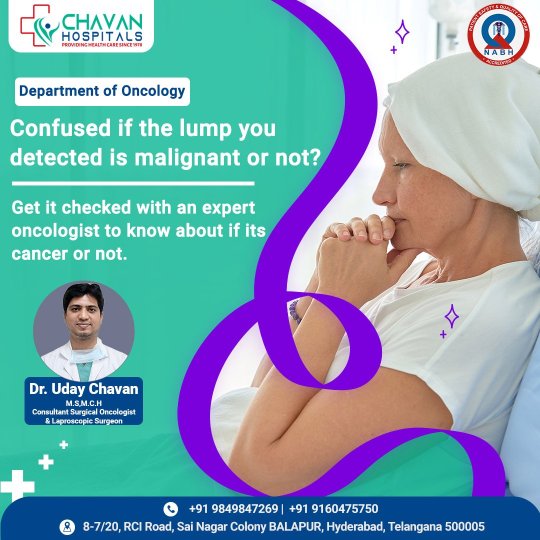
Oncology Hospitals in Hyderabad - Chavan Hospital
The oncology Department at Chavan Hospitals is one of the best Oncology Hospitals in Hyderabad that offers a wide range of cancer care as well as prevention, diagnosis, and treatment for all age groups.
Chavan Hospitals is one of the best cancer hospitals in Hyderabad, with comprehensive and cutting-edge facilities and a coordinated multidisciplinary approach to cancer treatment. The oncology team consists of renowned and experienced medical oncologists, surgical oncologists, radiation oncologists, pathologists, and radiologists who discuss each case to determine the best possible management for an individual patient while considering the patient's physical, social, psychological, and emotional status. These cancers are frequently managed through multidisciplinary cancer conferences, where each member of the team contributes his expertise in order to provide the best treatment.
Chavan Hospitals' Department of Oncology is concerned with the prevention, screening, diagnosis, and treatment of early and advanced cancers that affect various organs, muscles, bones, and connective tissue. To arrive at an accurate diagnosis and help tailor an appropriate and individualised treatment plan, the oncology team collaborates closely with colleagues specialised in plastic surgery, gastroenterology, urology, cardiothoracic surgery, radiology, medical genetics, pathology, palliative care, physiotherapy, and others.
Oncologists determine the evidence-based treatment strategy for each individual based on a variety of factors such as the location, extent, and stage of cancer, disease biology, patient performance status, and associated medical conditions, as well as the patient's involvement in decision-making.
Despite several advances in oncology, surgery remains the only modality with the potential to cure most solid cancers. Our surgical oncology team is composed of qualified surgeons who are committed to providing the best possible outcomes for cancer patients through the use of advanced surgical management techniques. Members of our surgical team are experts in sub-specialties that cover the entire body.
The team provides the following surgical oncology services at the institute:
Head and Neck Cancers:
Oral Cavity Cancers (Tongue, Buccal Mucosa, Alveolus, Hard Palate, Maxilla), Larynx, Hypopharynx Cancer, Thyroid Cancer
Breast Cancer:
Breast-Conserving Surgery, Modified Radical Mastectomy, Sentinel Lymph node Biopsy, Axillary Dissection, and Oncoplastic Breast Surgery are some of the procedures available.
Thoracic Cancer:
Lobectomy, pneumonectomy, dissection of mediastinal lymph nodes, and excision of a mediastinal tumour
Gynaecological Cancers:
For cancers of the Cervix, Ovary, Vulva, and Uterus
Skin and Soft Tissue:
Skin and soft tissue tumour excision, limb-saving surgery
Genitourinary:
For cancers of the Kidney, Testis, Urinary Bladder, Penis and Prostate
Gastrointestinal:
For cancers of the Oesophagus, Colorectal, Stomach, Liver and Bile Duct, Pancreas
Orthopaedic Oncology:
Amputation and limb-sparing surgeries
0 notes
Text
Juniper Publishers-Open Access Journal of Head Neck & Spine Surgery

Unusual Case of Nasopharyngeal Carcinoma with Intracranial Metastasis without Direct Invasion
Authored by Balamurugan Rajendran
Abstract
Intracranial metastasis from Nasopharyngeal Carcinoma (NPC) is an extremely rare occurrence, although direct intracranial invasion is not infrequent in patients with NPC at a locally advanced stage. Only a few patients have been reported in detail in the English literature. Here, we report a case of a man with left high parietal dural metastasis of NPC, detected 2 years after Completed Concurrent Chemoradiotherapy (CCRT).
Introduction
Nasopharyngeal Carcinoma (NPC) is a rare malignancy in Western countries but considered endemic in certain places such as southern China and Southeast Asia [1] In Malaysia, NPC is the fifth (4.9%) most common cancer among Malaysians and the third (8.1%) most common cancer among males [2]. Because of nasopharyngeal anatomical location, patients with locally advanced NPC, it is not infrequent that the primary tumor directly invades the intracranial sites such as cavernous sinus through the skull base. Distant metastases to Central Nervous System (CNS) either through hematogenous route or Cerebral Spinal Fluid (CSF) spread are rare and dural metastasis at parietal region is even extremely rarer [3]. In this article, we report a rare of an elderly man with left parietal dural metastasis from NPC.
Case Report
A 65-year-old male was diagnosed with T2N1M0-Stage II NPC (American Joint Committee on Cancer (AJCC, 7 edition)) back in 2015. A Magnetic Resonance Imaging (MRI) scan and fiber optic endoscopy revealed a small primary tumor in left lateral nasopharynx and the biopsy revealed undifferentiated carcinoma of the nasopharynx. There was no intracranial extension but presence of metastatic lymph node measuring 1.5cm in greatest dimension over right neck. Three cycles of cisplatin based combined chemotherapy regimen (cisplatin, 5-flourouracil) were given prior to radiotherapy and a complete response was obtained at both primary site and in the neck. The nasopharynx and regional lymphatics were treated to a total dose of 34 fractionwith conventional radiotherapy regimen. He subsequent received a Concurrent Chemoradiotherapy (CCRT), which was finally completed in May 2017. Patient had been on surveillance follow up with Oncology team since then.
He remained well until 2 years after the conclusion of the radiotherapy, when he presented with pain over the left chest region for which a Computed Tomography (CT) Thorax was performed at a private center. On assessment, there was no neurological deficit elicited during that time. On CT imaging, there was nodular left pleural thickening with enhancement with metastasis lesions in thoracic vertebral bodies. Fluorodeoxyglucose positron emission computed tomography (FDG-PET/CT) scan was done for detailed workup, revealed pathological uptakes at the left nasopharynx, left pleural, left lung and a new hypermetabolic hyperdense mass in left higher parietal lobe, measuring 4.5 x 3.2 cm in size (Figure 1). The extra axial mass enhanced homogenously at a contrasted enhance CT with minimal edema (Figure 2). Radiologically unable to rule out possibility of a convexity meningioma arising from left parietal dura. The brain lesion was excised totally with a craniotomy and pathological examination revealed metastatic undifferentiated carcinoma (WHO Grade III).
On microscopic examination, the tumor shows fragments of cellular tissue infiltrated by malignant tumor arranged in syncytial pattern of cohesive cells with indistinct cell margins. The cells were large, pleomorphic with hyperchromatic nuclei, prominent nucleoli and moderate amount of cytoplasm. There was focal necrosis, and high mitosis. Immunohistochemical study revealedthat the tumor cells showed membranous CK 5/6 (Cytokeratin 5/6) immunoreactivity (Figure 3&4). Original diagnosis of the nasopharyngeal mass made in 2015 was confirmed to be undifferentiated nasopharyngeal carcinoma, which has similar morphological characteristics as the metastatic carcinoma to the brain. Post-operative, he was right hemiplegic secondary to infarct with hemorrhagic transformation. Patient was opted for palliative care in view next line of chemotherapy is high risk for the patient with current poor performance status.
Discussion
Even though intracranial invasion by direct extension of locally advanced disease is not uncommon, metastases to the CNS, either through hematogenous route or through the Cerebrospinal Fluid (CSF), from NPC have rarely been reported. Metastatic lesions to occipital and temporal lobes have been reported in some cases but for this gentleman, metastatic lesion was located near the parietal lobe [4, 5]. There is no known lymphatic or vascular connection between the nasopharynx and parietal lobes. Ngan and colleagues, discussing the same issue for the patients who had occipital metastases, hypothesized that microenvironment in the occipital lobes might have facilitated the metastatic tumor cells to localize in that region [5]. This case has shown that the occipital lobe and temporal are not the only sites for NPC cells to metastasize, thus make this case the first to be reported of a parietal dural metastasis from NPC. The incidence of recurrence of NPC has been reported as high as 77.5% following a remission, in which 48.1% occurs after 2 years while 17.4% recurred after 7-12 months [6]. Concurrent Chemoradiotherapy (CCRT) is the most potent treatment combination for locoregional control as well as improve overall survival for the advanced disease [7]. However, the impact of CCRT upon distant control was inadequate [8]. As for this case even with a good locoregional control post CCRT, yet after 2 years patient develop new distant metastasis to lung, vertebral bodies and brain. The 5-year overall survival rate of non-metastatic locoregionally advanced NPC treated with radiotherapy is reportedly up to just 60%, while the presence of distant metastases decreases it further to be around 12-15 months at best, after chemotherapy [9].
Status of primary tumor, evidences of disseminated disease, performance status of the patient, treatment modalities already applied to the patient, histopathology of the tumor, and symptoms related to brain metastases are the factors effecting the therapeuticmanagement. Since there was locoregional recurrence and patient was hemiplegic after operation, palliative care was planned for this patient. Palliative chemotherapy was not given in view his family members refused. In conclusion, due to the rarity of dural metastasis from the NPC, a high index of suspicion should be practiced where 6 monthly or yearly radiological assessments should be performed to rule out distant metastases including a CT scan of the brain.
For more articles in Open access Journal of Head Neck & Spine Surgery | Please click on: https://juniperpublishers.com/jhnss/index.php
For more about Juniper Publishers | Please click on: https://juniperpublishers.com/pdf/Peer-Review-System.pdf
0 notes
Text
Medullary Carcinoma of Breast: A Case Report and Review of Literature in Open Access Journal of Medical and Clinical Surgery by Amani Saleh Hadi Saeed*

Abstract
Medullary carcinoma (MBC) of breast is rare variant of invasive ductal carcinoma of breast. represent < 5% of all invasive breast carcinomas. This breast cancer named because the tumor resembles the part of the brain known as the medulla. It can be difficult to diagnose and may be missed on conventional imaging as the finding may overlap with benign lesions i.e. fibroadenoma. These tumors tend to occur in younger women, with average age reported to range from 42 to 52 years although these tumors show aggressive pathological features they are often associated with a more favorable outcome. They are almost invariably negative for hormone receptors as well as Her-2/neu (Triple negative phenotype). It is also said to be particularly common in carriers of BRCA1/2 mutations. The prognosis for medullary carcinoma is better than the ordinary invasive ductal carcinoma. We report similar case in 60-year-old female with cytological, histological and immunohistochemical analysis of MBC.
Keywords: Medullary breast carcinoma, basal phenotype; immunohistochemistry, fibroadenoma
Introduction
Medullary carcinoma is rare and distinct subgroup of breast carcinomas accounting for less than 5% of all invasive breast cancers [1]. The 2012World Health Organization (WHO) up dated the classification of medullary carcinoma under an umbrella term ''carcinomas with medullary feature'', which includes atypical Medullary carcinoma and invasive carcinoma of no special type with medullary features [2]. The diagnosis of medullary carcinoma is usually defined by histologic diagnostic criteria proposed by Ridolfi [3]. Histologically medullary carcinoma demonstrates syncytial growth pattern of poorly differentiated tumor cells with a high mitotic rate. prominent lymphocytic infiltrates with circumscribed microscopic appearance of desmoplastic inflammatory reaction involving mainly the periphery was also diffusely present throughout the substance of the tumor-this is another characteristic feature, which may account for its clinical and biological behavior. Recent publications of breast cancer classification base on gene expression profile analyses indicate that medullary breast carcinoma can be consider part of basal -like carcinoma spectrum made up of estrogen receptor ER negative, progestogen receptor PR negative Her-2/neu negative (triple negative phenotype) [4]. However, there are also publication in which one my find that some MBC and atypical MBC are ER, PR and or. Her-2 positive, indicating the heterogeneity of this type of breast carcinomas [5,6]. The prognosis of MBC is still under dispute. Some studies have reported that survival rates do not differ from those of IDC [3,7]. Most published study have reported lower incidence of axillary lymph node involvement in patients with medullary carcinoma (19% to 46%) than those with atypical medullary carcinomas (30% to 52%) or invasive ductal carcinomas (29% to 65%) [8,9]. Some authors have shown that patients with medullary carcinoma of the breast with positive lymph nodes have 10year overall survival lower than those with N0 nodal disease (58.8% versus 97.1%) [10]. The 5-year survival rate for medullary carcinoma is approximately 78%. Death secondary to this disease is only 10% though. The 20year disease free survival for stage I and II patients are approximately 95% and 61% respectively [10].
Case Report
A60-year old female presented to breast surgeon with lump in the left breast since 6months. On physical examination it had hard mass pain less and fixed to skin with palpable axillary lymph node. She had non-family history of breast carcinoma, no history of nipple discharge or trauma. The clinical and Ultrasonography diagnoses were given as fibroadenoma. Sonommamogrphy for both breasts done show: both breast having normal parenchyma echopattern, the palpable mass in left breast is hypoechoic well-defined visualized mass about 15x11x11mm in size (suspicious mass for FNA).no focal breast mass or cystic lesion detected at right breast, normal nipple a retro- areolar areas. Both axillary regions scanned there is three lymph nodes at left axillary having normal center echogenic center with oval shape, they are 24x9mm and 11x5mm in size, the third lymph node is hypoechoic rounded shape (suspicious lymph node LN) of about (6x6 mm) in size. no evidence of significant axillary LN in right side. All other routine investigation was within normal limits.
6/11/2019 FNAB from left breast mass +left axillary LN was cohesive sheet of ductal epithelial cell showing moderate atypia mixed with myoepithelial cells lie against proteinaceous back ground, the smears contain heterogenous populations of lymphocytes dispersed as single cell are numerically dominated by small round lymphocytes. patient underwent Excisional biopsy for left breast mass 23/11/2019. Specimen was received for histopathological examination and IHC status. Excisional specimen of an irregular pieces of breast tissue 3.5x2x1.5 cm, gross measurement of mass 1.8x1.5 cm) and 1cm distance from close surgical margin. Cut section showed will circumscribed mass. Histopathology analysis of the mas was suggestive of medullary carcinoma. There was no evidence of vascular or capsular invasion. Microscopic grade III (with SBR score :9out 9). Immunohistochemistry of tumor was negative for ER, PR and Her-2/neu. we recommended for modified radical mastectomy with axillary clearance26/12/2019. A mastectomy specimen measure 18x12x5 cm, compose of entire breast parenchyma, the underlying and surrounding adipose tissue with normal nipple and normal surrounding skin, the overlying skin is 17x15 cm. serial section show unremarkable breast tissue with cavity 3x2.5 cm.
No residual tumor seen grossly. Left axillary clearance: several pieces of fibro-fatty tissue, in aggregate:8x8cm contain ten (10) loos lymph node the largest is 1x1x0.5 cm was recovered from axillary tail are all fee of metastasis 0/10. Histopathology was medullary carcinoma, TNM stage (T1N0M0 stage IIA, based on American joint committee on cancer staging system). All surgical margin and base of the specimen were free of dysplastic or malignant cell infiltration. Post-operative chemotherapy with Adriamycin and cyclophosphamide and 5FU complete 6 cycles, no evidence bases for radiotherapy in this case because N0 and surgical margin free. patient put on fellow up till writing this case she has no evidence of local tumor or recurrence or any suspicious distant metastatic lesion [11].
Discussion
According to WHO over 1.2 million women are diagnosis with breast cancer annually worldwide. Infiltrating ductal carcinoma is abroad entity which comprise of tumor that exhibit one or more characteristics of specific types of breast cancers. There is evidence of tubular, papillary, medullary or mucinous differentiation microscopically [3]. It has a predilection for women of younger age group and in a study by Rosen [12] it was found to constitute 11% of all breast malignancies among aged35 and younger. In gross appearance these lesions are well-circumscribed, soft and tan -brown to grey tumors that bulge above the cut surface of the specimen. A multinodular appearance may be appreciated in some spectrum (ER. PR and HER-2negative with high proliferative MIB-1) and apoptotic activity (P53) [4]. The prognosis of medullary carcinoma is better than invasive ductal carcinoma but like another invasive ductal carcinoma it depends on the tumor stage. Based on several studies done previously, medullary carcinoma of the breast is usually described as an irregular shaped mass with micro- lobulation and least frequently with posterior acoustic shadowing sonographically [13]. Calcification is usually not present on mammographic imaging as our case report, so sonographic features of medullary carcinoma and other subtypes group do not differ substantially. Treatment is based on disease stage, and in early stages conservative breast surgy, followed by chemotherapy and radiotherapy strongly recommended [8].
Radiotherapy and chemotherapy were common adjuvant therapies for invasive breast cancers. however, it was often suggested that MBC had good prognosis and therefore may be not benefit from systemic. There was a study found chemotherapy would improve 5- and 10-year overall survival (OS). However, the P value was 0.08, which might not be solid [11].
Conclusion
Medullary breast carcinoma is an uncommon type of infiltrative breast carcinoma that usually affects women around 50years old. It usually misdiagnosed clinically and grossly with fibroadenoma hence we should keep in mind this type of carcinoma in differential diagnosis of fibroadenoma.
Regarding our Journal: https://oajclinicalsurgery.com/ Know more about this article https://oajclinicalsurgery.com/oajcs.ms.id.10011/ https://oajclinicalsurgery.com/pdf/OAJCS.MS.ID.10011.pdf
#Medullary breast carcinoma#basal phenotype#immunohistochemistry#fibroadenoma#oajcs#Amani Saleh Hadi Saeed*#clinical surgery
0 notes
Text
Treatment Options for Breast Cancer proved to be effective.
Breast cancer remains one of the most researched cancers globally and there are numerous new advances in the Aplastic anemia . In India, breast cancer is now reaching persistent levels. In order to diagnose breast lumps at an early stage, it is possible to identify breast cancer using MRI mammograms and X-ray mammograms. Breast Biopsy can not only detect tumour form, but also receptor status. To stop lymphedema emergence, there are Breast Conserving Surgeries, Complete Breast Reconstruction for women requiring mastectomy, and Conservative Axillary Dissection. There are emerging radiation techniques for the treatment of breast cancer tumours, such as Accelerated Partial Breast Radiation (APBI), IGRT and Brachytherapy. With raising awareness of breast cancer, in the early stages, more and more women are being diagnosed, resulting in cure rates rising to up to 85-90 percent. In addition to care, this has made post-treatment quality of life and recovery very significant and appropriate.
You'll want to know the aplastic anemia treatment cost if you've been diagnosed with breast cancer. The answers will let you know more about what's ahead of you and your doctors and decide about your treatment. There are many ways for doctors to figure out which stage of breast cancer you have. Physical examinations, biopsies, X-rays, bone scans and other images, and blood samples give clues. Tissue tests from the breast and lymph nodes are put under the microscope by a specialist called a pathologist to find out any more.
Doctors thread together letters and numbers on the basis of these results to allocate a stage to any case of breast cancer. It may sound like a weird code, but it's actually just a way of finding out exactly what's going on with your cancer.

Breast Cancer Stages
The TNM system is the most common method that doctors use to describe the stage. To answer these questions, doctors use the findings from medical testing and scans:
Tumor (T): How big is the primary breast tumour? What are biomarkers of its?
Node (N): Has the tumour spread to the nodes of the lymph? If that's the case, when, what size, and how many?
Metastasis (M): Has the cancer spread to other body parts?
To decide the stage of cancer for each person, the outcomes are combined.
There are 5 stages of breast cancer that are used for invasive breast cancer: stage 0 (zero), which is non-invasive ductal carcinoma in situ (DCIS), and stages I through IV (1 through 4). A typical way to explain cancer is the stage, so doctors will work together to plan the right treatments.
About the Treatment
Your treating doctor will determine the best breast cancer treatment that will bring better outcomes, based on the pathology. As listed below, some of the methods of treatment are:
Breast cancer treatment involves breast conservation surgery or enhanced radical mastectomy with or without complete reconstruction of the breast.
Breast Cancer Oncoplastic Breast Conservation Surgery incorporates the concepts of plastic surgery and cancer surgery to promote cosmetic surgery in breast cancer patients and to enable reduction or augmentation surgery in the same sitting posture.
Partial and Complete Breast Reconstruction, with or without silicone implants, followed by nipple and areola reconstruction
Advising the patient and relatives on the histopathology report and addressing adjuvant therapy therapeutic options along with individual cost-benefit ratios and overall risk reduction following adjuvant therapy
Placement of indwelling chemotherapy ports for adjuvant chemotherapy administration and control of chemotherapy ports in patients
Adjuvant chemotherapy delivery and targeted treatment by a surgical oncologist specialising in chemotherapy for breast cancer
Specialized radiation therapy (radiotherapy of the whole breast or chest wall) administered by field specialists, using the latest technologies. In metastatic breast cancer, radiation therapy also entails palliative radiotherapy for bone and brain metastases
Detailed instructions by the respective experts on exercise and diet following completion of therapy and follow-up advice
Benign breast disorders such as cyclical mastalgia (breast pain), breast cysts, fibrocystic disease, diagnosis, therapy and medical/surgical treatment of (ANDI)
Important nipple discharge diagnosis and treatment
Breast abscess control with cautious care (if <3cm) or surgical drainage (if >3cm) Genetic counseling for patients with a history of breast cancer or inherited breast cancer in the family.
Non-Hodgkin's lymphoma, the disease-fighting network that spreads across the body, is cancer that originates throughout the lymphatic system. Tumors form in non-Hodgkin's lymphoma from lymphocytes, a type of white blood cell. Non-Hodgkin's lymphoma is more prevalent than Hodgkin's lymphoma, another general form of lymphoma. There are several different non-Hodgkin's lymphoma subtypes. Some of the most common subtypes include diffuse large B-cell lymphoma and follicular lymphoma. Progress in the diagnosis and aplastic anemia treatment cost in India has helped to boost the prognosis of this condition in individuals.
Non-Hodgkin lymphoma is typically treated with radiotherapy or chemotherapy, but certain patients may not be treated with chemotherapy.
In a few cases, there may be no further care required if the initial cancer is very small and can be removed through a biopsy. As many of the treatments can place a considerable strain on the body, the prescribed treatment plan will depend on your general health and age.
Discussions with many doctors and other health practitioners who specialise in all aspects of treating lymphoma will typically take place. This is a multidisciplinary team known as (MDT). The best care choices for you will be recommended by your MDT.
But you shouldn't be rushed into making a decision about your treatment plan.
Before deciding, you may wish to talk to friends, family and your partner.
You'll be invited back to see your care team for a full discussion about the risks and benefits of any treatments planned before treatment begins.
0 notes
Text
Juniper Publishers_The Etiologies behind the Scenes in Elderly Infection

Keywords: Necrotizing Fasciitis; Thigh; Cecum; Carcinoma
Abbreviations: ED: Emergency Department; WBC: White Blood Cell; CRP: C-Reactive Protein; CT: Computed tomography; AST: Aspartate Amino Transferase; BWL: Body Weight Loss; FOLFIRI: Folinic acid, Fluorouracil, and Irinotecan; ECOG: Eastern Cooperative Oncology Group
Introduction
Fever with lethargy, even altered mental status is commonly seen in daily practice, especially in infected elderly. Recently we saw a 63 years old man who complaints of right thigh redness and tenderness for two days. But the etiology behind the scenes is perforated cecal tumor. Prudent workup of infectious source is mandatory to prevent from misdiagnosis. We also briefly review the literatures of necrotizing fasciitis of thigh secondary to perforated colon cancer. This old man went to our emergency department (ED) and said he also felt right back pain and difficulty in standing for one week. There was no symptom of fever, abdominal pain, nausea and vomiting, recent weight neither loss nor bowel habit change. His vital signs showed body temperature of 36°C, respiratory rate of 20 breaths per minute, pulse rate of 93 beats per minute and blood pressure of 92/54 mm Hg. Physical examination revealed swelling, erythematous and mild tenderness with crepitus over the right thigh (Figure 1).
Abdomen was palpated soft with tenderness over the right lower quadrant of abdomen. Laboratory data revealed white blood cell (WBC) count of 8,400/μL, band of 8%, neutrophil of 82%, hemoglobin of 12.4g/dL, platelet count of 36,000/μL, aspartate aminotransferase (AST) of 79 U/L and C-reactive protein (CRP) of 32.25mg/dL. Computed tomography (CT) of right thigh showed scattered, abnormal air accumulation in the subcutaneous layer and muscle layer of right thigh (Figure 2). Further, abdominal contrast-enhanced CT scan showed right colon tumor with abscess formation involving right retro peritoneum and psoas muscle (Figure 3). We applied empiric antibiotic therapy with teicoplanin 200mg and meropenem 500 mg intravenously every 12 hours and fluid resuscitation. Patient received emergent laparotomy and debridement. A 5 cm x 4 cm ulcerative tumor found at cecum and perforated to retroperitoneal space.
Right hemicolectomy with side-to-side anastomosis, debridement and fasciotomy over right thigh were performed. Blood culture showed the presence of Bacteroides fragilis. Antibiotic was shifted to cefpirome 1gm every 12 hours and metronidazole 500mg every 8 hours. Abscess culture grew Bacteroides fragilis and Morganella morganii. After three weeks of admission, ventilator-associated pneumonia happened to him and sputum culture revealed Acinetobacter baumannii complex infection. We administrated tigecycline 50 mg every 12 hours for him. Pathological report of the specimen proved adenocarcinoma with one lymph node metastasis, classified as T4N1M0. After surgical intervention and drainage, he recovered from septic shock and was discharged 40 days later. Two months later, the followed chest computed tomography (CT) showed a 1cm nodular lesion with pleural dimpling at right middle lung.
Video-assisted thoracic surgery and wedge resection was performed and pathological report concluded a metastatic adenocarcinoma. Until now, post-operative 4month later, patient received adjuvant chemotherapy with FOLFIRI (Folinic acid, Fluorouracil, and Irinotecan). His ECOG (Eastern Cooperative Oncology Group) performance status is grade 0. Necrotizing fasciitis is a rare but life-threatening infectious disease needing early surgical and broad-spectrum antibiotic intervention. The etiology of necrotizing fasciitis in this case is unusual and rarely happened. Patient was healthy before and he had the symptoms with low grade fever, back pain and difficulty in standing for one week.
He denied of neither abdominal pain, bloody stool passage, body weight loss (BWL) nor bowel habit change. It is difficult to diagnose cecal tumor in the early stage, but devastating necrotizing fasciitis invaded rapidly to gluteal and thigh. Most of necrotizing fasciitis are associated with perforated colorectal cancer limited within peritoneum as Fournier's gangrene. In the English literatures review, there are only 13 reported cases of necrotizing fasciitis in thigh resulting from perforated colorectal malignancy (Table 1) [1-13]. Of them, mean age was 63.3±10.2 years old with male predominant (M:F = 5:1). The most common site of perforated colorectal cancer leading to necrotizing fasciitis is rectum (61.5%; 8/13), followed by sigmoid colon (23%; 3/13), descending colon (7.7%; 1/13) and cecum (7.7%, 1/13). Necrotizing fasciitis involving in right thigh (53.8%) is more commonly seen than left thigh (38.5%), and both thighs is 7.7%. The two-year survival rate is about 53.8%, and 46.2% of cases died 6 months later.
Note: List of necrotizing fasciitis secondary to perforated colon cancer cases. The most common site is rectum (8 cases), followed by sigmoid colon (3 cases), descending colon (1 case) and cecum (1 case).
Our patient is the second case worldwide of necrotizing fasciitis in thigh resulting from perforated cecum. Interestingly, sigmoid colon cancer tends to be associated with left thigh necrotizing fasciitis (100%, 3/3). In contrast, rectum cancer easily affected to right thigh necrotizing fasciitis (75%, 6/8). The most commonly seen pathogen is Escherichia coli (75%; 8/12); followed by Pseudomonas aeruginosa, Enterococcus, Bacteroides fragilis and anaerobics. The overall mortality rate was estimated to be 25% [14]. Other risk factors including old age, diabetes mellitus, hypoalbuminemia, alcohol, site of infection, delayed surgical intervention increased mortality rate [14]. In conclusion, necrotizing fasciitis of the thigh is rarely found secondary to perforated colon cancer. Delayed diagnosis and intervention of wound increase morbidity and mortality. Emergency physicians should keep in mind of the possible unusual causes and etiologies of necrotizing fasciitis, especially in infected elderly.
To read more articles in Journal of Gerontology & Geriatric Medicine
Please Click on: https://juniperpublishers.com/oajggm/index.php
For more Open Access Journals in Juniper Publishers
Click on: https://juniperpublishers.com/journals.php
0 notes
Text
Artificial Intelligence vs. Tuberculosis, Part 1

By SAURABH JHA, MD
Slumdog TB
No one knows who gave Rahul Roy tuberculosis. Roy’s charmed life as a successful trader involved traveling in his Mercedes C class between his apartment on the plush Nepean Sea Road in South Mumbai and offices in Bombay Stock Exchange. He cared little for Mumbai’s weather. He seldom rolled down his car windows – his ambient atmosphere, optimized for his comfort, rarely changed.
Historically TB, or “consumption” as it was known, was a Bohemian malady; the chronic suffering produced a rhapsody which produced fine art. TB was fashionable in Victorian Britain, in part, because consumption, like aristocracy, was thought to be hereditary. Even after Robert Koch discovered that the cause of TB was a rod-shaped bacterium – Mycobacterium Tuberculosis (MTB), TB had a special status denied to its immoral peer, Syphilis, and unaesthetic cousin, leprosy.
TB became egalitarian in the early twentieth century but retained an aristocratic noblesse oblige. George Orwell may have contracted TB when he voluntarily lived with miners in crowded squalor to understand poverty. Unlike Orwell, Roy had no pretentions of solidarity with poor people. For Roy, there was nothing heroic about getting TB. He was embarrassed not because of TB’s infectivity; TB sanitariums are a thing of the past. TB signaled social class decline. He believed rickshawallahs, not traders, got TB.
“In India, many believe TB affects only poor people, which is a dangerous misconception,” said Rhea Lobo – film maker and TB survivor.
Tuberculosis is the new leprosy. The stigma has consequences, not least that it’s difficult diagnosing a disease that you don’t want diagnosed. TB, particularly extra-pulmonary TB, mimics many diseases.
“TB can cause anything except pregnancy,” quips Dr. Justy – a veteran chest physician. “If doctors don’t routinely think about TB they’ll routinely miss TB.”
In Lobo, the myocobacteria domiciled in the bones of her feet, giving her heel pain, which was variously ascribed to bone bruise, bone cancer, and staphylococcal infection. Only when a lost biopsy report resurfaced, and after receiving the wrong antibiotics, was TB diagnosed, by which time the settlers had moved to her neck, creating multiple pockets of pus. After multiple surgeries and a protracted course of antibiotics, she was free of TB.
“If I revealed I had TB no one would marry me, I was advised” laughed Lobo. “So, I made a documentary on TB and started ‘Bolo Didi’ (speak sister), a support group for women with TB. Also, I got married!”
Mycobacterium tuberculosis is an astute colonialist which lets the body retain control of its affairs. The mycobacteria arrive in droplets, legitimately, through the airways and settle in the breezy climate of the upper lobes and superior segment of the lower lobes of the lungs. If they sense weakness they attack, and if successful, cause primary TB. Occasionally they so overpower the body that an avalanche of small, discrete snowballs, called miliary TB, spread. More often, they live silently in calcified lymph nodes as latent TB. When apt, they reappear, causing secondary TB. The clues to their presence are calcified mediastinal nodes or a skin rash after injection of mycobacterial protein.
MTB divides every 20 hours. In the bacterial world that’s Monk-like libido. E. Coli, in comparison, divides every 20 minutes. Their sexual ennui makes them frustratingly difficult to culture. Their tempered fecundity also means they don’t overwhelm their host with their presence, permitting them to write fiction and live long enough to allow the myocobacteria to jump ship.
TB has been around for a while. The World Health Organization (WHO) wants TB eradicated but the myocobacteria have no immediate plans for retirement. Deaths from TB are declining at a tortoise pace of 2 % a year. TB affects 10 million and kills 1.6 million every year – it is still the number one infectious cause of death.
The oldest disease’s nonchalance to the medical juggernaut is not for the lack of a juggernaut effort. Mass screening for TB using chest radiographs started before World War 2, and still happens in Japan. The search became fatigued by the low detection of TB. The challenge wasn’t just in looking for needles in haystacks, but getting to the haystacks which, in developing countries, are dispersed like needles.
The battleground for TB eradication is India, which has the highest burden of TB – a testament not just to its large population. Because TB avoids epidemics, it never scares the crap out of people. Its distribution and spread match society’s wealth distribution and aspirations. And in that regard India is most propitious for its durability.
Few miles north of Nepean Sea Road is Dharavi – Asia’s largest slum, made famous by the Oscar-winning film, Slumdog Millionaire. From atop, Dharavi looks like thousand squashed coke cans beside thousand crumpled cardboard boxes. On the ground, it’s a hot bed of economic activity. No one wants to stay in Dharavi forever, its people want to become Bollywood stars, or gangsters, or just very rich. Dharavi is a reservoir of hope.
Dharavi is a reservoir also of active TB. In slums, which are full of houses packed like sardines in which live people packed like sardines, where cholera spreads like wildfire and wildfire spreads like cholera, myocobacteria travel much further. Familiarity breeds TB. One person with active TB can infect nine – and none are any the wiser of the infection because unlike cholera, which is wildfire, TB is a slow burn and its symptoms are indistinguishable from the maladies of living in a slum.
Slum dwellers with active TB often continue working – there’s no safety net in India to cushion the illness – and often travel afar to work. They could be selling chai and samosas outside the Bombay Stock Exchange. With the habit of expectoration – in India, spitting on the streets isn’t considered bad manners – sputum is aplenty, and mycobacteria-laden droplets from Dharavi can easily reach Roy’s lungs. TB, the great leveler, bridges India’s wealth divide. Mycobacteria unite Nepean Sea Road with Dharavi.
Rat in Matrix Algebra
The major challenges in fighting tuberculosis are finding infected people and ensuring they take the treatment for the prescribed duration, often several months. Both obstacles can wear each other– if patients don’t take their treatment what’s the point finding TB? If TB can’t be found what good is the treatment?
The two twists in the battle against TB, drug resistant TB and concurrent TB and HIV, favor the mycobacteria. But TB detection is making a resurgence with the reemergence of the old warrior – the chest radiograph, which now has a new ally – artificial intelligence (AI). Artificial Intelligence is chest radiograph’s Sancho Panza.
Ten miles north of Dharavi in slick offices in Goregaon, Mumbai’s leafy suburb, data scientists training algorithms to read chest radiographs are puzzled by AI’s leap in performance.
“The algorithm we developed,” says Preetham Sreenivas incredulously, “has an AUC of 1 on the new set of radiographs!”
AUC, or area under the receiver operator characteristic curve, measures diagnostic accuracy. The two types of diagnostic errors are false negatives – mistaking abnormal for normal, and false positives – mistaking normal for abnormal. In general, fewer false negatives (FNs) means more false positives (FPs); trade-off of errors. A higher AUC implies fewer “false” errors, AUC of 1 is perfect accuracy; no false positives, no false negatives.
Chest radiograph are two-dimensional images on which three dimensional structures, such as lungs, are collapsed and which, like Houdini, hide stuff in plain sight. Pathology literally hides behind normal structures. It’s nearly impossible for radiologists to have an AUC of 1. Not even God knows what’s going on in certain parts of the lung, such as the posterior segment of the left lower lobe.
Here, AI seemed better than God at interpreting chest radiographs. But Sreenivas, who leads the chest radiograph team in Qure.ai – a start-up in Mumbai which solves healthcare problems using artificial intelligence, refused to open the champagne.
“Algorithms can’t jump from an AUC of 0.84 to 1. It should be the other way round – their performance should drop when they see data (radiographs) from a new hospital,” explains Sreenivas.
Algorithms mature in three stages. First, training – data (x-rays), labelled with ground truth, are fed to a deep neural network (the brain), Labels, such as pleural effusion, pulmonary edema, pneumonia, or no abnormality, teach AI. After seeing enough cases AI is ready for the second step, validation – in which it is tested on different cases taken from the same source as the training set – like same hospital. If AI performs respectably, it is ready for the third stage – the test.
Training radiology residents is like training AI. First, residents see cases knowing the answer. Then they see cases on call from the institution they’re training at, without knowing the answer. Finally, released into the world, they see cases from different institutions and give an answer.
The test and training cases come from different sources. The algorithm invariably performs worse on test than training set because of “overfitting” – a phenomenon where the algorithm tries hard fitting to the local culture. It thinks the rest of the world is exactly like the place it trained, and can’t adapt to subtle differences in images because of different manufacturers, different acquisition parameters, or acquisition on different patient populations. To reduce overfitting, AI is regularly fed cases from new institutions.
When AI’s performance on radiographs from a new hospital mysteriously improved, Sreenivas smelt a rat.
“AI is matrix algebra. It’s not corrupt like humans – it doesn’t cheat. The problem must be the data,” Sreenivas pondered.
Birth of a company
“I wish I could say we founded this company to fight TB,” says Pooja Rao, co-founder of Qure.ai, apologetically. “But I’d be lying. The truth is that we saw in an international public health problem a business case for AI.”
Qure.ai was founded by Prashant Warier and Pooja Rao. After graduating from the Indian Institute of Technology (IIT), Warier, a natural born mathematician, did his PhD from Georgia Tech. He had no plans of returning to India, until he faced the immigration department’s bureaucratic incompetence. Someone had tried entering the US illegally on his wife’s stolen passport. The bureaucracy, unable to distinguish the robber from the robbed, denied her a work visa. Warier reluctantly left the US.
In India, Warier founded a company which used big data to find preferences of niche customers. His company was bought by Fractal, a data analytic giant – the purchase motivated largely by the desire to recruit Warier.
Warier wanted to develop an AI-enabled solution for healthcare. In India, data-driven decisions are common in retail but sparse in healthcare. In a move unusual in industry and uncommon even in academia, Fractal granted him freedom to tinker, with no strings attached. Qure.ai was incubated by Fractal.
Warier discovered Rao, a physician-scientist and bioinformatician, on LinkedIn and invited her to lead the research and development. Rao became a doctor to become a scientist because she believed that deep knowledge of medicine helps join the dots in the biomedical sciences. After her internship, she did a PhD at the Max Planck Institute in Germany. For her thesis, she applied deep learning to predict Alzheimer’s disease from RNA. Though frustrated by Alzheimer’s, which seemed uncannily difficult to predict, she fell in love with deep learning.
Rao and Warier were initially uncertain what their start-up should focus on. There were many applications of AI in healthcare, such as genomic analysis, analysis of electronic medical records, insurance claims data, Rao recalled two lessons from her PhD.
“Diseases such as Alzheimer’s are heterogeneous, so the ground truth, the simple question – is there Alzheimer’s – is messy. The most important thing I realized is that without the ground truth AI is useless.”
Rao echoed the sentiments of Lady Lovelace, the first computer programmer, from the nineteenth century. When Lovelace saw the analytical engine, the first “algorithm”, invented by Charles Babbage, she said: “The analytical Engine has no pretensions whatever to originate anything. It can do whatever we know how to order it to perform. It can follow analysis; but it has no power of anticipating any analytical relations or truths.”
The second lesson Rao learnt was that the ground truth must be available immediately, not in the future – i.e. AI must be trained on diseases of the present, not outcomes, which are nebulous and take time to reveal. The immediacy of their answer, which must be now, right away, reduced their choices to two – radiology and pathology. Pathology had yet to be digitized en masse.
“The obvious choice for AI was radiology”, revealed Warier.
Why “Qure” with a Q, not “Cure” with a C, I asked. Was it a tribute to Arabic medicine?
“We’re not that erudite,” laughed Warier. “The internet domain for ‘cure’ had already been taken.”
Qure.ai was founded in 2016 during peak AI euphoria. In those days deep learning seemed magical to those who understood it, and to those who didn’t. Geoffrey Hinton, deep learning’s titan, famously predicted radiologists’ extinction – he advised that radiologists should stop being trained because AI would interpret the images just as well.
Bioethicist and architect of Obamacare, Ezekiel Emanuel, told radiologists that their profession faced an existential threat from AI. UK’s health secretary, Jeremy Hunt, drunk on the Silicon Valley cool aid, prophesized that algorithms will outperform general practitioners. Venture capitalist, Vinod Khosla predicted modestly that algorithms will replace 80 % of doctors.
Amidst the metastasizing hype, Warier and Rao remained circumspect. Both understood AI’s limitations. Rao was aware that radiologists hedged in their reports – which often made the ground truth a coin toss. They concluded that AI would be an incremental technology. AI would help radiologists become better radiologists.
“We were firing arrows in the dark. Radiology is vast. We didn’t know where to start,” recalls Rao.
Had Qure.ai been funded by venture capitalists, they’d have a deadline to have a product. But Fractal prescribed no fixed timeline. This gave the founders an opportunity to explore radiology. The exploration was instructive.
They spoke to several radiologists to better understand radiology, find the profession’s pain points, see what could be automated, and what might be better dealt by AI. The advice ranged from the flippant to the esoteric. One radiologist recommended using AI to quantify lung fibrosis in interstitial pulmonary fibrosis, another, knee cartilage for precision anti-rheumatoid therapy. Qure.ai has a stockpile of unused, highly niche, esoteric algorithms.
Every radiologist’s idea of augmentation was unique. Importantly, few of their ideas comprised mainstream practice. Augmentation seemed a way of expanding radiologist’s possibilities, rather than dealing with radiology’s exigencies – no radiologist, for instance, suggested that AI should look for TB on chest radiographs.
Augmentation doesn’t excite venture capitalists as much as replacement, transformation, or disruption. And augmentation didn’t excite Rao and Warier, either. When you have your skin in the commercial game, relevance is the only currency.
“Working for start-ups is different from being a scientist in an academic medical center. We do science, too. But before we take a project, we think about the return of investment. Just because an endeavor is academically challenging doesn’t mean that it’s commercially useful. If product don’t sell, start-ups have to close shop,” said Rao.
The small size of start-ups means they don’t have to run decisions through bulky corporate governance. It doesn’t take weeks convening meetings through Doodle polls. Like free climbers who aren’t encumbered by climbing equipment, they can reach their goal sooner. Because a small start-up is nimble it can fail fast, fail without faltering, fail a few time. But it can’t fail forever. Qure needed a product it could democratize. Then an epiphany.
In World War 2, after allied aircrafts sustained bullets in enemy fire, some returned to the airbase and others crashed. Engineers wanted the aircrafts reinforced at their weakest points to increase their chances of surviving enemy fire. A renowned statistician of the time, Abraham Wald, analyzed the distribution of the bullets and advised that reinforcements be placed where the plane hadn’t been shot. Wald realized that the planes which didn’t return were likely shot at the weakest points. On the planes which returned the bullets marked their strongest point.
Warier and Rao realized that they needed to think about scenarios where radiologists were absent, not where radiologists were abundant. They had asked the wrong people the wrong question. The imminent need wasn’t replacing or even augmenting radiologists, but in supplying near-radiologist expertise where not a radiologist was in sight. The epiphany changed their strategy.
“It’s funny – when I’m asked whether I see AI replacing radiologists, I point out that in most of the rest of the world there aren’t any radiologists to replace,” said Rao.
The choice of modality – chest radiographs – followed logically because chest radiographs are the most commonly ordered imaging test worldwide. They’re useful for a number of clinical problems and seem deceptively easy to interpret. Their abundance also meant that AI would have a large sample size to learn from.
“There just weren’t enough radiologists to read the daily chest radiograph volume at Christian Medical College, Vellore, where I worked. I can read chest x-rays because I’m a chest physician, but reading radiographs takes away time I could be spending with my patients, and I just couldn’t keep up with the volumes,” recalls Dr. Justy. Several radiographs remained unread for several weeks, many hid life-threatening conditions such as pneumothorax or lung cancer. The hospital was helpless – their budget was constrained and as important as radiologists were, other physicians and services were more important. Furthermore, even if they wanted they couldn’t recruit radiologists because the supply of radiologists in India is small.
Justy believes AI can offer two levels of service. For expert physicians like her, it can take away the normal radiographs, leaving her to read the abnormal ones, which reduces the workload because the majority of the radiographs are normal. For novice physicians, and non-physicians, AI could provide an interpretation – diagnosis, or differential diagnoses, or just point abnormalities on the radiograph.
The Qure.ai team imagined those scenarios, too. First they needed the ingredients, the data, i.e. the chest radiographs. But the start-up comprised only a few data scientists, none of whom had any hospital affiliations.
“I was literally on the road for two years asking hospitals for chest radiographs. I barely saw my family,” recalls Warier. “Getting the hospitals to share data was the most difficult part of building Qure.ai.”
Warier became a traveling salesman and met with leadership of over hundred healthcare facilities of varying sizes, resources, locations, and patient populations. He explained what Qure.ai wanted to achieve and why they needed radiographs. There were long waits outside the leadership office, last minute meeting cancellations, unanswered e-mails, lukewarm receptions, and enthusiasm followed by silence. But he made progress, and many places agreed to give him the chest radiographs. The data came with stipulations. Some wanted to share revenue. Some wanted research collaborations. Some had unrealistic demands such as share of the company. It was trial and error for Warier, as he had done nothing of this nature before.
Actually it was Warier’s IIT alumni network which opened doors. IITians (graduates of the Indian Institutes of Technology) practically run India’s business, commerce, and healthcare. Heads of private equity which funds corporate hospitals are often IITians, as are the CEOs of these hospitals.
“Without my IIT alumni network, I don’t think we could have pulled it off. Once an IITian introduces an IITian to an IITian, it’s an unwritten rule that they must help,” said Warier.
Warier’s efforts paid. Qure has now acquired over 2.5 million chest radiographs from over 100 sites for training, validation and testing the chest radiograph algorithm.
“As a data scientist my ethos is that there’s no such thing as ‘too much data.’ More the merrier,” smiled Warier.
“The mobile phone reached many parts of India before the landline could get there,” explains Warier. “Similarly, AI will reach parts of India before radiologists.”
Soon, a few others, including Srinivas, joined the team. Whilst the data scientists were educating AI, Rao and Warier were figuring their customer base. It was evident that radiologists would not be their customers. Radiologists didn’t need AI. Their customers were those who needed radiologists but were prepared to settle for AI.
“The secret to commercialization in healthcare is need, real need, not induced demand. But it’s tricky because the neediest are least likely to generate revenues,” said Warier in a pragmatic tone. Unless the product can be scaled at low marginal costs. An opportunity for Qure.ai arose in the public health space – the detection of tuberculosis on chest radiographs in the global fight against TB. It was an indication that radiologists in developing worlds didn’t mind conceding – they had plenty on their plates, already.
“It was serendipity,” recalls Rao. “A consultant suggested that we use our algorithm to detect TB. We then met people working in the TB space – advocates, activists, social workers, physicians, and epidemiologists. We were inspired particularly by Dr. Madhu Pai, Professor of Epidemiology at McGill University. His passion to eradicate TB made us believe that the fight against TB was personal.”
Qure.ai started with four people. Today 35 people work for it. They even have a person dedicated to regulatory affairs. Rao remembers the early days. “We were lucky to have been supported by Fractal. Had we been operating out of a garage, we might not have survived. Building algorithms isn’t easy.”
Finding Tuberculosis
Hamlet’s modified opening soliloquy, “TB or not TB, that is the question”, simplifies the dilemma facing TB detection, which is a choice between fewer false positives and fewer false negatives. Ideally, one wants neither. The treatment for tuberculosis – quadruple therapy – exacts several month commitment. It’s not a walk in the park. Patients have to be monitored to confirm they are treatment compliant, and though directly observed therapy, medicine’s big brother, has become less intrusive, it still consumes resources. Taking TB treatment when one doesn’t have TB is unfortunate. But not taking TB treatment when one has TB can be tragic, and defeats the purpose of detection, and perpetuates the reservoir of TB.
Hamlet’s soliloquy can be broken into two parts – screening and confirmation. When screening for TB, “not TB is the question”. The screening test must be sensitive –capable of finding TB in those with TB, i.e. have a high negative predictive value (NPV), so that when it says “no TB” – we’re (nearly) certain the person doesn’t have TB.
Those positive on screening tests comprise two groups – true positives (TB) and false positives (not TB). We don’t want antibiotics frivolously given, so the soliloquy reverses; it is now “TB, that is the question.” The confirmatory test must be specific, highly capable of finding “not TB” in those without TB, i.e. have a high positive predictive value (PPV), so that when it says “TB” – we’re (nearly) certain that the person has TB. Confirmatory tests should not be used to screen, and vice versa.
Tuberculosis can be inferred on chest radiographs or myocobacteria TB can be seen on microscopy. Seeing is believing and seeing the bacteria by microscopy was once the highest level of proof of infection. In one method, slide containing sputum is stained with carbol fuchsin, rendering it red. MTB retains its glow even after the slide is washed with acid alcohol, a property responsible for its other name – acid fast bacilli.
Sputum microscopy, once heavily endorsed by the WHO for the detection of TB, is cheap but complicated. The sputum specimen must contain sputum, not saliva, which is easily mistaken for sputum. Patients have to be taught how to bring up the sputum from deep inside their chest. The best time to collect sputum is early morning, so the collection needs discipline, which means that the yield of sputum depends on the motivation of the patient. Inspiring patients to provide sputum is hard because even those who regularly cough phlegm can find its sight displeasing.
Which is to say nothing about the analysis part, which requires attention to detail. It’s easier seeing mycobacteria when they’re abundant. Sputum microscopy is best at detecting the most infectious of the most active of the active TB sufferers. Its accuracy depends on the spectrum of disease. If you see MTB, the patient has TB. If you don’t see MTB, the patient could still have TB. Sputum microscopy, alone, is too insensitive and cumbersome for mass screening – yet, in many parts of the world, that’s all they have.
The gold standard test for TB – the unfailing truth that the patient has TB, independent of the spectrum of disease – is culture of mycobacteria, which was deemed impractical because on the Löwenstein–Jensen medium, the agar made specially for MTB, it took six weeks to grow MTB, which is too long for treatment decisions. Culture has made a comeback, in order to detect drug resistant mycobacteria. On newer media, such as MGIT, the mycobacteria grow much faster.
The detection of TB was revolutionized by molecular diagnostics, notably the nucleic acid amplification test, also known as GeneXpert MTB/ RIF, shortened to Xpert, which simultaneously detects mycobacterial DNA and assesses whether the mycobacteria are resistant to rifampicin – one of the mainline anti-tuberculosis drugs.
Xpert boasts a specificity of 98 %, and with a sensitivity of 90 % it is nearly gold standard material, or at least good enough for confirmation of TB. It gives an answer in 2 hours – a dramatically reduced turnaround time compared to agar. Xpert can detect 131 colony-forming units of MTB per ml of specimen – which is a marked improvement from microscopy, where there should be 10, 000 colony-forming units of MTB per ml of specimen for reliable detection. However, Xpert can’t be used on everyone, not just because its sensitivity isn’t high enough – 90 % is a B plus, and for screening we need an A plus sensitivity. But also its price, which ranges from $10 – $20 per cartridge, and is too expensive for mass screening in developing countries.
This brings us back to the veteran warrior, the chest radiograph, which has a long history. Shortly after Wilhelm Röntgen’s discovery, x-rays were used to see the lungs, the lungs were a natural choice because there was natural contrast between the air, through which the rays passed, and the bones, which stopped the rays. Pathology in the lungs stopped the rays, too – so the ‘stopping of rays’ became a marker for lung disease, chief of which was tuberculosis.
X-rays were soon conscripted to the battlefield in the Great War to locate bullets in wounded soldiers, making them war heroes. But it was the writer, Thomas Mann, who elevated the radiograph to literary fame in Magic Mountain – a story about a TB sanitarium. The chest radiograph and tuberculosis became intertwined in people’s imagination. By World War 2, chest radiographs were used for national TB screening in the US.
The findings of TB on chest radiographs include consolidation (whiteness), big lymph nodes in the mediastinum, cavitation (destruction of lung), nodules, shrunken lung, and pleural effusion. These findings, though sensitive for TB – if the chest radiograph is normal, active TB is practically excluded, aren’t terribly specific, as they’re shared by other diseases, such as sarcoid.
Chest radiographs became popular with immigration authorities in Britain and Australia to screen for TB in immigrants from high TB burden countries at the port of entry. But the WHO remained unimpressed by chest radiographs, preferring sputum analysis instead. The inter- and intra-observer variation in the interpretation of the radiograph didn’t inspire confidence. Radiologists would often disagree with each other, and sometimes disagree with themselves. WHO had other concerns.
“One reason that the WHO is weary of chest radiographs is that they fear that if radiographs alone are used for decision making, TB will be overtreated. This is common practice in the private medical sector in India,” explains Professor Madhu Pai.
Nonetheless, Pai advocates that radiographs triage for TB, to select patients for Xpert, which is cost effective because radiographs, presently, are cheaper than molecular tests. Using Xpert only on patients with abnormal chest radiographs would increase its diagnostic yield – i.e. percentage of cases which test positive. Chest radiograph’s high sensitivity compliments Xpert’s high specificity. But this combination isn’t 100 % – nothing in diagnostic medicine is. The highly infective endobronchial TB can’t be seen on chest radiograph, because the mycobacteria never make it to the lungs, and remain stranded in the airway.
“Symptoms such as cough are even more non-specific than chest radiographs for TB. Cough means shit in New Delhi, because of the air pollution which gives everyone a cough,” explains Pai, basically emphasizing that neither the chest radiograph nor clinical acumen, can be removed from the diagnostic pathway for TB.
A test can’t be judged just by its AUC. How likely people – doctors and patients – are to adopt a test is also important and here the radiograph outshines sputum microscopy, because despite its limitations, well known to radiologists, radiographs still carry a certain aura, particularly in India. In the Bollywood movie, Anand, an oncologist played by Amitabh Bachchan diagnosed terminal cancer by glancing at the patient’s radiograph for couple of seconds. Not CT, not PET, but a humble old radiograph. Bollywood has set a very high bar for Artificial Intelligence.
Saurabh Jha (aka @RogueRad) is a contributing editor for THCB. This is part 1 of a two-part story.
The post Artificial Intelligence vs. Tuberculosis, Part 1 appeared first on The Health Care Blog.
Artificial Intelligence vs. Tuberculosis, Part 1 published first on https://venabeahan.tumblr.com
0 notes
Text
Artificial Intelligence vs. Tuberculosis, Part 1

By SAURABH JHA, MD
Slumdog TB
No one knows who gave Rahul Roy tuberculosis. Roy’s charmed life as a successful trader involved traveling in his Mercedes C class between his apartment on the plush Nepean Sea Road in South Mumbai and offices in Bombay Stock Exchange. He cared little for Mumbai’s weather. He seldom rolled down his car windows – his ambient atmosphere, optimized for his comfort, rarely changed.
Historically TB, or “consumption” as it was known, was a Bohemian malady; the chronic suffering produced a rhapsody which produced fine art. TB was fashionable in Victorian Britain, in part, because consumption, like aristocracy, was thought to be hereditary. Even after Robert Koch discovered that the cause of TB was a rod-shaped bacterium – Mycobacterium Tuberculosis (MTB), TB had a special status denied to its immoral peer, Syphilis, and unaesthetic cousin, leprosy.
TB became egalitarian in the early twentieth century but retained an aristocratic noblesse oblige. George Orwell may have contracted TB when he voluntarily lived with miners in crowded squalor to understand poverty. Unlike Orwell, Roy had no pretentions of solidarity with poor people. For Roy, there was nothing heroic about getting TB. He was embarrassed not because of TB’s infectivity; TB sanitariums are a thing of the past. TB signaled social class decline. He believed rickshawallahs, not traders, got TB.
“In India, many believe TB affects only poor people, which is a dangerous misconception,” said Rhea Lobo – film maker and TB survivor.
Tuberculosis is the new leprosy. The stigma has consequences, not least that it’s difficult diagnosing a disease that you don’t want diagnosed. TB, particularly extra-pulmonary TB, mimics many diseases.
“TB can cause anything except pregnancy,” quips Dr. Justy – a veteran chest physician. “If doctors don’t routinely think about TB they’ll routinely miss TB.”
In Lobo, the myocobacteria domiciled in the bones of her feet, giving her heel pain, which was variously ascribed to bone bruise, bone cancer, and staphylococcal infection. Only when a lost biopsy report resurfaced, and after receiving the wrong antibiotics, was TB diagnosed, by which time the settlers had moved to her neck, creating multiple pockets of pus. After multiple surgeries and a protracted course of antibiotics, she was free of TB.
“If I revealed I had TB no one would marry me, I was advised” laughed Lobo. “So, I made a documentary on TB and started ‘Bolo Didi’ (speak sister), a support group for women with TB. Also, I got married!”
Mycobacterium tuberculosis is an astute colonialist which lets the body retain control of its affairs. The mycobacteria arrive in droplets, legitimately, through the airways and settle in the breezy climate of the upper lobes and superior segment of the lower lobes of the lungs. If they sense weakness they attack, and if successful, cause primary TB. Occasionally they so overpower the body that an avalanche of small, discrete snowballs, called miliary TB, spread. More often, they live silently in calcified lymph nodes as latent TB. When apt, they reappear, causing secondary TB. The clues to their presence are calcified mediastinal nodes or a skin rash after injection of mycobacterial protein.
MTB divides every 20 hours. In the bacterial world that’s Monk-like libido. E. Coli, in comparison, divides every 20 minutes. Their sexual ennui makes them frustratingly difficult to culture. Their tempered fecundity also means they don’t overwhelm their host with their presence, permitting them to write fiction and live long enough to allow the myocobacteria to jump ship.
TB has been around for a while. The World Health Organization (WHO) wants TB eradicated but the myocobacteria have no immediate plans for retirement. Deaths from TB are declining at a tortoise pace of 2 % a year. TB affects 10 million and kills 1.6 million every year – it is still the number one infectious cause of death.
The oldest disease’s nonchalance to the medical juggernaut is not for the lack of a juggernaut effort. Mass screening for TB using chest radiographs started before World War 2, and still happens in Japan. The search became fatigued by the low detection of TB. The challenge wasn’t just in looking for needles in haystacks, but getting to the haystacks which, in developing countries, are dispersed like needles.
The battleground for TB eradication is India, which has the highest burden of TB – a testament not just to its large population. Because TB avoids epidemics, it never scares the crap out of people. Its distribution and spread match society’s wealth distribution and aspirations. And in that regard India is most propitious for its durability.
Few miles north of Nepean Sea Road is Dharavi – Asia’s largest slum, made famous by the Oscar-winning film, Slumdog Millionaire. From atop, Dharavi looks like thousand squashed coke cans beside thousand crumpled cardboard boxes. On the ground, it’s a hot bed of economic activity. No one wants to stay in Dharavi forever, its people want to become Bollywood stars, or gangsters, or just very rich. Dharavi is a reservoir of hope.
Dharavi is a reservoir also of active TB. In slums, which are full of houses packed like sardines in which live people packed like sardines, where cholera spreads like wildfire and wildfire spreads like cholera, myocobacteria travel much further. Familiarity breeds TB. One person with active TB can infect nine – and none are any the wiser of the infection because unlike cholera, which is wildfire, TB is a slow burn and its symptoms are indistinguishable from the maladies of living in a slum.
Slum dwellers with active TB often continue working – there’s no safety net in India to cushion the illness – and often travel afar to work. They could be selling chai and samosas outside the Bombay Stock Exchange. With the habit of expectoration – in India, spitting on the streets isn’t considered bad manners – sputum is aplenty, and mycobacteria-laden droplets from Dharavi can easily reach Roy’s lungs. TB, the great leveler, bridges India’s wealth divide. Mycobacteria unite Nepean Sea Road with Dharavi.
Rat in Matrix Algebra
The major challenges in fighting tuberculosis are finding infected people and ensuring they take the treatment for the prescribed duration, often several months. Both obstacles can wear each other– if patients don’t take their treatment what’s the point finding TB? If TB can’t be found what good is the treatment?
The two twists in the battle against TB, drug resistant TB and concurrent TB and HIV, favor the mycobacteria. But TB detection is making a resurgence with the reemergence of the old warrior – the chest radiograph, which now has a new ally – artificial intelligence (AI). Artificial Intelligence is chest radiograph’s Sancho Panza.
Ten miles north of Dharavi in slick offices in Goregaon, Mumbai’s leafy suburb, data scientists training algorithms to read chest radiographs are puzzled by AI’s leap in performance.
“The algorithm we developed,” says Preetham Sreenivas incredulously, “has an AUC of 1 on the new set of radiographs!”
AUC, or area under the receiver operator characteristic curve, measures diagnostic accuracy. The two types of diagnostic errors are false negatives – mistaking abnormal for normal, and false positives – mistaking normal for abnormal. In general, fewer false negatives (FNs) means more false positives (FPs); trade-off of errors. A higher AUC implies fewer “false” errors, AUC of 1 is perfect accuracy; no false positives, no false negatives.
Chest radiograph are two-dimensional images on which three dimensional structures, such as lungs, are collapsed and which, like Houdini, hide stuff in plain sight. Pathology literally hides behind normal structures. It’s nearly impossible for radiologists to have an AUC of 1. Not even God knows what’s going on in certain parts of the lung, such as the posterior segment of the left lower lobe.
Here, AI seemed better than God at interpreting chest radiographs. But Sreenivas, who leads the chest radiograph team in Qure.ai – a start-up in Mumbai which solves healthcare problems using artificial intelligence, refused to open the champagne.
“Algorithms can’t jump from an AUC of 0.84 to 1. It should be the other way round – their performance should drop when they see data (radiographs) from a new hospital,” explains Sreenivas.
Algorithms mature in three stages. First, training – data (x-rays), labelled with ground truth, are fed to a deep neural network (the brain), Labels, such as pleural effusion, pulmonary edema, pneumonia, or no abnormality, teach AI. After seeing enough cases AI is ready for the second step, validation – in which it is tested on different cases taken from the same source as the training set – like same hospital. If AI performs respectably, it is ready for the third stage – the test.
Training radiology residents is like training AI. First, residents see cases knowing the answer. Then they see cases on call from the institution they’re training at, without knowing the answer. Finally, released into the world, they see cases from different institutions and give an answer.
The test and training cases come from different sources. The algorithm invariably performs worse on test than training set because of “overfitting” – a phenomenon where the algorithm tries hard fitting to the local culture. It thinks the rest of the world is exactly like the place it trained, and can’t adapt to subtle differences in images because of different manufacturers, different acquisition parameters, or acquisition on different patient populations. To reduce overfitting, AI is regularly fed cases from new institutions.
When AI’s performance on radiographs from a new hospital mysteriously improved, Sreenivas smelt a rat.
“AI is matrix algebra. It’s not corrupt like humans – it doesn’t cheat. The problem must be the data,” Sreenivas pondered.
Birth of a company
“I wish I could say we founded this company to fight TB,” says Pooja Rao, co-founder of Qure.ai, apologetically. “But I’d be lying. The truth is that we saw in an international public health problem a business case for AI.”
Qure.ai was founded by Prashant Warier and Pooja Rao. After graduating from the Indian Institute of Technology (IIT), Warier, a natural born mathematician, did his PhD from Georgia Tech. He had no plans of returning to India, until he faced the immigration department’s bureaucratic incompetence. Someone had tried entering the US illegally on his wife’s stolen passport. The bureaucracy, unable to distinguish the robber from the robbed, denied her a work visa. Warier reluctantly left the US.
In India, Warier founded a company which used big data to find preferences of niche customers. His company was bought by Fractal, a data analytic giant – the purchase motivated largely by the desire to recruit Warier.
Warier wanted to develop an AI-enabled solution for healthcare. In India, data-driven decisions are common in retail but sparse in healthcare. In a move unusual in industry and uncommon even in academia, Fractal granted him freedom to tinker, with no strings attached. Qure.ai was incubated by Fractal.
Warier discovered Rao, a physician-scientist and bioinformatician, on LinkedIn and invited her to lead the research and development. Rao became a doctor to become a scientist because she believed that deep knowledge of medicine helps join the dots in the biomedical sciences. After her internship, she did a PhD at the Max Planck Institute in Germany. For her thesis, she applied deep learning to predict Alzheimer’s disease from RNA. Though frustrated by Alzheimer’s, which seemed uncannily difficult to predict, she fell in love with deep learning.
Rao and Warier were initially uncertain what their start-up should focus on. There were many applications of AI in healthcare, such as genomic analysis, analysis of electronic medical records, insurance claims data, Rao recalled two lessons from her PhD.
“Diseases such as Alzheimer’s are heterogeneous, so the ground truth, the simple question – is there Alzheimer’s – is messy. The most important thing I realized is that without the ground truth AI is useless.”
Rao echoed the sentiments of Lady Lovelace, the first computer programmer, from the nineteenth century. When Lovelace saw the analytical engine, the first “algorithm”, invented by Charles Babbage, she said: “The analytical Engine has no pretensions whatever to originate anything. It can do whatever we know how to order it to perform. It can follow analysis; but it has no power of anticipating any analytical relations or truths.”
The second lesson Rao learnt was that the ground truth must be available immediately, not in the future – i.e. AI must be trained on diseases of the present, not outcomes, which are nebulous and take time to reveal. The immediacy of their answer, which must be now, right away, reduced their choices to two – radiology and pathology. Pathology had yet to be digitized en masse.
“The obvious choice for AI was radiology”, revealed Warier.
Why “Qure” with a Q, not “Cure” with a C, I asked. Was it a tribute to Arabic medicine?
“We’re not that erudite,” laughed Warier. “The internet domain for ‘cure’ had already been taken.”
Qure.ai was founded in 2016 during peak AI euphoria. In those days deep learning seemed magical to those who understood it, and to those who didn’t. Geoffrey Hinton, deep learning’s titan, famously predicted radiologists’ extinction – he advised that radiologists should stop being trained because AI would interpret the images just as well.
Bioethicist and architect of Obamacare, Ezekiel Emanuel, told radiologists that their profession faced an existential threat from AI. UK’s health secretary, Jeremy Hunt, drunk on the Silicon Valley cool aid, prophesized that algorithms will outperform general practitioners. Venture capitalist, Vinod Khosla predicted modestly that algorithms will replace 80 % of doctors.
Amidst the metastasizing hype, Warier and Rao remained circumspect. Both understood AI’s limitations. Rao was aware that radiologists hedged in their reports – which often made the ground truth a coin toss. They concluded that AI would be an incremental technology. AI would help radiologists become better radiologists.
“We were firing arrows in the dark. Radiology is vast. We didn’t know where to start,” recalls Rao.
Had Qure.ai been funded by venture capitalists, they’d have a deadline to have a product. But Fractal prescribed no fixed timeline. This gave the founders an opportunity to explore radiology. The exploration was instructive.
They spoke to several radiologists to better understand radiology, find the profession’s pain points, see what could be automated, and what might be better dealt by AI. The advice ranged from the flippant to the esoteric. One radiologist recommended using AI to quantify lung fibrosis in interstitial pulmonary fibrosis, another, knee cartilage for precision anti-rheumatoid therapy. Qure.ai has a stockpile of unused, highly niche, esoteric algorithms.
Every radiologist’s idea of augmentation was unique. Importantly, few of their ideas comprised mainstream practice. Augmentation seemed a way of expanding radiologist’s possibilities, rather than dealing with radiology’s exigencies – no radiologist, for instance, suggested that AI should look for TB on chest radiographs.
Augmentation doesn’t excite venture capitalists as much as replacement, transformation, or disruption. And augmentation didn’t excite Rao and Warier, either. When you have your skin in the commercial game, relevance is the only currency.
“Working for start-ups is different from being a scientist in an academic medical center. We do science, too. But before we take a project, we think about the return of investment. Just because an endeavor is academically challenging doesn’t mean that it’s commercially useful. If product don’t sell, start-ups have to close shop,” said Rao.
The small size of start-ups means they don’t have to run decisions through bulky corporate governance. It doesn’t take weeks convening meetings through Doodle polls. Like free climbers who aren’t encumbered by climbing equipment, they can reach their goal sooner. Because a small start-up is nimble it can fail fast, fail without faltering, fail a few time. But it can’t fail forever. Qure needed a product it could democratize. Then an epiphany.
In World War 2, after allied aircrafts sustained bullets in enemy fire, some returned to the airbase and others crashed. Engineers wanted the aircrafts reinforced at their weakest points to increase their chances of surviving enemy fire. A renowned statistician of the time, Abraham Wald, analyzed the distribution of the bullets and advised that reinforcements be placed where the plane hadn’t been shot. Wald realized that the planes which didn’t return were likely shot at the weakest points. On the planes which returned the bullets marked their strongest point.
Warier and Rao realized that they needed to think about scenarios where radiologists were absent, not where radiologists were abundant. They had asked the wrong people the wrong question. The imminent need wasn’t replacing or even augmenting radiologists, but in supplying near-radiologist expertise where not a radiologist was in sight. The epiphany changed their strategy.
“It’s funny – when I’m asked whether I see AI replacing radiologists, I point out that in most of the rest of the world there aren’t any radiologists to replace,” said Rao.
The choice of modality – chest radiographs – followed logically because chest radiographs are the most commonly ordered imaging test worldwide. They’re useful for a number of clinical problems and seem deceptively easy to interpret. Their abundance also meant that AI would have a large sample size to learn from.
“There just weren’t enough radiologists to read the daily chest radiograph volume at Christian Medical College, Vellore, where I worked. I can read chest x-rays because I’m a chest physician, but reading radiographs takes away time I could be spending with my patients, and I just couldn’t keep up with the volumes,” recalls Dr. Justy. Several radiographs remained unread for several weeks, many hid life-threatening conditions such as pneumothorax or lung cancer. The hospital was helpless – their budget was constrained and as important as radiologists were, other physicians and services were more important. Furthermore, even if they wanted they couldn’t recruit radiologists because the supply of radiologists in India is small.
Justy believes AI can offer two levels of service. For expert physicians like her, it can take away the normal radiographs, leaving her to read the abnormal ones, which reduces the workload because the majority of the radiographs are normal. For novice physicians, and non-physicians, AI could provide an interpretation – diagnosis, or differential diagnoses, or just point abnormalities on the radiograph.
The Qure.ai team imagined those scenarios, too. First they needed the ingredients, the data, i.e. the chest radiographs. But the start-up comprised only a few data scientists, none of whom had any hospital affiliations.
“I was literally on the road for two years asking hospitals for chest radiographs. I barely saw my family,” recalls Warier. “Getting the hospitals to share data was the most difficult part of building Qure.ai.”
Warier became a traveling salesman and met with leadership of over hundred healthcare facilities of varying sizes, resources, locations, and patient populations. He explained what Qure.ai wanted to achieve and why they needed radiographs. There were long waits outside the leadership office, last minute meeting cancellations, unanswered e-mails, lukewarm receptions, and enthusiasm followed by silence. But he made progress, and many places agreed to give him the chest radiographs. The data came with stipulations. Some wanted to share revenue. Some wanted research collaborations. Some had unrealistic demands such as share of the company. It was trial and error for Warier, as he had done nothing of this nature before.
Actually it was Warier’s IIT alumni network which opened doors. IITians (graduates of the Indian Institutes of Technology) practically run India’s business, commerce, and healthcare. Heads of private equity which funds corporate hospitals are often IITians, as are the CEOs of these hospitals.
“Without my IIT alumni network, I don’t think we could have pulled it off. Once an IITian introduces an IITian to an IITian, it’s an unwritten rule that they must help,” said Warier.
Warier’s efforts paid. Qure has now acquired over 2.5 million chest radiographs from over 100 sites for training, validation and testing the chest radiograph algorithm.
“As a data scientist my ethos is that there’s no such thing as ‘too much data.’ More the merrier,” smiled Warier.
“The mobile phone reached many parts of India before the landline could get there,” explains Warier. “Similarly, AI will reach parts of India before radiologists.”
Soon, a few others, including Srinivas, joined the team. Whilst the data scientists were educating AI, Rao and Warier were figuring their customer base. It was evident that radiologists would not be their customers. Radiologists didn’t need AI. Their customers were those who needed radiologists but were prepared to settle for AI.
“The secret to commercialization in healthcare is need, real need, not induced demand. But it’s tricky because the neediest are least likely to generate revenues,” said Warier in a pragmatic tone. Unless the product can be scaled at low marginal costs. An opportunity for Qure.ai arose in the public health space – the detection of tuberculosis on chest radiographs in the global fight against TB. It was an indication that radiologists in developing worlds didn’t mind conceding – they had plenty on their plates, already.
“It was serendipity,” recalls Rao. “A consultant suggested that we use our algorithm to detect TB. We then met people working in the TB space – advocates, activists, social workers, physicians, and epidemiologists. We were inspired particularly by Dr. Madhu Pai, Professor of Epidemiology at McGill University. His passion to eradicate TB made us believe that the fight against TB was personal.”
Qure.ai started with four people. Today 35 people work for it. They even have a person dedicated to regulatory affairs. Rao remembers the early days. “We were lucky to have been supported by Fractal. Had we been operating out of a garage, we might not have survived. Building algorithms isn’t easy.”
Finding Tuberculosis
Hamlet’s modified opening soliloquy, “TB or not TB, that is the question”, simplifies the dilemma facing TB detection, which is a choice between fewer false positives and fewer false negatives. Ideally, one wants neither. The treatment for tuberculosis – quadruple therapy – exacts several month commitment. It’s not a walk in the park. Patients have to be monitored to confirm they are treatment compliant, and though directly observed therapy, medicine’s big brother, has become less intrusive, it still consumes resources. Taking TB treatment when one doesn’t have TB is unfortunate. But not taking TB treatment when one has TB can be tragic, and defeats the purpose of detection, and perpetuates the reservoir of TB.
Hamlet’s soliloquy can be broken into two parts – screening and confirmation. When screening for TB, “not TB is the question”. The screening test must be sensitive –capable of finding TB in those with TB, i.e. have a high negative predictive value (NPV), so that when it says “no TB” – we’re (nearly) certain the person doesn’t have TB.
Those positive on screening tests comprise two groups – true positives (TB) and false positives (not TB). We don’t want antibiotics frivolously given, so the soliloquy reverses; it is now “TB, that is the question.” The confirmatory test must be specific, highly capable of finding “not TB” in those without TB, i.e. have a high positive predictive value (PPV), so that when it says “TB” – we’re (nearly) certain that the person has TB. Confirmatory tests should not be used to screen, and vice versa.
Tuberculosis can be inferred on chest radiographs or myocobacteria TB can be seen on microscopy. Seeing is believing and seeing the bacteria by microscopy was once the highest level of proof of infection. In one method, slide containing sputum is stained with carbol fuchsin, rendering it red. MTB retains its glow even after the slide is washed with acid alcohol, a property responsible for its other name – acid fast bacilli.
Sputum microscopy, once heavily endorsed by the WHO for the detection of TB, is cheap but complicated. The sputum specimen must contain sputum, not saliva, which is easily mistaken for sputum. Patients have to be taught how to bring up the sputum from deep inside their chest. The best time to collect sputum is early morning, so the collection needs discipline, which means that the yield of sputum depends on the motivation of the patient. Inspiring patients to provide sputum is hard because even those who regularly cough phlegm can find its sight displeasing.
Which is to say nothing about the analysis part, which requires attention to detail. It’s easier seeing mycobacteria when they’re abundant. Sputum microscopy is best at detecting the most infectious of the most active of the active TB sufferers. Its accuracy depends on the spectrum of disease. If you see MTB, the patient has TB. If you don’t see MTB, the patient could still have TB. Sputum microscopy, alone, is too insensitive and cumbersome for mass screening – yet, in many parts of the world, that’s all they have.
The gold standard test for TB – the unfailing truth that the patient has TB, independent of the spectrum of disease – is culture of mycobacteria, which was deemed impractical because on the Löwenstein–Jensen medium, the agar made specially for MTB, it took six weeks to grow MTB, which is too long for treatment decisions. Culture has made a comeback, in order to detect drug resistant mycobacteria. On newer media, such as MGIT, the mycobacteria grow much faster.
The detection of TB was revolutionized by molecular diagnostics, notably the nucleic acid amplification test, also known as GeneXpert MTB/ RIF, shortened to Xpert, which simultaneously detects mycobacterial DNA and assesses whether the mycobacteria are resistant to rifampicin – one of the mainline anti-tuberculosis drugs.
Xpert boasts a specificity of 98 %, and with a sensitivity of 90 % it is nearly gold standard material, or at least good enough for confirmation of TB. It gives an answer in 2 hours – a dramatically reduced turnaround time compared to agar. Xpert can detect 131 colony-forming units of MTB per ml of specimen – which is a marked improvement from microscopy, where there should be 10, 000 colony-forming units of MTB per ml of specimen for reliable detection. However, Xpert can’t be used on everyone, not just because its sensitivity isn’t high enough – 90 % is a B plus, and for screening we need an A plus sensitivity. But also its price, which ranges from $10 – $20 per cartridge, and is too expensive for mass screening in developing countries.
This brings us back to the veteran warrior, the chest radiograph, which has a long history. Shortly after Wilhelm Röntgen’s discovery, x-rays were used to see the lungs, the lungs were a natural choice because there was natural contrast between the air, through which the rays passed, and the bones, which stopped the rays. Pathology in the lungs stopped the rays, too – so the ‘stopping of rays’ became a marker for lung disease, chief of which was tuberculosis.
X-rays were soon conscripted to the battlefield in the Great War to locate bullets in wounded soldiers, making them war heroes. But it was the writer, Thomas Mann, who elevated the radiograph to literary fame in Magic Mountain – a story about a TB sanitarium. The chest radiograph and tuberculosis became intertwined in people’s imagination. By World War 2, chest radiographs were used for national TB screening in the US.
The findings of TB on chest radiographs include consolidation (whiteness), big lymph nodes in the mediastinum, cavitation (destruction of lung), nodules, shrunken lung, and pleural effusion. These findings, though sensitive for TB – if the chest radiograph is normal, active TB is practically excluded, aren’t terribly specific, as they’re shared by other diseases, such as sarcoid.
Chest radiographs became popular with immigration authorities in Britain and Australia to screen for TB in immigrants from high TB burden countries at the port of entry. But the WHO remained unimpressed by chest radiographs, preferring sputum analysis instead. The inter- and intra-observer variation in the interpretation of the radiograph didn’t inspire confidence. Radiologists would often disagree with each other, and sometimes disagree with themselves. WHO had other concerns.
“One reason that the WHO is weary of chest radiographs is that they fear that if radiographs alone are used for decision making, TB will be overtreated. This is common practice in the private medical sector in India,” explains Professor Madhu Pai.
Nonetheless, Pai advocates that radiographs triage for TB, to select patients for Xpert, which is cost effective because radiographs, presently, are cheaper than molecular tests. Using Xpert only on patients with abnormal chest radiographs would increase its diagnostic yield – i.e. percentage of cases which test positive. Chest radiograph’s high sensitivity compliments Xpert’s high specificity. But this combination isn’t 100 % – nothing in diagnostic medicine is. The highly infective endobronchial TB can’t be seen on chest radiograph, because the mycobacteria never make it to the lungs, and remain stranded in the airway.
“Symptoms such as cough are even more non-specific than chest radiographs for TB. Cough means shit in New Delhi, because of the air pollution which gives everyone a cough,” explains Pai, basically emphasizing that neither the chest radiograph nor clinical acumen, can be removed from the diagnostic pathway for TB.
A test can’t be judged just by its AUC. How likely people – doctors and patients – are to adopt a test is also important and here the radiograph outshines sputum microscopy, because despite its limitations, well known to radiologists, radiographs still carry a certain aura, particularly in India. In the Bollywood movie, Anand, an oncologist played by Amitabh Bachchan diagnosed terminal cancer by glancing at the patient’s radiograph for couple of seconds. Not CT, not PET, but a humble old radiograph. Bollywood has set a very high bar for Artificial Intelligence.
Saurabh Jha (aka @RogueRad) is a contributing editor for THCB. This is part 1 of a two-part story.
The post Artificial Intelligence vs. Tuberculosis, Part 1 appeared first on The Health Care Blog.
Artificial Intelligence vs. Tuberculosis, Part 1 published first on https://wittooth.tumblr.com/
0 notes