
🔬 Bringing together the microscope community 🌎 Sharing our love of microscopy 💚 Visit BoliOptics.com for all your microscope needs
Don't wanna be here? Send us removal request.
Statistics
We looked inside some of the posts by bolioptics and here's what we found interesting.
Average Info
Notes Per Post
5K
Likes Per Post
3K
Reblog Per Post
1K
Reply Per Post
55
Time Between Posts
2 days
Number of Posts By Type
Photo
8
Text
9
Last Seen Tumblr Blogs


Fun Fact
Tumblr is used by 21% of adults online aged 18-29 years.

Photo

Geckos are famous for their climbing abilities due to their special feet lined with microscopic hairs called setae. Each foot contains over 500,000 setae, which are 10x smaller than a human hair. [@microscopicvision]
436 notes
·
View notes
Photo

Snowflake
H2O is a fascinating substance. It is literally the only common chemical compound found on Earth in liquid, solid, and vapor states under common conditions. H2O ice meets all the definitions of a mineral – solid, crystalline structure, etc.; but it’s a pretty neat mineral since it occasionally falls from the sky!
Keep reading
339 notes
·
View notes
Photo

C.L.Frost, untitled photograph of cream cheese mould
1K notes
·
View notes
Photo

Human fingerprint. The smaller circles within the epidermal ridges are sweat gland ducts. P.S. Don’t touch me ever again
821 notes
·
View notes
Text

Any guesses?
4x magnification on a dissecting scope. Hint is in the tags.
133 notes
·
View notes

Text


Entomology people!
What is this? Found in a kicknet sample in late fall in a small urban stream. It's about 2 mm long.
I've sent pictures of a few other bug people but I though someone on here might recognize it.
Possibly a beetle larva? It does have 6 legs, mandibles, and a pronotum that doesn't cover the head.
361 notes
·
View notes

Text
These are the tips of the long things that come out of the bugambilia flowers

9 notes
·
View notes
Text


This was one of the fluffy tubes that come out of those elongated flowers. And its hairs.
33 notes
·
View notes
Photo

“Electron mirror micrograph of magnetic domain pattern of barium ferrite.” Encyclopedia of microscopy. 1961.
Internet Archive
193 notes
·
View notes
Photo

Data analysis today.
The green-orange thing is a membrane vesicle (like a tiny ball) produced by a cancer cell. I put these membranes under the AFM microscope, scan their shape and height with a tiny needle (AFM tip), then pinch them with this needle, and record how much force I need to bend them and pinch through.
Now I am trying to analyze those data to get to know how stiff these membranes secreted from cancer cells are. These membrane vesicles are so small that they are not visible under any optical microscope! They are literally about 100 nm = 0.000 000 1 m in diameter or even smaller. The needle I use to scan their surface is only 10 nm = 0.000 000 01 m wide.
154 notes
·
View notes
Photo

“Self-shadowed carbon replica of spherical polysterene latex particles.” Encyclopedia of microscopy. 1961.
Internet Archive
156 notes
·
View notes


Text


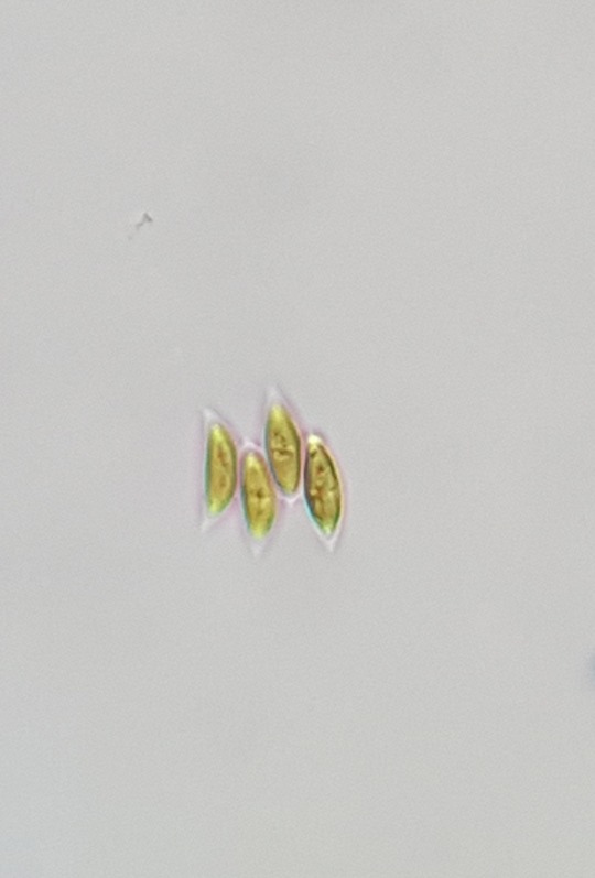
My love for algae just keeps growing.
These are all from one tiny sample of lake water.
229 notes
·
View notes