
Statistics
We looked inside some of the posts by bozonkwark and here's what we found interesting.
Average Info
Notes Per Post
6K
Likes Per Post
4K
Reblog Per Post
2K
Reply Per Post
7
Time Between Posts
5 days
Number of Posts By Type
Photo
12
Text
3
Video
1
Link
1
Last Seen Tumblr Blogs


Fun Fact
Tumblr was named as a finalist in Lead411’s New York City Hot 125 in Aug 2010.
Photo
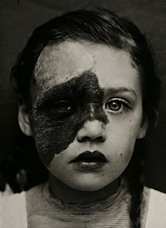
Utrecht goitre (Utrechtse Krop) is the title of a collection of photographs by Paul Kooiker. The book includes images of the file Utrecht University Hospital, some of them around 1890.
“Utrechtse Krop” (Utrecht goitre) was the name given to a thyroid condition once common in the Dutch town of Utrecht, due to a deficiency of iodine in drinking water.
1K notes
·
View notes
Photo

Over the past couple decades, microfluidic devices have become a staple of medical and biological diagnostics and analysis. Tests that once required large and specialized equipment can now be completed closer to a patient, using only a few drops of sample fluid. Running multiple tests on a single chip can become difficult, though, since flow through the device tends to dissolve and mix the dried reagents used for tests. But a new method cleverly uses fluidic forces to keep reagents separated without the need for complicated microfluidic structures.
The basic concept is outlined in the illustration above. You’re looking down on a microfluidic channel that’s long and very thin. A shallow groove down the middle serves as a barrier by pinning the contact line of the incoming fluid. So when the sample fluid flows in through the inlet on the left, it will only fill the top half of the cell. When it reaches the far right side, it turns the corner and flows to the left, encountering the first of the dried reagents it must dissolve for the device’s tests. The fluid will fill the lower channel quickly and then come to rest while the reagents dissolve.
With both sides of the channel full of liquid, the shallow barrier can no longer hold, and the fluid will take up the full width of the channel, with two well-dispersed – but separated – regions of reagents. Once that’s happened, a valve – represented by the pale blue line near the right side of the illustration – releases the fluid into the next section of the chip, allowing the analysis to proceed. (Image credit: Nature; research credit: O. Gökçe et al.; submitted by Kam-Yung Soh)
114 notes
·
View notes
Text
Researchers have shown for the first time that fatty tissue accumulates in the airway walls.
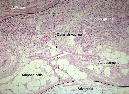
[Pic: Micrographs (x200) of the (A.) outer airway wall, between the airway smooth muscle (ASM) layer and the airway adventitia (dashed line) showing adipose tissue and mucous glands and (B.) inner airway wall (submucosa), between the basement membrane and ASM layer (dashed line) in a case of fatal asthma stained with haematoxylin and eosin. Inflammatory cells were counted within the inner airway wall.
Credit: European Respiratory Journal]
We already know how fatty build-ups in the arteries can increase the chances of developing heart problems, but now scientists have found early evidence the same sort of clogging could happen in the lungs – and it might be linked to asthma.
Asthma is known to be more common in obese people. Scientists analyzed lung samples from 52 people post-mortem. They found the amount of fat in the vital organ increased alongside the deceased’s BMI.
Sir Charles Gairdner Hospital and the University of Western Australia in Perth found that excess adipose tissue swelled the thickness of the airway walls and caused them to become inflamed. Of these lungs, 15 belonged to people who did not suffer asthma, 21 who had asthma but died of other causes, and 16 who died of asthma. A total of 1,373 airways were examined under the microscope using dyes to expose any fatty tissue.
The team is looking for new ways to study and measure fatty tissue in the lungs. They want to confirm the relationship with respiratory disease and to find out whether the effect can be reversed by weight loss therapy.
100 notes
·
View notes

Photo

Windmill, a process: start with a suitable line through some point. Rotate the line clockwise around the pivot until it contains another point, which now takes over as the new pivot. This process loops and visits all points infinitely often.
Via 3Blue1Brown who is a hip math educator and a terrific math animation engineer. My gif lacks his clickety-clacks, yes, among other things.
631 notes
·
View notes

Photo

For large creatures, it’s not hard to feel the evidence of someone else swimming nearby. But to tiny swimmers water is incredibly viscous and hard to move. These creatures have to swim very differently than their larger cousins, and evidence of their motion dies out quickly. But at least one microorganism, Spirostomum ambiguum, has discovered a method for overcoming the limits of size and viscosity.
The single-celled swimmer, when threatened, contracts its body in milliseconds, generating accelerations greater than those seen by fighter pilots. That acceleration is strong enough that it generates a burst of turbulence powerful enough to overcome the natural damping of its viscous surroundings. Within their colonies, S. ambiguum seem to use contraction to send out hydrodynamic signals to neighbors, who pass on the call to arms. To see the colonies in action, check out this previous article. (Image and research credit: A. Mathijssen et al.; via Physics Today; submitted by Kam-Yung Soh)
220 notes
·
View notes
Photo

Light it Up
Cardiovascular diseases, conditions affecting the heart and blood vessels, are the leading cause of death worldwide. Most commonly, these problems are linked to atherosclerosis, the build-up of fatty compounds, collectively known as plaque, in the walls of arteries. Fat molecules, or lipids, in these deposits are linked to a protein named apolipoprotein-B (ApoB), which may be key to understanding how and why harmful plaque forms. Researchers recently developed a technique to monitor the levels and distribution of ApoB-containing lipid complexes, or lipoproteins, in transparent zebrafish larvae, by fusing ApoB to a luciferase enzyme, closely related to that responsible for light production in fireflies. In the developing larvae, pictured from one (top) to five days after fertilisation (bottom), the bright blue glow reveals where and when lipoproteins are most abundant. Operating in a versatile model organism, this technique could unlock new ways of investigating plaque formation and facilitate drug testing.
Written by Emmanuelle Briolat
Image adapted from work by James H. Thierer, Stephen C. Ekker & Steven A. Farber
Carnegie Institution for Science Department of Embryology, Baltimore, MD, USA
Image originally published under a Creative Commons Licence (BY 4.0)
Published in Nature Communications, July 2019
You can also follow BPoD on Instagram, Twitter and Facebook
15 notes
·
View notes
Video
vimeo
Artist Susi Sie explores fluidic worlds through her macro lens. In “Emergence,” her focus is on ferrofluids immersed in other liquids. Beginning with tiny droplets traversing the thin fluid channels of a foam, she allows the unique qualities of ferrofluids to slowly take center stage. Dark blobs grow into curvy labyrinths as a magnetic fields come into play. Until ultimately the magnetic nature of the fluid becomes undeniable as scattered droplets elongate into miniature compass needles and swing around to follow the field lines. (Video and image credit: S. Sie)


286 notes
·
View notes
Photo

Cooperate or Cheat?
Using maths to study microbes might sound strange but these computer simulations could help to explain how bacteria grow and spread. In any population there’ll be co-operators – individuals that share their resources for the greater good of the group – as well as cheaters, who take more than their fair share. By testing out different scenarios based on how cheats (red) and co-operators (green) interact and move around, researchers can see what kinds of patterns emerge over time. In some cases, the cheats and co-operators end up segregating into separate areas (top), in others they mix (middle) and sometimes the co-operators win (bottom). Figuring out how these populations interact could shed light on how colonies of harmful antibiotic-resistant bacteria emerge and spread. More widely, the same equations apply to all kinds of species from bacteria all the way up to birds and animals, and perhaps our own human communities too.
Written by Kat Arney
Image adapted from work by Felix Funk and Christoph Hauert
Department of Mathematics, University of British Columbia, Vancouver, British Columbia, Canada
Image originally published under a Creative Commons Licence (BY 4.0)
Published in PLOS Computational Biology, August 2019
You can also follow BPoD on Instagram, Twitter and Facebook
10 notes
·
View notes
Photo

Stitched Up
The brain’s processing power easily surpasses the most advanced supercomputer, yet information technology can still help us to understand our nervous systems. Mapping out connections of this developing fruit fly’s nervous system, the sophisticated computer algorithms of a new software called BigStitcher work alongside two microscopy techniques: expansion microscopy, to swell and inflate the tiny tissues under the lens, and light sheet microscopy to take thousands of virtual ‘slices’ through the cells. Each coloured area is a huge 3D image (carrying many gigabytes of data) virtually reassembled into a mosaic ‘map’ of the whole nervous system. The next challenge is interpreting the information – perhaps calling on algorithms again to help follow the snaking paths of each individual nerve cell or neuron – and deciphering clues about common neuronal patterns found in ‘higher’ organisms like humans.
Written by John Ankers
Image from Janelia / MDC
Berlin Institute for Medical Systems Biology, Max Delbrück Center for Molecular Medicine in the Helmholtz Association, Berlin, Germany
Image copyright held by the original authors
Research published in Nature Methods, August 2019
You can also follow BPoD on Instagram, Twitter and Facebook
8 notes
·
View notes
Photo

Being hot isn’t always enough to make water boil. To form vapor bubbles, water and other liquids need imperfections that serve as seeds. In the absence of these, the liquid can become superheated, reaching temperatures higher than its boiling point without forming bubbles. Superheated water can be quite dangerous because it appears to be cooler, but once it’s disturbed – thereby breaking its surface tension – vapor bubbles form rapidly and explosively. You can see in the animation above just how quickly and unsteadily a sudden vapor bubble expands as it rises to the surface. (Image credit: C. Kalelkar and K. Raj, source)
351 notes
·
View notes
Photo

Many substances – like the cornstarch and water mixture above – exhibit a property called shear-thickening. In these fluids, deforming them quickly causes the viscosity to increase dramatically. That shear-thickening occurs when particles inside the fluid jam together, creating large chains able to resist the force being applied. That’s why the oobleck on this vibrating speaker can sustain these “cornstarch monsters”.
Shear-thickening is useful in many contexts, but it’s problematic during manufacturing, when pumping these substances can become incredibly difficult due to the fluid’s innate resistance to flowing. A new study, though, finds that it’s possible to temporarily suppress shear-thickening using acoustic waves. The researchers used piezoelectric devices to generate acoustic waves at a frequency around 1 MHz while shearing the cornstarch mixture. The acoustic waves disrupt the formation of particle chains inside the mixture, keeping its viscosity 10 times lower than during regular shear-thickening. (Image credit: bendhoward, source; research credit: P. Sehgal et al.; submitted by Brian K.)
882 notes
·
View notes
Photo
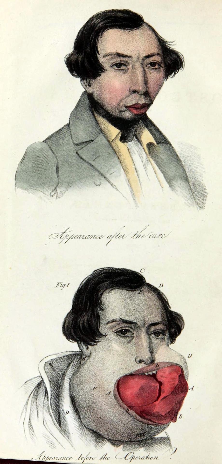
Osteosarcoma, before and after operation
Osteosarcoma are cancerous bone tumors found in immature bone, most often in those over 10 and under 25 years old. This particular case is in a man named Robert Penman, who was twenty-four.
These days, in those whose tumor is non-malignant (has not spread beyond its original tumor site), the five-year survival rate is about 75%. Back in 1839, I cant imagine this lad was quite as lucky, but it appears that at least the primary tumor was removed for a time.
Case of Osteo-sarcoma of the lower jaw, as operated upon Robert Penman, aged twenty-four years. 1839.
613 notes
·
View notes
Text
Scientists Use Nanoparticles to transport anti-cancer agent to cells

Small interfering ribonucleic acid (siRNA), has the potential to inhibit overexpressed cancer-causing genes and has become an increasing focus for scientists on the hunt for new cancer treatments. siRNA binds to specific gene messenger molecules and destroys them before they can tell the cell to produce a particular protein. This process is known as ‘gene knockdown’.
As a result, RNA interference (RNAi) has become a widely used tool for cellular genetic knockdown. However, RNA instability and susceptibility to enzymatic degradation have prevented its widespread clinical use. Thus, research efforts are seeking methods to protect the fragile RNA payload during delivery.
Scientists at the University of Cambridge think they have the solution. Led by Dr. David Fairen-Jimenez, from the Cambridge Department of Chemical Engineering and Biotechnology, they have created a platform that uses nanoparticles to get the anti-cancer agent [small interfering ribonucleic acid (siRNA)] to the cells. Their work was published in journal Chem.
The nanoparticles, known as metal-organic frameworks or MOFs could be a viable way of delivering the agent. To come up with the platform the team of scientists used simulations to pinpoint a MOF that has the ideal size to carry the siRNA agent. Once it’s inside the cell it breaks down releasing the agent.
A problem with using MOFs or other vehicles to carry small molecules into cells is that they are often stopped by the cells on the way to their target. This process is known as endosomal entrapment and is essentially a defense mechanism against unwanted components entering the cell. Fairen-Jimenez’s team added extra components to their MOF to stop them being trapped on their way into the cell, and with this, could ensure the siRNA reached its target.
63 notes
·
View notes
Text
Refuting the Dogma: Donor Cells Pass Through Blood-Brain-Barrier and Target Tumor

Medulloblastoma (glowing white) in 15-year-old boy. Image contributed to Radiopaedia by Paresh K. Desai, MD.
Located in the cerebellum, medulloblastoma is one of the most common cancerous brain tumors in children and can cause behavioral changes, nausea, vomiting and decreases in coordination. Most patients with medulloblastoma can be cured, but unfortunately, a few do not respond to traditional radiation and chemotherapies. Researchers led by Hisham Abdel-Azim, MD, from The Saban Research Institute of Children’s Hospital Los Angeles, recently demonstrated a new treatment option for these cases of high-risk recurrent medulloblastoma. The findings were published on April 20 in Pediatric Blood & Cancer.
In the reported case, investigators transplanted healthy donor stem cells into a patient with medulloblastoma. This procedure, called an allogenic, or “donor” hematopoeitic stem cell transplant, is performed with the idea that the stem cells will differentiate and become cancer-fighting immune cells. But just as in organ donation, receiving donor stem cells greatly increases the chance of rejection and a serious condition known as graft-versus-host disease (GVHD).
GVHD is dangerous and occurs when the transplanted stem cells attack the recipient’s body; however, previous studies showed that these transplants may also offer a beneficial “graft-versus-tumor” effect when the immune cells from the transplant attack the cancerous cells.
Abdel-Azim and colleagues were the first to report the “graft-versus-tumor” effect in the brain, refuting the dogma that immune cells could not pass through the blood-brain-barrier.This was also the first time that whole bone marrow was used as a source of donor stem cells to treat a child with a brain tumor. They were also the first to describe an allogenic hematopoeitic stem cell transplant using whole bone marrow to treata child with a brain tumor.
“We demonstrated that, despite all opposing views, a hematopoietic stem cell transplant from donor cells may be a viable thereapeutic option for select patients with medulloblastoma,” said Abdel-Azim. “Through this study, we suspect that the donor T lymphocytes and natural killer cells play a crucial role in the ‘graft-versus-tumor’ effect. This gives us a specific vehicle to explore in order to develop cell based, targeted therapies for brain tumors.”
200 notes
·
View notes
Link
16 notes
·
View notes