
Author: Koenders, Laura et al. Citation: Koenders, Laura, et al. "Grey Matter Changes Associated with Heavy Cannabis Use: A Longitudinal sMRI Study." PLoS ONE, vol. 11, no. 5, 2015, https://doi.org/10.1371/journal.pone.0152482. Accessed 30 Nov. 2022.
Don't wanna be here? Send us removal request.
Statistics
We looked inside some of the posts by palmanzar and here's what we found interesting.
Average Info
Notes Per Post
1
Likes Per Post
1
Reblog Per Post
0
Reply Per Post
0
Time Between Posts
3 minutes
Number of Posts By Type
Text
5
Last Seen Tumblr Blogs


Fun Fact
Tumblr was created by web developers David Karp and Marco Arment.
Text
Introduction
Cannabis is commonly known as the most frequently used illicit** drug throughout the world, and the use of cannabis has been associated with a higher risk of experiencing mood and anxiety-based disorders, cognitive impairment, and psychotic symptoms. Previous neuroimaging studies have questioned if chronic cannabis exposure and cannabis abuse can affect the brain’s morphology. However, when the paper was released, there had not been conclusive evidence or a distinction between the cause and effect using neuroimaging.
This study aims to determine if constant cannabis use and high level of cannabis exposure in young adults are correlated with grey matter reduction. This three year long study was done using healthy controls (HC) (N=22, age M=21.6, Sd2.45) and heavy cannabis users (CB)(N=20, age M=20.5, SD=2.1). The goal of the study was to examine if grey matter changed over time in the cannabis users group when compared with the healthy controls, as well as investigate the association between change in grey matter volumes and change in the severity of cannabis use-related issues when looking at the CB group. The hypothesis was there would be reductions in grey matter volumes in the CB group when compared to the stable volumes in the HC group in the orbitofrontal cortex, anterior cingulate cortex, insula, striatum, thalamus, amygdala, hippocampus, and cerebellum. They also hypothesized there would be changes in grey matter volume over time in the CB group that would be directly correlated with cannabis use issues and exposure.

0 notes
Text
Participants/Methods
The participants of the study were recruited through advertisements via the Internet and cannabis outlets such as coffee shops. All the participants were told not to consume alcohol or any drugs 24 hours before the study assessments were completed. A participant was considered heavy cannabis user if they had been using cannabis for two or more years and more than 10 days per month. In contrast, participants were considered to be in the control group if they had used cannabis 30 times or less in their entire lifetime and had not used it in the past year. The participants were asked to complete a detailed history of cannabis use as well.
These groups took part in a comprehensive psychological assessment, T1- structural MRI scan at baseline with a three-year follow-up MRI scan. The grey matter volumes were measured using SPM (VBM-8-module), a software package. The severity of cannabis use was measured using the Cannabis Use Disorder Identification Test (CUDIT), which is a 10-item screening on cannabis use frequency and severity of use-related problems. Cannabis use was also measured in grams. Nicotine dependence was also measured using the Fagerstrom Tolerance Questionnaire, and the severity of alcohol use was identified using the Alcohol Use Disorder Identification Test. In addition, psychologists assessed the prevalence of mental disorders using the DSM-IV requirements. MRI data processing was performed in the longitudinal analyses by using the VBM8 longitudinal batch. In order to look at the relationship between grey matter change over time and change in CUDIT scores and amount of grams weekly, the grey matter difference images were calculated within ImCalc, which involved subtracting each subject’s probability map at baseline from the map at follow-up.

The grey matter results were normalized using the diffeomorphic image registration algorithm. In addition, each image of regional grey matter volume was corrected for individual brain size.
In assessing cross-sectional group differences in grey matter volume, two sample t-tests were performed to compare the control group and cannabis user group both at baseline and follow-up. When assessing if grey regional grey matter volume changed throughout the time period in the CB group compared to the HC group, the study used a full factorial model. To find the relationship between cannabis-use-related problems (CUDIT) and the grams consumed per week with grey matter volume, multiple regression analyses were performed within the CB groups both at baseline and follow-up. Some covariates considered age and gender in all the analyses, and the confounding effects of comorbid nicotine use, alcohol dependence, and other drug use or psychiatric disorders were assessed. In all the analyses, the region of interest (ROI) images were performed as well as exploratory whole-brain analyses.

0 notes
Text
Results
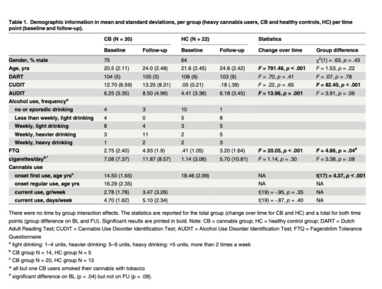
The table above (Table 1), shows the demographic variables. We can see that the heavy cannabis users showed significantly higher cannabis and nicotine dependence scores compared to the healthy controls. This is reflected by higher CUDIT and FTQ scores for the CB group compared to the HC group at both baseline and follow-up points. As shown, the CUDIT scores appeared stable throughout time but the FTQ scores were higher at the follow-up point in both groups. All except one subject in the CB group smoked their cannabis with tobacco. 2 subjects in the HC group used cannabis 20-25 times in their life, while the rest used cannabis less than 5 times. The AUDIT scores, representing alcohol problem severity, increased over time as well; however, comparing the groups to each other, the changes were not significant. The difference in consumed alcohol at baseline was not significant at baseline (p=.23) however, it was at follow-up (p=0.02).
GM volume group comparisons:
The results showed a cluster with larger grey matter volume in the CB group when compared to the HC group in the cerebellum, which was significant at baseline but not at follow-up. This cluster was also not significant after the inclusion of control covariates except for the frequency of alcohol use. The results also did not show any group differences in other ROI or at the whole-brain level at both time periods.
GM volume associations with cannabis use:
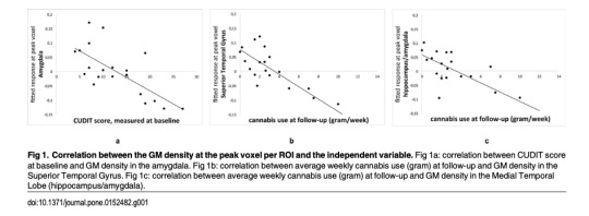
The results showed a significant inverse correlation between amygdala volume and cannabis-related problems. There was a significant negative correlation at baseline between CUDIT scores and grey matter volume in a cluster in the left amygdala. However, the cluster was no longer significant after including control covariates except for the frequency of alcohol use. There was a significant negative correlation between the grams per week and grey matter volume in a cluster comprising the amygdala and hippocampus at follow-up, which remained significant after controlling for the severity of nicotine use, the severity of alcohol use, and frequency of alcohol use and without subjects with comorbid substance or psychiatric disorders. The whole brain analysis showed a significant negative correlation between the amount of grams weekly and grey matter volume in a cluster in the superior temporal gyrus, which was no longer significant after control. The linear regression analysis found no significant correlation between the CUDIT score and cannabis use measures and any of the ROIs or whole-brain analyses.
0 notes
Text
Discussion/Conclusion
The results of the study did not show an effect of cannabis on grey matter volume when looking at the baseline and follow-up scans. When cross-sectional analyses were performed, there were consistent negative correlations between cannabis-related issues and cannabis use, which was measured in grams. Ultimately the results supported that reduced grey matter volumes in the medial temporal lobe are indeed a risk factor for heavy cannabis use, and this effect is limited to adolescents with no further damage from continued use after early adulthood.

The grey matter columns in the OFC, ACC, insula, striatum, thalamus, amygdala, hippocampus, and cerebellum were compared between the HC and CB groups. The study showed no differences between CB and HC groups which contrasted the hypothesis. Additionally, there were no significant changes in weekly cannabis use and cannabis use-related problems related to grey matter volumes. The cross-sectional analysis showed correlations between grey matter volumes in the left medial temporal lobe (hippocampus, amygdala, STG) and cannabis-related problems and cannabis use at baseline and follow-up. It is possible that in cannabis users, the cerebellar decrease, which is typical during development, occurs at a later age which explains group differences at baseline but not at follow-up. Ultimately, the lack of significant changes supports that cannabis use during later adolescence and early adulthood does not change grey matter morphology.
0 notes
Text
Limitations
Several of the participants developed DSM-IV diagnoses over time. In the future, studies should include healthy, nicotine, and/or alcohol-dependent controls, and to account for comorbid psychiatric disorders, future longitudinal studies should take into account psychiatric symptoms to include a larger sample and have enough power to control the nuisance factors. In addition, the study does not allow for definite conclusions to be drawn regarding causality since it did not have a group of cannabis users that was cannabis-naive at the start of the study.
1 note
·
View note