
Don't wanna be here? Send us removal request.
Statistics
We looked inside some of the posts by rnedicalimaging and here's what we found interesting.
Average Info
Notes Per Post
598
Likes Per Post
422
Reblog Per Post
176
Reply Per Post
0
Time Between Posts
7 days
Number of Posts By Type
Text
17
Last Seen Tumblr Blogs


Fun Fact
In Q3 of 2020, 31% of US users access the Tumblr app daily.
Text

Another great snippet from "Adult Children of Emotionally Immature Parents"
598 notes
·
View notes
Text
11th July 2024
CC - A&P: Taste & Smell Terminology
Olfaction - Our sense of smell, or transduction of chemical signals in our nasal passage to perceptible action potentials.
Gustation - Our sense of taste, or transduction of chemical signals in the mouth (mostly) to perceptible action potentials.
Taste Pore - A small hole in the stratified squamous epithelium of the tongue that leads to the gustatory epithelial receptors.
Gustatory Epithelial Receptors - The kind of cell that actually senses molecules dissolved in saliva, but does not transmit those signals to the brain.
Gustatory Hairs - A small extension of the cell membrane from a gustatory epithelial cell that reaches through the taste pore of a taste bud.
Fungiform Papillae - Most taste buds are found between these small bumps across the main surface of the tongue.
Salty - This taste is sensed when sodium ions dissolved in saliva enter open sodium channels in specific gustatory epithelial receptors.
Sour - This taste is sensed when hydrogen ions dissolved in saliva enter open proton channels in specific gustatory epithelial receptors.
Glomerulus - Meaning "ball of yarn", this clump of cells is where olfactory neurons converge after they enter the cranium.
Ethmoid - The bone through which olfactory neurons pass from the nasal cavity to the brain.
Olfactory Epithelium - A small area at the back of the nasal cavity where a thin membrane of columnar cells is penetrated by millions of neurons that receive odorants and transduce the sense of smell.
Olfactory Cortex - The part of the temporal lobe of the cerebrum that receives and initially processes signals about smell.
Mitral Cell - The name of the cell that receives signals from the olfactory neurons and transmits those signals to the brain.
Limbic System - The part of the brain that receives some information about smell and associates it with emotional responses.
Transduction - The action of a sensory cell translating chemical, electromagnetic, or mechanical stimuli into action potentials.
Anosmia - A partial or complete loss of smell.
0 notes
Text
10th July 2024
Crash Course - Anatomy & Physiology: Taste & Smell
youtube
Summary:
Anatomy & Physiology of Smell
The Olfactory Sensory Neurons
Receptors > Glomerulus > Mitral Cells > Brain
Taste Receptor Epithelial Cells (Taste Buds)
Receptors Trigger Action Potentials To 4 Different Cranial Nerves
Taste & Smell
Anosmia - Partial or full loss of smell.
caused by head trauma, respiratory infections, aging
Without being able to smell, you can't access emotional memories wrapped in particular scents, or sniff out dangers in her environment.
Transduction - Sensory cells translating chemical, electromagnetic, and mechanical stimuli into action potentials that our nervous system can make sense of."
Photoreceptors
detect light waves
Mechanoreceptors
detect sound waves and pressure on the skin and in the inner ear
Chemoreceptors = Gustation (Taste), Olfaction (Smell)
detect the molecules in our food and the air around us
Tastes and smells are powerful at activating memories, triggering emotions, and alerting us to danger - as well as enjoying the small things, like pizza.
How Smell Works
In order to smell something, the odarant must be volatile, or in a gaseous state to enter the nostrils.
Olfactory Epithelium
small yellowish patch of tissue on the roof of the nasal cavitiy
olfactory system's main organ
contains millions of bowling pin-shaped olfactory sensory neurons surrounding by insulating columnar supporting cells
Most molecules are caught by protective nose hairs, but a few make it to the back of the nose and hit the olfactory epithelium and dissolce in the mucus coating.
Olfactory Sensory Neurons
Once in mucus, they're able to bind to receptors on olfactory sensory neurons, which, assuming they hit the necessary threshold, fire action potentials up the long axons, through the ethmoid bone into the brain's olfactory bulb.
Each olfactory neuron has receptors for just one kind of smell.
E.g. thymol of oregano, butyric acid of cheese, acetylpyridine of crust
Glomerulus
After each smell-specific neuron is triggered, the signal travels down its axon and converges with other cells in the glomerulus.
Latin - "ball of yarn".
tangle of fibers that serves as a transfer station, where nose information turns into brain information.
Inside the glomerulus, olfactory axons meet up with the dendrites of another kind of nerve cell, mitral cells, which relays the signal to the brain.
For each mitral cell, there are any number of olfactory axons synapsing with it, each representing and identifying a single volatile chemical.
As a result, every combination of an olfactory neuron and mitral cell is like a single note, with smell triggering countless combinations, forming a musical chord of smells.
40 million different olfactory receptor neurons help us identify approximately 10,000 different smells.
How The Brain Processes Smell
Once a mitral cell picks up its signal from an olfactory neuron, it sends it alongthe olfactory tract to the olfactory cortex of the brain.
From there the smell hits the brain through two avenues:
Frontal Lobe - conscious identification. E.g "melted mozeralla!"
Emotional Ground Control - hypothalamus, amygdala and other parts of the limbic system.
The emotional pathway is fast, intense, and quick to trigger memories.
E.g. If the odour is associated with danger, like the smell of smoke, it quickly activates your sympathetic system’s fight or flight response.
Taste is 80 percent smell, so the same intellectual and emotional dynamics of smell apply to taste.
As you chew food, air is forced up the nasal passages, so the olfactory receptor cells register information at the same time as taste receptors are, so you’re both smelling and tasting simultaneously.
How Taste Works
After taking a bite of food, sensory information is quickly sorted by the 10,000 or so taste buds covering the tongue, mouth, and upper throat.
Most taste buds are packed deep down between the fungiform papillae -- those little projections that make your tongue kinda rough.
Fungiform papillae are not taste buds.
What Are Taste Buds? (Taste Receptor Epithelial Cells)
Taste Sensations
sweet
salty
sour
bitter
umami
All tastes register in all parts.
Taste buds are tucked into tiny pockets hidden behind the stratified squamous epithelial cells on the tongue.
Each taste bud has 50 to 100 taste receptor epithelial cells which register and respond to different molecules in your food.
Taste buds are specialised epithelial cells, not nervous tissue, so they still have to synapse to sensory neurons that carry information about the type and amount of taste back to the brain.
Types of Taste Receptor Epithelial Cells: Gustatory & Basal
The epithelial receptor cells come in two major types:
Gustatory
do the tasting
Basal
stem cells that replace gustatory cells
Basal epithelial cells are extremely dynamic and replace gustatory cells every week or so, which is why a terribly burnt tongue will feel better in a couple of
Each gustatory cell project a thread-like protrusion of the cellular membrane, a gustatory hair, which runs down to a taste pore, a small hole in the stratified squamous epithelium covering the taste bud and rest of the tongue.
Food chemicals, or tastants, must dissolve in saliva to diffuse through taste pores, bind to receptors on gustatory cells, and then trigger an action potential.
How Different Tastants Are Sensed
Each tastant is sensed differently.
E.g. Salty food is full of positively-charged sodium ions that cause sodium channels in the gustatory cells to open, which generate a graded potential, and spark an action potential. Sour-tasting acidic foods are high in hydrogen ions and take a different route by activating proton channels.
Taste, like all senses, is about how action potentials get triggered.
Once an action potential is activated, that taste message is relayed through neurons via the 7th, 9th and 10th cranial nerves to the taste area of the cerebral cortex, at which point the brain makes sense of it all, and begins releasing digestive enzymes in saliva and gastric juices in the stomach to help break down food to be used.
0 notes
Text
9th July 2024
CC: A&P - Peripheral Nervous System Terminology
Peripheral Nervous System - The part of the body that carries signals between the brain and spinal cord and the rest of the body.
Stimulus - A general term for the internal or external event that is sensed by the PNS.
Sensation - A general term for the detection of an internal or external stimulus by the nervous system.
Perception - How the brain receives, processes, interprets, and consciously experiences information sent by the PNS.
Reflexes - Immediate reactions that don't need conscious processing from the brain, these involve sensation, transmission to the CNS, integration, transmission to an effector, and response.
Intrinsic Reflex - A reflex action that we are often born with, often processed in the spinal cord, involving simple movements like withdrawing from pain.
Acquired Reflex - A reflex action that we are not born with, that takes time and exposure to develop into a fast response that doesn't require conscious input.
Motor Division - The parts of the PNS responsible for carrying signals from the CNS to the body.
Sensory Division - The parts of the PNS responsible for carrying signals from the body to the CNS.
Thalamus - The part of the brain that acts as a switchboard, first receiving sensory signals and sending them out to the appropriate regions of the cerebrum.
Integration Center - In a sensation-process-motor-loop, the CNS functions as this.
Thermoreceptors - The class of receptors in the PNS that pick up signals about temperature, both inside and outside your body.
Effector - Any structure in the body that receives and reacts to a motor neuron's signal, like muscles or glands.
Photoreceptors - The class of receptors in the PNS that pick up signals about light.
Nociceptors - The class of receptors in the PNS that send signals of pain.
Chemoreceptors - The class of receptors in the PNS that pick up signals about chemicals, like pH and oxygen concentrations in the blood.
Mechanoreceptors - The class of receptors in the PNS that pick up signals about pressure, touch and vibration,
0 notes
Text
4th July 2024
Crash Course - Anatomy & Physiology: Peripheral Nervous System
youtube
Summary:
Peripheral Nervous System
Afferent and Efferent Divisions
Information and Responses to Pain
Fives Steps of the Reflex Arc
Different Kinds of Reflexes
What The Brain Says About Pain
Peripheral Nervous System
puts your brain in touch with the physical environment, and allows it to respond
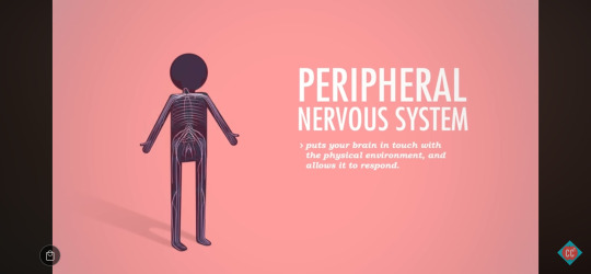
Sensory Nerve Receptors: Thermoreceptors, Photoreceptors, Chemoreceptors, Mechanoreceptors, Nociceptors
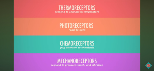
Thermoreceptors
respond to change in temperature
Photoreceptors
react to light
Chemoreceptors
pay attention to chemicals
Mechanoreceptors
respond to pressure, touch and vibration
Nociceptors
fire only to indicate pain
What is Pain?
Pain helps protect ourselves, and from the outside world.
How Pain is Processed
Stimulus
distinct feeling of discomfort
Nociceptors
provided base sensation
Spinal Cord
immediate reflexive action of retracting
Brain
interprets awareness into the perception of pain, decides to move, probably cusses
Pain Threshold vs Pain Tolerance
Pain is thought of as the perception of pain
We all have the same pain threshold, but not tolerance.

Stimulating Event
E.g. foot meets pin
Reception
E.g. nociceptors in foot sense stimulus
Transmission
transmission of signal through nerves to spainl cord and eventually brain
How The Brain Processes Pain
Mechanically Gated Channels
open in response to the physical stretching of the membrane

Ligand-Gated Channels
open when histamine or potassium ions released by damaged tissue bind to receptors

Afferent and Efferent Divisions
Sensory Division (Afferent)

collects data and sends to Central Nervous System
1 - receptor senses a stimulus
2 - sensory neuron transmits signal up the PNS to the CNS
3 - integration center decodes the signal
Motor Division (Efferent)
4 - motor neuron sends directions to effector organs to respond to stimulus
5 - effector cells respond by contracting or secreting
Five Steps of The Reflex Arc
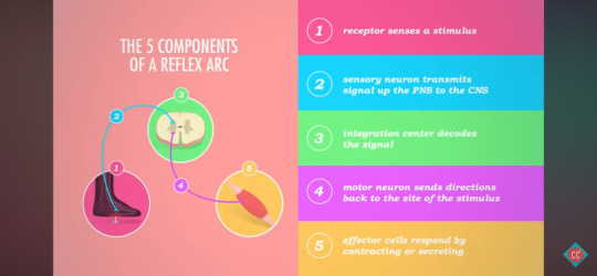
1 - receptor sense a stimulus
2 - sensory neuron transmits signal up the PNS to the CNS
3 - integration center decodes the signal
4 - motor neuron sends directions back to the site of the stimulus
5 - effector cells respond by contracting or secreting
Reflexes
immediate reactions that can either be innate or learned, but don't need much conscious processing in the brain
Innate or Intrinsic Reflex Action
a super fast motor response to a starting stimulus
E.g. lifting your foot when standing on a thumb tack
Processing happens in the spinal cord, so that the control of the muscles can be initiated before the pain is actually perceived by the brain
Learned or Acquired Reflexes
come from experience
E.g. learn to dodge obstacles while riding a bike/driving a car
Reflex arcs stimulate some muscles, while inhibiting others.
E.g. right foot activates motor neurons in right hip flexors and hamstrings, causing that knee to bend your foot and lift up, as well as telling quad muscles in left leg to extend and stand tall, allowing you to shift your bodys weight off thumb tack
Muscle & Tendon Spindles
If triggered by an overstretch, they generate a reflex arc that contracts the muscle to keep it from stretching further.
What The Brain Says About Pain
1) Thalamus
information switchboard, splits the signal

2) Somatosensory Cortex
identifies and localises the pain E.g. "sharp", "foot"

3) Limbic System
registers emotional suffering

Frontal Cortex
assigns meaning to pain. E.g 'Oh, the thumb tack must've fallen off the corkboard"
0 notes
Text
21st May 2024
CC: A&P - Central Nervous System Terminology
Central Nervous System - Integrating sensory information and deciding how to respond to it are the primary functions of this part of the body.
Spinal Cord - This part of the central nervous system conducts signals between your brain and the rest of the body, and governs many reflexes and muscle patterns while walking.
Cerebral Cortex - The outer layer of gray matter of the cerebrum with all of the folds and grooves.
Neural Tube - In a developing embryo, the first structure that will go on to form the central nervous system.
Corpus Cullosum - The "large body" that connects the left and right cerebral hemispheres.
Mesencephalon - The middle of the three primary brain vesicles during brain development.
After about five weeks of development, when the primary brain vesicles divide into secondary vesicles, the prosencephalon divides into the diencephalon and... telencephalon.
After about five weeks of development, when the primary brain vesicles divide into secondary vesicles, the rhombencephalon divides into the myelencephalon and... metencephalon
Cerebellum - The metencephalon develops into the pons and this part of the adult brain responsible for coordinating muscle activity by comparing sensory and motor information.
Rhombencephalon - The most posterior or caudal of the three primary brain vesicles during brain development.
Gyri - On the cerebral cortex, the ridges that result from an embryological increase in surface area and folding of the cortex.
Sulci - On the cerebral cortex, the grooves or valleys that result from an embryological increase in surface area and folding
Cerebrum - The embryological telencephalon develops into this largest part of the brain, that is so big it covers most of the rest of the brain.
Cerebrospinal Fluid - The fluid in your central nervous system that cushions the brain and spinal cord and provides some nourishment.
Midbrain - The general region of the brain responsible for quickly processing sensory information and responding with visual and auditory reflexes.
Diencephalon - The thalamus, hypothalamus, epithalamus and mamillary bodies, which regulate homeostasis, make up this region of the brain.
Cerebrum - The part of the brain responsible for voluntary movement, conscious thought, thinking learning and recognising emotions.
Cerebellum - The metencephalon develops into the pons and this part of the adult brain reesponsible for coordinating muscle activity by comparing sensory and motor information.
Frontal Lobe - This region of the cerebrum, responsible for controlling voluntary muscle movement, and higher cognitive functions like planning and social inhibitions.
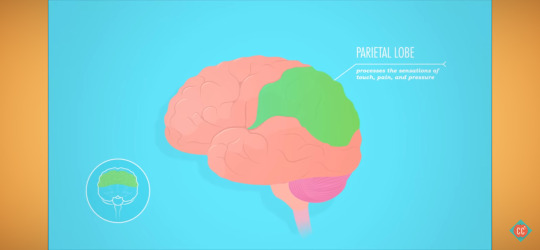
Parietal Lobe - The superior posterior regin of the cerebrum processes sensory information from the body like touch, pressure and pain.
Occipital Lobe - The most posterior part of the cerebrum, this processes visual information.
Temporal Lobe - This lateral region of the cerebrum processes auditory information like language and helps with balance
Temporal Lobe - This marked region of the cerebrum processes auditory information like language and helps with balance.
Brain Stem - The midbrain, pons, and medulla oblongata make up this general region of the brain, which regulates basic, vital, involuntary body functions.
Hippocampus - This part of the brain is the center of short term memory.
Frontal Lobe - The most anterior part of the cerebrum, this controls voluntary muscle movement, and higher cognitive functions like planning and social inhibitions.
Parietal Lobe - The region of the cerebrum processes sensory information for the body like touch, pressure and pain.
Amygdala - This part of the brain is a center of emotions, like fear, and controls sexual and social behaviours.
0 notes
Text
21st May 2024
Crash Course: Anatomy & Physiology - Central Nervous System
youtube
Summary:
Central Nervous System: Locating and Brain Function
Brain Development
Neural Tube
Three Primary Vesicles
Five Secondary Vesicles
Four Adult Structures And Their Functions
Broca's Aphasia
Broca's Area
area in left hemisphere for the brain
partly responsible for ability to produce and process language
Broca's Aphasia
some ability to understand speech, but an inability to produce intelligible words
Broca's aphasia can sometimes be treated by teaching patients to sing, because singing uses a different region of the brain on the right side - analogous to Broca's area on the left.
A lot of knowledge about the brain ahs arisen from studying injuries.
The brain is made up of super-specific areas that have super-specific functions.
Central Nervous System Structure & Function
Central Nervous System
Brain
Spinal Cord
integrating sensory information (collected from peripheral nervous system)
coordinating both conscious and unconscious activity
Brain
Carries out most complex functions:
thinking
feeling
remembering
sorts out sensory information and gives orders
Spinal Cord
conducts two-way signals between brain and rest of body
governs basic muscle reflexes and patterns that don't need brains permission
The brain and spinal cord are made of fragile, jelly-like nervous tissue that is extremely suscepitble to injury.

The nervous tissue is protected by the bones of the vertebrae and cranium, as well as membrane layers, or meninges, before being bathed in clear cerebrospinal fluid.
Cerbrospinal fluid allows the brain to float somewhat in the skull, reducing its weight
"Your brain is divided into specialised regions that may, or may not, interact with each other to produce a given action."
Brain Development: Neural Tube
Inside a developing embryo, the central nervous system starts as a neural tube.
The caudal, or lower, end of the tube stretches to form the spinal cord, while the cranial end expands, divides and enlarges into three primary brain vesicles, or interconnected chambers.
Prosencephalon (Forebrain)
Mesencephalon (Midbrain)
Rhombencephalon (Hindbrain)
Brain Development: 3 Primary Vesicles
By an embryo's fifth week of development, these vesicles morph into five secondary vesicles that form the roots of your mature brain structures.
Brain Development: 5 Secondary Vesicles
Prosencephalon
The prosencephalon divides into two sections:
Telencephalon
Diencephalon
Rombencephalon
The rombencephalon forms into another pair:
Metencephalon
Myelencephalon
Mesencephalon
The mesencephalon remains undivided.
The five secondary vesicles develop into the major adult brain regions. Each vesicle grows in different ways, some developing more than others.
Brain Development: Major Adult Brain Regions
Brainstem
Cerebellum
Diencephalon (Interbrain)
Cerebral Hemispheres
Brain Development: Cerebellum & Brain Stem
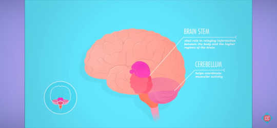
Cerebellum
helps coordinate muscular activity
Brain Stem
Midbrain
vital role in relaying information between the body and higher regions of the brain
carries out higher-level functions
receives and processes sensory information and sends out reflexive motor signals
passes data to cerebral cortex
Pons
Medulla Oblongata
Together, they regulate many basic, vital, involuntary functions. E.g. keeping heart on pace, keeping lungs working, controlling sleep, appetite, pain sensitivity and awareness.
Brain Development: Reptillian Brain
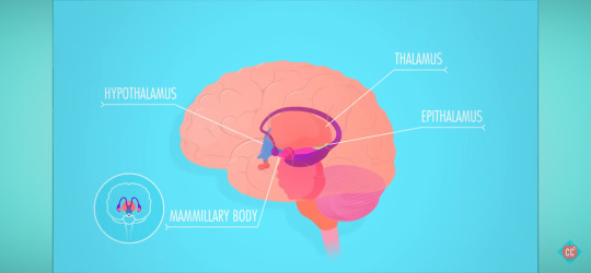
Diencephalon:
Thalamus
Hypothalamus
Epithalamus
Mamillary Bodies
= Homeostasis, alertness, reproductive activity.
Part of The Limbic System
Strong emotions - fear
Reptillian Brain
eat
drink
sleep
mate
stay safe
Brain Development: Cerebral Hemispheres
The telencephalon develops into the two classic walnut-looking hemispheres - the cerebrum.
Cerebrum
largest region of brain
performs highest functions
rules voluntary movements
thinking
learning
regulating and recognising emotions
experiencing consciousness
Grey Matter = wrinkly outer layer
White Matter = inner squishy layer
Higher processing requires lots of synapses, which requires lots of nervous tissue.
In order to squeeze the cerebrum into the skull, the brain forms creases:
Gyri - little creases
Sulci - larger grooves
The Brain's Lobes
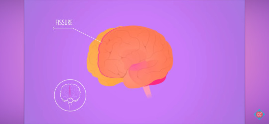

Although a big fissure seperates the left and right hemispheres, the two halves communicate, through a series of myelinated axon fibers called the corpus callosum.
Each hemisphere has other, smaller fissures that divide it into lobes - each with a different set of major functions.
Frontal Lobe
governs muscle control
cognitive functions. E.g. planning for the future, concentration, preventing socially unacceptable behaviours.
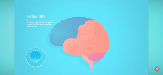
In most people, the frontal lobe doesn't finish developing until after teenage years.
The Broca's area lives in this lobe in the left hemisphere, it's also important in language comprehension and speech.
Occipital Lobe
processing bright visual cues

Parietal Lobe
processes sensations of touch, pain and pleasure
Temporal Lobe
sorts auditory information, including language
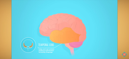
It contains Wernicke's area - an important region of the brain associate with production of written and spoken language.
Part of the Limbic System

Hippocampus
short-term memory keeper
Amygdala
emotional, controls sexual and social behaviour
0 notes
Text
20th May 2024
CC: A&P - NT, Synapses - Terminology
Axon Terminal - The structure at the end of a neuron's axon at a chemical synapse that contains and releases neurotransmitter.
Presynaptic Neuron - The cell that is sending a signal releasing the neurotransmitter in a chemical synapse.
Postsynaptic Neuron - The cell in a chemical synapse that is receiving the neurotransmitter.
Voltage-Gated Calcium Channels - The channels that are opened by an action potential reaching an axon terminal, sending the final trigger to release neurotransmitters.
Synapse - The general term for a structure where one cell can directly communicate with another cell.
Synaptic Cleft - The name for a very narrow gap between the two cells in a synapse.
Synaptic Vesicle - The structure within the presynaptic neuron's axon terminal that contains neurotransmitter waiting to be released.
Chemical Synapse - A structure that requires the use of a neurotransmitter to allow one cell to communicate directly with another cell.
Electrical Synapse - A structure including gap junctions that allows an ion current to pass from one cell directly into another cell.
Reuptake - The term for the reabsorption from a synaptic cleft of neurotransmitters by the pre-synaptic neuron that released it.
Neurotransmitter - A chemical released by neurons into a synapse to send a signal to another cell.
Excitatory Neurotransmitters - Neurotransmitters that lead to depolarisation of the postsynaptic neuron.
Inhibitory Neurotransmitters - Neurotransmitters that lead to hyperpolarisation of the postsynaptic neuron.
Serotonin - A typically inhibitory neurotransmitter, associated with regulating mood, appetite, circadian rhythm and sleep.
Dopamine - A typically excitatory neurotransmitter that influences emotion and attention, and is active in reward centers in the brain.
Norepinephrine - An excitatory neurotransmitter important in the fight or flight response to increase heart rate, alertness and mobilise energy reserves.
0 notes
Text
20th May 2024
Crash Course: Anatomy & Physiology - Nervous System, Synapses
youtube
Summary:
Electrical Synapses Use Ion Currents Over Gap Junctions To Transmit Neurological Signals
Chemical Synapses Turn Electrical Signals Into Chemical Ones
Chemical Synapses Use Neurotransmitters
Effects of Cocaine in the Electrochemical System
What are Synapses?
Synapse
the meeting point between two neurons
Greek - "to clasp or join"

If neurons form the structure of the nervous system, then synapses, the communication links between them, make it an actual system
"Neurons strength and purpose lies in their connection."
When an action potential sends an electrical message to the end of an axon, that messages hits a synapse that then translates, or converts it, into a different type of signal and flings it to another neuron. The human brain alone has 100 billion neurons, with each neuron having 1000 - 10,000 synapses.
100 - 1,000 trillion synapses in the brain.
"Synapses are able to change and adapt in response to neuron firing patterns" - either strengthening or weakening over time, depending how much they're used.
Synapses allow you to learn and remember.
Synapses are the root of many psychiatric disorders.
Synapses are why illicit drugs, and addictions to them, exist.
Electrical vs Chemical Synapses
Nerve cells have two main different communication settings, depending on how fast news needs to travel.
Electrical Synapses
the immediate group texts
Chemical Synapses
controlled, slower and more personal texts
How Electrical Synapses Work: Gap Junctions
"Electrical synapses are much more abundant in embryonic tissue where they help guide neural development, as the nervous system matures, many electrical synapses are replaced by chemical ones."
Electrical synapses send an ion current directly from the cytoplasm of one nerve cell to another through gap junctions.
The signal isn't converted from it's pure electrical state to another singla, like chemical synapses.
Instead, one cell and one synapse can trigger thousands of cell that act in synchrony.
Why Aren't All Synapses Electrical?
Control
With such a direct connection between cells, "An action potential in one neuron will generate an action potential in other cells across the synapse."
E.g The heart.
If every synapse in the body activated the neurons around it, the nervous system would be in "group text reply all" mode.
How Chemical Synapses Work: Neurotransmitters
Chemical Synapses
much more abundant
slower
more precise
more selective
Chemical synapses use neurotransmitter, or chemical signals, that diffuse across a synaptic gap to deliver a message.
effectively convert signal in steps: electrical > chemical > electrical
allows for different ways to control impulse.
"This sort of long term translation could be the result of:
Potentiation - the strengthening of a synapse through classical conditioning.
Habituation - when a synapse decreases its response to a common stimulus.
Sensitization - when a reaction to one stimulus causes other synapses to be more sensitive to reactions."
At the synapse, signal can be modified, amplified, inhibited or split, either immediately or over longer time periods.
Set-up - 2 Principal Parts:
Presynaptic Neuron Presynaptic Terminal
axon terminal
The cell that sends the signal is presynaptic neuron and it transmits through a knoblike structure called the presynaptic terminal, usually the axon terminal.
Synaptic Vesicles

The axon terminal holds a whole bunch of tiny synaptic vesicle sacs, each loaded with thousands of molecules of a given neurotransmitter.
Postsynaptic Neuron

Receptor Region

The receiving cell is the postsynaptic neuron, which accepts the neurotransmitters in its receptor region, which is usually on the dendrite or the cell body itself.
Synaptic Cleft
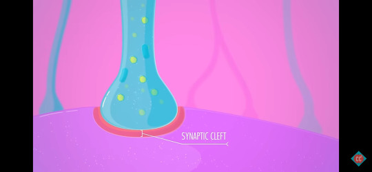
The neurons communicate, despite never touching, through a tiny gap called the synaptic cleft.
< five millionths of a cm apart
Messages that travel via chemical synapses are technically not transmitted directly between neurons, like in electrical synapses.
Instead, a chemical event that involves the release, diffusion and reception of neurotransmitters in order to transmit signals.
How Neurotransmitters Work

When an action potential travels along the axon of a neuron, activating sodium and potassium channels, it eventually comes down to the presynaptic terminal, and activates the voltage-gated calcium (Ca2+) channels to open and release calcium into the neuron's cytoplasm.
Voltage Gated Calcium Channels
-Ca2+

The flow of positively-charged calcium ions causes the tiny synaptic vesicles to fuse with the cell membrane and pure their chemical messengers.
It's these neurotransmitters that act like couriers diffusing across the synaptic gap and binding to receptor sites on the postsynaptic neuron.
The first neuron converts the electrical signal into a chemical signal.
In order for it to become an action potential again in the receiving neuron, it has to be converted back to an electrical signal.
This happens once a neurotransmitter binds to the receptor, causing the ion channels to open
"Depending on which particular neurotransmitter binds to which receptor, the neuron might either get excited or inhibited."
Excitatory Neurotransmitters
Excitatory Post-Synaptic Potentials (EPSPs): graded potentials that push the neuron closer to threshold, and make an action potential more likely.

Excitatory neurotransmitters depolarise the postsynaptic neuron by making the inside more positive and bringing it closer to its action potential threshold, making it more likely to fire the messageon to the next neuron.
Inhibitory Neurotransmitter
Inhibitory Post-Synaptic Potentials (IPSPs): graded potentials that push the neuron further from threshold, make an action potential harder to achieve.

Inhibitory neurotransmitters hyperpolarises the postsynaptic neuron by making the inside more negative, driving its charge down - away from the message. The message not only doesn't get passed down, it's harder to excite that portion of the neuron.
Any region of a single neuron may have hundreds of synapses, each with different inhibitory or excitatory neurotransmitters.
"The likelihood of that post-synaptic neuron developing an action potential depends on the sum of all of the excitations and inhibitions in that area."
100 Different Types of Naturally-Occurring Neurotransmitters
move around
keep vital organs humming along
amp us up
calm us down
make us hungry, sleepy or more alert
simply make us feel good
Neurotransmitters don't stay bonded to their receptors for more than a few milliseconds.
After delivering message, they pop back out and either degrade or recycled.
Some kinds diffuse back across the synapse are immediately re-absorbed by the sending neuron in a process called reuptake.

Others are broken down by enzymes in the synaptic cleft, or sent away from the synapse by diffusion.
How Cocaine Works
Illicit drugs either excite or inhibit the production, releasse and reuptake of neurotransmitters.
They can simply mimic neurotransmitters, tricking a neuron into thinking its receiving a natural chemical signal, when it's anything but.
Cocaine
Once in bloodstream, it targets 3 major neurotransmitters:
Serotonin
mainly inhibitory
role in regulating, mood, appetite, circadian, rhythm and sleep
Antidepressants can stabilise moods by stabilising serotonin levels.
Dopamine
influences emotion and attention
makes you feel awesome
E.g. hugging a loved one, sex, donuts
Norepinephrine
triggers fight or flight response
increases heart rate
primes muscle
Undersupply can depress mood.
In a sober state, once these neurotransmitters deliver their chemical payloads, they're usually diffused back out across the synapse to be absorbed by the neuron that sent them.
Cocaine blocks the reuptake, especially of dopamine, allowing these chemicals to float around and accumulate - causing euphoria, paranoia and "jitters".
As the body has a limited supply of these neurotransmitters, the body needs time to brew more.
Flooding synapses like this eventually depletes your supply, making you feel terrible in a number of ways.
Cocaine, and other drugs, that target neurotransmitters trick the brain, and after prolonged use may eventually cause it to adapt, as those synapses remember how great the extra chemicals feel.
As a result, you lose receptors, so it takes more cocaine, and finally cocaine, to function normally.
In a natural state, synapses know when to excite, when to inhibit, when to use electricity and when to dispatch chemical messengers.
0 notes
Text
19th May 2024
Crash Course: Anatomy & Physiology - Nervous System, Action Potential
youtube
Summary:
Ion Channels Regulate Electrochemistry To Create Action Potential
Resting State
Depolarisation
Repolarisation
Hyperpolarisation
What are Neurons?
When a neuron is stimulated enough, it fires an electrical impulse down its axon to neighbouring neurons.
"They've only got one signal they can send and it only transmits at one uniform strength and speed."
Frequency varies.
Action Potential
buzz, nerve impulse
Your Body is a Sack of Batteries
Electricity
Electrically Neural
The body has equal amounts of positive and negative charge, with certain areas more positively or negatively charged than others.
Membranes, acting as barriers, separate the opposite-attracting positive and negative charges until ready to use the energy.
"We keep em separated to build potential."

Voltage
the measure of potential energy generated by separated charges
membrane potential
measured in millivolts
The bigger the difference between positive and negative areas, the higher the voltage, the large the potential.
Current
the flow of electricity from one point to another
The amount of charge in a current is related to both its voltage and resistance.
Resistance
whatever's getting in the way of the current
"Currents indicate the flow of positively or negatively charged ions across the resistance of cells' membranes."
Membranes separate the charges, so provide potential to convert electricity for use.
How Electricity Works Inside The Nervous System
Resting Neuron
more negative inside, relative to extracellular space around it

Resting Membrane Potential
The difference between the inside and outside charges.
-70 mV
Outside Cell
positive sodium ions
Inside Cell
positive potassium ions
bigger negative proteins
As there're more positive sodium outside than positive potassium ions inside, the cell's interior has an overall negative charge.
negative membrane potential = polarised
Sodium-Potassium Pump

Protein straddles the membrane of the neuron.
Tons all along the axon.
For every two potassium ions it pumps into the cell, it pumps three sodium ions out.
Electrochemical Gradient
This creates a difference in the concentration of sodium and potassium, and a difference in charges, making it more positive outside the neuron.
Types of Ion Channels: Voltage-Gated, Ligand-Gated and Mechanically-Gated
Ion Channels
Ion channels are large protein, riddle among the membrane, that provide passages across the membrane when their respective gates are open.
The gate open and close for different reasons, depending on structure and purpose.
Voltage-Gated Channels
most abundant
open and close in response to changes in membrane potential

E.g. Sodium channels in neurons open at -55 mV.
Ligand-Gated Channels
open when a neurotransmitter latches onto its receptor.
E.g serotonin, a hormone.

Mechanically-Gated Channels
open in response to the physical stretching of the membrane

When gates open, ions diffuse across that membrane down their electrochemical gradient, even out the concentrations and moving away from other positively charged ions.
"The movement of ions is the key to all electrical events in neurons."
Graded Potential vs. Action Potential
Graded Potential
E.g. If only a few channels open, and only a bit of sodium enters the cell, that causes just a little change in the membrane potential in a localised part of the cell.
Action Potential
E.g. In order to send long-distance signals all the way along an axon, you need a bigger change, one big enough to trigger those voltage-gated channels.
Depolarisation
Start:
neurons sitting at resting state
ion channels closed
inner voltage resting = -70 mV


An environmental stimulus occurs, E.g. like a spider brushing against your knee, triggering the sodium channels to open and increasing the charge inside the membrane.
The stimulus, and resulting change, have to be strong enough to cross a threshold for the true action potential to kick in.
-55 mV
all or nothing phenomenon
If the stimulus is too weak, and the change doesn't hit -55 mV, the neuron returns to its resting state.

At -55 mV, the many voltage-gated sodium channels open, so the positive sodium ions rush in, making the cell massively deporalised - so much so that it becomes positive, up to +40 mV.
"A brief depolarisation caused by changes in currents."
Unlike graded potentials, small and localised, an action potential kicks off a biological chain reaction, which sends the electrical signal down the axon.
Repolarisation
As each neuron has many voltage-gated sodium channels, so when a few in one area open, the local current is strong enough to change the voltage around them.
This striggers the neighbours, which triggers the volttage around them and so on.
Once underway, the process of repolarisation occurs.
The voltage-gated potassium ion channels open up, letting the potassium ions flow out in an attempt to rebalance the charges.
Hyperpolarisation
The voltage drops to -75 mV beofere the gates close and the sodium-potassium pump take over and bring things back to resting level.
Refractory Period
An axon during this process, with its ion channels open, can't respond to any other stimulus.
It prevents signals from travelling in both directions down the axon at once.
"The first phase of this period, from depolarisation to repolarisation, is called the absolute refractory peirod. It makes sure that each action potentials is its own unique all-or-nothing event. The phase immediately following the period, the one that spans from repolarisation through hyperpolisation and back to resting potential is the relative refractory period."
Voltage
Initial Threshold = -55 mV
Peak of Depolarisation = +40 mV
"A weak stimulus tends to trigger less frequent action potentials.
E.g.
Low Frequency - delicate action, picking up an egg
High Frequency - intense signal, crushing a can
Conduction Velocity
Action potentials can vary by speed.
The factor that affects a neuron's transmission speed most is whether there's a myelin sheath on its axon.
Axons coated in insulating myelin conduct impulses faster than non-myelin axons.
Instead of triggering one channel at a time in a chain reaction, a current can leap from one gap in the myelin to the next.
Nodes of Ranvier - little gaps
salatory conduction - propogation
Latin - "leaping"

"Axons with a larger diameter conduct signals faster because they offer less resistance, so allow action potentials to happen quicker."
0 notes
Text
19th May 2024
CC: A&P - NS, Into to Nervous System Terminology
Central Nervous System - The part of the nervous system consisting of the brain and spinal cord.
Soma - Neuron's cell body that contains nucleus, DNA, mitochondria, etc.
Axon - Long extension or fiber that transmits electrical impulses away from the cell body to other cells.
Dendrites - Branch-like processes that pick up messages from other cells.
Astrocytes - Most abundant and versatile glial cells. They govern the exchange of materials between neurons and capillaries.
Neurons - In nervous tissue, the type of cell that can transmit signals from one cell to another.
Unipolar Neurons - A neuron with a single extension from the soma that leads to an axon with dendrites at one and and axon terminals at the other, peripheral sensory neurons are this type.
Bipolar Neuron - A neuron with one axon and a single dendrite extending from its soma, found in limited places like the retina of the eye.
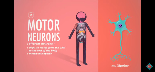
Multipolar Neuron - A neuron with one axon and many dendrites extending directly from the soma, this is the most common type of neuron in the body.
Afferent Neurons - In the peripheral nervous system, these pick up messages from sensory receptors and transmit impulses toward the central nervous system.
Efferent Neurons - In the peripheral nervous system, these receive messages from the central nervous system and transmit impulses out to peripheral effector organs, like muscles and glands.
Interneurons - In the central nervous system, these transmit impulses between other neurons.
Schwann Cells - Produce insulating barrier called myelin sheath in the peripheral nervous system.
Satellite Cells - Surround and support cell bodies.
Microglial Cells - Main source of immune defense against invading microorganisms.
Ependymal Cells - Create, secrete and circulate cerebrospinal fluid.
Oligodendrocyte - Produce insulating barrier called myelin sheath in the central nervous system.
Schwann Cells - Produce insulating barrier called myelin sheath in the peripheral nervous system.
Integration - When the central nervous system processes an input and decides what should be done with it.
Somatic (Voluntary) Nervous System -The part of the nervous system that carries signals from your central nervous system to only your skeletal muscles.
Peripheral Nervous System - All the nerves that branch off from your brain and spine and allow your central nervous system to communicate with the rest of your body.
Autonomic Nervous System - Part of the nervous system that carries signal from your central nervous system to regulate things like your heart beating, lungs breathing, and stomach churning.
Motor (Efferent) Division - The part of the peripheral nervous system that carries signals from your central nervous system to your muscles and glands.
Sympathetic Division - The part of the autonomic nervous system that mobilises energy and gets the body ready to fight or flee.
Parasympathetic Division - The part of the autonomic nervous system that encourages digestion, relaxes the body, and stores excess energy.
Sensory Division (Afferent) - The part of the peripheral nervous system that picks up sensory stimuli and transmits these signals to the central nervous system.
Sensory Input - The first function of the nervous system, describes the reception of stimuli by our senses and transmission of the signal to to central nervous system.
Motor Output - The signal that the central nervous system sends out to activate certain parts of the body.
0 notes
Text
19th May 2024
Crash Course: Anatomy & Physiology - Nervous System, Intro to the Nervous System
youtube
Summary:
Sensory Input, Integration and Motor Output
Organisation of Central and Peripheral Systems
Glial Cells
Role, Anatomy and Function of Neuron Types
Structure and Function of Neurons
Nervous System Fundamentals
"Its anatomy and organisation, how it communicates and what happens when it gets damaged."
Nervous System Functions: Sensory Input, Integration and Motor Output
Sensory Input, Integration & Motor Output
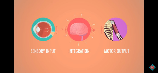
Sensory Input
E.g. Sensory receptors detect a spider on your knee.
Integration
"From there your nervous system processes that input and decides what should be done about it."
E.g. "Should I let it walk over me?" "Should I freak out and scream?"
Motor Output
"The response that occurs when the nervous system activates certain parts of your body."
E.g. Swatting the spider away.
Organisation of Central and Peripheral Nervous Systems
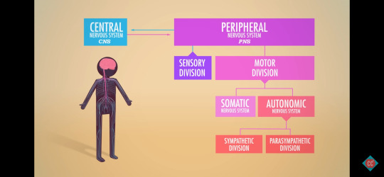
Central Nervous System (CNS)
brain
spinal cord
main control center
E.g It decided to remove the spider and gave the order to your hand.
Peripheral Nervous System (PNS)
nerves that branch off from brain and spine that allow your CNS to communicate with rest of body
It's job is communication, so is set up to work in both directions.
Sensory Division (Afferent)
picks up sensory stimuli
E.g. "There's a spider on you."
Motor Division (Efferent)
sends directions from your brain to muscles and glands
E.g. "Hand, how bout you do something about the spider?"
Somatic Nervous System (Voluntary)
rules skeletal muscle movement
Autonomic Nervous System (Involuntary)
keeps heart beating, lungs breathing, stomach churning
Autonomous Nervous System - Sympathetic & Parasympathetic Division
Sympathetic Division
mobilises body into action
E.g. "Gah! Spider!"
Parasympathetic Division
relaxes the body
E.g. "It's not a white tail. Breathe."
Neurons & Glial Cells
Neurons, or nerve cells, respond to stimuli and transmit signals.
Glial Cell Types
provide support, nutrition, insulation, and help with signal transmission in the nervous system

Central Nervous System
Astrocytes
support, regulate ions
Microglia Cells
defend
Ependymal Cells
line cavities
Oligodendrocytes
wrap and insulate, form myelin sheath
Peripheral Nervous System
Satellite Cells
surround neuron cell bodies
Schwann Cells
insulate, help form myelin sheath
Glial cell types serve many functions and make up about half of the brain's mass.
Central Nervous System Glial Cells: Astrocytes, Microglial, Ependymal and Oligodendrocytes
Astrocytes
exchange of materials between neurons and capillaries
Star-shaped astrocytes are found in the central nervous system.
most abundant and versatile glial cell
anchor neurons to their blood supply
govern the exchange of materials between neurons and capillaries
Microglial Cells
immune defense against invading microganisms
thorny-looking
Main source of immune defense against invading microorganisms in brain and spinal cord.
Ependymal Cells
create, secrete and circulate cerebrospinal fluid
line cavities in brain and spinal cord
Oligodendrocytes
produce an insulating barrier called the myelin sheath
Peripheral Nervous System Glial Cells: Satellite and Schwann
Satellite Cells
surround and support neuron cell bodies
Schwann Cells
produce an insulating barrier called the myelin sheath
Neuron Fun Facts
Neurons are some of the longest-lived cells in the body.
Neurons are irreplaceable.
Most neurons are amitotic, so once they take on given roles in the nervous system, they lose the ability to divide.
3. Neurons have huge appetites.
Neurons have a high metabolic rate, requiring a steady and abundant supply of glucose and oxygen.
"About 25% of the calories that you take in every day are consumed by your brain's activity."
Neuron Structure
Soma (Cell Body)
Neuron's life support.
nucleus
DNA
mitochondria
ribosomes
cytoplasm
Dendrites
listeners
Axon
talker
"The axons transmit electrical impulses away from the cell body to other cells.
Classifying Neuron Structures: Multipolar, Bipolar and Unipolar

"how many processes extend out from the cell body".
Process - a projecting part of an organic structure.
Multipolar
multiple processes
99% of neurons are multipolar neurons, with 3 or more processes sticking out of the soma.
E.g axon, multiple dendrites.
Bipolar
2 processes
E.g. axon, dendrite - extending from opposite ends of the cell body.
Bipolar neurons are rare, found only in special sensory places - like eye retina.
Unipolar Neurons
1 process
E.g. sensory receptors
Classifying Neuron Functionality: Sensory (Afferent), Motor (Efferent), Interneurons (Association)
"Which way an impulse travels through a neuron in relation to the brain and spine."
Sensory Neurons (Afferent Neurons)
transmit impulses from sensory receptors toward the CNS
mostly unipolar
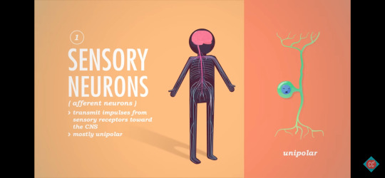
E.g. neurons pick up messages and transmit impulses from sensory receptors, like skin or internal organs, and send them through the CNS.
Motor Neurons (Efferent Neurons)
impulse moves from the CNS to the rest of the body
mostly unipolar
Interneurons (Association Neurons)
impulse moves between sensory and motory neurons
mostly multipolar
most abundant neurons

0 notes
Text
18th May 2024
CC: A&P - Skin Deeper Terminology
Skin - This organ functions as an immune barrier, blood storage unit, temperature regulator, vitamin producer, waste extractor, environment sensor, and odor producer.
Tactile Corpuscle - A type of receptor in the epidermis that is sensitive to light touch.
Lamellar Corpuscle - A type of receptor in the dermis that is sensitive to vibration and wide pressure.
Cutaneous Sensory Receptors - The collective name for all the parts of the nervous system that sense touch and pressure in the skin.
Sebaceous Glands - The type of skin glad that produces oil in hair follicles.
Ceruminous Glands - The type of modificed apocrine sweat gland that produces ear wax.
Mammory Glands - The type of modified apocrine sweat gland that produces milk in lactating women.
Sudoriferous Glands - A technical name for sweat glands, and located all over the body, these skin glands may produce either watery or sticky sweat.
Eccrine Sweat Glands -The type of sudoriferous glands located all over the body but concentrated on the palms of the hands and soles of the feet, these produce salty watery sweat.
Apocrine Sweat Glands - The type of sudoriferous glands located in the axillary and inguinal regions of the body, these produce viscous sweat, containing lipids and proteins that contributes to body odor.
Insensible Perspiration - A constant level of low level sweat produced constantly by the skin, at about 0.5 liters per day, to regulate temperature.
Sensible Perspiration - A variable and potentially high - up to 12 liters per day - amount of sweat produced as needed to cool the body.
Hair Root - The center of a hair in a follicle, where keratinisation is still active.
Hair Follicle - The part of every hair embedded in the skin.
Hair Shaft - The part of every hair that extends beyond the surface of the skin.
Hair Cuticle - The outermost layer of a hair, made of overlapping dead keratinocytes.
Vasodilate - Dermal blood vessels do this to dump excess body heat.
Vasoconstrict - Dermal blood vessels do this to reduce heat loss and send blood deeper to keep vital organs warm.
Jaundice - A yellow of skin, usually indicating liver dysfunction and a buildup of bile in the blood.
Cyanosis - Meaning "blue appearance", this term describes the colour of skin - usually the lips - when blood oxygen levels are low.
Erythema - Reddened skin caused by increased blood flow due to high body temperature, fever, inflammation, or embarrassment.
Vitamin D - The only vitamin that can be made in the body, its synthesis starts when sun hits the skin.
Melanin - The primary human skin pigment that mostly protects deeper layers of the skin from damage from UV light from the sun.
0 notes
Text
18th May 2024
Crash Course: Anatomy & Physiology - The Integumentary System, Skin Deeper
youtube
Summary:
Integumentary System Protects Your Body
Senses The Outside World
Helps Excrete Waste
Stores Blood
Regulates Temperature
Makes Vitamin D
Indicates Signs of Poor Health
Integumentary System
Functions
Protection
Sensation
Blood Storage
Temperature Regulation
Vitamin D Synthesis
Excretion
"The first and most vital purpose of the integumentary system is to act as a protective barrier."
Protective & Sensory Functions of the Integumentary System
Skin, hair, nails, sweat and oil glands work to shield us from threats: excessive sunlight, infections, abrasions etc.
"The integumentary system is also vital to how you sense the world around you."
Cutaneous Sensory Receptors
Structures, part of the nervous system, that receive stimuli from outside environment and send them to the brain.
Corpuscles (Receptors)
register different sensations associated with touch
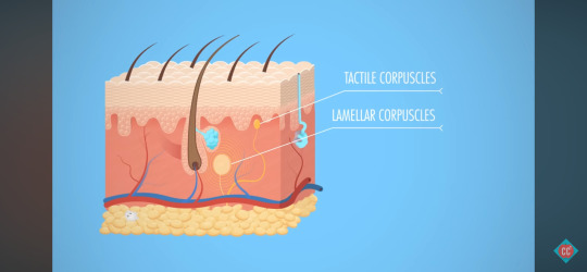
Tactile Corpuscles
transmits sensations of light touch and low frequency vibrations from periphery > central nervous system
E.g. Makes you aware of a tag scratching the back of your neck.
Lamellar Corpuscles
register the sense of pressure
E.g. Someone puts their hand on your shoulder.
Hair Follicle Receptors
feeling the breeze on skin/through your hair
More Functions: Removing Waste, Storing Blood & Regulating Body Temperature
"The integumentary system also plays a role in the excretion of waste."
Waste Removal
The integumentary system excretes small amounts of waste elimanted through the skin via sweat.
Urea, uric acid and ammonia, nitrogen-containing waste, are disposed via urine.
There's no evidence that heavy sweating rids the body of extra toxins, just water loss.
Blood Storage Unit
"About 5 percent of the entire blood volume is retained in your skin at any given time."
When more bloody supply is needed, like exercising, the nervous syste constricts dermal blood vessels to squeeze extra blood into circulation.
Body Temperature Regulation
During exertion, both blood and sweat glands regulate body temperature.
Insensible Perspiration
unnoticable sweat
The body excretes about 1.5L of sweat daily to maintain a comfortable body temperature.
Sensible Perspiration
noticeable sweat
Heavy exertion can cause 12L of sweat per day.
Vasoconstriction
To regulate heat loss, dermal blood vessels constrict, causing causing blood to head deeper into tissues to keep vital organs warm
Vasodilation
Once heat returns, blood vessels relax, allowing blood to return to the surface.
Skin Dyscolouration: Cyanosis, Jaundice or Erythema
Skin colour can indicate homeostatic imbalances.
As human skin colour spans, some conditions are easier to diagnose by discolouration of other tissues - mucous membranes, finger and toenail beds.
Cyanosis (Blue Skin)
In Caucasian people:
may indicate heart failure
poor circulation
severe respiratory issues
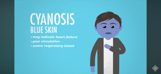
Blood depleted of oxygen turns darker in colour, which seen through the tissue of lips or skin, can look blue-ish.
Jaundice (Yellow Skin)
may indicate liver disorder

Yellow bile accumulates in the blood stream.
Erythema (Red Skin)
may indicate fever, inflammation, allergy
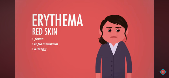
Fever, inflammation and allergy cause blood vessels expand and cause more blood flow to the skin's surface.
Melanin, Vitamin D & Skin Tone
Melanocyte
synthesizes melanin
Melanin is a pigment produced by melanocyte cells in the epidermis.
Melanin
produces reddish yellow pigments
produces brownish black pigments
It's main job is to protect against sun's ultraviolet rays.
Vitamin D
Bones require Vitamin D to produce new bone cells, which is the only vitamin the body can produce on its own.
Skill cells contain a molecule that converts to Vitamin D when in contact with UV light.
Vitamin D Path:
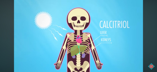
Skin > Bloodstream > Liver & Kidneys
In the liver and kidneys, Vitamin D becomes Activated D (Calcitriol).
Calcitriol circulated to all bones of the body.
How Does Hair Conditioner Work?
Skin Appendages
Hair
Nails
Sweat
Sebaceous Glands
Oil Glands
Hair
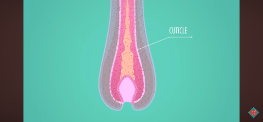
Hair, or pili, are flexible strands of dead keratin protein - like fingernails.
The outermost layer of the dead cells, the cuticle, look like overlapping roof shingles.
Shaft
keratinisation is complete
Root
keratinisation is still happening
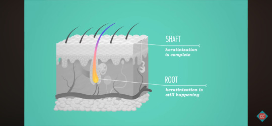
Each follicle is a tube of epidermal cells.
Like the epidermis, cells at the bottom of each follicle are young, continually dividing and pushing older cells up through the skin's surface.
Finger & Toe Nails
root of nail
nail body
nail bed
At the back of the nail bed, new cells divide at the root and are pushed forward, creating scaly-hard keratin.
Types of Sweat Glands: Eccrine, Apocrine, Mammary and Ceruminious
Sweat & Oil Glands
"There are up to three million toiny sudoriferous, or sweat glands, distributed throughout the body."
secrete salty, watery sweat
Eccrine & Apocrine Glands
Eccrine Glands
palms
forehead
soles of feet
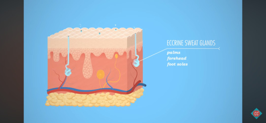
Eccrine glands are abundant.
They're simple coiled tubes that start in the dermis, extend through a duct and open into a pore on the skin's surface.
Apocrine Glands
armpit hair follicles
groin hair follicles
"deluxe" sweat - with fats and proteins
viscous
yellow-ish colour
odor
"You only have about 2000 of these, and they start cookin' around puberty, emptying into the hair follicles around the armpits and groin."
When the bacteria on the skin gets a hold of this sweat, it gets odorific, creating body odor.
Deodorants don't affect how much you sweat, but they reduce the smell by attacking odor-making bacteria.
Antiperspirants, using aluminum, block the sweat glands and prevent perspiration.
Modified Apocrine Sweat Glands
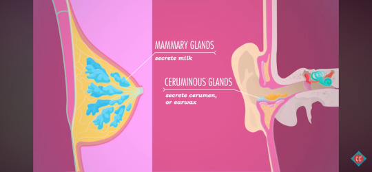
Mammary Glands
secrete milk
Ceruminious Glands
cerumen (earwax)
Sebaceous (Oil) Glands
Sebaceous Glands
oil glands
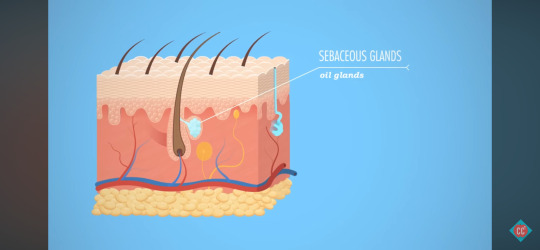
Sebaceous glands are everywhere but in thick skin on palms and foot soles.
Ducts are smaller on limbs but bigger on face, neck and upper chest.
Most sebaceous glands secrete sebum, an oily substance, into hair follicles that can travel to the skin's surface.
Although they cause pimples, their primary function is to soften and lubricate skin and hair, and slow skin's water loss in dry environments.
0 notes
Text
18th May 2024
CC: A&P - Skin Deep Terminology
Integumentary System - The organ system that includes your skin, hair, nails, sweat glands and oil glands.
Friction Ridges - Structures supported by dermal papilae, also known as fingerprints.
Keratinocytes - The main type of cells in the epidermis, these make a lot of the protein keratin which makes the skin durable.
Melanocytes - The spider shaped cells in the epidermis that are responsible for producing the brown pigment melanin.
Tactile Cells (Merkel Cells) - Epidermal cells that work with neurons to provide sensation for light touch.
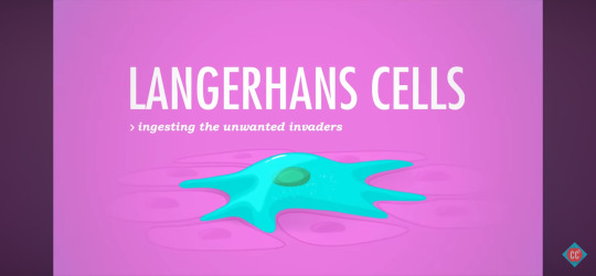
Dendritic Cells (Langerhans Cells) - Epidermal cells that are part of the immune system, and try to gobble up invading bacteria before they can break into the deeper layers of the skin.
Statified Squamous Epithelium - The tissue type found in the epidermis.
Thin Skin - The type of epidermis with just four named layers, and found everywhere on the body other than the palms of the hands and soles of the feet.
Thick Skin - The type of epidermis with five named layers, and only found on the palms of the hands, and the sole of the feet.
Epidermis - The most superficial of the three main layers of the skin.
Stratum Corneum - The most superficial layer of the epidermis, made of 20 - 30 layers of dead keratinocytes that eventually slough off, its name means "horny layer".
Stratum Lucidum - The so called "clear layer" of the epidermis, these 2 - 3 layers of keratinocytes are only found in thick skin on the palms of the hands and soles of the feet.
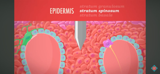
Stratum Granulosum - This layer of epidermal cells looks grainy because the cells are producing large amounts of keratin before they die.
Stratum Spinosum - This layer of epidermal cells is held together by protein fibers, and is the layer just superficial to the layer where active cell division is occuring.
Stratum Basale - This is the deepest layer of the epidermis, is made of just one layer of cells that are actively diving to produce cells that get pushed out to the other layers.
Dermis - The middle of the three main layers of the skin, where you find blood vessels, sweat glands, and most touch receptors.
Dermis - The layer of the skin just deep to the epidermis, containing blood vessels, and making skin elastic.
Papilary Layer - Made of loose areolar connective tissue, this is the layer of the dermis just deep to the epidermis.
Reticular Layer - The dermal layer just deep to the papilary layer, made of dense irregular connective tissue.
Hypodermis - The deepest of the three main layers of the skin, made mostly of adipose tissue.
0 notes
Text
18th May 2024
Crash Course: Anatomy & Physiology - The Integumentary System, Skin Deep
youtube
Summary:
All About Skin
Epidermis, Dermis and Hypodermis
Melanin and Keratin Cells
Ensure You Get A Good Tattoo
Skin
Skin protects your body against infection and extreme temperatures, maintains your balance of fluids, and even synthesizes Vitamin D for personal use.
The nerve endings allow you to sense the outside world.
Sweat glands and blood vessels help maintain proper temperature and communicate - from health to emotions - through things like blushing, flushing and sweating
Integumentary System
Hair
Nails
Sweat
Oil Glands
Skin
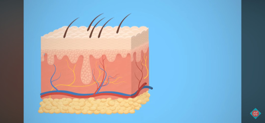
Skin Layers: Epidermis, Dermis & Hypodermis
Epidermis
stratified squamous epithelial tissue
Dermis
sweating, blood circulation, feeling
Hypodermis (Subcutis)
adipose tissue
Types of Epidermal Cells: Keratinocytes, Melanocytes, Langerhan Cells and Merkel Cells
Keratinocytes
building blocks of the tough, fibrous protein keratin
make up the bulk of the epidermis

Provide structure, durability and waterproofing to hair, nails and outer skin.
Kertatinocytes are constantly dying and being replaced, losing milling of them daily, enough to completely replace the epidermis every 4 - 6 weeks.
Melanocyte
spider-shaped cell that synthesizes melanin
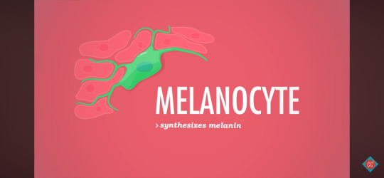
Skin colour isn't about the number of melanocytes but instead about the breath of their spidery cellular extensions, which in turn affect the amount of melanin that they contain.
Langherhan Cells (Denritic)
star-shaped
originate in bone marrow
defend the epidermis
Merkel Cells (Tactile)
combine with nerve endings to create a sensory receptor for touch

Found deep down at the boundary between the epidermis and dermis.
Thick Skin
Thick skin, found on the palms of hands and sole of feet, consists of five epidermal layers.
Thin Skin
Thin skin, found everywhere else, contains four epidermal layers.
Layers of Skin: Stratum Corneum, Stratum Lucidum, Straum Granulosum, Stratum Spinosum, and Stratum Basale
The deeper the epidermal layer, the younger the cells are.
Regeneration happens in the lower layers, with new cells moving up, maturing along the way, until they die on the surface.
This is because the epidermal is epithelial, so it's avascular, with all oxygen and nutrients coming from the dermis, so as they mature and move towards the surface, they lose their oxygen and nutrient supply and die.
Stratum Corneum
"horny layer"
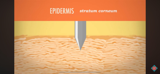
Outermost, roughest layer.
20 - 30 sheets of dead keratinocyte cells.
Stratum Lucidum
"clear layer"
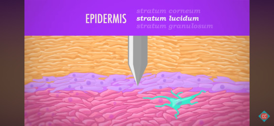
2 - 3 rows of clear, flat, dead keratinocytes that are only found in the thick skin of palms and soles.
Thin skin is only missing the stratum lucidum.
Stratum Granulosum
"granular layer"
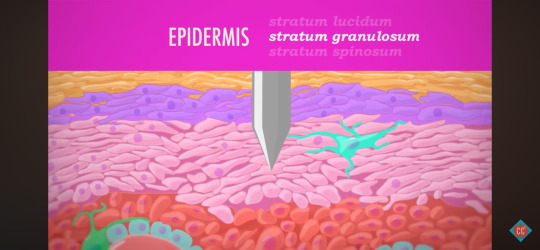
Layer contains living keratinocytes that form keratin like "crazy".
Looks grainy as cells are compressed and flattened as they move up through the epidermal layers, maturing as they go.
Stratum Spinosum
"spiny layer"
Prickly appearance under a microscope.
Closer to the point where regeneration, or mitosis, is active.
Stratum Basale
"basal layer"
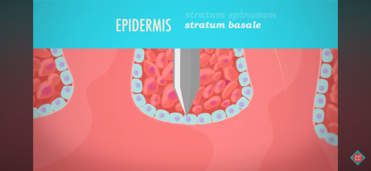
A single layer of columnar cells.
Cell factory - where most cell production happens.
It connects the epidermis to the layer of skin below it, the dermis.
Pneumonic - Come, Let's Get Sun Burned!
C - Stratum Corneum
L - Stratum Lucidum
G - Stratum Granulosum
S - Stratum Spinosum
B - Stratum Basale
Sunburn
The ultraviolet radiation in sunlight can damage the epidermis, cause elastic fibers to clump up to form a leathery appearance.
Sunburn temporarily depresses the immune system, with immune cells in the epidermis - and radiation can alter skin cell's DNA, leading to skin cancer.
Layers of the Dermis: Papillary, Reticular and Hypodermis
Dermis
collagen fibers, elastin fibers
capillaries
blood vessels
nerve fibers
parts of hair follicles, oil glands/sweat glands with ducts that lead to the skin's surface
Papillary Layer
areolar connective tissues
dermal pippilae
In thick skin, papillae are tiny protrusions that form unique friction ridges that press up through the epidermis to help grip - fingerprints.
Reticular Layer
dense irregular connective tissue
The reticular layer, deeper and thicker, make up 80% of the dermis.
All the dynamic parts contained within the dermis, like nerve fibers and capillaries, are distributed between both its layers.
Any time you're cut deep enough to bleed or feel pain, you've broken through the epidermis layer and lacerated the dermis.
Tattoos must reach the dermis for permanency.
Hypodermis (Subcutis)
adipose connective tissue
Basically a seam of fat, it provides insulation, energy storage, shock absorption and anchors the skin.
0 notes
Text
17th May 2024
CC: A&P - Types of Connective Tissues Terminology
Loose & Dense - The two subclasses of connective tissue proper.
Loose Connective Tissue - The subclass of connective tissue proper with relatively fewer protein fibers but more ground substance and cells.
Dense Elastic - The type of stretchy connective tissue proper found in the walls of many of our large arteries.
Fibrocartilage - The connective tissue with thick bundles of collagen fibers that is found in the menisci in our knees, and in our intervertebral discs.
Dense Regular - The type of connective tissue proper dominated by thick collections of collagen fibers all running in different directions.
Dense Regular - The type of connective tissue proper found in ligaments and tendons.
Dense Irregular - The type of connective tissue proper with thick bundles of collagen fibers running in lots of different directions; it is good at resisting forces in lots of different directions.
Elastic Cartilage - The connective tissue with extra elastic protein fibers, it provides the structure for our ear pinnae.
Hyaline Cartilage - The glassy connective tissue found in our nose, between the ribs and sternum, covering the ends of bones in joints.
Areolar - The most common type of loose connective tissue, found just under the epidermis and wrapped around every organ.
Areolar - The type of loose connective tissue defined by a random arrangement of fivers, few fibroblast cells, and lots of space of watery ground substance.
Reticular Tissue - The type of loose connective tissue proper that is dominated by reticular fibers instead of elastic or collagen.
Reticular Tissue - The type of loose connective tissue proper that provides the structure and space in the liver, spleen, lymph nodes and bone marrow.
Compact Bone - The type of connective tissue that uses calcium and other minerals to form very hard and durable structures.
Osseous Tissue - The type of connective tissue that uses calcium and other minerals to form very hard and durable structures.
Osseous Tissue - Very dense osseous tissue that acts as a shell over every bone, provides the strength of the shafts of long bones, and stores calcium for the body.
Spongy Bone - Porous but strong osseous tissue that provides space for bone marrow and is found inside the ends of long bones and throughout flat bones.
Fibrocartilage - The connective tissue with thick bundles of collagen fibers that is found in the menisci in our knees, and in our intervertebral discs.
Adipose - The type of loose connective tissue that functions as insulation and a storage for energy and in the form of fat.
Adipocytes - The name of the cells that take up most of the space in adipose tissue.
Blood - Type of connective tissue that transports materials to all parts of the body, in which the ground substances is called plasma, and the extracellular fibers are usually invisible.
Erythrocytes - The name of red blood cells, the dominant cell type in blood.
0 notes