
Don't wanna be here? Send us removal request.
Statistics
We looked inside some of the posts by shajismri and here's what we found interesting.
Average Info
Notes Per Post
0
Likes Per Post
0
Reblog Per Post
0
Reply Per Post
0
Time Between Posts
7 days
Number of Posts By Type
Text
17
Last Seen Tumblr Blogs


Fun Fact
In 2020, 44% of users from Denmark used Tumblr daily.
Text
Understanding Jaw Pain: When an OPG Scan Is the Right Diagnostic Tool
Jaw pain is more than just an inconvenience — it can be a sign of underlying dental, skeletal, or nerve-related issues that need prompt diagnosis. Whether it’s a persistent ache, pain while chewing, or stiffness in the jaw, understanding the root cause is essential for effective treatment. One of the most recommended and non-invasive tools for diagnosing jaw-related problems is the Orthopantomogram, commonly known as an OPG scan.
What is an OPG Scan?
An OPG (Orthopantomogram) is a panoramic dental X-ray that captures a single, comprehensive image of your entire mouth. It includes the upper and lower jaws, all the teeth, the temporomandibular joints (TMJ), and the surrounding bones and structures. Unlike traditional X-rays that only show a small section of the mouth, an OPG scan offers a broad view that’s crucial for evaluating jaw conditions.
Common Causes of Jaw Pain
Jaw pain can stem from various issues, including:
Temporomandibular Joint Disorders (TMJ/TMD): This is one of the most frequent causes of jaw discomfort. The temporomandibular joint acts like a sliding hinge connecting your jawbone to your skull. Issues here can cause pain, clicking sounds, or difficulty in movement.
Impacted Wisdom Teeth: When wisdom teeth don’t erupt properly, they can push against adjacent teeth or become trapped in the jawbone, causing pain and swelling.
Dental Infections or Abscesses: Untreated cavities or gum infections can spread to the jaw, causing severe pain and even bone loss if left unchecked.
Jaw Fractures or Trauma: Injuries to the jaw due to accidents, falls, or sports incidents can cause lingering pain and structural damage.
Orthodontic Issues or Misalignment: Poor bite alignment can strain the jaw muscles and joints, leading to chronic discomfort.
Identifying the exact cause of pain is the first step toward relief — and that’s where an OPG scan becomes invaluable.
How an OPG Scan Helps Diagnose Jaw Pain
Comprehensive Jaw View: The panoramic nature of the OPG scan allows the dentist or specialist to see the complete jaw structure, including both joints, which is essential in evaluating TMJ disorders and fractures.
Detecting Bone Abnormalities: The scan can reveal signs of bone infections, cysts, or tumors that may not be noticeable in a standard dental check-up.
Wisdom Tooth Evaluation: It’s the preferred imaging tool to detect impacted or misaligned wisdom teeth and their effect on the jaw and neighboring teeth.
Monitoring Dental Development: For younger patients, an OPG can be used to assess the development and positioning of teeth and jawbone growth, helping in early orthodontic planning.
Non-Invasive and Quick: The procedure is quick (usually completed in under 2 minutes), safe, and doesn’t require any invasive instruments or injections.
Who Should Consider an OPG Scan for Jaw Pain?
Individuals with persistent jaw discomfort, stiffness, or clicking sounds.
Patients advised to undergo wisdom tooth extraction.
Those with a history of facial trauma or jaw injury.
People experiencing unexplained headaches, ear pain, or facial swelling.
Anyone beginning orthodontic or dental implant treatments.
What to Expect During an OPG Scan
The procedure is simple and painless. You will be asked to bite gently on a small plastic piece to keep your mouth steady. The machine rotates around your head, taking a panoramic image. There’s no need for special preparation, and the entire scan takes just a few moments.
Conclusion
Jaw pain should never be ignored, as it may point to serious dental or structural conditions that require early intervention. An OPG scan is a vital diagnostic tool that offers a wide, clear view of the jaw and surrounding structures, enabling healthcare professionals to diagnose and treat issues effectively.
If you’re experiencing persistent jaw pain and looking for a reliable diagnostic center, Dr. Shaji’s MRI & Medical Research Centre offers one of the best OPG scan services with advanced imaging technology and expert care. Schedule your consultation today and take the first step toward a pain-free, healthier smile.
0 notes
Text
Choosing the Right CBCT Scan Centre in Calicut: Factors to Consider
With several CBCT scan centres available in Calicut, it can be challenging for patients to decide which one offers the most reliable services. While all CBCT machines offer 3D imaging, not all centres provide the same level of precision, image quality, or patient experience. Important factors to consider include the age and brand of the equipment, radiation safety measures, experience of the radiologist, and the centre's reputation for report accuracy and turnaround time.
0 notes
Text
#ct scan#mri scanning services in kotakkal#mri scanning services in wayanad#mri scan#ct scan services#mri scanning services in kannur
0 notes
Text
What Is CBCT and Why Might Your Dentist or Doctor Recommend It?
In modern dentistry and medical imaging, precision is everything. Whether you’re preparing for a dental implant, dealing with chronic sinus issues, or evaluating jaw pain, having detailed insights into your anatomy is crucial for accurate diagnosis and treatment. That’s where CBCT — or Cone Beam Computed Tomography — comes into play.
This advanced imaging technology is quickly becoming a standard tool for dentists, oral surgeons, ENT specialists, and other healthcare providers. But what exactly is CBCT, how is it different from a traditional CT scan, and why might your healthcare provider recommend one?
What Is CBCT?
CBCT stands for Cone Beam Computed Tomography, a specialized type of X-ray equipment that produces 3D images of the teeth, soft tissues, nerve pathways, and bone in a single scan. Unlike traditional CT scans used in hospitals, which take multiple slices of an area and reconstruct them into a 3D image, CBCT uses a cone-shaped X-ray beam to capture the entire area in a single rotation.
This technology provides high-resolution images with much lower radiation exposure compared to conventional CT scans. It is specifically designed for imaging smaller body areas, particularly the head, neck, face, and jaw, making it ideal for dental and ENT (ear, nose, and throat) applications.
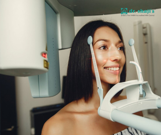
How Does a CBCT Scan Work?
When you go in for a CBCT scan, the procedure is quick and non-invasive. You’ll either sit or stand, depending on the machine design, while the scanner rotates around your head. In less than a minute, the machine captures hundreds of images from different angles, which are then reconstructed into a 3D model.
There’s no need for contrast dyes, no discomfort, and in most cases, no need for extensive preparation.
Why Might Your Dentist or Doctor Recommend a CBCT Scan?
A CBCT scan is typically recommended when your provider needs more detailed information than a regular X-ray can provide. Here are some of the most common reasons you might be referred for one:
1. Dental Implants
Before placing a dental implant, precise measurements of your jawbone and surrounding structures are needed to avoid nerve damage and ensure implant success. CBCT provides the depth and clarity necessary for precise surgical planning.
2. Impacted Teeth
If a tooth is trapped under the gum or pushing against other teeth — like wisdom teeth — CBCT can show the exact location and orientation to plan safe removal.
3. Root Canal Treatment
In endodontics, CBCT helps identify root canal anatomy, hidden infections, or missed canals that don’t show up on 2D X-rays.
4. Jaw Disorders (TMJ)
For patients with jaw pain or temporomandibular joint (TMJ) issues, CBCT offers a detailed look at joint structure and alignment, aiding in diagnosis and treatment planning.
5. Sinus & ENT Issues
ENT specialists often use CBCT scans to evaluate chronic sinusitis, airway obstructions, and nasal cavity issues, as the 3D detail helps in planning minimally invasive procedures.
6. Orthodontics & Facial Reconstruction
In orthodontics, CBCT scans are used to study jaw alignment, tooth position, and bone density. It’s also useful in surgical planning for facial trauma or reconstruction cases.
Advantages of CBCT Over Traditional Imaging
Lower radiation dose compared to hospital CT scans
Faster scan times, usually under 30 seconds
High-resolution 3D images for greater diagnostic accuracy
Better visualization of hard and soft tissues
Increased comfort with non-invasive and quick procedures
Is CBCT Safe?
Yes, CBCT is considered safe for most patients. Although it does involve radiation, the dose is significantly lower than conventional CT, and the benefits often far outweigh the risks — especially when used appropriately. However, as with any imaging involving radiation, it should be used only when necessary and under professional guidance.
Conclusion: Where to Get a High-Quality CBCT Scan
If your dentist or doctor has recommended a CBCT scan, it’s essential to choose a center that offers advanced technology, skilled staff, and fast, reliable service. Dr. Shaji’s CBCT Scan Centre stands out as one of the best CBCT facilities, known for its state-of-the-art equipment, expert radiologists, and commitment to patient comfort. With a reputation for accuracy, quick turnaround, and affordable pricing, Dr. Shaji’s ensures that you receive the diagnostic clarity needed for the best possible care.
0 notes
Text
Why Same-Day MRI Appointments Are a Game-Changer in Local Imaging Services
In today’s fast-paced world, waiting days or even weeks for an MRI appointment can be frustrating and stressful—especially when you're in pain or need answers quickly. That’s why same-day MRI services are becoming a major breakthrough in local diagnostic imaging. This article explores how same-day appointments are changing the game for patients and providers alike.
0 notes
Text
https://www.drshajimri.com/whats-mri/
#mri scanning services in kotakkal#ct scan#mri scanning services in kasargod#MRI Scan#MRI#MRI Scanning Services
0 notes
Text
MRI Scanning Services
#mri scanning services in kotakkal#MRI Scan#MRI#mri scanning services in kannur#mri scanning services in kasargod
0 notes
Text
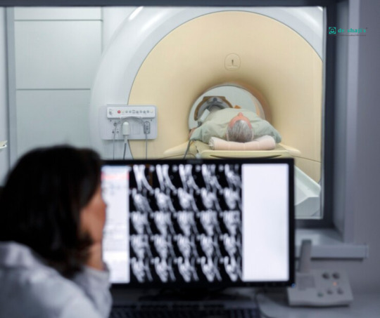
Ultrasonography in Acute Abdominal Pain: A First-Line Diagnostic Tool
One of the most prevalent issues in emergency and outpatient settings is acute abdominal discomfort, which can be difficult to diagnose because there are so many potential underlying reasons. Effective therapy depends on a timely and precise diagnosis, which can sometimes mean the difference between emergency surgery and conservative maintenance. Ultrasonography (USG) has become a first-line imaging modality in this clinical setting because of its accessibility, safety, and diagnostic versatility.
Why Start with Ultrasonography? A non-invasive, radiation-free, and reasonably priced imaging technique is ultrasound. Because of these qualities, it is particularly appropriate for vulnerable groups, including elderly people, pregnant women, and children. In contrast to CT scans, which use ionizing radiation, USG is safe and can be performed again without causing any problems.
Ultrasonography is also useful in critical care and emergency situations because it may be done at the patient’s bedside. Point-of-care ultrasonography (POCUS) has become more popular, which has enhanced its function as a real-time diagnostic tool that supports clinical judgment and physical examination.
Typical Uses in Acute Abdomen The evaluation of various intra-abdominal organs and tissues can be accomplished efficiently with ultrasound. The most frequent causes of acute abdominal pain, for which USG is an essential diagnostic tool, are listed below:
1. The biliary tree and gallbladder One of the most frequent indications for abdominal USG is suspected cholelithiasis (gallstones) or cholecystitis. USG can detect gallstones, gallbladder wall thickening, pericholecystic fluid, and a positive sonographic Murphy’s sign — all indicators of acute inflammation.
2. The disease of appendicitis For acute appendicitis, USG has a good specificity and a fair sensitivity in younger, leaner individuals. An inflamed appendix may be indicated by a non-compressible, blind-ending tubular formation in the right lower quadrant that is larger than 6 mm in diameter.
3. Ureteric and Renal Colic USG is often the first imaging test for flank pain or suspected renal colic. It helps detect hydronephrosis, which may suggest obstruction due to a ureteric stone, even if the stone itself isn’t visible.
4. Gynecological Emergencies In women of reproductive age, USG is the modality of choice for evaluating ectopic pregnancy, ovarian torsion, or ruptured ovarian cysts. Transvaginal ultrasonography, in particular, offers superior resolution for pelvic organs.
5. Aortic aneurysm in the abdomen (AAA) Finding a AAA requires quick bedside USG, particularly in elderly patients with hypotension and stomach pain. An intervention that saves lives can result from an early diagnosis.
6. Perforation and Obstruction of the Intestine Although CT is more definitive, USG can show dilated bowel loops, free fluid, or reduced peristalsis, raising suspicion for obstruction. It can also detect free air under the diaphragm in some perforation cases, particularly with careful scanning.
Restrictions and Complementary Applications Despite being a great first-line tool, USG has drawbacks. Image clarity can be decreased by bowel gas interference, operator dependence, and patient body habits (such as obesity). In some situations, further CT or MRI imaging might be required for confirmation.
Nonetheless, the first use of ultrasonography frequently greatly reduces the differential diagnosis, directing the clinician’s subsequent actions in a safe and effective manner.
The Changing Function of POCUS
The management of acute abdominal patients has changed as a result of emergency physicians’ and surgeons’ growing use of point-of-care ultrasonography (POCUS). Better patient outcomes, early therapy initiation, and quick decision-making are all made possible by bedside USG. It has a modest learning curve and can be a vital tool for frontline healthcare practitioners with the right training.
In conclusion When it comes to assessing acute abdominal discomfort, ultrasonography is still a vital, first-line diagnostic technique. Its real-time imaging capability, lack of radiation, and rapid diagnostic yield make it ideal for both emergency and outpatient settings. From gallbladder disease to gynecological emergencies, USG often provides the first crucial clue in complex clinical puzzles.
For patients seeking high-quality, accurate, and compassionate diagnostic care, Dr. Shaji’s USG Scan Center stands out as one of the best in the region. With state-of-the-art equipment and a reputation for excellence, Dr. Shaji’s center combines technology and experience to deliver timely, reliable results — ensuring that patients receive the care they need without unnecessary delays.
0 notes
Text
Temporomandibular Joint (TMJ) Evaluation
CBCT is a valuable diagnostic tool for evaluating the temporomandibular joint (TMJ), particularly when bony abnormalities are suspected. It allows for high-resolution 3D imaging of the condylar head, glenoid fossa, and surrounding bony structures, which is essential for diagnosing degenerative joint diseases like osteoarthritis, ankylosis, or trauma-induced changes. CBCT can detect changes in joint morphology, such as flattening, erosion, or osteophyte formation, which are not easily visible on standard panoramic or cephalometric X-rays. Although CBCT does not image soft tissues like the articular disc, it provides critical information on bone health and joint space, guiding further investigation or referral for MRI if soft tissue pathology is suspected. TMJ-focused CBCT scans are often used to evaluate asymmetry, pain, limited range of motion, or pre-surgical assessment.
0 notes
Text
What is CBCT and How Can It Benefit Your Dental Health?
Dental technology has made tremendous strides in recent years, giving patients better diagnosis and more efficient treatments. Cone Beam Computed Tomography (CBCT), a state-of-the-art imaging method that has completely changed how dentists handle complicated dental problems, is among the most noteworthy developments. You may be wondering what CBCT is and how it might improve your dental health if you've recently seen a dentist and heard about it.
Understanding A specialized form of X-ray technology, cone beam computed tomography (CBCT) is mostly utilized in orthodontics and dentistry. In contrast to conventional X-rays, which provide flat, 2D images, CBCT creates 3D images that offer fine-grained cross-sectional views of your soft tissues, bones, and teeth. In order to produce a three-dimensional (3D) image of the area being inspected, the device revolves a cone-shaped X-ray beam around the patient's head, taking numerous pictures from various perspectives.
Compared to normal 2D X-rays, the 3D images produced by CBCT are significantly more complete, allowing dentists to assess your dental health more thoroughly. This enables better treatment planning and more precise diagnosis, particularly for intricate dental treatments.
CBCT's advantages for dental health Both patients and dentists can benefit greatly from CBCT technology. It can considerably enhance your dental care in the following ways:
1. Improved Diagnosis Limited images of the teeth and surrounding structures can occasionally be obtained with traditional dental X-rays. The 360-degree, detailed 3D image that CBCT gives, on the other hand, enables dentists to spot issues that conventional X-rays might miss. CBCT can provide more accurate information about the underlying problem, whether it's an infection, impacted tooth, or concealed cavity.
2. Better Planning for Treatment When it comes to treatment planning for operations like orthodontic treatments, root canals, and dental implants, CBCT is essential. For instance, the dentist must evaluate blood vessels, nerve location, and bone density before implanting dental implants. By helping the dentist plan the treatment more precisely, CBCT lowers the chance of difficulties and ensures that the implant is placed correctly.
CBCT scans give orthodontic patients comprehensive details about the position of their teeth, the alignment of their jaw, and even the structure of their airways. This makes it possible to plan braces or other orthodontic treatments more effectively.
3. Accuracy in Complicated Situations CBCT can provide a more detailed image of the affected area for patients with complex dental problems such tumors, cysts, or abnormalities of the jaw. This enables more precise diagnosis and aids dentists in choosing the best treatment plan. Additionally, it aids in the planning of procedures, such as those for temporomandibular joint (TMJ) disorders and reconstructive jaw surgery.
4. Less Exposure to Radiation CBCT is a safer alternative for dental imaging because, although it uses radiation, it often emits a far lower dose than conventional CT scans. In order to protect patients from needless radiation exposure, the technology is made to take high-resolution pictures with the least amount of radiation possible. CBCT is a safer alternative for both patients and dental professionals since it produces higher-quality images with less radiation than traditional X-rays.
5. Quicker Outcomes CBCT scans are non-invasive and rapid. Patients don't have to wait long for findings because the full scan usually only takes a few minutes. The dentist may examine the pictures right away, which helps speed up treatment choices and prevent care delays.
CBCT Applications in Dentistry Many different dental specializations can benefit from CBCT. Among the most widely used applications are:
Dental implants: The dentist can determine the ideal position for implant implantation with the use of CBCT's precise 3D images of the bone structure.
Endodontics: CBCT provides a better image of the internal structure of the tooth, including the root canals and surrounding tissues, during root canal therapy.
Orthodontics: By giving more precise measurements for braces or other treatments, CBCT scans assist orthodontists in evaluating the alignment of the teeth and jaws.
TMJ and Jaw Issues: For patients with temporomandibular joint (TMJ) disorders or jaw pain, CBCT helps analyze the bone structure, muscles, and joints to determine the root cause of the issue.
Oral Pathology: CBCT is invaluable in detecting cysts, tumors, and other abnormal growths in the mouth and jaw.
In conclusion
Cone beam computed tomography, or CBCT, is a cutting-edge technology that has significantly improved dentistry. Particularly in difficult dental cases, CBCT enables more precise diagnosis, better treatment planning, and better results by providing thorough, three-dimensional images of the teeth, bones, and soft tissues. It guarantees a more successful and efficient course of treatment, minimizes radiation exposure, and eliminates the need for several imaging modalities.
Dr. Shaji's Clinic is among the greatest in Calicut for a dependable and cutting-edge CBCT scan. Dr. Shaji's Clinic is well-known for providing excellent CBCT scanning services. They make sure that every patient gets the best care possible by combining state-of-the-art technology with skilled care. Dr. Shaji's Clinic offers top-notch care in a welcoming and expert setting, whether you require a basic scan or are getting ready for a more involved dental surgery.
0 notes

Text
Find the Best MRI Centers Near You
Looking for a reliable MRI center nearby? Finding the best MRI center near you is essential for getting accurate and timely results. Whether you’re seeking a routine scan or need imaging for a medical condition, it’s important to choose a facility that offers advanced technology, professional staff, and convenient scheduling options. In this guide, we’ll help you navigate local MRI centers, offering tips on how to select the right one based on factors like location, quality of service, insurance compatibility, and patient reviews. Discover the top-rated MRI centers in your area and book your appointment with confidence.
#mri scanning services in kotakkal#mri scanning services in wayanad#mri scanning services in kasargod#MRI Scan
0 notes
Text
Finding Reliable CT Scan Centers in Calicut: Your Guide to Local Diagnostic Services
If you're searching for a CT scan facility in Calicut, this guide will help you find trustworthy centers offering advanced imaging services. Learn about the latest technology, pricing options, and what to expect during your scan.
0 notes