
An independent publication that visualises the complexities of science for the creative spirit. Launching soon.
Don't wanna be here? Send us removal request.
Statistics
We looked inside some of the posts by sirprintpublishing-blog and here's what we found interesting.
Average Info
Notes Per Post
20K
Likes Per Post
10K
Reblog Per Post
10K
Reply Per Post
2
Time Between Posts
9 hours
Number of Posts By Type
Photo
17
Last Seen Tumblr Blogs


Fun Fact
The KCSC sent more than 20K requests to delete posts related to prostitution and porn to Tumblr from January to June 2017.
Photo

#Issue00 is wrapped up and all ready for tomorrow's launch! Can't wait to share 8 months of hard work with you all! #particlemag #scienceillustrated #beautiful #science #magazine #independentpublishing #independentmagazine #risograph @dizzy.ink @jessrosepictures
#beautiful#independentpublishing#risograph#issue00#science#particlemag#magazine#independentmagazine#scienceillustrated
3 notes
·
View notes
Photo

There’s a hexagonal cloud at Saturn’s north pole
It’s a six-sided jet stream (dubbed as the “Hexagon”) that spans 30,000 kilometers (20,000 miles) across. The hexagon is a wavy jet stream of 200-mile-per-hour winds (about 322 kilometers per hour) with a massive, rotating storm at the center. There is no weather feature exactly, consistently like this anywhere else in the solar system. (Source)
9K notes
·
View notes

Photo

The many faces of histology… by i-heart-histo
320 notes
·
View notes
Photo


The images above were taken with the Cytell, a machine developed at GE Healthcare Life Sciences that combines the microscope with the cell analyzer. Click through the photos to learn more about what you’re seeing, and view more images at GE Reports.
172 notes
·
View notes
Photo
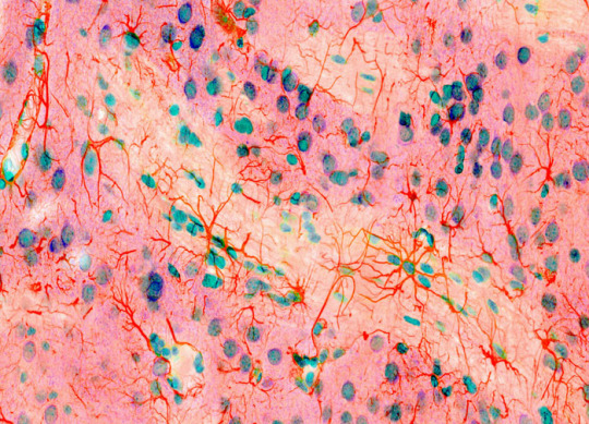
rat brain: a mosaic of glia and neurons
fluorescence, 600x
credit: Thomas Deerinck
220 notes
·
View notes
Photo


Images from the book Science Is Beautiful by Colin Salter. Click on images for descriptions.
About the book:
Take a fantastic journey through the human body, thanks to the magic of technology and the most spectacular microscopic images ever created. These pictures, as beautiful as any art, provide a window into the wonder of our brains, the work of a white blood cell, the power of hormones, the tiny hairs on our arms, the movement of human cancer cells, the jagged edges of caffeine crystals, and more. Enjoy the collection purely as a visual voyage or as a means of understanding the science behind the images—which all include the scale of the photography as well as the scientific details in layman’s terms.
643 notes
·
View notes
Photo

A Histology Slideshow by i♡histo
Heart valve
Lip
Umbilical cord
Nerve
Endometrium
Developing teeth
Mesentery
Tongue
Eyelid
Placenta
Finger
Hard palate
Lymph node
Mammary gland
Prostate
(repeat)
i♡histo
421 notes
·
View notes
Photo

Ovary of a parsley plant
Plant fertilization takes place when pollen from the male part of a plant lands on the female-receiving part called the stigma. Within minutes after making contact, the pollen will germinate and grow a long tube down into the plant before reaching the ovary. Once at the ovary, the pollen will discharge its sperm into the embryo sac, fertilizing an egg that will develop into seed-containing fruit. Plants have evolved many mechanisms to prevent self-pollination, which hinders adaptation to changing environments and spreads genetic mutations to new plant progeny. Earlier this year, researchers uncovered a 100-million-year old amber fossil that perfectly preserved plant fertilization in action, suggesting this ancient process led to the huge diversity of plants we see today.
Image by Meritxell Vendrell, Universitat Autònoma, Spain.
398 notes
·
View notes
Photo


Growing Crystals: Under Cross-Polarised Light.
Polarized light microscopy is an optical microscopy technique involving the use of light polarization. Simple techniques include illumination of the sample with polarized light. More complex microscopy techniques include differential interference contrast microscopy and interference reflection microscopy.
These illumination techniques are most commonly used on birefringent samples where the polarized light interacts strongly with the sample and so generating contrast with the background. Polarized light microscopy is used extensively in optical mineralogy.
This optical microscopy technique is capable of providing information on absorption color and optical path boundaries between minerals of differing refractive indices, in a manner similar to brightfield illumination, but the technique can also distinguish between isotropic and anisotropic substances.
Furthermore, the contrast-enhancing technique exploits the optical properties specific to anisotropy and reveals detailed information concerning the structure and composition of materials that are invaluable for identification and diagnostic purposes.
~Giffed by: rudescience From: This video
1K notes
·
View notes
Photo


The scientific definition of ice is that it has a regular crystalline structure based on the molecule geometry of water, which consists of a single oxygen atom covalently bonded to two hydrogen atoms, or H-O-H. Normal ice has a hexagonal symmetry. However, it shows under polarized light a large variety of forms and colors. Even in samples of ‘normal’ crystalline ice it has diverse manifestations. Normal means ice is crystallized under atmospheric pressure and by temperatures below 0°C.
Some samples are shown here. The photography’s are made of ice that was crystalized on microscope slides and placed in polarized light.
531 notes
·
View notes
Photo

Zebrafish embryo
Just 22 hours after fertilization, this zebrafish embryo is already taking shape. By 36 hours, all of the major organs will have started to form. The zebrafish’s rapid growth and see-through embryo make it ideal for scientists studying how organs develop.
Image courtesy of Philipp Keller, Bill Lemon, Yinan Wan and Kristin Branson, Janelia Farm Research Campus, Howard Hughes Medical Institute, Ashburn, Va. Part of the exhibit Life:Magnified by ASCB and NIGMS.
source
1K notes
·
View notes
Photo

Leucophosphite
Locality:
Tip Top Mine (Tip Top pegmatite), Fourmile, Custer District, Custer Co., South Dakota, USA
Gianfranco Ciccolini’s Photo
857 notes
·
View notes
Photo

Have you ever noticed something swimming in your field of vision? It may look like a tiny worm or a transparent blob, and whenever you try to get a closer look, it disappears, only to reappear as soon as you shift your glance. But don’t go rinsing out your eyes!
What you are seeing is a common phenomenon known as a floater. Floaters are tiny objects that cast shadows on the retina, the light-sensitive tissue at the back of your eye. They might be bits of tissue, red blood cells, or clumps of protein that are suspended within the vitreous humor, the gel-like liquid that fills the inside of your eye. As a result, floaters drift along with your eye movements,and seem to bounce a little when your eye stops.
To learn more about floaters, watch the TED-Ed Lesson What are those floaty things in your eye? - Michael Mauser
Animation by Reflective Films
2K notes
·
View notes
Photo

We're a sucker for a good nebula pic. This one's called the Tarantula as seen in infrared light. Photo by @nasa #alwaysaclassic #nevergoesoutofstyle #beautiful #science #space #nebula #particlemag 🚀🌌🌠💫
0 notes
Photo

An image of the Home Plate area on Mars taken by the one of the rovers called Spirit! The colours show the differences in rock weathering. Mars looks kinda fun to live in so in #Issue00 out tomorrow we created a guide on how it might compare to life on earth! Have a read and make an informed decision yourself 👽🚀 #PARTICLEMAG #scienceillustrated #beautiful #science #mars #marsrover
0 notes