#Research microsope
Explore tagged Tumblr posts
Visit Tumblr Blog
Explore Tumblr blogs with no restrictions, modern design and the best experience.
Last Seen Tumblr Blogs


Fun Fact
The Tumblr office adopted Tommy, an 11-year-old Pomeranian.
Text
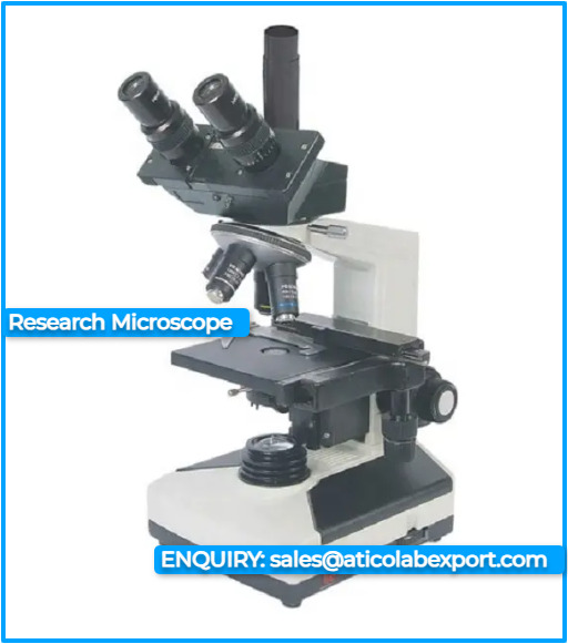
Research Microscopes Manufacturer and Supplier In India
Explore a comprehensive range of research microscopes for your scientific investigations. From biological to industrial research, find the perfect microscope to meet your specific needs. We are leading Research Microscopes manufacturer, supplier and exporter from India.
0 notes
Link
2 min readPreparations for Next Moonwalk Simulations Underway (and Underwater) ESI24 Zhai Quadchart Lei ZhaiUniversity of Central Florida Lunar dust, with its chemical reactivity, electrostatic charge, and potential magnetism, poses a serious threat to astronauts and equipment on the Moon’s surface. To address this, the project proposes developing structured coatings with anisotropic surface features and electrostatic dissipative properties to passively mitigate lunar dust. By analyzing lunar dust-surface interactions at multiple scales, the team aims to optimize the coatings’ surface structures and physical properties, such as Young’s modulus, electrical conductivity, and polarity. The project will examine tribocharging, external electric fields, and the effects of particle shapes and sizes. Numerical sensitivity analyses will complement simulations to better understand lunar dust dynamics. Once fabricated, the coatings will be tested under simulated lunar conditions. The team will employ a state-of-the-art nanoscale force spectroscopy system, using atomic force microsope (AFM) microcantilevers functionalized with regolith to measure dust-surface interactions. Additional experiments will assess particle adhesion and removal, with scanning electron microscopy used to analyze remaining dust. This project aims to provide insights into surface structure effects on dust adhesion, guiding the creation of lightweight, durable coatings for effective dust mitigation. The findings will foster collaborations with NASA and the aerospace industry, while offering training opportunities for students entering the field. Back to ESI 2024 Keep Exploring Discover More Topics From STRG Space Technology Mission Directorate STMD Solicitations and Opportunities Space Technology Research Grants About STRG
1 note
·
View note
Text
DNA

Ghostpoet: Shedding Skin, 2015 Studio album cover Ghostpoet, a British singer, songwriter and musician, wanted an album cover to reflect his music content directly. Since the album was called “Shedding Skin”, he collaborated with researchers at UCL to get a skin biopsy. When his biopsy samples were returned in uniquely tinted colours, the singer said “they just looked like paintings. I was really blown away by the detail”. The microsopic cell structures make for eye-catching art that hold a much deeper meaning
https://www.itsnicethat.com/articles/ghostpoet-shedding-skin
2 notes
·
View notes
Text
How to choose a right camera for your Fluorescence Microscope ?
It’s a challenge to choose a right camera for your research. There are various cameras in the market . So, what should you focus on when choosing a digital camera for fluorescence imaging .
At first , you need to know about your imaging requirements . Secondly, it’s also important to know the technical characteristics of each camera .
Here are some options to considering :
>Imaging cells and tissues
For a wide-field fluorescence microscope, the camera will be used to image stationary cells and tissues . In the below picture,two different cameras were used to image the Alexa Fluor 488-labeled actin filaments immobilized in endothelial cells . There are some key technical difference between the two cameras that make for a four-fold difference in cost . Using standard imaging conditions, both cameras produce sharp , clear images ,and the noise and background seem to make no difference . Well, you may ask what is the difference between the two cameras and is it necessary to buy the more expensive one ?

The main difference between the two images is that the one on the left was captured with an uncooled camera , while the one on the right was captured with a cooled camera . In the past ,the fluorescent imaging has to be cooled, however, advances in camera sensor technology over the last 25 years have meant that cooling is no longer required for conventional fluorescent imaging . For challenging fluorescent samples that require sophisticated microsopes(such as confocal microscopes) ,cooling cameras are ideal choice .But for most fluorescent imaging ,cooling is not required . Uncooled cameras also produce great images at a lower cost .
>Nosie
For fluorescence microscopes, noise affects signal-to-noise ratio and dynamic range .
A high signal-to-noise ratio(SNR) results in sharp contrast and good sharpness ,while as SNR decreases, background signals increase and image sharpness and contrast decrease . Pixel merging can be used to reduce noise and improve SNR , but, it will affect space resolution. The main source of noise in a fluorescence microscopes is the read noise, which is much higher than the dark current noise .
The dark current noise is independent of temperature of the sensor . Cooling cameras reduce dark current noise ,which can be considered for use in situations requiring long exposures or very low light intensity . In general,dark currents have no significant effect on standard fluorescent imaging. So, unless you are imaging in a situation that requires a long exposure or low light intensity, the effect of dark current on the image is minimal .
>Other considerations
Of course, other imaging variables also affect image quality . To maximize the image quality,we should use a uniform light sourc,a high NA objective and a good optical filter combination .
Combining the right camera technology with your microscope system will ensure you get excellent images .
In addition , some challenging applications, such as confocal imaging, calcium signal imaging and near-infrared fluorescence, have more stringent requirements for the camera to achieve the desired results . However, for general fluorescent imaging ,a high quality , uncooled camera can also produce sensitive images . That way , you can spend your precious money on other equipments .
You may also need fluorescence filter set for your fluorescence microscope .
Optolong optics(www.optolong.com) supply filter set DAPI , FITC , TRITC,TexasRed, Cy5 , dual-band green/orange , which you may interesting .

0 notes
Text
The microbiome: Images and visualisations
On Monday 26 June I went to Oxford to participate in a workshop on the microbiome organised by The Oxford Interdisciplinary Microbiome Project (IMP). This was what one might call a meta-workshop. Its aim was to find questions that social scientists can sensibly ask about the microbiome, or in the words of the organisers, this was about establishing a “social-scientific research agenda about the microbiome and its implications for public policy and social change”. The workshop ties in with some of my older and newer reflections on the microbiome.
The Oxford English Dictionary defines the microbiomes in two ways, as “[a] population of microorganisms inhabiting a specific environment; a microbial community or ecosystem, now esp. that of the body” (first attested in 1952) and as “[t]he collective genomes of all the microorganisms inhabiting a specific environment, esp. that of the body” (first attested in 2001).
The workshop was held at Wolfson College (see featured image), where I did my JRF in the history of linguistics between 1985 and 1988. It was really nice to be there again and see all the new additions and extensions.
Questions, questions questions
In advance of the workshop we had been sent 120 questions and were asked to vote for 10. At the workshop itself each of four tables/working groups was given a list of themed questions and we were asked to select two high-level ones and two reserve questions. The general aim was to establish about 20 questions overall.
At our table we dealt with the issue of how the microbiome is being conceptualised. One question we found interesting in this context was how the microbiome, a still rather abstract and contested concept, is being imagined and visualised in the public sphere (which is, of course, not the same as visualising microbiome datasets).
While we were discussing this, I quickly had a peak on Google Images and what I found was quite intriguing. So when I came back from Oxford, I had a closer look. Of course, I cannot provide an answer to the broad question of how the microbiome is visualised in the public sphere in one blog post, but perhaps I can stimulate some discussion.
People can also look at other image sources in the future, such as Pinterest, Shutterstock, or the Science Photo Library, for example, which have images of the microbiome that seem to be quite different to the Google Images sample.
Thematic clusters
When you search for ‘microbiome’ on Google Images, you get not only images, but Google also provides you with a list of ‘themes’ (in various colours). (I searched on 27 June)
I am not sure how these themes are generated, but they are really interesting. Here they are without the colours – followed by my attempts at interpreting these thematic clusters:
skin, gut, lung, human skin, colon, stomach – this theme seems to deal with loci of various types of human microbiome
nature, role, health, diet, environmental, food – this one hints at various contexts of the human gut microbiome and how it is fed or maintained
plant, animal, host – here we are dealing with a simple (almost) dichotomy between plant and animal hosts of microbiomes
obesity, disease, cancer, diabetes, dysbiosis [microbial imbalance or maladaptation on or inside the body, such as an impaired microbiota], ibd [inflammatory bowel disease] – this theme encapsulates various illnesses or diseases linked to a disturbed microbiome
Scientific American, New Yorker, Time Magazine, National Geographic, Nature Review, The Economist – here we have a list of the main outlets for microbiome images
core, cell, body, root, fibre, sequence – this list leaves me a bit perplexed – comments welcome
infographic, map, heatmap, pie chart – here we have a list of various types of visualisations of the microbiome
bacterial, microbiota, fungal, candida, candida albicans [a type of yeast that is commonly used as a model organism for biology], C. difficile – this list homes in on various types of microbiome as well as one of the most talked-about afflictions, Cdiff, linked to a disruption of the microbiome in the human gut
human gut, rhizosphere, human intestine, insect gut, phyllosphere [a term used in microbiology to refer to the total above-ground portions of plants as habitat for microorganisms] – here the focus is on the human gut on the one hand but also the insect gut and plant habitats for microorganism – a bit of a strange mix I have to confess
fish, chicken, lefse [a traditional soft Norwegian flatbread], milk, poultry – this might refer to ways of enhancing the microbiome through various types of food
immune system, metabolism, digestive system, antibiotic resistance, hygiene hypothesis – these are scientific topics discussed in the context of microbiome research, and they were also discussed at the workshop
hospital, home, stable – these are, it seems, some places where microbiomes like to evolve
alpha diversity, relative abundance, beta diversity – comments welcome! Alpha diversity refers, it seems, to the diversity of species at a specific site (local species pool), while beta diversity represents the differences in species composition among sites
metagenomics, metabolomics, next generation sequencing – these are high-level science topics as well as technologies relating to research in microbiomics
human milk, saliva, blood – this might refer to interactions between a mother’s breast milk and a baby’s saliva; I am not so sure about the blood
dog, ruminant, pig, swine, bovine, equine – this indicates that microbiomics deals not only with humans but also animals that humans keep and eat
short chain fatty acid, tmao [Trimethylamine N-oxide (TMAO) is a small colorless amine oxide generated from choline, betaine, and carnitine by gut microbial metabolism], bile acid – comments welcome!
drosophila, human genome, coral, zebrafish, termite, mosquito – these might be model organisms used in the study of microbiomes (apart from the human genome)
sem [structural equation modelling or scanning electon microsope?], 16s rrna sequencing [the component of the 30S small subunit of a prokaryotic ribosome that binds to the Shine-Dalgarno sequence] – science!
pca [principal component analysis], hierarchical clustering, gastric bypass, otu [operational taxonomic unit] – I am not sure what gastric bypass is doing in this list!
Overall, these themes provide some insights not only into who generates most of the images, i.e. which journals are involved, or which types of images and visualisations are used, but also, and more importantly, what topics these images and visualisations are supposed to illuminate, from types of (human) microbiome, through various loci for microbiomes, through (domestic) animal microbiomes, to ways of studying the microbiome in certain fields of science and by using certain techniques, technologies and machines. Surprisingly, these themes did not mention Faecal Microbial Transplants, which are increasingly being discussed in the context of the microbiome.
Types of images and visualisations
I then looked at the first fifty images that were displayed when I searched for ‘microbiome’. I sorted them roughly into the following groups and I list them below in order of importance:
Outlines of human body/bodies/head/hands
Rod-shaped bacteria milling about
Microbiome as a word with or without other text
Various types of bacteria or cells milling about (sometimes) under a microscope
One cartoon
Surprisingly, I could not immediately see an image of a human gut – the most popular location of the microbiome. (Such images seem to be much more prominent on Shutterstock; while rod-shaped bacteria seem to be the domain of the Science Photo Library).
The images based on outlines of the human body were mostly outlines with arms slightly raised; sometimes there were two outlines, for example, of a man and a woman (skirt) standing beside each other; sometimes there were several outlines; sometimes the outline merged with an image of the double helix; sometimes there were outlines of a head or of two hands. The outlines were mostly inhabited or surrounded by round or rod-shaped outlines of microbes. This type of image also included the logo of Human Microbiome Project which adapts Leonardo de Vinci’s Vitruvian Man to the genomic age:
The images that displayed the word ‘microbiome’ were also interesting. Sometimes I just saw the word ‘microbiome’ surrounded by bacterial fuzziness, or else as part of a wordle; or ‘Microbiome 101’ against a background of swirling bacterial soup. There were also images that displayed texts such as: ‘What is the Microbiome?’, ‘Gut Feeling’, ‘The importance of your gut microbiome for your optimal health’, ‘Your body is mostly microbes’, ‘How to feed your microbiome’, and so on. Interestingly, the focus of the words, rather than the images, was on the human gut.
Conclusion
The images and visualisations of the microbiome desplayed on Google Images provide a unique and also biased insight into what types of images are out there of the microbiome. There were three surprises: the absence of faecal microbial transplants as topic or theme; the absence of the human bowel as an image; and the focus on the human gut in the words but not in the images. At the moment I can’t provide an explanation for this. More research needed!
Images: Wikimedia commons
The post The microbiome: Images and visualisations appeared first on Making Science Public.
via Making Science Public http://ift.tt/2snkAS0
0 notes