#fMRI visual system
Explore tagged Tumblr posts
Visit Tumblr Blog
Explore Tumblr blogs with no restrictions, modern design and the best experience.
Last Seen Tumblr Blogs


Fun Fact
Tumblr.com rank in the US is 25.
Text
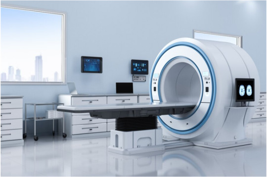
Innovative MRI Technology With Stunning Life Impact
Magnetic Resonance Imaging (MRI) is a modern diagnostic healthcare standard. Its radiation-free, noninvasive imaging capabilities have transformed how physicians see and treat complex medical conditions. Yet, MRI is still out of reach for many people worldwide due to its high costs and unique requirements.
However, this limitation disproportionately affects low and middle-income countries, where MRI healthcare systems are preoccupied with resource balancing between advanced imaging technologies. A gap in accessibility is created because standard superconducting MRI scanners are limited by the availability of robust infrastructure, such as radio frequency (RF) shielding and high power consumption, for use. This article explores how the next wave of innovation in low-power and ultra-low field (ULF) MRI will go far in democratising life-saving diagnostic tools.
The Cost and Infrastructure Barriers
Conventional MRI systems typically require high magnetic fields (1.5 Tesla (T) to 7 T). However, these robust systems create high-resolution images at a steep price and operational demands like working in an RF shield room and consuming more than 25,000 watts of energy. On the other hand, recent advances in ULF MRI have brought a low-power, low-cost alternative to ULF MRI, as demonstrated by Yujiao Zhao and colleagues.
These innovative systems are operated utilising 0.05 T field strength and on standard wall power outlets with only 1,800 watts of a current draw during scanning. These ULF MRI systems solve electromagnetic interference problems using active sensing and deep learning technologies to provide image quality comparable to high-field MRI devices. The possibilities of such advancements enable their utilisation of smaller medical facilities or rural hospitals or clinics located in resource-constrained settings.
MRI Ambience for Enhancing Patient Experience
Technological advancements streamline costs and increase accessibility, but patient comfort in MR evaluations is fundamental to care. Improvements in patient experience have been made by innovations such as In-Bore MRI and MRI cinema for healthcare. These technologies integrate immersive audiovisual solutions, including MRI projectors and MRI-compatible displays, to create a soothing environment which reduces anxiety and increases cooperation during scans.
Patient relaxation virtual skylights, for example, create relaxing outdoor views in the confined MRI bore where the patient is situated. In addition, an MRI-compatible monitor prevents real-time visuals from interfering with the system’s magnetic field, providing patients with comfort and reassurance.
Functional MRI Systems Applications
Functional MRI systems measuring brain activity with blood flow changes have also undergone significant changes. Unlike traditional diagnostic MRI, advanced fMRI visual systems and fMRI monitors allow researchers and clinicians to perform detailed studies of neurological conditions through MRI. The MIT team demonstrates the adaptability of MRI technology to meet new healthcare and research needs with these innovations.
Portable MRI Solutions
Another game-changing development is the portability of low-field MRI systems. Their compact and lightweight designs require little logistical planning, and specialised reports are unnecessary. The MRI-compatible stretcher and wheelchair facilitate seamless interfacing with patient workflows, even in mobile or temporary medical locations.
MRI-compatible cameras and TVs further simplify operations by removing some of the obstacles that medical teams face in seeing, communicating, and doing their jobs, but without compromising the safety and operation of the MRI healthcare system.
Future Directions and Challenges
Even so, ULF MRI technology is in its infancy. Honest challenges are resolution refinement, noise reduction, and scalability. In addition, robust training programs for healthcare professionals are necessary to ensure effective use. Experts such as Udunna Anazodo and Stefan du Plessis point out that while low-field MRI has great promise, its success depends on the ability to prove its reliability and cheap enough for use in multiple clinical settings. The potential for ULF MRI to become a universally accessible diagnostic tool increases exponentially as these challenges are met.
Significant Changes with Small Impact
Healthcare developments include a shift towards patient-centred and environmentally sustainable practices by integrating new technologies, such as MRI-compatible displays and virtual skylights for hospitals. These advances improve diagnostics and address gaps in healthcare equity.
Kryptonite Solutions exemplifies this forward momentum. They can provide comprehensive MRI solutions that embrace innovation and the potential for accessibility. By focusing on supporting the advancement of healthcare technology, they are in the game to transform the way MRI systems serve global communities.
With this paradigm shift in MRI accessibility, comfort and innovation firmly underway, we are charting the future of a healthcare world that closes the diagnostic inequality gap.
#virtual skylights for healthcare#mri ambience#mri compatibles#fmri visual system#mri compatible products#mri compatible stretcher#mri compatible wheelchair#mri compatible#virtual skylights
0 notes
Text
A dog’s eye view: Exploring the visual system from the kennel to the clinic
A dog’s eye view: Exploring the visual system from the kennel to the clinic gene therapyblindnessconesdogrodscolor blindnesscolor visiondog fMRIdog visionvisual system organization

View On WordPress
#blindness#color blindness#color vision#cones#Dog#dog fMRI#dog vision#gene therapy#rods#visual system organization
0 notes
Text
Neuroradiology Imaging Techniques: What Patients and Doctors Need to Know

What Is Neuroradiology?
Neuroradiology is a highly specialized area of medical imaging that focuses on diagnosing and treating disorders of the brain, spinal cord, and nervous system using advanced technologies. These techniques are crucial in identifying strokes, brain tumors, multiple sclerosis, and traumatic injuries.
Trusted resources like uevolve.in provide excellent educational material and healthcare updates that can help patients better understand these complex procedures.
Essential Imaging Techniques in Neuroradiology
🧠 1. Computed Tomography (CT)
Used For: Acute head trauma, bleeding, and stroke.
How It Works: X-rays produce cross-sectional images of the brain.
Benefits: Fast, reliable, and ideal in emergencies.
🧠 2. Magnetic Resonance Imaging (MRI)
Used For: Tumors, degenerative diseases, and soft tissue evaluation.
How It Works: Uses magnetic fields to generate high-resolution images.
Benefits: No radiation, excellent detail of brain and spinal cord.
Advanced Techniques That Offer Deeper Insights
🔬 3. Functional MRI (fMRI)
Detects brain activity and blood flow.
Supports surgical planning and cognitive mapping.
🔬 4. Diffusion Tensor Imaging (DTI)
Visualizes white matter tracts in the brain.
Important for traumatic brain injury and surgical planning.
🔬 5. Magnetic Resonance Spectroscopy (MRS)
Analyzes chemical composition of brain tissues.
Helps differentiate tumor types and grades.
🔬 6. Perfusion Imaging
Evaluates blood flow within the brain.
Used to assess stroke severity and tumor viability.
💡 Learn more about patient care advancements at uevolve.in, a platform committed to personalized healthcare knowledge.
Interventional Neuroradiology Techniques
These procedures go beyond diagnosis and are minimally invasive:
Angiography: Maps blood vessels in the brain.
Embolization: Treats aneurysms and AVMs.
Vertebroplasty: Strengthens spinal fractures.
FAQs: Neuroradiology Imaging
✅ Are these procedures safe?
Yes. MRI avoids radiation, and CT scans use low, medically necessary doses.
✅ Will I need contrast material?
Sometimes, yes. Your doctor will explain if contrast is needed and any potential side effects.
✅ How long does each scan take?
CT: 10–30 minutes
MRI: 30–60 minutes, depending on complexity
✅ When will I get my results?
Usually within 24–48 hours. Your doctor will review and explain the results.
0 notes
Text
Hello! Neuroscience is a fascinating and multidisciplinary field devoted to the study of the nervous system, ranging from the molecular and cellular level to the complex systems that govern behavior and cognition.
Important Parts of Neuroscience:
Neuroscience can be divided into several areas of study, each with its own specific focus:
* Neuroanatomy: Dedicates itself to the study of the structure of the nervous system, including the brain, spinal cord, and peripheral nerves.
* Neurophysiology: Investigates the functioning of the nervous system, such as the electrical and chemical activity of nerve cells and how they communicate.
* Neurochemistry: Examines the chemical processes that occur in the nervous system, including neurotransmitters and other signaling molecules.
* Molecular and Cellular Neurobiology: Focuses on the molecular and cellular mechanisms that underlie neuronal function.
* Cognitive Neuroscience: Explores the neural basis of higher mental processes, such as perception, attention, memory, language, and reasoning.
* Neuroscience: Explores the neural basis of higher mental processes, such as perception, attention, memory, language, and reasoning.
* Neuroscience: * Behavioral Neuroscience: Investigates the relationships between the nervous system and behavior, including emotions, motivation, and social interactions.
* Clinical Neuroscience: Dedicated to the study of diseases and disorders of the nervous system, seeking to understand their causes, develop treatments, and improve diagnosis.
* Computational Neuroscience: Uses mathematical models and computer simulations to understand how the nervous system works.
Where Neuroscience Wants to Go:
Neuroscience has ambitious and high-impact goals:
* Understanding the normal functioning of the brain: Uncovering the mechanisms that enable cognition, emotion, consciousness, and behavior.
* Understanding the causes of neurological and psychiatric diseases: Identifying the biological processes that lead to conditions such as Alzheimer's, Parkinson's, depression, schizophrenia, and others, in order to develop more effective treatments.
* Developing new therapies and interventions: Creating innovative approaches to prevent, treat, and rehabilitate individuals with neurological and mental disorders.
* Advances in neurotechnology: Develop brain-machine interfaces, neural prosthetics, and other technologies to restore lost functions and enhance human capabilities.
* Apply neuroscience knowledge to other areas: Inform practices in education, law, marketing, and other disciplines, optimizing learning, decision-making, and social interaction.
Key Points of Neuroscience:
* Brain Plasticity: The discovery that the brain is malleable and capable of adapting and changing throughout life has revolutionized the understanding of learning and recovery from injury.
* Brain-Behavior Connection: Neuroscience increasingly demonstrates the intricate relationships between brain activity and our actions, thoughts, and feelings.
* Advances in Neuroimaging: Techniques such as functional magnetic resonance imaging (fMRI) and electroencephalography (EEG) allow us to visualize brain activity in real time, providing valuable insights into how the brain works. * Brain-Based Therapies: Neuroscientific knowledge is leading to the development of more targeted and effective therapeutic interventions, such as deep brain stimulation and neurofeedback.
* Neuroethics: As neuroscience advances, important ethical issues related to mental privacy, consent, and the use of neurotechnologies are increasingly being discussed.
In summary, neuroscience is a dynamic and essential field for understanding ourselves and for addressing challenges related to brain health and well-being. The ongoing search for knowledge promises significant transformations in many areas of science and society.
0 notes
Text
¶ … literacy specialist who works in Adult Education. You have noticed that reading is very difficult for people to learn in adulthood, and you wonder whether this might be related to brain plasticity. Gaillard, WH et al. (2002) Language dominance in partial epilepsy patients identified with an fMRI reading task, Neurology, 59, 2, 256-265 https://www.paperdue.com/customer/paper/literacy-specialist-who-works-in-adult-education-56222#:~:text=Logout-,LiteracyspecialistwhoworksinAdultEducation,-Length2pages Search words:reading + fMRI; Google Scholar; neuroscience. Poldrack, RA, Desmond, JE., Glover, GH & Gabrieli, JD. (1998) . The neural basis of visual skill learning: an fMRI study of mirror reading. Cereb. Cortex, 8, 1-10. Search words:reading + fMRI; Google Scholar; neuroscience. Guinevere F.E. et al. (2004) Neural Changes following Remediation in Adult Developmental Dyslexia, Neuron, 44, 3, 411-422 Search words: reading + fmri + adults + neuroplasticity; Google Scholar; neuroscience. Brain imaging studies have investigated the neural mechanisms of recovery in adults following acquired disorders and childhood developmental disorders. However, there are few to no studies that investigate the neural systems that underlie adult rehabilitation of neural-ingrained learning disorder (i.e. those from birth) despite their high incidence. In this study, researchers identified the differences in brain activity during a phonological manipulation task before and after a behavioral intervention in adults who had developmental dyslexia. Phonologically targeted training resulted in improvements in reading in the instructed participants as compared to the dyslexics who received no instruction, and fMRI showed concordant increases in activation in the bilateral parietal and right perisylvian cortices. Authors concluded that behavioral plasticity in adult developmental dyslexia involves two neural mechanisms (or regions in the brain) 24 participants were used. A sublecxical sound deletion task was employed where researchers tested for phonological processing. They tested 19 dyslexic patients who did not receive reading instruction with 19 dyslexic patients who had received instruction (112 hours of structured multi-sensory phonological intervention) and using FMRI identified regions of the brain that were activated during the study. . . Adults who received instruction benefited significantly in their phonological processing skills as compared to those who had not received instruction. This improved understanding of the phonological characteristics of language led, in turn, to improving other aspects of reading ability such as nonword decoding and oral paragraph reading. The study is helpful in that it provides enhanced understanding of the neural characteristics of adults with dyslexia as well as how to structure interventions that can improve their neural plasticity. Skalicky, AE. (2002) . The relationship between early visual processing and reading ability: investigation of temporal processing in adults and children . University of Georgia Theses and Dissertations . http://hdl.handle.net/10724/5745 Search words: reading + fmri + adults + neuroplasticity; Google Scholar; neuroscience. 5. Turkeltaub et al. (2003). Development of neural mechanisms for reading. Nature Neuroscience 6, 767-773 Search words: reading + fMRI in adults; Google Scholar; neuroscience Pediatric brain imaging has generally excluded studies that investigate the neural development of cognitive skills acquired during childhood. Researchers here compared pediatric brain development during reading tasks to those of adults using fMRI. Their task involved an implicit word-processing task that required detection of a visual feature (tall letters) within both words and matched false font strings. Reading was done using eye-motion. Researchers studied an unrelated amount of participants whose ages ranged from 6 to 22 years and found that learning to read is associated with two patterns of change in brain activity: increased activity in the left-hemisphere middle temporal and inferior frontal gyri and decreased activity in the right inferotemporal cortical areas. Activity in the left-posterior superior temporal sulcus of the youngest readers was associated with the maturation of their phonological processing abilities. These discoveries are relevant to the focus of this assignment's focus in that younger children may indeed have an easier time learning to read than adults. They also found that learning to read was associated with engagement of left inferior frontal and middle temporal cortices as well as disengagement of right inferotemporal cortex It remains to investigate whether neuroplasticity can be strengthened to make up for these natural conditions and whether it can be strengthened using mechanisms that would employ these neural regions. Read the full article
0 notes
Text
**Mechanism of Migraine with Aura: Step-by-Step Brain Tissue Changes**
Migraine with aura involves a cascade of neurological events, primarily driven by **cortical spreading depression (CSD)** and subsequent activation of pain pathways. Here’s a breakdown of the process:
---
### **1. Initiation of Cortical Spreading Depression (CSD)**
- **Trigger**: Neuronal hyperexcitability in the cortex (often genetic or due to ion channel dysfunction) leads to abnormal electrical activity.
- **Mechanism**: A sudden surge of neuronal depolarization (excessive firing) begins in a localized brain region, typically the **occipital lobe** (visual cortex).
- **Key Players**:
- **Glutamate**: Excess release of this excitatory neurotransmitter triggers depolarization.
- **Potassium (K⁺) ions**: Leak from neurons, propagating the wave.
---
### **2. Wave of Depolarization Spreads**
- **Process**: The depolarization wave spreads across the cortex at ~3–5 mm/minute, temporarily disrupting normal brain activity.
- **Symptoms**:
- **Visual aura**: Flashing lights, zigzag patterns (scintillations) or blind spots (scotoma) as the wave affects the visual cortex.
- **Sensory aura**: Tingling/numbness (parietal lobe involvement) or speech difficulties (Broca’s area).
---
### **3. Neurotransmitter & Ionic Shifts**
- **Glutamate Surge**: Sustained depolarization increases extracellular glutamate, overstimulating NMDA receptors and causing excitotoxicity.
- **Calcium Influx**: Neuronal calcium overload disrupts mitochondrial function, generating **reactive oxygen species (ROS)** and oxidative stress.
---
### **4. Cerebral Blood Flow Changes**
- **Initial Hyperemia**: Brief increase in blood flow (due to vasodilation) during depolarization.
- **Oligemia**: Prolonged reduced blood flow (vasoconstriction) following the wave, potentially contributing to hypoxia-like symptoms.
- **Imaging**: fMRI studies show a "spreading oligemia" matching the aura’s progression ([*Annals of Neurology*](https://doi.org/10.1002/ana.410280502)).
---
### **5. Trigeminovascular System Activation**
- **Trigeminal Nerve Stimulation**: CSD activates pain-sensitive trigeminal nerve terminals in the meninges (brain’s protective membranes).
- **Inflammatory Mediators**:
- **CGRP (Calcitonin Gene-Related Peptide)**: Released, causing vasodilation and neurogenic inflammation.
- **Substance P**: Enhances pain signaling and sensitizes blood vessels.
---
### **6. Central Sensitization & Pain**
- **Pain Pathways**: Trigeminal signals reach the thalamus and cortex, perceived as throbbing headache (often unilateral).
- **Sensitization**:
- **Allodynia**: Normal touch becomes painful (due to hyperexcitable neurons).
- **Photophobia/Phonophobia**: Brainstem nuclei (e.g., locus coeruleus) amplify sensitivity to light/sound.
---
### **7. Resolution & Postdrome**
- **Recovery**: Neurons repolarize, blood flow normalizes, and inflammatory mediators clear.
- **Postdrome**: Fatigue, cognitive fog, or residual sensitivity due to lingering neurotransmitter imbalances.
---
### **Key Factors Influencing Aura**
- **Genetics**: Mutations in ion channels (e.g., CACNA1A) increase neuronal excitability.
- **Hormones**: Estrogen fluctuations may modulate CSD susceptibility (common in women).
- **Triggers**: Stress, dehydration, or sleep changes lower the threshold for CSD.
---
### **Clinical Implications**
- **Preventive Therapies**: CGRP inhibitors, beta-blockers, or antiepileptics (e.g., topiramate) target CSD or trigeminal activation.
- **Acute Relief**: Triptans constrict dilated meningeal vessels and block CGRP release.
---
*Migraine with aura reflects a complex interplay of neuronal, vascular, and inflammatory processes. While CSD is central to aura, ongoing research aims to clarify genetic and molecular drivers.* 🧠⚡
0 notes
Text
Radiology Technology: Breaking New Ground with Modern Innovations
Radiology technology has made tremendous progress, delivering faster, safer, and more precise ways to diagnose and treat illnesses. With advancements in artificial intelligence (AI), imaging techniques, and portable devices, the role of radiology in healthcare has expanded beyond traditional methods. These innovations are transforming the way medical professionals detect diseases, manage treatments, and offer personalized care. This article delves into the most exciting developments reshaping radiology and their impact on the future of healthcare.
The Digital Revolution in Radiology
Gone are the days of film-based X-rays and analog systems. The shift toward digital radiology has introduced new efficiencies into medical imaging. Digital radiography (DR) allows healthcare providers to capture, view, and share high-resolution images in real-time. Radiologists no longer need to wait for physical films to be developed, speeding up diagnosis and treatment decisions.
Additionally, digital radiology reduces radiation exposure for patients, offering safer diagnostic procedures. Cloud storage systems make it easier for healthcare teams to access imaging data anytime, anywhere, enabling better collaboration among specialists.
AI and Machine Learning: The New Allies in Radiology
AI and machine learning (ML) are having a profound effect on radiology by automating tasks and boosting diagnostic accuracy. AI-powered tools like Computer-Aided Detection (CAD) systems are being used to flag subtle abnormalities that might be missed by human eyes. These tools play a crucial role in early detection of diseases like cancer and cardiovascular conditions.
AI also helps manage radiology workflows more efficiently. It can prioritize urgent cases by evaluating scan results, allowing radiologists to focus on critical patients. Machine learning algorithms continuously improve by learning from each case, ensuring enhanced performance and fewer errors over time.
New Frontiers in Imaging Techniques
Advancements in imaging technology have led to more detailed and functional images, improving diagnostic precision.
1. Hybrid Imaging Systems
Hybrid imaging, such as PET-CT and PET-MRI, provides a comprehensive view by combining two imaging methods into one system. This dual approach helps detect diseases at earlier stages and monitor treatment progress with greater accuracy. PET-CT is now a gold standard for diagnosing and staging cancers, while PET-MRI offers enhanced soft tissue visualization.
2. Functional MRI (fMRI)
Functional MRI (fMRI) takes imaging beyond anatomy by showing brain activity in real-time. This technology is invaluable in neurology, helping doctors understand brain functions and plan surgeries with precision. It is also being explored for use in mental health diagnostics, including conditions like depression and schizophrenia.
3. Low-Dose Imaging Techniques
The need for safer diagnostic tools has driven the adoption of low-dose imaging protocols. Low-dose CT (LDCT) scans are transforming lung cancer screening, enabling early detection with minimal radiation exposure. This innovation promotes preventive care, especially among high-risk patients, while minimizing health risks associated with radiation.
4. 3D and 4D Ultrasound Imaging
Ultrasound technology has advanced significantly, offering 3D and even 4D imaging capabilities. 3D ultrasound provides detailed visuals of organs, while 4D imaging allows real-time monitoring of fetal movements during pregnancy. These technologies are improving diagnostic precision and making ultrasound an indispensable tool for many medical fields.
Portable Imaging Devices: A New Era of Mobility
Portable imaging devices are changing the landscape of healthcare, especially in emergency care and rural settings. Mobile X-ray units, handheld ultrasound devices, and compact MRI systems enable healthcare professionals to perform scans at the patient’s location. This reduces delays in diagnosis and eliminates the need to transfer critically ill patients to imaging centers.
Point-of-care ultrasound (POCUS) devices have become essential tools in emergency departments and intensive care units. They allow quick evaluations of cardiac, abdominal, and vascular conditions, ensuring faster decision-making in critical situations. During the COVID-19 pandemic, portable ultrasound devices played a vital role in identifying lung abnormalities on-site.
Teleradiology and Cloud Integration: Imaging without Borders
The integration of teleradiology and cloud-based systems has unlocked new possibilities for radiology. Teleradiology allows radiologists to provide expert interpretation of scans from any location, making it possible to deliver diagnostic services 24/7. This is especially beneficial for hospitals and clinics in remote areas that lack access to specialized radiology expertise.
Cloud-based Picture Archiving and Communication Systems (PACS) streamline image sharing across healthcare networks, improving collaboration between medical teams. With instant access to imaging data, physicians can make informed treatment decisions more efficiently. The flexibility of cloud systems also supports radiology departments by reducing storage costs and maintenance challenges.
Interventional Radiology: Precision without Surgery
Interventional radiology (IR) has emerged as a game-changer, offering minimally invasive alternatives to surgical procedures. Using imaging guidance, radiologists can perform treatments like tumor ablation, embolization, and angioplasty through tiny incisions. These procedures minimize trauma, reduce recovery times, and decrease the risks associated with open surgeries.
Robotic-assisted interventions are the latest advancement in interventional radiology. Robotic systems enable radiologists to perform complex procedures with exceptional precision, making them highly effective for delicate tasks like stent placement. As robotic technology continues to improve, it will open new possibilities for treating previously inoperable conditions.
Radiomics: A Data-Driven Approach to Personalized Medicine
Radiomics is an emerging field that involves extracting vast amounts of data from medical images to uncover hidden patterns. By analyzing data beyond visual interpretation, radiomics can provide valuable insights into tumor characteristics and disease progression.
When combined with genomic data, radiomics offers a personalized approach to treatment. Oncologists can predict how a patient’s tumor will respond to specific therapies, enabling more targeted and effective treatment plans. This approach is transforming cancer care by improving outcomes and reducing unnecessary treatments.
Ensuring Safety: Radiation Dose Management
One of the key priorities in modern radiology is minimizing radiation exposure. New imaging systems are equipped with dose optimization features to ensure patients receive the lowest radiation necessary for accurate diagnosis. Pediatric imaging, in particular, benefits from these advances, as children are more sensitive to radiation.
Dose-tracking software also plays a vital role in patient safety by monitoring cumulative radiation exposure. Healthcare providers can use this data to make informed decisions about imaging frequency, balancing diagnostic needs with patient safety.
Radiology’s Role in Precision Oncology
Precision oncology relies heavily on advanced imaging for diagnosis, treatment planning, and monitoring. PET-CT and PET-MRI provide detailed insights into tumor behavior, helping oncologists personalize therapies based on individual patient profiles.
Liquid biopsies, which analyze tumor DNA from blood samples, complement imaging techniques by offering non-invasive diagnostic options. Together, radiology and molecular diagnostics provide a comprehensive picture of the patient’s condition, guiding treatment decisions and improving outcomes.
Challenges and Future Directions
While radiology technology offers exciting possibilities, there are challenges to address. Integrating AI systems requires extensive validation to ensure reliability. Additionally, adopting advanced equipment involves significant investment, making it difficult for smaller healthcare facilities to keep pace.
Data security is another pressing concern. As radiology departments increasingly rely on cloud-based platforms, safeguarding patient information from cyber threats is essential. Robust security measures are necessary to protect sensitive data while ensuring accessibility.
The future of radiology lies in continuous innovation. Researchers are exploring new ways to enhance imaging quality, improve AI algorithms, and unlock the full potential of radiomics. Collaboration between radiologists, data scientists, and medical researchers will be key to advancing the field further.
Radiology technology is undergoing a remarkable transformation, with innovations that enhance diagnostic accuracy, improve patient care, and support personalized treatment strategies. From AI-powered diagnostics to portable imaging devices and robotic interventions, the latest advancements are redefining the healthcare landscape.
As these technologies become more accessible, healthcare providers can offer faster, safer, and more precise care to patients. Radiology will remain at the forefront of medical innovation, ensuring that future generations benefit from improved diagnostics and more effective treatments. With ongoing research and collaboration, the possibilities for radiology are limitless, making it an essential pillar of modern healthcare.
0 notes
Text
Neurosurgery in the Digital Age: Leveraging Technology for Better Outcomes
In recent years, the field of neurosurgery has witnessed a remarkable transformation, largely driven by advancements in technology. This evolution has not only improved surgical techniques but has also redefined the way neurosurgeons interact with their patients and optimize outcomes. As we delve into the intricacies of this digital age, we will explore how these technological innovations impact minimally invasive procedures, enhance diagnostic capabilities, and ultimately lead to better patient care in the realm of neurosurgery.

The Rise of Minimally Invasive Techniques
One of the most significant advancements in neurosurgery is the shift towards minimally invasive techniques. Traditional open surgeries often require large incisions, resulting in longer recovery times and increased risk of complications. In contrast, minimally invasive procedures, such as those pioneered by leading Orange County neurosurgeon Robert Louis, MD, utilize advanced imaging and surgical tools to access the brain and spine through smaller openings.
These techniques emphasize the use of keyhole surgery and neuroendoscopy, allowing surgeons to perform complex procedures with minimal disruption to surrounding tissues. For instance, Dr. Louis specializes in endoscopic approaches for treating brain tumors, which significantly reduce postoperative pain and shorten hospital stays. By leveraging sophisticated technologies like the CyberKnife® Robotic Radiosurgery System, neurosurgeons can precisely target tumors with high-dose radiation while preserving healthy tissue. This approach exemplifies how technology not only enhances surgical precision but also fosters quicker recovery and improved patient outcomes.
Enhanced Diagnostic Capabilities
In addition to surgical techniques, technological advancements have revolutionized diagnostic capabilities in neurosurgery. Neuroimaging technologies, such as MRI and CT scans, have become indispensable tools for neurosurgeons, enabling them to visualize brain and spinal structures in remarkable detail. The advent of functional MRI (fMRI) allows surgeons to assess brain activity in real-time, providing critical information before and during surgery.
Moreover, the integration of machine learning and artificial intelligence in diagnostic processes has the potential to enhance accuracy and efficiency. Algorithms can analyze vast amounts of imaging data, identifying patterns that may be missed by the human eye. This capability is particularly crucial in diagnosing complex conditions, such as brain tumors or vascular malformations, where early detection can significantly influence treatment options and outcomes. By harnessing these advanced diagnostic tools, neurosurgeons like Dr. Louis are better equipped to develop personalized treatment plans that align with each patient's unique needs.
Patient-Centric Care Through Technology
The digital age has also fostered a shift towards patient-centric care in neurosurgery. The rise of telemedicine, mobile health applications, and online patient portals has transformed how patients interact with their healthcare providers. These technologies facilitate remote consultations, allowing patients to receive expert opinions without the need for extensive travel, which is particularly beneficial for individuals in Orange County and surrounding areas.
Moreover, patient education has become more accessible through online platforms. Educational videos and informational resources empower patients to understand their conditions and treatment options better. For instance, Dr. Louis's website offers a wealth of information on various neurologic conditions, including brain tumors and cervical spine disorders. This transparency not only alleviates patient anxiety but also fosters a sense of partnership between the patient and the neurosurgeon.
Robotic Assistance in Neurosurgery
The incorporation of robotic technology in neurosurgery marks another significant milestone in the digital age. Robotic systems assist surgeons in performing intricate procedures with unparalleled precision. For example, robotic-assisted surgery allows for improved dexterity and visualization in delicate operations, such as those involving the skull base or spinal cord.
Dr. Louis utilizes robotic systems to enhance the accuracy of minimally invasive surgeries, leading to reduced trauma and quicker recovery times for patients. The ability to navigate complex anatomical structures with robotic assistance is a game-changer, ensuring that even the most challenging surgeries can be executed with confidence and safety. This innovation exemplifies how technology enhances the surgeon's capabilities, ultimately translating into better outcomes for patients.
The Future of Neurosurgery: Data-Driven Decision Making
As we look to the future, the role of data analytics in neurosurgery will continue to expand. The collection and analysis of patient data can inform evidence-based practices, enabling neurosurgeons to make more informed decisions regarding treatment approaches. By leveraging data from various sources, including surgical outcomes and patient demographics, neurosurgeons can refine their techniques and improve overall patient care.
Incorporating predictive analytics into surgical planning allows neurosurgeons to anticipate potential complications and adjust their strategies accordingly. This proactive approach enhances patient safety and contributes to better overall outcomes. As the field of neurosurgery continues to evolve, the integration of data-driven decision-making will be paramount in shaping the future of patient care.
Conclusion
In conclusion, the digital age has ushered in a new era for neurosurgery, characterized by technological innovations that enhance surgical techniques, improve diagnostic capabilities, and promote patient-centric care. As highlighted by the expertise of Orange County neurosurgeon Robert Louis, MD, minimally invasive approaches and advanced imaging technologies are at the forefront of this transformation. By embracing these advancements, neurosurgeons are better equipped to deliver exceptional care, ensuring that patients experience optimal outcomes on their journey to recovery. As we move forward, the continued integration of technology in neurosurgery holds immense promise, paving the way for a future where patient care is more personalized, efficient, and effective.
0 notes
Text
The Future of Radiology: Innovations and Advancements
Radiology, the medical discipline that uses imaging to diagnose and treat diseases within the body, has witnessed remarkable innovations and advancements over the years. These advancements have revolutionized the way we understand and treat various health conditions, making radiology an indispensable part of modern medicine. As we look to the future, the landscape of radiology is set to evolve even further, driven by cutting-edge technologies and groundbreaking research. The Best Radiology Hospital in Jaipur exemplifies these advancements, consistently incorporating the latest technologies and research findings to provide superior diagnostic and treatment services. This article explores the future of radiology and highlights some of the most promising innovations and advancements shaping the field.
Artificial Intelligence in Radiology
Artificial Intelligence (AI) is at the forefront of transforming radiology. AI algorithms can analyze vast amounts of imaging data with unprecedented speed and accuracy, assisting radiologists in detecting abnormalities that may be missed by the human eye. Machine learning models are being trained to recognize patterns in medical images, aiding in early diagnosis and improving treatment outcomes. The integration of AI in radiology promises to enhance diagnostic precision, reduce human error, and optimize workflow efficiency.
Advanced Imaging Techniques
The development of advanced imaging techniques is another significant advancement in radiology. Techniques such as functional MRI (fMRI), positron emission tomography (PET), and dual-energy CT scans provide more detailed and comprehensive images of the body's internal structures. These techniques allow for better visualization of complex conditions, leading to more accurate diagnoses and tailored treatment plans. Furthermore, the advent of 3D and 4D imaging technologies offers dynamic views of organs and tissues, enabling real-time assessment of physiological functions.
Interventional Radiology
Interventional radiology is a rapidly growing subspecialty that utilizes minimally invasive procedures to diagnose and treat various medical conditions. Guided by imaging techniques such as fluoroscopy, ultrasound, and CT, interventional radiologists perform procedures like angioplasty, embolization, and tumor ablation with precision. These minimally invasive procedures often result in shorter recovery times, reduced risk of complications, and improved patient outcomes. The future of interventional radiology holds immense potential for further innovations, including the use of robotics and advanced imaging guidance systems.
Personalized Medicine and Radiogenomics
The concept of personalized medicine is gaining traction in radiology, with radiogenomics playing a crucial role. Radiogenomics involves the correlation of imaging features with genetic and molecular data to understand the underlying biology of diseases. This approach allows for the identification of specific biomarkers that can predict disease progression and response to treatment. By integrating radiogenomics into clinical practice, radiologists can provide personalized treatment plans tailored to each patient's unique genetic makeup, leading to more effective and targeted therapies.
The Role of the Best Radiology Hospital in Jaipur
In India, Jaipur stands out as a hub for advanced medical facilities, and the best radiology hospital in Jaipur is at the forefront of embracing these innovations. Equipped with state-of-the-art imaging technologies and staffed by highly skilled radiologists, this hospital is committed to providing top-notch diagnostic and interventional radiology services. By leveraging cutting-edge technologies and staying abreast of the latest advancements in the field, the best radiology hospital in Jaipur ensures that patients receive accurate diagnoses and optimal treatment outcomes.
Conclusion
The future of radiology is undeniably bright, with continuous innovations and advancements shaping the field. From the integration of AI and advanced imaging techniques to the rise of interventional radiology and personalized medicine, radiology is poised to play an even more critical role in healthcare. As we move forward, institutions like the best hospital in Jaipur will continue to lead the way, ensuring that patients benefit from the latest technological breakthroughs and receive the highest standard of care. The evolution of radiology promises a future where early detection, precise diagnosis, and effective treatment are the norm, ultimately improving patient outcomes and transforming the landscape of modern medicine.
#best hospital in jaipur#child hospital in jaipur#best orthopedic hospital in jaipur#best urology hospital in jaipur
0 notes
Text
Things to consider before MRI Scan!
Metal Objects: Remove all metal items, including jewelry and piercings, as MRI uses strong magnets.
Medical Implants: Inform your doctor about any implants, pacemakers, or metal fragments in your body.
Pregnancy: Notify the technician if you are pregnant or suspect you might be.
Claustrophobia: Discuss options like sedation if you are uncomfortable in small spaces.
Dietary Restrictions: Follow any fasting instructions if contrast dye will be used.
Comfort: Wear loose clothing or a gown provided by the facility.
To know more visit: How to ensure patient safety during MRI scan?
0 notes
Text
INNOVATIONS IN DIAGNOSTIC IMAGING: WHAT THE FUTURE HOLDS
In the ever-evolving landscape of healthcare, diagnostic imaging stands at the forefront of technological advancements. As we move further into the 21st century, innovations in this field promise to revolutionize the way we detect, diagnose, and treat medical conditions. From artificial intelligence to molecular imaging, the future of diagnostic imaging holds exciting prospects that will enhance precision, reduce risks, and improve patient outcomes.
ARTIFICIAL INTELLIGENCE AND MACHINE LEARNING
One of the most transformative innovations in diagnostic imaging is the integration of artificial intelligence (AI) and machine learning. These technologies enable the analysis of vast amounts of imaging data with unprecedented speed and accuracy. AI algorithms can identify patterns and anomalies that may be imperceptible to the human eye, leading to earlier and more accurate diagnoses.
For instance, AI-powered tools are being developed to assist radiologists in detecting conditions such as cancer, cardiovascular diseases, and neurological disorders. These tools can highlight areas of concern, prioritize cases based on urgency, and even suggest potential diagnoses, thus streamlining the diagnostic process and reducing the likelihood of human error.
ADVANCED IMAGING TECHNIQUES
Advancements in imaging techniques are continuously pushing the boundaries of what is possible. Techniques such as functional MRI (fMRI), positron emission tomography (PET), and single-photon emission computed tomography (SPECT) are becoming more sophisticated, providing detailed insights into the functioning of organs and tissues.
Functional MRI, for example, measures brain activity by detecting changes in blood flow, offering valuable information about brain function and aiding in the diagnosis of neurological conditions like epilepsy, stroke, and Alzheimer's disease. Similarly, PET and SPECT imaging are instrumental in understanding metabolic processes and can be used to diagnose and monitor diseases like cancer and heart disease.
MOLECULAR IMAGING
Molecular imaging represents a significant leap forward in diagnostic imaging. Unlike traditional imaging methods that focus on the structure of organs and tissues, molecular imaging visualizes the biological processes at the molecular and cellular levels. This allows for the detection of diseases at their earliest stages, often before symptoms appear.
One of the most promising applications of molecular imaging is in oncology. By targeting specific molecules associated with cancer, molecular imaging can identify tumors with high specificity and sensitivity. This not only aids in early detection but also in monitoring the effectiveness of treatments, enabling personalized and adaptive therapy strategies.
HYBRID IMAGING SYSTEMS
Hybrid imaging systems, which combine two or more imaging modalities, are gaining traction in the medical field. Technologies like PET/CT and PET/MRI integrate the strengths of different imaging techniques, providing comprehensive information about both the anatomy and the metabolic activity of tissues.
PET/CT, for instance, combines the metabolic imaging capabilities of PET with the detailed anatomical imaging of CT. This allows for precise localization and characterization of lesions, improving diagnostic accuracy and aiding in treatment planning. PET/MRI, on the other hand, offers superior soft tissue contrast and reduced radiation exposure compared to PET/CT, making it particularly useful in pediatric and neurological imaging.
PORTABLE AND POINT-OF-CARE IMAGING
The future of diagnostic imaging also includes a shift towards portability and point-of-care solutions. Portable imaging devices, such as handheld ultrasound machines and compact MRI scanners, are making it possible to bring advanced imaging capabilities to remote and underserved areas. This democratization of imaging technology can significantly improve access to healthcare and facilitate timely diagnosis and treatment.
Point-of-care imaging, integrated with telemedicine, allows for real-time consultations and remote diagnostics. For example, a handheld ultrasound device connected to a smartphone can capture and transmit images to a specialist for immediate interpretation. This not only saves time but also reduces the need for patient transfers, making healthcare more efficient and accessible.
3D AND 4D IMAGING
The advent of 3D and 4D imaging is adding new dimensions to diagnostic imaging. 3D imaging provides a more accurate representation of anatomical structures, which is particularly useful in planning surgical procedures and guiding minimally invasive interventions. It also enhances the visualization of complex structures, such as the intricate networks of blood vessels in the brain or heart.
4D imaging, which incorporates the dimension of time, offers dynamic views of physiological processes. This is particularly valuable in cardiology, where 4D echocardiography can capture the motion of the heart in real-time, providing detailed information about cardiac function and aiding in the diagnosis of heart diseases.
PERSONALIZED MEDICINE AND THERANOSTICS
The future of diagnostic imaging is closely linked to the concept of personalized medicine. Advanced imaging techniques are enabling the development of theranostics, a field that combines diagnostics and therapeutics. Theranostics involves the use of imaging to guide targeted therapies, ensuring that treatments are tailored to the individual patient's condition.
For example, in oncology, theranostics can involve using molecular imaging to identify specific biomarkers on cancer cells and then delivering targeted therapies that bind to these markers. This approach not only enhances the effectiveness of treatments but also minimizes side effects by sparing healthy tissues.
As we look to the future, the innovations in diagnostic imaging promise to transform healthcare in profound ways. The integration of AI, the development of advanced and hybrid imaging techniques, the advent of molecular and portable imaging, and the rise of personalized medicine are just a few of the exciting trends shaping the field. These advancements will not only improve the accuracy and efficiency of diagnoses but also pave the way for more effective and personalized treatments, ultimately enhancing patient care and outcomes. The future of diagnostic imaging is bright, and its potential to revolutionize healthcare is immense.
Orignal Source: https://av-imaging.com/innovations-in-diagnostic-imaging-what-the-future-holds.html
0 notes
Text
Innovations in Neuroimaging: What Patients Need to Know

Neuroimaging technologies have transformed the field of neurosurgery, enabling more accurate diagnoses and improved surgical outcomes. As the best neurosurgeon doctor in Jaipur, Dr. Himanshu Gupta leverages these cutting-edge advancements to provide superior patient care. This article explores the latest innovations in neuroimaging and their impact on neurosurgical practices.
The Role of Neuroimaging in Neurosurgery
Neuroimaging is crucial for diagnosing neurological conditions and planning surgeries. Techniques such as MRI, CT scans, and PET scans provide detailed images of the brain and spinal cord, helping neurosurgeons like Dr. Himanshu Gupta to identify abnormalities and plan precise interventions.
Advanced Imaging Techniques
Functional MRI (fMRI): This technique measures brain activity by detecting changes in blood flow. fMRI helps in mapping brain functions and is essential for planning surgeries that avoid critical areas, thus preserving neurological function.
Diffusion Tensor Imaging (DTI): DTI visualizes white matter tracts in the brain, crucial for understanding brain connectivity. It aids in the assessment of conditions like brain tumors and traumatic brain injuries.
3D Imaging and Navigation: Advanced 3D imaging allows for the creation of detailed anatomical models. Combined with navigation systems, it enables Dr. Himanshu Gupta to perform minimally invasive surgeries with higher precision.
Benefits for Patients
Innovations in neuroimaging offer numerous benefits, including:
Improved Accuracy: Enhanced imaging techniques provide clearer, more detailed views of the brain and spine, leading to more accurate diagnoses.
Minimally Invasive Procedures: Better imaging supports less invasive surgical approaches, reducing recovery times and minimizing risks.
Personalized Treatment Plans: Detailed imaging allows for tailored treatment plans, improving overall outcomes for patients.
Dr. Himanshu Gupta: Pioneering Advanced Neuroimaging in Jaipur
As the best neurosurgeon doctor in Jaipur, Dr. Himanshu Gupta is at the forefront of integrating these neuroimaging innovations into clinical practice. His expertise and commitment to utilizing the latest technologies ensure that patients receive the highest standard of care.
Conclusion
Neuroimaging advancements have revolutionized neurosurgery, providing significant benefits to patients. With experts like Dr. Himanshu Gupta, the best neurosurgeon doctor in Jaipur, leading the way, patients can be assured of receiving top-notch care through the use of state-of-the-art imaging technologies.
0 notes
Text
Visualize Success - The Billionaire Lifestyle for Ultimate Motivation
Visualization is a powerful tool that successful people employ to keep them motivated and on track with their goals. Visualization is an effective mental technique which involves visualizing desired outcomes such as winning a race or receiving promoted. Pay attention to the sensory details in order to bring your ideas to life. You should think about your goals when you wake up or before you go to sleep. 1. Affirmations Positive affirmations are statements you can say to yourself to boost your confidence, increase positivity and encourage you to pursue your dreams. They can be used to put your goals in motion to make something happen They require repeated repetition in order for them to take root. They can also be more effective if they're founded on values that focus on the future. Researchers have found that fMRI scans showed that focusing on future-oriented affirmations engages brain systems which are associated with positive valuation and self-processing to a higher degree than affirmations relating to prior events. These findings offer a new insight into the way affirmations work within the brain. Bathgate suggests you look for affirmations that are meaningful to you and make them your own. Make an affirmation you repeat every morning to help keep your goals in the forefront of your head. There are numerous applications, such as ThinkUp and Shine which can assist you in finding positive affirmations, but you can also simply note them down on a tiny piece of paper, or on a notecard that you can carry all day. 2. Visualization Visualization is the act of creating a mental picture of your goals. This is often referred to "thought rehearsal" and has been shown to improve the odds to achieve your desired goal. Many successful individuals--from CEOs to athletes--report that their visualization techniques are a key part of their achievement. They utilize visualization techniques to envision themselves getting to where they want to be. They'll use all of their senses to imagine the possibility of success. For example they will imagine what it feels like winning, how it smells like to celebrate and what it must sound like to hear the cheers of the crowd. Then, they'll work backwards from the desired outcome to determine the most effective steps. It is possible to consult a professional mental health to assist you in visualizing or experiment on your own. They can assist you in learning about the law, and also help you develop a vision of success.
youtube
3. Motivation To become a billionaire requires curiosity, openness, and a willingness to learn. Anyone who is able to see things differently and recognize the potential for change or profit in old objects can generate enormous wealth with their creative business ideas or inventing ideas. Even the most inventive entrepreneurs must work hard to establish their businesses. It is not always easy. Millionaires are often loners because of the expectations they have for themselves as well as those they spend time with and have difficulties maintaining healthy relationships. Success visualization is the use of mental images of a desired future outcome as a source for motivation, guidance, and neural programming to help make that goal real. It's like how a piano player's practice on a instrument can help to improve their performance. fMRI research shows that visualization practices have physiological impacts. They can, for example help reduce anxiety reactions in unpredictable outcomes. 4. Act Now Visualization works by tricking your brain to believe that a desired outcome -- like delivering the big stage or getting a big promotion--is already true. This makes you more likely make the necessary changes to get there. However, it's important to keep in mind that visualization exercises by themselves aren't enough. It's crucial to mix them with tangible actions. Otherwise, you'll only be thinking about your success but not moving forward towards your goals. Close your eyes, and picture the desired outcome as clearly as you can. While you are visualizing, utilize all your senses and experiences. Another great way to practice visualization is to write your objectives on index cards and then reading them daily (in the morning and before going to getting ready for bed). Create a visionboard, a collection images and words that symbolize your goals. It can be used as a constant reminder. The vision board should be placed in a place you will regularly see it. Go to the video
1 note
·
View note
Text
Latest Advances in Healthcare Technology. Transforming Patient Care and Outcomes

Remarkable advancements in healthcare technology have been witnessed lately, revolutionizing how we diagnose, treat, and prevent diseases. These innovations have not only improved patient outcomes but also enhanced the efficiency and effectiveness of healthcare delivery. Here, we explore some of the latest breakthroughs in healthcare technology that are shaping the future of medicine.
Artificial Intelligence and Machine Learning are at the forefront of healthcare innovation. These technologies are being used to analyze vast amounts of medical data, leading to more accurate diagnoses and personalized treatment plans. AI-powered algorithms can quickly identify patterns in medical images, such as X-rays and MRIs, aiding radiologists in detecting conditions like cancer at an early stage. Additionally, machine learning models are helping predict patient outcomes and potential complications, allowing for proactive intervention.
Telemedicine has become an essential component of modern healthcare, especially highlighted during the COVID-19 pandemic. This technology enables patients to consult with healthcare providers remotely, reducing the need for in-person visits and expanding access to care, particularly in rural or underserved areas. Remote monitoring devices, such as wearable sensors and smartwatches, continuously track vital signs and health metrics, allowing patients to manage chronic conditions from the comfort of their homes. These devices alert healthcare providers to any concerning changes, facilitating timely medical intervention.
Personalized Medicine. Advancements in genomics and biotechnology have paved the way for personalized medicine, where treatments are tailored to an individual’s genetic makeup. By analyzing a patient’s DNA, healthcare providers can identify genetic predispositions to certain diseases and develop targeted therapies. This approach has shown promise in treating conditions like cancer, where personalized treatments can improve efficacy and reduce side effects. Pharmacogenomics, the study of how genes affect a person’s response to drugs, is also helping to optimize medication regimens for better outcomes.
Robotics and Minimally Invasive Surgery. Robotic surgery has revolutionized surgical procedures, offering greater precision, flexibility, and control than traditional techniques. Robotic systems, such as the da Vinci Surgical System, allow surgeons to perform complex operations through small incisions, reducing recovery times and minimizing complications. These advancements have expanded the possibilities of minimally invasive surgery, benefiting patients with quicker recoveries and less postoperative pain.
3D printing technology is making significant strides in the healthcare industry, enabling the creation of custom prosthetics, implants, and even organs. This technology allows for the production of patient-specific medical devices that fit precisely, improving comfort and functionality. Researchers are also exploring bioprinting, a process that involves printing layers of living cells to create tissue structures. While still in experimental stages, bioprinting holds the potential to revolutionize organ transplantation by addressing the shortage of donor organs.
Advances in imaging technology are providing unprecedented insights into the human body, improving the accuracy of diagnoses and treatment planning. Techniques such as functional MRI (fMRI) and PET scans allow for detailed visualization of brain activity and metabolic processes, aiding in the study and treatment of neurological disorders. Additionally, enhanced imaging modalities are facilitating more precise targeting in radiation therapy, increasing the effectiveness of cancer treatments while sparing healthy tissue.
Blockchain technology is emerging as a solution for securing health data and ensuring patient privacy. By providing a decentralized and immutable ledger, blockchain can protect sensitive medical information from unauthorized access and tampering. This technology also enables secure sharing of health records among providers, improving care coordination and reducing administrative burdens. As concerns about data breaches and cyber threats grow, blockchain offers a promising approach to safeguarding health information.
Michael Shvartsman, a prominent expert in the investment sector, provides his perspective on the impact of these technological advancements. “The integration of AI and machine learning into healthcare has been a game-changer,” Michael Shvartsman observes. “These technologies are not only enhancing diagnostic accuracy but also enabling personalized treatment plans that were previously unimaginable.”
Michael Shvartsman also emphasizes the transformative potential of remote monitoring and telemedicine. “Telemedicine and wearable health tech have made healthcare more accessible, especially in remote areas,” he notes. “These innovations empower patients to manage their health proactively and reduce the burden on healthcare facilities.”
Commenting on the future of personalized medicine and bioprinting, Michael Shvartsman adds, “The ability to tailor treatments based on an individual’s genetic profile and the advancements in 3D printing for medical applications are paving the way for a new era in patient care. These technologies promise to revolutionize how we approach treatment and recovery.”
The latest advances in healthcare technology are transforming the landscape of medicine, offering new tools and approaches to improve patient care and outcomes. From AI-driven diagnostics to personalized treatments and innovative surgical techniques, these technological breakthroughs are enhancing the precision, efficiency, and accessibility of healthcare. As research and development continue to push the boundaries of what is possible, the future of healthcare promises to be even more dynamic and patient-centric, ushering in a new era of medical innovation.
Read more:
0 notes
Text
How MRI Machines Update Medical Imaging
How MRI Machines Update Medical Imaging
Magnetic Resonance Imaging (MRI) machines have revolutionized medical imaging, offering detailed and non-invasive visualization of internal structures in the human body. Over the years, advancements in MRI technology have led to significant improvements in image quality, diagnostic accuracy, and patient comfort and safety.
One of the key areas of innovation in MRI technology is in imaging sequences and techniques. MRI machines use a variety of imaging sequences, such as T1-weighted, T2-weighted, and diffusion-weighted imaging, to visualize different types of tissues and pathologies in the body. These sequences can be tailored to specific clinical indications, providing radiologists with valuable insights into the underlying anatomy and pathology.
Another area of advancement in MRI technology is in hardware and software enhancements. Modern MRI machines are equipped with high-field magnets, advanced gradient systems, and multi-channel receiver coils, allowing for faster imaging acquisition, higher spatial resolution, and improved signal-to-noise ratio. In addition, software algorithms and image reconstruction techniques have been developed to further enhance image quality and diagnostic performance.
Patient comfort and safety are also key considerations in MRI technology. Recent innovations in MRI machine design have focused on improving patient experience and reducing scan times. Open-bore MRI machines with wider and shorter bores offer a more spacious and less claustrophobic environment for patients, while faster imaging sequences and motion correction techniques help minimize motion artifacts and improve image quality.
Furthermore, advancements in MRI technology have expanded its clinical applications beyond traditional anatomical imaging. Functional MRI (fMRI) techniques can map brain activity by measuring changes in blood flow and oxygenation, providing insights into cognitive function, language processing, and neurological disorders. Diffusion tensor imaging (DTI) can visualize white matter tracts in the brain, aiding in the diagnosis and treatment of conditions such as stroke, multiple sclerosis, and traumatic brain injury.
In conclusion, MRI technology continues to evolve and innovate, driving advancements in medical imaging and patient care. With ongoing research and development efforts focused on improving image quality, diagnostic accuracy, and patient comfort and safety, MRI machines are poised to remain indispensable tools in the diagnosis and treatment of a wide range of medical conditions.
0 notes
Text
A Comprehensive Overview of Spinal Cord and Brain Tumour Removal Surgeries
Neurosurgery has witnessed remarkable advancements in recent years, especially in procedures involving spinal cord and brain tumour removal. These surgeries, once considered highly invasive and risky, have been revolutionized by cutting-edge technology, leading to improved outcomes and patient safety. This comprehensive exploration delves into the techniques, tools, and innovations shaping the landscape of spinal cord and brain tumour removal surgeries, highlighting the role of technology in enhancing surgical precision, minimizing invasiveness, and optimizing patient care.
Understanding Spinal Cord Surgery: Spinal cord surgery encompasses a range of procedures aimed at treating various spinal conditions, including herniated discs, spinal stenosis, spinal cord tumours, and spinal deformities. Traditional spinal surgeries often involved large incisions, prolonged recovery times, and significant tissue damage. However, advancements in technology, such as minimally invasive spine surgery techniques and intraoperative imaging, have transformed the way spinal cord surgery is performed.

Technological Innovations in Spinal Cord Surgery:
Minimally Invasive Approaches: Minimally invasive spine surgery (MISS) utilizes small incisions, specialized instruments, and advanced imaging techniques to access the spine with minimal disruption to surrounding tissues. This approach reduces postoperative pain, speeds up recovery, and lowers the risk of complications compared to traditional open surgeries.
Intraoperative Imaging: Technologies like intraoperative MRI (iMRI) and intraoperative CT (iCT) allow surgeons to visualize the spine in real-time during surgery, enabling precise navigation and ensuring optimal placement of implants or instrumentation. These imaging modalities enhance surgical accuracy and help surgeons make informed decisions during the procedure.
Navigation Systems: Computer-assisted navigation systems use 3D imaging and GPS-like tracking to guide surgeons during spinal surgeries, ensuring precise placement of screws, rods, or other spinal implants. Navigation systems improve surgical accuracy, reduce the risk of complications, and enhance patient outcomes.
Brain Tumour Removal Surgery: Brain tumour removal surgery, also known as craniotomy in Bangalore, involves the surgical removal of abnormal growths or tumours located within the brain tissue. These tumours can be benign or malignant and may pose a risk to the patient's neurological function if left untreated. The goal of brain tumour removal surgery is to safely resect as much of the tumour as possible while preserving neurological function and minimizing complications.

Technological Innovations in Brain Tumour Removal Surgery:
Neuroimaging Techniques: Advanced neuroimaging techniques such as MRI, CT scans, and functional MRI (fMRI) provide detailed images of the brain's structure and function, allowing surgeons to precisely locate and characterize brain tumours before surgery. This information helps in surgical planning and improves the accuracy of tumour resection.
Intraoperative Neuromonitoring: Intraoperative neuromonitoring (IONM) involves the continuous monitoring of neurological function during brain surgery. Techniques such as electromyography (EMG), somatosensory evoked potentials (SSEP), and motor evoked potentials (MEP) help surgeons identify and protect critical neural structures, reducing the risk of postoperative neurological deficits.
Surgical Navigation Systems: Similar to spinal surgeries, computer-assisted navigation systems are used in brain tumour removal surgeries to guide surgeons in navigating complex brain anatomy and accurately targeting tumour tissue. These systems enhance precision, improve surgical outcomes, and minimize the risk of damage to surrounding healthy brain tissue.
Integration of Robotics and AI: The integration of robotics and artificial intelligence (AI) holds immense potential in advancing the field of neurosurgery. Robotic-assisted surgery systems allow for greater precision and dexterity in performing delicate maneuvers during spinal cord and brain tumour removal surgeries. AI algorithms can analyze preoperative imaging data, predict surgical outcomes, and assist surgeons in planning and executing complex procedures more effectively.
Patient Benefits and Future Directions:
Improved Outcomes: Technological advancements in spinal cord and brain tumour removal surgeries have led to shorter hospital stays, faster recovery times, and reduced morbidity rates for patients. Minimally invasive techniques and intraoperative imaging technologies have minimized surgical trauma and optimized patient outcomes.
Enhanced Safety: Advanced navigation systems, intraoperative imaging modalities, and neuromonitoring techniques contribute to safer surgeries by helping surgeons navigate complex anatomy, avoid critical structures, and minimize the risk of complications.
Future Directions: The future of neurosurgery is poised for further innovation, with ongoing research in areas such as robotic-assisted surgery, AI-driven decision support systems, and targeted drug delivery methods. These advancements aim to further improve surgical precision, patient outcomes, and quality of life for individuals undergoing spinal cord and brain tumour removal surgeries.
Technological advancements have revolutionized the field of neurosurgery, particularly in brain tumour removal and spinal cord surgery in Bangalore. From minimally invasive approaches and intraoperative imaging to robotics and artificial intelligence, these innovations have transformed surgical techniques, improved patient outcomes, and enhanced safety. As technology continues to evolve, the future holds promising opportunities for further innovation and advancement in the field of neurosurgery, ultimately benefiting patients worldwide.
#Minimally Invasive Spine Surgery in Bangalore#Craniotomy in Bangalore#Brain tumour removal in Bangalore#Spinal Cord surgery in Bangalore
0 notes