#Follow on Linkedin : https://www.linkedin.com/company/lupinepublishers Follow on Twitter : https://twitter.com/lupine_online
Explore tagged Tumblr posts
Visit Tumblr Blog
Explore Tumblr blogs with no restrictions, modern design and the best experience.
Last Seen Tumblr Blogs


Fun Fact
In Q3 of 2020, 31% of US users access the Tumblr app daily.
Text
Lupine Publishers | The Current Approach to the Hepatocellular Carcinoma; A Mini Review of Etiology, Prognosis and Treatment

Lupine Publishers |Current Trends in Gastroenterology and Hepatology
Abstract
Hepatocellular carcinoma (HCC) is the most common liver malignancy worldwide and is one of the major causes of cancerrelated deaths. HCC is reported to be the second most fatal malignancy. The major risk factors for HCC are well known; the known risk factors include hepatitis C virus (HCV) and hepatitis B virus (HBV). Major advances have been reported in the treatment of HCC. Success of early diagnosis increases when these risk factors are identified, and the cases are followed up. It is reported that in the treatment of early-diagnosed HCC cases, ethanol injection or radiofrequency ablation methods as well as surgical resection should be preferred, particularly in cases without liver cirrhosis and in cases where the tumor is restricted. Similarly, liver transplantation may be an option for patients that meet specific criteria.
Keywords: Hepatocellular Carcinoma; Liver Carcinoma; Liver; Cirrhosis
Introduction
Hepatocellular carcinoma (HCC) is one of the leading causes of cancer-related deaths in the world. HCC is reported to be the second most fatal malignancy after pancreatic carcinoma [1-3]. In the United States of America, 5-year survival of patients with HCC is reported to be approximately 9% [4]. In contrast to several other malignancies, the major risk factors for HCC are well known. These risk factors include hepatitis C (HCV) and hepatitis B virus (HBV). Major advances have been reported in the treatment of HCC. Success of early diagnosis increases when these risk factors are identified, and the cases are followed up [1]. A glance at the epidemiology of HCC reveals that it is more common in developing regions [5,6]. A 2008 study reported extremely high mortality rates for HCC [7]. Owing to the high mortality rate, HCC is one of the most common causes of cancer-related deaths. There are several predisposing factors in the etiology and pathogenesis of HCC. Following are the few examples of these factors: Hepatotropic viruses, HBV and HCV, are the most common cause [8]. Cirrhosis: Considering its annual incidence, approximately 3% of the patients with cirrhosis are expected to develop HCC [9]. Liver cell dysplasia (Large cell dysplasia and small cell dysplasia): Although both large and small cell dysplasia’s are risk factors for HCC, some authors argue that the presence of small cell dysplasia is a more important risk factor for HCC [10,11]. Thorotrast: It has been reported to play a role in HCC development [12]. Alpha1 antitrypsin deficiency: It is reported that individuals born with this metabolic disorder have a predisposition for HCC [13]. Tyrosinemia: There is a high risk of HCC in individuals born with this metabolic problem [14]. Aflatoxin: Aflatoxin, derived from the metabolic wastes of the fungus Aspergillus flavus, is associated with HCC [15].
HCC can be radiographically diagnosed by computed tomography (CT) or magnetic resonance imaging (MRI). Moreover, dynamic imaging maintains contrast in the early arterial phase, which is then washed or released in the next portal phase. This imaging approach is 90% sensitive and 95% specific for HCC [16]. Ultrasound (US) findings are similar to those of CT and MRI. However, in recent years, contrast-enhanced US is no longer accepted as a diagnostic tool owing to the fact that cholangiocarcinoma cases cannot be distinguished from HCC and further investigation methods are recommended. In terms of laboratory findings, alpha fetoprotein (AFP) elevation in patients with HCC has been known for more than 40 years. AFP elevation can also be detected in pregnancy, normal fetal yolk sac, and fetal liver tissues. Other than HCC, it is also detected in the malignancies of the biliary tract, pancreas, and gastrointestinal system, as well as in nonseminamatous tumors. AFP level is expected to decrease in infants at 300 days after birth, and any AFP elevation detected after this period is a cause to suspect malignancy [17]. However, it has been reported that approximately one-third of the diagnosed cases may have normal AFP levels at the time of diagnosis [18]. Despite the advances in imaging systems and the support provided by laboratory findings, final diagnosis of HCC is still made on the basis of histopathological examination. US or CT-guided cytological fine-needle aspiration biopsy (FNAB) or histological tru-cut biopsy might be preferred in cases suspected with HCC. It has been reported that the diagnostic accuracy of concurrent FNAB and tru-cut biopsy is higher than that of either procedure on its own, with a sensitivity of 96% and specificity of 95% [19]. In microscopic examination of HCC, neoplastic hepatocytes mimic normal liver tissue depending on the degree of differentiation. Well-differentiated tumors that are almost similar to normal tissue are generally difficult to histopathologically distinguish from differentiated liver adenoma tissues. Less differentiated anaplastic tumors can be identified using certain additional immunohistochemical or histochemical analyses because their similarity with normal tissue is reduced.
The most common histological patterns in microscopy are trabecular (sinusoidal), solid, and pseudoglandular (acinar) patterns [20]. Most HCC cases are immunohistochemically positive for AFP, epithelial membrane antigen, alpha1 antitrypsin, fibrinogen, IgG, ferritin, Heppar 1, MOC 31, glypican-3, and polyclonal CEA [21]. HCC staging is generally dependent on many criteria such as tumor size, number of tumor nodules, and the presence/absence of vascular invasion [21]. There are various treatment approaches available for HCC cases. Considering the HCC stage, the functional status of liver and the accompanying medical problems, the treatment decision for HCC should be made with a multidisciplinary team comprising a surgeon, oncologist, pathologist, radiologist, and hepatologist [1]. The most effective treatment in HCC cases is surgical resection and liver transplantation (Figures 1 & 2) [22]. Ablative therapies such as radiofrequency ablation (RF), microwave ablation, or percutaneous ethanol injection are among the treatments that should be primarily used for masses smaller than 2 cm. However, ablative therapies are also preferred in patients with advanced age and poor clinical condition, and in whom surgical resection or transplantation cannot be employed [23]. In cases of HCC, many factors such as the number of tumors, tumor size, presence of cirrhosis, and surgical experience should be taken into account before performing resection and transplantation procedures. In case of partial resection of cirrhotic livers, patient’s condition may deteriorate owing to impaired function and low regeneration capacity. For this reason, liver functions should be comprehensively evaluated in patients with cirrhosis, and then decision regarding surgery should be taken [22]. In patients in whom surgery cannot be performed, neoadjuvant therapies such as transarterial embolization (TAE), transarterial chemoembolization (TACE), RF ablation, and percutaneous acid injection can be employed. In addition to these techniques, the use of microwave therapy, transarterial radioembolization, and cryotherapy applications has also been reported. Furthermore, sorafenib, a tyrosine kinase inhibitor used for molecular therapy, can be preferred in patients with advanced stage HCC [24].
Figure 1: Right Hepatectomy for Hepatocellular Carcinoma.
Figure 2: Liver Hiler dissection.
0 notes
Text
Lupine Publishers | Hydrogen Peroxide and Cancer

Lupine Publishers | Open Access Journal of Oncology and Medicine (OAJOM)
Introduction
I was asked to submit a minireview to this journal on oncology. I’ll share an antidotal observation that may prove useful to cancer researchers. I studied hyper-ferritism and Ferric Chloride to see its possible link to schizophrenia. Those results are well published [1,2]. However, I noticed from that research a trend that 12 patients who should have had high hydrogen peroxide (H2O2) also universally had had some form of cancer in their medical history. I wonder if H2O2 is not a carcinogen? Could the H2O2 break down that cell walls leading to cell mutation? I’m not an expert on cancer, nor do I intend to be, but perhaps this clue may lead to further research into to the causes of cancer. I provide here some possible chemical reactions that could take place in a system in balance. The cell walls are fatty acids [3,4].
Lipids =Fatty Acids COOH
COOH-+O2+H2O+ H2
(COOH)2H2+O2+H2 →
(COOH)2H2+O2+H2O→
CH4O2+O2+H2O →
CH4O4+H2O→
CH2O2+H2O→
C+4+H2O2+ H2O
COOH+H2O2+H2→
(COOH)H2 +O2+H2O→
C+4+H2O2+H2O
Carbon Ion + Hydrogen Peroxide + Water
Carboxylic Acid
R- COOH = CH3-COOH +H2+H2O
CH3-COOH +H2+H2O+O2 +Cl→
3CH2-COOH +2H2+2H2O +2O2+Cl2→
CH3+Cl2 →Dichloromethane=Carcinogen
Animal studies have shown increases in liver and lung cancer and benign mammary gland tumors following the inhalation of methylene chloride (Figure 1).
Evidence for Carcinogenicity
Classification: B2; probable human carcinogen. Basis for Classification: Based on inadequate human data and sufficient evidence of carcinogenicity in animals; increased incidence of hepatocellular neoplasms and alveolar/bronchiolar neoplasms in male and female mice, and increased incidence of benign mammary tumors in both sexes of rats, salivary gland sarcomas in male rats and leukemia in female rats. This classification is supported by some positive genotoxicity data, although results in mammalian systems are generally negative. Human Carcinogenicity Data: Inadequate. Animal Carcinogenicity Data: Sufficient.
Conclusion
So we see that dichloride methane, a carcinogen, could be the culprit in allowing cancers (leukemia; Prostate; Breast; and lung cancers) to form. I reiterate; I’m not an expert in the field. I simply provide an observation that I hope is helpful to other researchers.
For More Lupine Publishers Open Access Journals Please visit our Website: https://lupinepublishers.us/ For More Open Access Journal of Oncology and Medicine Articles Please Click Here: https://lupinepublishers.com/cancer-journal/index.php To Know More About Open Access Publishers Please Click on Lupine Publishers Follow on LinkedIn: https://www.linkedin.com/company/lupinepublishers Follow on Twitter: https://twitter.com/lupine_online
11 notes
·
View notes
Text
Lupine Publishers | A Discussion on How Otolaryngologists Now Correlate Sinusitis with Air Pollution?

Lupine Publishers | Journal of Otolaryngology Impact factor
Opinion
Scientific writer on food and environment issues
A person inhales daily almost 14 kiloliters of air, breathing on average 18 times a minute, or consumes around 540 milliliters of air per breath; and makes every cell of the body receive oxygen essential for respiration, a biochemical process that releases energy from food within the cells [1]. The nose, the first organ which comes in contact of air in the respiration process, conceptually should be considered the most worthy of attention, when the patients suffering from respiratory diseases are checked by a physician. But the lungs are often x-rayed and sinus is seldom diagnosed. The nasal membrane, inner part is checked if the patient goes to ENT (ear, nose and throat) department. And if he or she approaches to general OPD (out patients department), he or she is generally referred to heart department where he might be x-rayed for the purpose of lung malfunctioning diagnosis. The medical world is perhaps late enough in studying air pollution sinusitis correlation. However, now, the otolaringologists have begun to correlate sinusitis with air pollution. In 1996, Dr. RJ Trevino of Louisiana State University Medical Centre situated at Shreveport (USA) tried to prove that industrial chemical pollutants, present in air, are accumulated in the nasal mucosa and lead to sinus irritation, inflammation, edema, swelling and blocked sinuses resulting in acute and chronic sinusitis; and advised regular intake of dietary antioxidants for sinusitis control [2]. In 2009, N Bhattacharya of otolaringology division of women’s Hospital at Boston (USA), conducting a cross-sectional study, found out regression between air quality and prevalence of hay fever and sinusitis; and concluded that prevalence of hay fever and sinusitis are correlated with air quality, that means improved air quality (via removal of pollutants carbon monoxide, nitrous dioxide, sulfur dioxide and particulate matter from the air) leads to decreased prevalence of both the diseases [3]. Some population case studies carried out to know impact of urban air pollution on health reveal the same correlation of air quality with chronic sinusitis [4].
However, the biological and molecular evidences for correlation of air pollution with sinusitis or cause and effect could be provided in 2017, when a group of researchers associated with John Hopkins Medicine reported that experiments in mice continuously exposed to dirty air revealed the direct biological effect [5]. Nineteen mice breathed in filtered air, and 19 breathed polluted air for 6 hours per day, 5 days a week for 16 weeks. And after examinations of used water to flush out noses and sinuses of the both Dr. Murugappan Ramanathan, associated professor at the Johns Hopkins University School of Medicine, and his coworkers concluded that more white blood cells that signal inflammation, including macrophages, neutrophils and eosinophils were found in mice that breathed polluted air compared with those that breathed in filtered air [5]. However filtered air, for example air-conditioning using filtration, of course might prove a good time being remedy; but it is not the permanent solution for sinusitis problem. According to Dr. H. Ganpati, consultant ENT surgeon, Apollo Hospital, Chennai (India), ‘People who have a sensitive nose, who are predisposed to allergies, those who are already suffering from an infection, say, common cold, are prone to the ill-effects of air-conditioning [6]. Dr. John Ohman, chief of the division of allergy at Tufts Medical Centre in Boston, opines that the cold air of air-conditioner seems to trigger nervous system reflexes in the nose that cause glands in the nasal membrane to produce mucus and cause sinus congestion; the problem may be particularly in those with allergies [7].
Conclusion
This brief discussion, on how otolaringologists now correlate sinusitis with air pollution, leads to following conclusions.
a) Filtered air is a time being remedy for respiratory diseases, but air-conditioners equipped with air-filters should not be considered as proper environment for sinusitis control.
b) The natural pollution free air, that does not contain particulate matter as well as gaseous pollutants in appreciable amounts, might be proper healthy environment for patients suffering from respiratory and cardiovascular diseases; such a good air quality demands dense bio-diversified forestation.
c) A patient suffering from respiratory and cardiovascular diseases should be investigated for sinusitis too in hospitals. In the opinion of author of this article, WHO (World Health Organization) can make such guideline.
For more Lupine Publishers Open Access Journals Please visit our website: h http://lupinepublishers.us/ For more Journal of Otolaryngology-ENT Research articles Please Click Here: https://lupinepublishers.com/otolaryngology-journal/
To Know More About Open Access Publishers Please Click on Lupine Publishers
Follow on Linkedin : https://www.linkedin.com/company/lupinepublishers Follow on Twitter : https://twitter.com/lupine_online
#Lupine Publishers#Lupine Publishers Group#Open Access Publishers#Otolaryngology High Impact Factor Journal#ENT Research Journal#Rhinology Journal
6 notes
·
View notes
Text
LupinePublishers | Women in Advertisements: Women or Objectified Thin Bodies?

LupinePublishers | Scholarly Journal Of Psychology And Behavioral Sciences
Abstract
Since the beginning of Humanity history, women are one of the main focuses of attention. Women have to deal with beauty standards which are dynamic and are changing over time. Thus the actual beauty standard is very thin: to be beautiful, women have to be very thin. But women bodies in ads are retouched and unrealistic. Hence, when women with real bodies are confronted to thin-ideal bodies, they are, in fact, confronted to an unattainable standard of women beauty. By now, many findings suggest that repeated exposure to such unrealistic standards have negative and significative implications on women. Some of these effects could certainly be avoided if individuals, and especially women, were better informed about the unrealistic and harmful nature of this norm and its consequences.
Keywords: Objectivation; Thin Ideal; Advertisements; Women
Introduction
Since the beginning of Humanity history, women are one of the main focuses of attention. They have a lot of duties; have to adopt specific behaviors and to be good mothers and good wives. Specifically, women’s bodies are, since several decades, in the spotlight too. Women have to deal with beauty standards which are dynamic and are changing over time. With the apparition of mass media, the social pressure doesn’t only come from pairs and parents but also from media [1]. By now, mass media is considered as the most powerful and persuasive source of influence [2-4] as they are constantly surrounded by advertisements even when we do not necessarily pay much attention to Most ads (whatever the promoted product) use women bodies which depict the ideal norm of feminine beauty. Women in ads are presented as perfect, thin, beautiful, with smooth skin, very white teeth and unrealistic measurements [5]. Since the 1980s to the 1990s, we observe a significant decrease in the weight of female models [6] as models are now mostly underweight. Thus the actual beauty standard is very thin: to be beautiful, women have to be very thin. But women bodies in ads are retouched and unrealistic. Hence, when women with real bodies are confronted to thin ideal bodies, they are, in fact, confronted to an unattainable standard of women beauty. By now, many findings suggest that repeated exposure to such unrealistic standards have negative and significative implications on women. By setting what is appreciable or desirable in a woman [7], standards provide women with indicators to assess their own bodies. Social comparison with the weight standard contributes to the construction of women’s image of their own bodies [8] and since the standard is excessively thin , many women overestimate their weight, and even perceive themselves as overweight when they are objectively not [9]. Moreover, a perception of excess weight lead women to suffer from “normative discontent” [8] and a body dissatisfaction [10,11] which can affect women’s quality of life by generating low self-esteem [12], anxiety and even depression [13] and a greater accessibility of suicidal thoughts [14]. It also generates negative behavioral consequences: women who are dissatisfied with their bodies tend to use fast, harmful and unhealthy weight loss eating and physical practices [15], and weight overestimation predicts the use of behaviors that lead individuals to gain weight [16-19].
Conclusion
In addition, the thinness standard makes overweight synonymous with normative deviance. People who are overweight are the target of stereotypes: they are suspected of eating in secret, refusing to control their diet, losing control of themselves when eating, and over-consuming [20-22]. Overweight women are specifically considered as not being feminine and sensual [23]. Moreover, overweight individuals experience significant stigmatization [23-25] which obviously affects their quality of life and food behaviors. It also may also lead women who perceive themselves as overweight to make poor food choices and to consume more fatty and sweet foods through stereotype threat [25,26]. Some of these effects could certainly be avoided if individuals, and especially women, were better informed about the unrealistic and harmful nature of this norm and its consequences. Unfortunately, only a few are. We believe that public policies should address this limit and provide women the key elements to limit the negative consequences of exposure to the slimming standard, particularly in advertising.
https://lupinepublishers.com/psychology-behavioral-science-journal/fulltext/women-in-advertisements-women-or-objectified-thin-bodies.ID.000151.php
https://lupinepublishers.com/psychology-behavioral-science-journal/pdf/SJPBS.MS.ID.000151.pdf
For more Lupine Publishers Open Access Journals Please visit our website: https://lupinepublishersgroup.com/
For more Psychology And Behavioral Sciences Please Click Here:https://lupinepublishers.com/psychology-behavioral-science-journal/
To Know more Open Access Publishers Click on Lupine Publishers
Follow on Linkedin : https://www.linkedin.com/company/lupinepublishers Follow on Twitter : https://twitter.com/lupine_online
2 notes
·
View notes
Text
Lupine Publishers | Fast Food Pork in North America

Lupine Publishers | Agriculture Open Access Journal
Introduction
Figure 1:
Try to remember the last time you bought a pork item at a fast food restaurant in North America. Not a ham sandwich from Subway or a breakfast sandwich from Tim Hortons but a pork burger from MacDonald's, Wendy's, A&W, or Burger King. They have tried in the past offering the McRib from McDonalds but the closest permanent pork menu item, is an offer of bacon to act as a flavor accent but no real pork burgers. Now in 2018, we have the pulled pork sandwich which comes lathered in BBQ sauce, which actually might make a lasting impression but is it really a pork product or a sauce product? (Figure 1). Pork vs Beef Burger, What's the Difference FAT. Ground pork is relatively cheap when compared to beef burgers, sometimes almost half the price of regular ground hamburger (beef burger) but pork burgers have not made much progress into the fast menu in North America. Maybe it is the amount or type of fat? In Japan, they describe a pork and beef burger mixture as `aibiki niku'. The mixture can reach 50/50 and the flavour is described as being `un-detectable' from standard 100% beef burger. If it is 100% beef, then it is often labelled as `100% Wagyu' beef burger. Fat is assumed to be the main influence on flavour and beef has much less polyunsaturated fat (PUFA) than pork. Beef products typically have 15% more saturated fat (SFA), 7% less monounsaturated fat (MUFA) and 8% less polyunsaturated fat (PUFA) than pork [1] because Beef cattle naturally convert much of the MUFA and PUFA in their feed to SFA, by bacteria in the rumen. The USDA list the typical 28g beef burger of having 0.4g of PUFA and 6.2g of SFA. Pigs, being monogastrics, do not have this conversion ability in their gut, so the composition of their fat reflects the type of fats in their diet.
An attempt to get the lipid profile in pork to mirror the profile of beef has been tried. The premise is that by feeding pigs a diet high in SFA fat (eg. beef tallow), pigs would have a more beefy flavour but the experimental evidence shows that when feeding pigs either, beef tallow, coconut oil, or palm oil, which all have a much higher SFA content than normal pig feed, the PUFA to SFA ratio stays relatively the same in the pork [2]. For instance, feeding 5% coconut oil increased the SFA content ~3% [3] but the fat in a typical 28g ground pork burger is still ~1.6g of PUFA and 6.6g of SFA, which is 4X the amount of PUFA in a beef burger, according to USDA nutrition data (http:// nutritiondata.self.com) TASTE and ODOUR. The PUFA and MUFA fats are generally considered as being healthier than SFA, unless it is trans-fat. The MUFA oleic acid (C18:1) is an 18 carbon molecule with one cis unsaturated bond at the 9 position and is the most common MUFA beef fat. It brings a pleasant taste to meat and is also found in olive oil. Of the PUFAs, linoleic acid (C18:2 omega-6) and linolenic acid (C18:3 omega-3) are essential to the human diet and a balance of the two are needed for health. Pork fat naturally has a greater amount of PUFA than beef, and the proportion in the fat can be altered more easily than in cattle, making it easier to market a burger that matches the dietary requirements and can be marketed as 'healthy'.
Pork may have a better healthy MUFA and PUFA content than beef but PUFAs also have a greater susceptibility to oxidation. Oxidation leads to undesirable warmed-over-flavours (WOF) and rancidity [4]. The SFA to PUFA ratio is about the same in sheep as beef but meat from older sheep also has a distinct undesirable odour or taste described as `mutton'. Mutton PUFAs have a combination of unique branch chain fatty acids and phenols and lanolin and iso-stearic acid Wong et al. 1975. Poultry PUFAs are particularly susceptible to WOF which is due to a combination of oxidized fatty acids and sulfur compounds, dimethyl sulfoxide. There are other undesirable smells and flavours associated with pork and beef beside WOF. Pork from intact male pigs can have an odor called 'boar taint' which is a combination of a steroid `androst-16-enone' which is formed in the testis and skatole which is metabolized from the bacteria mainly present in the hindgut of pigs. Beef, on the other hand, can have liver flavours which occur with high myoglobin content and extra PUFA content. In 1986, the identifiable volatile beef aromatic components counts reached over 600 [5]. However, the influence of these components is undetectable by consumers, unless they are significantly concentrated by poor raising, age or storage conditions.
Colour
Figure 2:
There is a redder colour in the beef burger but this is diluted out as you go from extra-lean ground meat to the fatter ground meat used in a regular burger. The amount of red is caused mainly by the myoglobin. In pork, myoglobin is much lower at 2mg/g versus 8mg/g to 12mg/g for beef. This led pork advertiser in the 1980s and 90s to try the slogan `pork, the other white meat' for many years. In contrast, a bison hamburger has very high myoglobin content at ~15mg/g but this can give purple colour and early discolouration/ browning on air exposure. A direct comparison of myoglobin between beef and bison show very little difference in the sequence and even tuna (5mg/g) has essentially the same basic sequence Levy 1985. The higher myoglobin content has also been associated with liver flavours [6]. Protecting the color is maintained by a using modified gas packaging like carbon monoxide and treating it with alpha-tocopherol (vitamin E). Vitamin E also helps to prevent WOF and oxidations of unsaturated fats COST. A big factor consumer's use in deciding the choice of beef verses pork is price (Figure 2). Traditionally in North America, there may be a preference for the beef burger over the pork burger because of the expected value difference. Beef steaks costs are much higher than pork chops, almost double the price [6]. In 2017, the cost of ground beef was between $5 and $6 dollar a pound and pork was approximately at less than $4/lbs.
Conclusion
The bottom line is consumer acceptance. With the recent trend of hormone- and antibiotic-free label on the fast food burger gaining popularity, the cost of raising the meat will have to be absorbed by the fast food chain. More pressure on the fast food companies will results in renewed efforts to try and market ground pork burger products. In places like China and Indonesia, it has already met some success. Today (2017) in North America there is a demand for adding a pork bacon condiment on the standard beef burger for flavour. Pork bellies, from which bacon is made, are enjoying such a premium price that they are competing with pork loins, on a price per pound basis. The fast food industry may have to reformulate for a leaner pork burger to compensate for the bacon in the toppings, and for the extra 2g of fat in a typical 28g pork burger over the traditional beef burger.
https://lupinepublishers.com/agriculture-journal/pdf/CIACR.MS.ID.000145.pdf
For more Lupine Publishers Open Access Journals Please visit our website
https://lupinepublishersgroup.com/
For more Agriculture Open Access Journal articles Please Click Here:
https://www.lupinepublishers.com/agriculture-journal/
To know more about open access publishers click on Lupine Publishers.
Follow on Linkedin : https://www.linkedin.com/company/lupinepublishers Follow on Twitter : https://twitter.com/lupine_online
4 notes
·
View notes
Text
Lupine Publishers | Varied Presentation of Unusual Soft Tissue Lesions- A Case Series
For more Lupine Publishers Open Access Journals Please visit our website: http://lupinepublishers.us/ For more Research and Reviews on Healthcare articles Please Click Here: https://lupinepublishers.com/research-and-reviews-journal/ To Know More About Open Access Publishers Please Click on Lupine Publishers
Follow on Linkedin : https://www.linkedin.com/company/lupinepublishers
Follow on Twitter : https://twitter.com/lupine_online

Abstract
Malignant chest wall tumors are broadly classified into eight main diagnostic categories: muscular, vascular, fibrous and fibrohistiocytic, peripheral nerve, osseous and cartilaginous, adipose, hematologic and cutaneous. Some other malignant chest wall tumors that do not fit well in any of such category are synovial sarcoma and ewing’s sarcoma. Sarcomas of soft tissues, particularly those from the deep sites of the extremities raise a problem of diagnosis and treatment. Hemangiopericytoma (HPC) is a rare vascular tumor, and is most controversial, because earlier it was thought to represent a neoplasm of the pericytes of Zimmerman. Histiocytic sarcomas, including malignant fibrous histiocytoma (MFH), represent a group of neoplasms with an unpredictable course and for which treatment varies widely. Pleomorphic malignant fibrous histiocytoma (MFH) which is also known as undifferentiated high-grade pleomorphic sarcoma according to the latest World Health Organization classification is a diagnosis of exclusion. Myxoid liposarcoma (LS) is the most common subtype of liposarcoma and occurs predominantly in the extremities. Different cytogenetic features and their underlying molecular alterations define distinct entities among LS. Myxoid LS has a strong and specific association of the (12;16). Inflammatory fibrosarcoma, commonly referred to as inflammatory myofibroblastic tumor (IMT) has become as part of a spectrum of inflammatory myofibroblastic proliferation. It is potentially locally aggressive tumor of the mesentery of children and young adults. Immunohistochemistry plays an important role to distinguish different types of soft tissue tumors with similar morphology.
Keywords: Hemangiopericytoma; Pleomorphic malignant fibrous histiocytoma; Myxoid liposarcoma; Inflammatory fibrosarcoma.
Introduction
Hemangiopericytoma behaves aggressively with a high rate of local recurrence and distant metastases [1]. Hemangiopericytomas also represent rare intracranial tumors that have a tendency to recur locally and have the unique characteristic of extracranial metastases [2]. It has two histologic forms: conventional HPC and lipomatous HPC. Both the forms show a sponge‐like sinusoidal vasculature and staghorn‐shaped blood vessels which are haphazardly bounded and surrounded by ovoid and short spindle shaped cells. Histologic identification of lipomatous HPC is readily achieved because of an HPC like appearance with the added finding of a lipomatous component. Clinical presentation of conventional HPC is nonspecific. Pain is a late symptom associated with an enlarging mass; though symptoms vary depending on the site of disease. Characteristically, HPC is a well‐circumscribed, brown, spongioform lesion, surrounded by a pseudo‐capsule, often with small satellite nodules separate from the main tumor mass, whereas synovial sarcoma is grossly cream‐colored on gross examination [3].
Malignant fibrous histiocytoma presents with a rapid tumorous growth as the major symptom. The lower extremities are the most frequent anatomic site (±50%). This tumor may occur at any age but has a predilection for the 6th and 7th decades in males. The most important clinical prognostic features include site, depth, volume and number of muscles involved and the integrity of the neurovascular structures. The inflammatory component, mitotic index, cellular polymorphism and paraneoplastic syndromes are some of the other prognostic factors. Liposarcoma (LPS) is considered as one of the most common histologic subtypes of adult soft tissue sarcoma. Myxoid liposarcoma is a painless, slowly growing mass present for several months to several years. These tumors are encapsulated, non-infiltrating, nodular masses of varying size and usually septated. Myxoid liposarcoma of the extremities and trunk wall rarely show distant metastasis [4]. Few cases present with a painful nodule [5]. Inflammatory fibrosarcoma which is commonly referred to as inflammatory myofibroblastic tumor is a potentially locally aggressive myofibroblastic tumor that occurs predominantly in the mesentery of children and young adults. They are characterized as solitary, well-demarcated fibrous tumors with numerous inflammatory cells, mainly lymphoid or plasma cells along and may have associated reactive lymphadenopathy.
Case Summary
Case 1: Hemangiopericytoma: A 26-year-old female patient presented to the Surgical Clinic with complaints of pain and mass in the right anterior chest wall for two months. On physical examination, pulmonary auscultation showed decreased respiratory sounds on right side. A hard mass of approximately 10x10cm was palpated in the midline of right chest wall. Other physical examination findings with medical and familial history was non-contributory. The complete blood count and routine biochemical analysis were normal. On PA chest radiography, a mass of approximately 10x10cm was seen in the right lung with right pleural effusion. On thoracic computerized tomography, a heterogeneous mass with lobular contour localized in right mid lobe with continuity to the anterior chest wall without forming a costal destruction was observed. The mass was seen pushing forward the pectoral muscle without invading the breast tissue. An incisional biopsy was obtained from the mass and the histopathologic examination showed a malignant mesenchymal tumor, rich in vessels. Thereafter, an operation was planned for the patient and the vascular tumor mass disseminating to extrapleural space and the bottom tip of the sternum and the 4th, 5th, 6th, and 7th costae was excised.
Figure 1: Hemangiopericytoma: Tissue section showed a high cellularity mass with uniform tumor cells with minimal pleomorphism, spindle to round to oval nuclei with vesicular to hyperchromatic chromatin and eosinophilic cytoplasm with indistinct cell borders and richly vascularized with staghorn-appearing vessels, with high mitotic activity. Hematoxylin and Eosin x 40X.
The histopathologic examination showed a high cellularity mass with uniform tumor cells with minimal pleomorphism, spindle to round to oval nuclei with vesicular to hyperchromatic chromatin and eosinophilic cytoplasm with indistinct cell borders. The tumor was richly vascularized with staghorn-appearing vessels, with high mitotic activity (Figure 1). There was no evidence of tumor tissue infiltrating the bone and the cartilaginous tissues. On immunohistochemistry, the tumor cells were diffusely positive for CD34 and MIC-2 whereas staining for actin and EMA was negative. Our patient is doing well after 12 months of follow up.
Figure 2: Malignant fibrous histiocytoma: Microscopically, the lesion showed marked architectural and cytologic pleomorphism with haphazardly arranged malignant plump to spindle shaped cells admixed with giant cells. Hematoxylin and Eosin x 40X.
Figure 3: Malignant fibrous histiocytoma: The neoplastic cells were positive for vimentin. IHC Vimentin x 40X.
Case 2: Pleomorphic Malignant Fibrous Histiocytoma (Mfh): A 63-year-old man presented with a thigh mass for 10 months. On local examination, the mass was 12x 9cm, firm in consistency with ill-defined margins. The specimen of emergent debridement was submitted for pathologic and bacteriologic examination. Microscopically, the lesion showed marked architectural and cytologic pleomorphism with haphazardly arranged malignant plump to spindle shaped cells admixed with giant cells (Figure 2). The neoplastic cells were positive for vimentin (Figure 3), but negative for all lineage-specific markers. The diagnosis of pleomorphic MFH was made. Thoracic computed tomography scan showed bilateral multiple pulmonary nodules. The patient died 1 month later.
Case 3: Myxoid Liposarcoma: A 25-year-old man presented to the Surgical Clinic with complaints of soft tissue mass in the left thigh for the last 3 months. On local examination, the mass was soft to firm in consistency, well circumscribed of 12x10cm size. Blood parameters were normal and there was no functional abnormality. He was operated upon with wide local excision. The excised mass was 10x9.5cm, soft to gelatinous in consistency without necrosis or haemorrhage. Histopathological examination revealed a myxoid tumor comprising of small dark oval cells in a myxoid background. An extensive capillary network with typical lipoblasts were also seen with mitotic activity of 5 mitosis/ 10HPF (Figure 4). A diagnosis of myxoid liposarcoma was given. The initial surgery was accompanied with adjuvant chemotherapy and complementary radiotherapy. The patient was well after 12 months of follow up.
Figure 4: Myxoid liposarcoma: Histopathological examination showed a myxoid tumor comprising of small dark oval cells in a myxoid background. An extensive capillary network with typical lipoblasts were also seen with mitotic activity of 5 mitosis/ 10HPF. Hematoxylin and Eosin x 40X.
Case 4: Inflammatory Fibrosarcoma: A 42 years old male presented to the Surgery Out-patients Department with complaints of generalised severe pain in the abdomen, localized mainly in the right iliac fossa for 2 days. He was previously fit and healthy and started with generalised feeling of being unwell for four months with mild recurrent pain abdomen, bloating sensation, loss of weight over half a stone and loss of appetite. There was no history of bladder or bowel disturbance, fever, jaundice or vomiting. His pain was worse in the last 2 days associated with nausea. On clinical examination he appeared anxious and sweaty with mild dehydration. Abdominal examination showed tenderness and guarding in the right iliac fossa with the rest of the abdomen soft and there were no masses palpable. Rectal examination was unremarkable. A possible diagnosis of appendicitis was made. Haematological investigations showed a raised white cell count of 17.800/cc and biochemical investigations were within the normal limits. Chest X-Ray was normal with the abdomen plain film showing stones in the gall bladder and a few dilated small bowel loops. Urgent ultrasound scan of abdomen confirmed a small collection of fluid in the right iliac fossa and gallstones with no gas in the biliary tree. In view of the uncertain diagnosis a laparotomy was carried out. This revealed a large mass in the right iliac fossa at the Ileo-caecal junction. The distal ileum was adhered to caecum and adjacent mesentery with thick pus between the loops of small bowel. Right hemicolectomy and an end to side ileo-transverse anastomosis was performed. The postoperative recovery of the patient was uneventful. The histopathology was initially reported as highly cellular spindle cell tumour, with frequent mitotic activity with the most likely site of origin of tumour being bowel wall and spreading in to the surrounding areas including the mesentery. Tumour markers SMA, desmin and vimentin were positive there by indicating leiomyosarcoma. In view of the positive cytokeratin immunostaining, a second opinion was sought by pathologists. On further review, an “Inflammatory fibrosarcoma of the colon” was given due to the marked pleomophism and associated inflammatory cell infiltrate (Figure 5).
Figure 5: Inflammatory Fibrosarcoma: The histopathology revealed a cellular spindle cell tumour, with frequent mitotic activity with marked pleomophism and associated inflammatory cell infiltrate. Hematoxylin and Eosin x 40X.
Discussion
Hemangiopericytoma is a rare tumor of adult, found mainly in the fifth decade of life. It is most frequently mimicked by synovial sarcoma which occasionally presents as a pure hemangiopericytoma like lesion. These tumors can originate anywhere in the body where there are capillaries. The most common locations reported are the brain, lower extremities, pelvis and head & neck. It originates in the pericytes, the cells normally arranged along specific types of blood vessels. It can be broadly classified as intracranial and extracranial. Extracranially, it can occur at any site throughout the body in soft tissues and bone. They have a grade 2 or 3 behavior and needs to be distinguished from benign meningiomas because of their high rate of recurrence (41%) and metastases (12-20%) [6]. They are highly cellular and mitotically active neoplasm that is rich in pericellular reticulin and stains with anti-type IV collagen. They can be distinguished from benign meningiomas by their hypercellularity, higher mitotic index and microscopically bulge into vascular lumens without bursting through the endothelium, exhibiting a characteristic well-developed “staghorn” branching vascular pattern [7]. They are painless masses and may not have any associated symptoms. They can remain undetected for long periods of time due to the fact that they originate in soft tissue except when intracranial it can cause neurological disturbances. Exhibiting a characteristic well-developed “staghorn” branching vascular pattern. Differential diagnosis includes synovial sarcoma (similar vascular pattern, characteristic translocation), mesenchymal chondrosarcoma (islands of mature cartilage; malignant chondrocytes present), fibrous histiocytoma (storiform pattern, fibrohistiocytic lesion) and solitary fibrous tumor (more prominent collagen, less prominent vessels). Hemangiopericytoma on immunohistochemistry stain positive for CD99, vimentin, CD 34 and negative for Factor VIII and CD31. In adults, complete surgical resection remains the mainstay of treatment. Malignant fibrous histiocytoma has more recently been classified as pleomorphic undifferentiated sarcoma (PUS). It is considered as the most common type of soft tissue sarcoma in adults and has an aggressive biological behaviour with poor prognosis [8,9]. Typically occurs in adults with a slight male predilection. The presentation is usually with a painless, enlarging and well circumscribed palpable mass. They are usually confined to the soft tissues having predilection for extremities but occasionally may arise in or from bone also (1-5%). Some of its histological subtypes includes storiformpleomorphic, myxoid, myxofibrosarcoma, inflammatory, giant cell and angiomatoid. Pleomorphic malignant fibrous histiocytoma which is the most common subtype is considered a diagnosis of exclusion for sarcomas that cannot be more precisely categorized [10]. Imaging typically shows a well-circumscribed mass that is dark on T1-weighted images and bright on T2-weighted images. Histomorphology is characterized by high cellularity, marked nuclear pleomorphism accompanied by abundant mitotic activity including atypical mitoses and a spindle cell morphology. Necrosis is commonly present. Treatment consists of surgical excision and in almost all cases radiation eliminates the need for limb amputation.
Liposarcoma is a malignant tumor that arises from deep soft tissue fat and not from common lipomas. It accounts for up to 20% of all soft tissue sarcomas and commonly affects adult [11]. It can occur in almost any part of the body, commonly involving thigh and retroperitoneum. There are four subtypes, each having its unique characteristics: well-differentiated liposarcoma (most common subtype), myxoid/round cell liposarcoma, pleomorphic liposarcoma(rarest subtype) and dedifferentiated liposarcoma. Dedifferentiated liposarcoma and pleomorphic liposarcoma are considered as highly malignant [12]. Myxoid liposarcoma (MLS) is considered as a low grade tumor but the presence of areas of round cells more than 5% is associated with a worse prognosis [4]. MLS presents as a slow-growing, deep-seated tumor in the lower extremity of a relatively young adult. Specific chromosomal translocations have been discovered in MLS which consists of the fusion of the FUS and CHOP genes [(t12;16)(q13;p11)] in 90% of tumor [13]. Extremity myxoid liposarcomas have an unusually high predilection for extra-pulmonary metastases often deep soft tissue locations such as retroperitoneum or extrimities without any pulmonary metastases [14,15]. Imaging of the abdomen, retroperitoneum, and extrapleural chest should be performed for accurate staging and post-treatment follow-up of patients with myxoid liposarcoma. In all patients surgical management of the tumor is curative. It is radiosensitive as compared with other soft tissue sarcomas [16]. After treatment of the primary tumor, such patients should be followed with regular chest X-ray and abdominal/pelvic computed tomography (CT) scans.
Fibrosarcoma is a malignant neoplasm of mesenchymal origin in which histologically the predominant cells are fibroblasts that divide excessively without cellular control. Inflammatory fibrosarcoma is rare and was originally described in the lung by names such as pseudotumor, inflammatory pseudotumor and plasma cell granuloma. It is typically considered as a benign tumor with aggressive behavior (low-grade tumor) that can occur anywhere in the body. They are solitary, well-demarcated fibrous tumors and are characterized by storiform pattern of fibrous tissue along with of mixture of inflammatory cells such as plasma cells, lymphocytes and eosinophils as well as spindle cells without nuclear atypia [17]. These tumors may also have necrosis, hemorrhage, focal calcification and mitotic activity. Some of the histologic differential diagnosis includes: calcifying fibrous pseudotumor, inflammatory fibroid tumor and nodular fasciitis. Immunohistochemically, the tumor shows intense immunoreactivity for vimentin, muscle actin, and α-smooth muscle actin but are negative for desmin and highmolecular- weight caldesmon. In addition, tumor cells are not labeled by antibodies against AE1/3 and CAM5.2. The prognosis is generally good on tumor removal, but rarely some tumors are known to metastasize.
For more Lupine Publishers Open Access Journals Please visit our website: http://lupinepublishers.us/ For more Research and Reviews on Healthcare articles Please Click Here: https://lupinepublishers.com/research-and-reviews-journal/ To Know More About Open Access Publishers Please Click on Lupine Publishers Follow on Linkedin : https://www.linkedin.com/company/lupinepublishersFollow on Twitter : https://twitter.com/lupine_online
2 notes
·
View notes
Text
Lupine Publishers | Twine and Twine or Lose the Plug- Dislodged Left Atrial Appendage Closure Device

Lupine Publishers | Journal of Cardiology & Clinical Research
Abstract
Embolization is a known complication of LAA closure. It may be caused by insufficient anchoring of the device to the appendage or to the delivery system. We present a case of embolization of amplatz cardiac plug.
Keywords: Amplatzer cardiac plug; Left atrial appendage occlusion; Device embolization
Twine and Twine or Lose the Plug- Dislodged Left Atrial Appendage Closure Device
A 70-year-old hypertensive male, post CABG and post PTCA presented with permanent AF and history of cardio embolic stroke (CHA2DS2-VASc score 5) with a significant bleeding risk (HAS BLED score 4). The patient was planned for percutaneous Left Atrial Appendage occlusion (LAAO) using a 28mm Amplatzer cardiac plug (ACP). The device after being loaded on the cable, was advanced into the sheath that was placed trans-sept ally into the Left Atrial Appendage (LAA) (Figure 1). A final check before advancing the device into LAA was a mandatory tug to the cable to ensure that the plug was securely connected to it. During this tug the ACP device was found disconnected from the cable shaft (Figure 2). At this moment we were left with two options: first was to entirely withdraw the sheath along with the dislodged plug followed by a fresh vascular access as well as a repeat puncture of the atrial septum; or the second option was to snare the plug out while retaining the transseptal position of the sheath. We chose the second option and using a snare (Figure 3) we retrieved the device by holding its proximal hub, and once out we were able to reload it securely on the cable shaft for reuse. Device embolization is a well-known complication of LAA closure with an average reported rate of less than 4% [1,2] mainly caused by insufficient anchoring. Most of them could be retrieved by snares looping techniques [3]. Embolization of the device can also happen if it is inadequately mounted on the cable shaft. A final mandatory tug given before advancing the plug out of the sheath prevents such eventuality.
Figure 1: The sheath placed trans-septally into the Left Atrial Appendage (LAA).
Figure 2: The ACP device found disconnected from the cable shaft.
Figure 3: Retrieval of the device by snare.
The following are the defined steps for ACP plug preparation:
a) Immerse device & hub end of loader in sterile saline to remove air. b) Actively manipulate device in saline by hand to eliminate air bubbles on device. c) Pull loading cable vice until lobe is fully retracted within loader, & stop before disc is recaptured d) Insert distal end of delivery cable through haemostasis valve. e) Connect delivery cable to exposed proximal end screw of device. f) Grasp hub & rotate delivery cable clockwise until device is fully threaded on delivery cable. g) Rotate delivery cable counter clockwise 1/8 of one turn to ensure that cable is not over tightened.
Numerous complications can be avoided if these steps are the done carefully and with utmost attention to every minute detail. In our case, we assume that excessive reverse rotation of the cable might be responsible for the device being loosely connected to the cable which got easily disconnected by the tug.
https://lupinepublishers.com/cardiology-journal/pdf/ACR.MS.ID.000124.pdf
For more Lupine Publishers Open Access Journals Please visit our website
https://www.lupinepublishers.com/
For more Journal of Cardiology & Clinical Research Please Click Here: https://www.lupinepublishers.com/cardiology-journal/
To Know more Open Access Publishers Click on Lupine Publisher
Follow on Linkedin : https://www.linkedin.com/company/lupinepublishers Follow on Twitter : https://twitter.com/lupine_online
5 notes
·
View notes
Text
Lupine Publishers | Cluster Against Climate Lupine Publishers|Open access Journal of Civil and Architecture

Life on Earth depends on the circuit of water, carbon and everything that rises into the sky. In the atmosphere, their mixing, concentration, various chemical-physical processes, transformations take place, and with precipitation everything returns to the earth. On the soil, substances become the building material of bodies and plants, which, as they grow and multiply, produce waste, eat up, and waste again rise into the sky.
Humanity, in its development and the pursuit of the comfort of its existence, destroys the circuits. Vapors without changing the properties of vapors are clearly visible on the water - it came with precipitation and immediately went back to the atmosphere. In nature, such fumes come from the surfaces of water bodies, from stones, and other hard surfaces. But with the development of the industry, there are a lot of hard and impermeable surfaces, their volumes grow daily with the growth of technology and productivity. Alienated from nature territories - already more than 70% of the earth’s land. This is deep plowing agriculture, reservoirs, deforestation, asphalt and concrete pavement, garbage and ore landfills. Vapors new to nature from destroyed surfaces can be called artificial. More details can be found in: https://www. actascientific.com/ASMI/pdf/ASMI-SI-01-0009.pdf
One of the types of impermeable surfaces is garbage and ore landfills. Household garbage, in which a lot of chemical elements are concentrated, becomes the endpoint of many substances - the circuits open. At garbage landfills, rainwater moistens the upper layers of paper, plastic, metal, glass, does not find its consumers and returns to the atmosphere without fulfilling its natural functions. The main impact on nature - reducing the water cycle - is the destruction of underground infrastructure, which is located under layers of garbage. Each hectare of natural soil contains up to 20 tons of underground living creatures. All this lives, consumes water, oxygen, carbon dioxide and other gases, converts minerals and organics, releases moisture vapor, and everything else in processed form, is an important link in a single cycle of substances. It was determined that the total garbage area of the whole world occupies the same area as the entire territory of Mexico, which is growing with the development of technology (Figure 1).
All cities in the world solve the problems of recycling and recycling. The greatest success in this was achieved by the Scandinavian countries and Japan. They were able to create zero emissions, eliminated all landfills. All garbage is sorted and recycled; its own circuit of things has formed. These include metals, glass, paper, plastic, organics. All this goes to recycling and returns to reusable items. The destructive effects of these countries on natural processes are significantly reduced.
Examining garbage heaps, we find in their environment most of all plastic containers and bags that are on shelves and shop windows with liquid and bulk products, detergents, chemicals from many industries. The technology for manufacturing plastic dishes has achieved high results in terms of productivity, production volumes, and cost. Is it possible to combine or combine the action and reaction of these two categories: production and utilization. One of the main advantages of plastic products is durability - hundreds of years. But this advantage turns into a disadvantage after using the contents of the container. A variety of processes and studies are underway to reduce plastic because of this durability. Strategically protecting the environment requires reducing or eliminating the production of any kind of plastic, processing it after use, and using it in the production of other items. A variety of methods are actually being introduced to reduce the use of plastic bags and separate garbage collection.
We see a significant scatter in volumes, destinations, and packaging forms. The multitude of parameters of only utensils for filling and selling liquid products suggests the unification and standardization of all containers. Combining the efforts of designers of all types of utensils in a single standardization could reduce garbage many times and eliminate landfills. If plastic dishes do not decompose for hundreds of years, then can this factor be used for construction?
The simplest conclusion suggests itself - to make dishes in the form of a hollow brick. There are such cans with handles and corks recessed in the depths of one of the sides. In the construction of walls, these containers after use can be filled with sand, sawdust, crumpled or crushed plastic bags, crushed non-decaying waste of all other types of garbage, and lay the walls out of them. To do this, it is enough to standardize all types of dishes for a single series of sizes, choose the main size of one side of a rectangular shape, for example length, and make the others multiple in width and height. It is not necessary to do this only for plastic - this one Incip can also be used for glassware. Walls are loadbearing, but frame construction with filling annular space with lightly loaded or nonbearing walls is also known.
It is widely known that craftsmen and enthusiasts build houses from ordinary bottles. https://zen.yandex.ru/media/aleksandrov/ v-rossii-uzakonen-jiloi-dom-iz-butylok-chto-esce-mojno-sooruditiz- brosovogo-stroimateriala-5d837f573f548700ad0b38e0. A new form of utensils would allow such an army of builders to lead to the construction of their houses like these so that the replenishment of landfills would stop immediately (Figure 2). In addition, the useful area for food storage, warehousing, transportation would increase. But most importantly, the growth of landfills would cease. And coupled with the reclamation of existing ones, a significant contribution to reducing artificial and increasing organic fumes would appear - the main factor in stopping climate change.
For more Lupine Publishers Open Access Journals Please visit our website: http://lupinepublishers.us/ For more Open Access Journal of Civil Engineering and Architecture articles Please Click Here: https://lupinepublishers.com/civil-engineering-journal/index.php
To Know More About Open Access Publishers Please Click on Lupine Publishers
Follow on Linkedin : https://www.linkedin.com/company/lupinepublishers Follow on Twitter : https://twitter.com/lupine_online
#Lupine publishers#Lupine Publishers LLC#Lupine Publishers review#lupine publishers group#Open access journal of Civil Engineering
1 note
·
View note
Text
Lupine Publishers | Research on Fishery Water Quality Detection System of Unmanned Aerial Vehiclee
Lupine Publishers | Advances in Robotics & Mechanical Engineering

Lupine Publishers | Advances in Robotics & Mechanical Engineering
Abstract
Many lands in China are close to rivers and oceans and they are very suitable for developing fisheries. Water quality monitoring is indispensable to the development of fisheries. Therefore, a method of detecting, evaluating and predicting fishery water quality using unmanned aerial vehicle electrode sensor array is proposed. The results show that this method can timely monitor fishery water quality and predict the future trend of water quality and improve the detection efficiency.
Keywords: Fishery water; Prediction; Electrode Sensor Array; UAV
Introduction
Among the standards for measuring fishery water quality [1], the PH value of fishery water, ammonia nitrogen content, temperature, dissolved oxygen are important data indicators for traditional fish farming, which have a direct impact on fish farming. In the traditional analysis and prediction of fishery water quality, it takes a long time to sample fishery water and then carry out physical and chemical analysis, which will greatly delay the timely management of fishery water and resulting in irreversible losses. Therefore, it is necessary to find a faster, more convenient and reliable method. As far as we know, the electrode electrochemical sensors have the characteristics of fast equivalent circuit network, high selectivity of measurement, the excellent waterproof performance and corrosion resistance. It is an excellent choice for fishery water quality monitoring. At the same time, due to UAV’s [2] high mobility and flexibility, outstanding performance and practical application in the agricultural field, UAV has expanded our thinking of using it in the fishery field. In this paper, we focus on the application of electrodetype chemical water quality sensors in fishery production practice [3], and carry them on the platform, and transmit the data to the cloud database through GPRS network for LSTM [4], in order to achieve more accurate and rapid assessment and prediction of fishery water quality in modern aquaculture.
MethodsUAV Suitable for Water Surface
In order to ensure UAV to float smoothly on the water surface and leave the water quickly, the floating structure can be made of ethylene-vinyl acetate copolymer (EVA). EVA [5] is a polymeric material whose molecular formula is (C2H4) x. (C4H6O2) y, which is made of foam after foaming. It has the characteristics of closed cell structure, no water absorption, good water resistance, corrosion resistance, high resilience and high tensile resistance and EVA material has a good processing property and is easy to process floating structure.
Electrode Chemical Sensor Array
Most of the electrode chemical sensors are based on chemical electrolyte as the medium, belonging to the primary battery system. After contacting fishery water, the substances to be measured are exchanged through the thin film, and the electrolyte inside can take place electrolytic reaction to produce corresponding electrical signals. For different detection components, the electrolyte inside the electrode chemical sensor is different, and the film material is selective, so the electrode chemical sensor has the characteristics of high selectivity and reliability. Although the temperature will affect the sampling results of multiple electrode chemical sensors. However, by integrating the electrode chemical sensor with the temperature sensor, the data measured by the electrode chemical sensor can be corrected by the temperature sensor.
Collection of Fishery Water Data
We use remote controllers, computer terminal ground stations and GPS satellite positioning to allow UAV to fly on planned routes and land accurately in the water quality area to be measured. After the UAV is parked on the water surface, sensors hidden in EVA materials begin to monitor the water quality. Each group of data contains temperature and content. There are three groups of data. After average processing, the data are transmitted to the cloud server database, which is aggregated into the server database at intervals. During floating sampling, data is transmitted only to the cloud server. After floating sampling, the collected data are sent back to the back end, and the data are evaluated and processed. The final results will be displayed at the corresponding ports of the ground station. The original working data obtained in an evaluation operation are shown in the following Figure 1.
Figure 1: The original data measured by the sensor. .
Data Processing and Results
The four indicators measured by the sensor are uploaded to the cloud server after taking the average value, and the data are uploaded every 3 seconds. After every period of time, the data uploaded during that period are aggregated at the back end, and the aggregated data are processed. The processed experimental data are compared with the fishery water quality measurement standard, and the possible results are predicted, and the phase is given. The corresponding solution. Data processing mainly uses LSTM algorithm. LSTM can perform better in a longer sequence than ordinary RNN. LSTM differs from RNN in that it adds a “processor” to the algorithm to judge whether information is useful or not. The structure of the processor is called cell. According to the fishery water quality measurement standard, we divide the water quality grade into the following tables: (Table 1). Through the analysis of the original working data, it can be concluded that PH is basically stable between 7.7 and 7.9, DO is about 3.8–3.9, NO2 is between 0.09–0.1, NH4+ is between 0.1 and 0.11, the temperature is increasing, and the fishery water level is 2. The experimental results after treatment are extracted as follows: (Table 2). It can be seen from the table that the experimental results after treatment are basically consistent with the analysis.
Table 1: Classification.
Table 2: Analysis result.
Summary
This paper reviews the composition of UAV water quality monitoring system and its corresponding algorithm, analyses in detail the details of UAV water quality monitoring system operation and introduces the application of LSTM algorithm in it. From the current performance effect, the UAV water quality monitoring system has a significant effect, which also confirms the shortcomings of the traditional fishery water quality monitoring system.
For more Lupine Publishers Open Access Journals Please visit our website: http://lupinepublishers.us/ For more Advances in Robotics & Mechanical Engineering Please Click Here: https://lupinepublishers.com/robotics-mechanical-engineering-journal/
To Know More About Open Access Publishers Please Click on Lupine Publishers
Follow on Linkedin : https://www.linkedin.com/company/lupinepublishers Follow on Twitter : https://twitter.com/lupine_online
#Lupine publishers#Lupine Publishers Group#robotics engineering#Mechanical Engineering#robotics journal
2 notes
·
View notes
Text
Lupine Publishers| Cerebral Autosomal Dominant Arteriopathy with Subcortical Infarcts and Leukoencephalopathy (CADASIL) Syndrome: A Case Report

Lupine Publishers | Journal of Neurology and Brain Disorders
Abstract
Cerebral autosomal dominant arteriopathy with sub cortical infarcts and leukoencephalopathy (CADASIL) is a rare autosomal dominant genetic disease. A35 year old women presented with progressive loss of memory left sided weakness and hemiplegic and non insulin dependent diabetes mellitus with characteristic MRI findings is reported due to its rarity.
Keywords: CADASIL Small vessel disease; Cognitive impairment
Introduction
Cerebral small vessel disease (SVD) is increasingly being recognized as the cause of stroke, impaired cognitive function and mood disorders in geriatric age group. Commonly SVD is sporadic due to old age and high blood pressure, but occasionally SVD has a monogenic cause. The best known among these is cerebral autosomal dominant arteriopathy with sub cortical infarcts and leuko encephalopathy(CADASIL), a rare type of autosomal dominant cerebral angiopathy. It causes recurrent sub cortical ischemic events and vascular dementia which appear as diffuse white matter abnormality on neuro imaging. It involves mainly the small cerebral vessels; histopathology reveals non-atherosclerotic and non-amyloidal angiopathy. [1] The 1st case of aforementioned symptoms was reported 30 years ago in a Swedish family [2] and the term CADASIL was coined in early 1990's [3] Since then CADASIL has been diagnosed in number of families and ethnic groups all over the world.
The prevalence of the condition varies between 2 and 5 in 100,000. Clinically the condition manifests in early or middle adulthood with variety of complaints ranging from migraine, with or without aura, which occur in 30-40% individual, mood disorders, usually depression seen in 30 % people, along with recurrent ischemic events, transient or permanent, accompanied by progressive cognitive impairment leading to dementia and premature death [4-5].
Case Report
This 35 years old married female patient was brought to the psychiatry outpatient department by her family members with the complaint of gradual decline of memory, difficulty in walking due to residual left sided motor weakness and co morbid Non Insulin dependent Diabetes Mellitus. There was no history of hypertension, depression or psychosis or impaired pain sensation. No history of seizures. EEG done earlier did not reveal any abnormality. There was past history of two episodes of CVA in last 5 years. Family history was not significant for headache but there was history of premature death of uncles. Physical examination showed thin built poorly nourished lady. There was no pallor, cyanosis, icterus, edema or generalized lymphadenopathy. Systemic examination of central nervous system revealed grade 3 motor weakness in left upper and lower limbs. Cranial nerves and fundi were normal. There were no sensory deficits or cerebellar signs. Examination of other systems was normal. Score on Mini Mental Status Examination was 12 out of 20. Relevant investigations including hemoglobin, blood sugar, HbA1c, liver function tests, SGOT, SGPT, alkaline phosphatase, blood urea, serum creatinine, were within normal limits. CT Scan brain showed a lacunar infarct in left external capsular region and age inappropriate diffuse cerebral atrophy. MRI of brain revealed diffuse cerebral atrophy with prominent sulcal spaces with periventricular and subcortical arteriosclerotic white matter changes. Small areas of acute to sub acute infarct in rest of the bilateral parietal and frontal lobes support diagnosis of CADASIL Syndrome.
Discussion
CADASIL gene was first identified on the chromosome 19 in the year 1996 [6] Subsequent studies revealed the mutation of NOTCH3 gene on chromosome 19q12. This gene codes for transmembrane receptor protein which is located on surface of smooth muscle cell which surrounds arteries. When pathological NOTCH3 receptor protein got accumulated in cerebral arteries it resulted in varied symptoms of CADAIL [7].
Patient with CADASIL can present with a variety of psychiatric manifestation in 20-41% of cases. The psychiatric manifestations range from agoraphobia, psychoses, personality disorders, alcohol and substance use disorders, adjustment disorder to episodic mood disturbances and bipolar affective disorder [4,8].
CADASIL should be suspected in individual with history of transient ischemic attacks, migraine, severe mood disturbances or late onset affective disorder with headache and neurological symptoms [8-9]. MRI studies shows hyper-intense lesion and ischemic lesion in Basal Ganglia and frontal location of white matter is associated with mood disturbances in patient of CADASIL [10-11].
To conclude it is advisable to do MRI brain in patients presenting with late onset Psychiatric Disturbances and a diagnosis of CADASIL should be considered as a possible differential diagnosis whenever a marked leukoencephalopathy is reported [12].
https://lupinepublishers.com/neurology-brain-disorders-journal/fulltext/cerebral-autosomal-dominant-arteriopathy-with-sub-ortical-infarcts-and-leukoen-cephalopathy-syndrome-a-case-report.ID.000101.php
For more Journal of Neurology and Brain Disorders articles Please Click Here: https://lupinepublishers.com/neurology-brain-disorders-journal/
To Know More About Open Access Publishers Please Click on Lupine Publishers
Follow on Linkedin : https://www.linkedin.com/company/lupinepublishers
Follow on Twitter : https://twitter.com/lupine_online
2 notes
·
View notes
Text
Lupine Publishers- Clinical and Community Medicine

The Journal of Clinical & Community Medicine (JCCM) is a peer reviewed journal which deals with Clinical and community medicine and its associated topics. JCCM publishes the innovative research work with quality and informative data which is helpful for newbie’s for the experienced researchers in order to gain more knowledge regarding the medicine field. We include some of major topics like Addiction, Brain Conditions, Health Disparities, Magnetic Resonance Imaging, Community Health, Communicable diseases, Mental health education, Public health, Adolescent health, Community drug abuse and alcoholism and many other areas which fall under the scope of our journal. We invite authors to submit articles like Research, Review, Mini Review, Opinion, Short Communication, Editorials, E-books, Video Articles etc.
For More Lupine Publishers Open Access Journals Please visit our Website:
https://lupinepublishers.us/
For More Articles Clinical and Community Medicine Please Click Here:
https://lupinepublishers.com/clinical-community-medicine/
To Know More About Open Access Publishers Please Click on Lupine Publishers
Follow on Linkedin : https://www.linkedin.com/company/lupinepublishers Follow on Twitter : https://twitter.com/lupine_online
#Lupine publishers#Lupine Publishers Group#Journal of Clinical & Community Medicine#Public Health Journal#Journal of Community Medicine & Public Health
1 note
·
View note
Text
Lupine Publishers- Anthropological and Archaeological Sciences

Journal of Anthropological and Archaeological Sciences is an open access journal welcomes all archaeologists and scientists from different scientific backgrounds. This journal covers all areas of archaeology and anthropology including geoarchaeology, geochronology, Sociocultural anthropology, Philosophical Anthropology, archaeozoology, genetics, material analysis and conservation science. JAAS journal helps to learn the basic scientific principles and how scientific methodology can be used to analyze real world problems, including societal issues that can be informed by studies of past cultures. This journal helps to cultivate an informed awareness of the peoples, cultures, languages, and nations of the world, gaining a greater understanding and appreciation of the varieties of human experience across time and space and a basic understanding of evolutionary concepts in general, and human evolution specifically. This journal welcomes all the Original Research Papers, Mini Reviews, Review Articles, Opinions, Editorials, etc. in all major fields of the anthropological and archaeological sciences.
For More Lupine Publishers Open Access Journals Please visit our Website:
https://lupinepublishers.us/
For More Anthropological and Archaeological Sciences Articles Please Click Here:
https://lupinepublishers.com/anthropological-and-archaeological-sciences/
To Know More About Open Access Publishers Please Click onLupine Publishers
Follow on Linkedin : https://www.linkedin.com/company/lupinepublishers Follow on Twitter : https://twitter.com/lupine_online
#Lupine Publishers#Lupine Publishers Group#Journal of Anthropological Sciences#Journal of Archaeological Sciences#Journal of Anthropological and Archaeological Sciences
1 note
·
View note
Text
Lupine Publishers | Cancer: Our Body’s Global Warming Warning

Lupine Publishers | Open Access Journal of Oncology and Medicine (OAJOM)
Opinion
Monday 4 February 2019 was yet another World Cancer Day. We celebrated our efforts to find and eradicate cancer without admitting to ourselves that cancer isn’t a single disease, but rather a group of diseases all caused by our bodies responding to the toxic environment we are exposing ourselves to, just as our planet is responding to the toxic wastes we are dumping into it. The question is WHY do our bodies run amok. The answer lies in the same answer to why Global Warming exists. It is the untold unimaginable continued damage and destruction we cause to the world and to ourselves that accounts for the destruction of the planet and the development of cancer within us. While some individuals may have a genetic predisposition [1] for certain types of cancers, it is nonetheless this constant bombarding of ourselves with toxins, which our bodies try to react to and when overwhelmed the climate change of our bodies occur and call for the eradication of first the damaged cells and then eventually ourselves. Just as the continued bombardment of our Earth with environmental toxins is resulting in violent changes on the planet in an attempt to respond to and eradicate the cause of the toxins to the plant, so too are our bodies reacting to try to eradicate these toxins and their effect within our bodies. This process on a biological basis is reflected in how the cells of our body respond to the cellular environment as shown in Figure1. The process of developing cancer or not developing cancer is not a sudden change but rather a transitional series of events resulting from the interaction between the specific responses and expression of the genome of the cell involved and the specific environment in which the cell is immersed. This environment includes both carcinogenic and non-carcinogenic factors. As the insults occur, the cellular mechanisms to respond to those insults include a variety of responses, including cellular repair and immunologic reactions. The outcome is determined by the interaction between these two opposing sets of factors. Progression or regression is determined by these responses. Clearly no one wants to have cancer or for the planet to be destroyed. Confusion comes from not knowing what is helpful and what is hurting us. Until recently [2], we have been limited by testing (qualitative imaging, biomarkers, etc.), which at best can only provide a yes/no answer to the question of whether a person has cancer or not. Many of these tests do not even provide a yes/no answer, but rather infer there could be a problem. These qualitative approaches are unable to provide us with information warning us that these transitional changes are happening [3]; changes which we could act upon if we only knew they were occurring. Changes which when measured could be used to determine if a given treatment is working [4], harming us or having no effect. Measurable changes which can show us cancer in its early stages [5]. These transitional changes can now be measured using FMTVDM [2]. The only question is whether we use this tool to help find these transitional changes and guide our treatment regimens or whether we will continue to pretend that what we are doing is working?
For More Lupine Publishers Open Access Journals Please visit our Website: https://lupinepublishers.us/
For More Open Access Journal of Oncology and Medicine Articles Please Click Here: https://lupinepublishers.com/cancer-journal/index.php
To Know More About Open Access Publishers Please Click on Lupine Publishers Follow on LinkedIn: https://www.linkedin.com/company/lupinepublishers
Follow on Twitter: https://twitter.com/lupine_online
9 notes
·
View notes
Text
Lupine Publishers | Spontaneous Pneumothorax and Cavitated Lesions as First Manifestation of Metastatic Lung Adenocarcinoma to Ovary and Peritoneum in Young Patient?
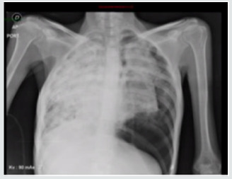
Lupine Publishers | Journal of Otolaryngology Impact Factor
Abstract
We present the report of a case of lung cancer of atypical manifestation with important challenges in its diagnosis in a young woman who came in with pneumothorax tension, cavitated lung lesions, ovarian, hepatic and peritoneal masses and a biopsy compatible with pulmonary adenocarcinoma, whose initial approach raised the differential diagnosis of a tumor of gynecological origin. A brief literature review was conducted.
Keywords: Pneumothorax; Pulmonary Cavitation; Pulmonary Adenocarcinoma; Ovarian Metastasis; TTF1
Introduction
A 28-year-old woman who consulted the emergency department of the National Cancer Institute (Instituto Nacional de Cancerología) for a 7-month history of asthenia, adinamia, progressive dyspnea, low cough, loss of 17 kilograms of weight and the appearance of a painful mass at the level of the iliac fossa and right flank. Initially studied as an outpatient with presumptive diagnosis of granulomatous disease with negative serial baciloscopy and chest x-ray showing right atelectasis + right pneumothorax. In the tomography multiple solid nodules some with hypodense center involving both pulmonary fields, caverns of thickened walls in both apices, posterior segments of the lower lobes and upper segments of the lower lobes. In the abdominal tomography there is presence of mass surrounding uterus of 185*100*85 mm of density of soft tissues, which takes the contrast IV and cystic area of 85 mm in its right aspect of probable ovarian origin and hepatic compromise with apparent peritoneal sowings. Positive tumor markers AFP 1.2, ACE 392, CA 125 257. A right thoracostomy, fibro bronchoscopy with cultures for M. negative tuberculosis and biopsy of pelvic mass that reported lesion characterized by cords and neuroglandular formation of epithelioid cells of neoplastic aspect with a marked desmoplasic response were initially performed. For which the diagnosis of metastatic ovarian cancer to lung, liver and peritoneum is made and send to initiate management.
Case Report
Upon admission to the institution, pulmonary thromboembolism is ruled out and extensive pulmonary parenchymatous involvement (Figure 1(a)) is confirmed by nodular areas, most of them with central cavitation and frosted glass halo, which are accompanied by paramilitary consolidations, making it necessary to consider neoplastic metastatic involvement with cavitation, less likely infectious (angioinvasive aspergillosis). At abdominal level, extensive infiltrative involvement of the peritoneum, hepatic subcapsular with extension to the parenchyma in segment 6, gastrohepatic ligament, transverse mesocolon, descending mesocolon. Heterogeneous bilateral adnexal masses of neoplastic aspect (Figure 1(b&c)). Scarce ascites. We reviewed pathology material of ovarian mass biopsy with report of adenocarcinoma with reactive immunopurified for CK7, TTF1 and negative for GATA 3, WT1, RE, CDX2, PAX 8 and CK20, we added NAPSIN which was positive, confirming lung metastatic origin (Figure 2(a&b)). The new fibro bronchoscopy with biopsy had immunohistochemistry that showed reactivity in tumor cells for TTF-1 and Napsin with absence of reactivity for p40, RE and RP. Compatible with compromise by non-small cell carcinoma, favors acinar pattern primary pulmonary adenocarcinoma. Less than 10% of all lung carcinomas debut with ��radiological cavitations, considered secondary to tumor necrosis by ischemia and/or bronchial obstruction [1]. Tokito et al. presented a cohort of lung cancer patients, with an incidence of cavitated lesions of 5.5% meanwhile in the Sing series this report is much higher near 9.6%. In both cohorts it was more frequent to find cavitations in men, over 60 years old, ex-smokers and with squamous cell histology [2].
Figure 1: (a) Chest X ray; (b) Axial computed tomography with extensive pulmonary parenchymatous involvement. (c) Computed tomography of the abdomen and pelvis with mass surrounding uterus.
Figure 2: (b) Immunohistochemistry of Napsine A in lung tissue; (b) Immunohistochemistry of TTF1 in ovarian tissue.
Figure 3 Chest X ray.
a greater extent induced by oncologic management are infection and hemoptysis, very infrequently, the presence of spontaneous pneumothorax which is estimated between 0.03 and 0.05% of primary lung cancer with a poor prognosis [3,4]. Among 1200 patients with spontaneous pneumothorax between 1970 and 2007, 37 (3%) had lung cancer. In all patients, the pneumothorax occurred on the same side of the carcinoma. The main cause of spontaneous pneumothorax was rupture of a necrotic tumor nodule or necrosis of subpleural metastases (21 ptes)., As well as the communication between the bronchus and the pleural cavity, producing a bronchopleural fistula that results in pneumothorax [5]. Spontaneous pneumothorax in the context of lung cancer occurs in 75% of cases as an initial manifestation and the remaining 25% during the course of the disease in patients with the known diagnosis, sometimes after the onset of management [6]. In this case, the first clinical manifestation of oncological disease was the presence of dyspnea secondary to spontaneous pneumothorax, infectious causes were ruled out and required surgical intervention with subsequent need for high oxygen flow. The main sites of lung cancer metastasis are pleura, brain, bone and liver. Ovarian metastases are rare, accounting for 5% of all ovarian cancers. The main tumors that metastasize to the ovary are from the gastrointestinal tract: colon and gastric, or originated in the breast. Lung cancer alone is the cause of these metastases by 0.3% [7]. The metastases of an adenocarcinoma of the lung are difficult to distinguish from a primary carcinoma of the ovary. Although there is no specific lung marker, TTF-1 can be used to discriminate between primary pulmonary and ovarian. TTF-1 is positive in approximately 63% of lung cancers [8]. Irving and Young reported 32 cases of metastatic to ovarian lung carcinoma in women aged 26 to 76. A history of lung carcinoma was documented in 53% of cases (17 out of 32), with detection of ovarian involvement at an interval of one year. In 10 cases (31%) ovary and lung occurred synchronously, in 5 (16%) ovarian tumors were detected 26 months before lung injury. The most frequent histologic subtype was small cell carcinoma 44%, adenocarcinoma in 34% and 16% in large cell carcinoma in 16% of patients. One third had bilateral presentation and the most frequent morphological characteristics were: multinodular growth, necrosis, lymphovascular invasion with rare involvement of the ovarian surface [9]. The management decision in this case was made considering the presence of pulmonary visceral crisis, the patient’s age, her ECOG and the high risk of rapidly deteriorating. Chemotherapy with palliative intention was started with carboplatin paclitaxel with a good clinical response in the two initial cycles, with which the requirement of supplementary oxygen and control of dyspnea was reduced. 40 days later, the patient was admitted with subite dyspnea of 3 days of evolution. with chest x-ray (Figure 3) that evidences left pneumothorax, ventilatory failure and dies despite rescue procedure.
Conclusion
Patients with cavitated tumors develop serious complications during or after chemotherapy or concomitance such as infection or massive hemoptysis. There are no reports of safety and efficacy of the use of chemotherapy in patients with advanced lung cancer with cavitated lesions. Some retrospective reports that have evaluated toxicity in this group of patients in the a 9% study developed hemoptysis, considering it as acceptable toxicity for this group of patients. Sandler et al reported that 30% of patients treated with carboplatin+paclitaxel+bevacizumab presented hemoptysis vs 6% of those without cavitations. In our case, taxane and platinumbased chemotherapy were initiated while obtaining studies of EGFR, ALK, Ros 1 and PDL1 to optimize management, with the initial cycles the palliation objective was achieved. However, the subsequent occurrence of pneumothorax is a possible consequence of the effect of cytotoxic treatment on existing lung lesions. It is not clear from the literature what would be the safest scheme, dose or frequency for this population to avoid the development of this type of complications. It is necessary to increase the reporting of these cases in order to try to elucidate their management.
For more Lupine Publishers Open Access Journals Please visit our website: h http://lupinepublishers.us/ For more Journal of Otolaryngology-ENT Research articles Please Click Here: https://lupinepublishers.com/otolaryngology-journal/
To Know More About Open Access Publishers Please Click on Lupine Publishers
Follow on Linkedin : https://www.linkedin.com/company/lupinepublishers Follow on Twitter : https://twitter.com/lupine_online
#Lupine Publishers#Lupine Publishers Group#Otolaryngology journal with impact factor#ENT Research Journal
2 notes
·
View notes
Text
Lupine Publishers | Disability in the Margin of Loneliness Research and Policy
For more Lupine Publishers Open Access Journals Please visit our website: https://lupinepublishersgroup.com/https://lupinepublishers.com/psychology-behavioral-science-journal/pdf/SJPBS.MS.ID.000139.pdfhttps://lupinepublishers.com/psychology-behavioral-science-journal/fulltext/disability-in-the-margin-of-loneliness-research-and-policy.ID.000139.php

Lupine Publishers | Scholarly Journal Of Psychology And Behavioral Sciences
Abstract
Loneliness is a key deficiency in well-being and health for the people of our time and, as a result, a major social policy issue. In Finland, the link between loneliness, poor health, ability to work and recovery from different lifestyles has been noticed in surveys of both population levels and individual groups, such as mental health rehabilitators, homeless people, and bread-line customers. However, the loneliness of people with disabilities has not been studied in Finnish society. The aim of the UN Convention on the Rights of Persons with Disabilities is to include people with disabilities and other marginalized groups of disability in national health, lifestyle and similar research. The samples selected for research are usually removed, for example, by persons in institutions or service homes. The living conditions, quality of life and many other issues of the various groups of injuries have been studied primarily - and almost exclusively - within these groups and even using ‘disability-specific’ indicators, making comparison with the rest of the population impossible. It is also worrying that people with disabilities do not have the opportunity to influence their position in the current change in the service structure, including housing and everyday life. For example, residents who move from institutions have not been listened to in the abolition of institutional care. It can be rightly asked whether people with disabilities will be thrown away and lobbied when major structural reforms are underway.
Introduction
Disability information and practical solutions based on it for the everyday life of people with disabilities are deficient and there is a risk of marginalization and alienation of people with disabilities. At worst, it is possible to talk about a cycle of negativity, where abandonment, marginalization and alienation can be related to psychoactive substance use, crimes or suicide. The fight against loneliness is the prevention of such a thread. But how and why should we study the loneliness of disabled people? Research on the subject is scattered and scarce. Material focused exclusively on loneliness has not been collected by people with disabilities. Existing studies have focused on materials where loneliness has been touched in one way or another. When studying the loneliness of people with disabilities, it is worthwhile to consider at least the following: disability itself does not tell or explain anything, and disabled people are either a homogeneous group. In the 2013 Regional Health and Welfare Questionnaire (ATH), the question of loneliness has been asked by “Do you feel lonely?”. The answers have been “Never”, “Very rarely”, “Sometimes”, “Quite often” or “Continuously”. A large fifth (22%) of the needs of disabled people (N = 1044) felt quite often or constantly lonely when about one in ten (9%) felt alone as the lonely ones [1]. In a survey conducted by [2], 39% of people with disabilities reported being lonely.
Conclusion
The knowledge base on the loneliness of people with disabilities is very fragmented, but on the basis of individual results, people with disabilities are more lonely than other people. Written material gathered in 2003 explains the factors behind the loneliness of disabled people. In the light of the material, the loneliness of people with disabilities appears to be experiences of the other and the outside: a much deeper experience than just a lack of people. Rather, it is represented as a lack of relevance [3,4]. As a societal issue about the loneliness of people with disabilities, not only factors related to the individual’s social standing, but also the habitat factors that bring about and maintain the loneliness experienced by disabled people, but also can contribute to inclusion and lasting contact with other people can be presented. Politically, in particular, the body issues and structures and values of the society determine how people with disabilities can participate in various activities such as independent living, work and social relationships; to determine her/his own life; make free choices according to one’s wishes and abilities; and live in an accessible environment, if necessary with the help of special services. These segments of social policy have also been highlighted as key areas in the Finnish Disability Policy Programmed. Such policy papers do not in themselves oblige or act as a clear justification for disability policy or individual empowerment but reveal that the fight against loneliness seems to have a deep gap between ideologies and practices that should not be ignored.
https://lupinepublishers.com/psychology-behavioral-science-journal/fulltext/disability-in-the-margin-of-loneliness-research-and-policy.ID.000139.php
https://lupinepublishers.com/psychology-behavioral-science-journal/pdf/SJPBS.MS.ID.000139.pdf
For more Lupine Publishers Open Access Journals Please visit our website: https://lupinepublishersgroup.com/
For more Psychology And Behavioral Sciences Please Click Here: https://lupinepublishers.com/psychology-behavioral-science-journal/
To Know more Open Access Publishers Click on Lupine Publishers
Follow on Linkedin : https://www.linkedin.com/company/lupinepublishers Follow on Twitter : https://twitter.com/lupine_online
4 notes
·
View notes
Text
Lupine Publishers | Autism and Food Selectivity- Mini Review

Lupine publishers | Scholarly Journal of Food and Nutrition
Introduction
The label “autism” has become one of the most important linguistic and conceptual creations in medical and psychological nomenclature, and has two names linked to pioneering studies: Austrian pediatrician Hans Asperger and Austrian psychiatrist Leo Kanner. Seventy years have passed since the publication of Asperger’s text on autism (1944), Kanner’s article (1943) had been known since its publication. Asperger was totally ignored until 1976 when English psychiatrist Lorna Wing published an article summarizing the work [1]. Kanner (1943), who first described and named early childhood autism, believed it was a distinct entity to be clearly distinguished from mental retardation, but there are many people who would argue that the two cannot be separated [2].
Autism and Asperger’s syndrome [3-5] are best known among invasive developmental disorders (PDD), a family of conditions marked by the early onset of delays and deviations in the development of social, communicative and cognitive skills. too many skills [6]. The first reports were made in 1943 by Leo Kanner, who first described 11 cases of what he called autistic affective contact disorders [7] as a condition with very specific behavioral characteristics, such as: disturbances of affective relationships. with the environment, extreme autistic loneliness, inability to use language for communication, presence of good cognitive potentialities, apparently normal physical appearance, ritualistic behaviors, early onset and predominant incidence in males.
Knickerbocker proposed that oral sensitivity, which is common in children with ASD, may be another example of a sensory system that is not only hyperresponsive to oral input, but also has a deficit in the modulation capacity of a varied sensory information input [8].
Oral hyperresponsiveness is more common in children with ASD than in children with typical development. Children with oral hyperresponsiveness are described as picky eaters, eat few vegetables and fruits, refuse to eat the same meal as their family, refuse to try and have aversion to certain tastes, textures and smells, do not like foods with low temperatures. or tall [9]. Parents of children with ASD often report that their children have a very restricted diet, with limited and limited food selection and acceptance and may be restricted to five foods [10].
In several of the studies analyzed by the authors, reports of food-based refusal of food have been identified, and difficulties in ingesting certain food textures have been attributed to changes in sensory processing or may be indicative of oro-motor difficulties, as well., sensory or oromotor changes may contribute to food refusal, with difficulty in chewing food or tolerating texture mix [11].
https://lupinepublishers.com/food-and-nutri-journal/fulltext/autism-and-food-selectivity-mini-review.ID.000135.php
For more Lupine Publishers Open Access Journals Please visit our website: https://twitter.com/lupine_online
For more Food And Nutrition Please Click
Here: https://lupinepublishers.com/food-and-nutri-journal/
To Know more Open Access Publishers Click on Lupine Publishers
Follow on Linkedin : https://www.linkedin.com/company/lupinepublishers Follow on Twitter : https://twitter.com/lupine_online
0 notes