#Optical Microscope
Explore tagged Tumblr posts
Visit Tumblr Blog
Explore Tumblr blogs with no restrictions, modern design and the best experience.
Last Seen Tumblr Blogs


Fun Fact
When “GIF” was named word of the year in 2012, Oxford Dictionaries U.S.A. credited Tumblr for pushing the word.
Text
Microscopy
Microscopy Microscopy is the scientific discipline which involves magnification of objects which may or may not be seen with naked eyes. Read more about Microscopy... Read Article...
(more…)

View On WordPress
#Component of Microscope#Easy notes on microscope#Forensic microscope notes#forensic microscopy#History of Microscope#microscope#Microscopy#Optical microscope#Parts of Microscope#Short notes on microscope#uses of microscope
2 notes
·
View notes
Text
Optical Microscope for testing available at Lab AtoZ
Lab AtoZ offers Optical Microscopes that can transform your research and product development. The offered Optical microscopes range from regular light microscopes to confocal and birefringence microscope and are provided in various models at industry leading prices. Connect with http://www.labatoz.com/instrumentcompany/31/Optical%20Microscope and know more about our extensive range of Optical Microscopes.
1 note
·
View note
Photo


bugün bunlarla geçti. sabah 10′dan akşam 17′e kadar uğraştık ama ortaya bir şey çıkmadı çünkü birisi mikroskopu kirletmiş ve temizlemeden gitmiş. bir de şu eldivenlerin 200 tanesi için 45 lira verdim, üniversitenin verdiği eldivenler elimden fırlayacak gibi emanet duruyordu. mecbur daha esnek eldiven almak zorunda kaldım. fotoğraflarda gördüğünüz şeyler ise agat kristalinden bir havanda şişe camlarına yaptığımız birkaç işlem sonrasında elde ettiklerimizi dövmek. günün yarısını havan döverek geçirdik diğer yarısı da işte mikroskopla uğraşıp görüntüler alarak geçti. sabah içtiğimden başka kahve içemedim, mal gibi dolanıyorum.
#material science#ceramics#sls glass#optical microscope#laboratory#ceramic science#grain size#agglomeration#glass particles#agate mortar#metallurgical and material science
3 notes
·
View notes
Text

#microscope#types of microscope#microscopy#history#optical microscope#beam paths#svg#diagram#electron microscope
0 notes
Text
Research On World Optical Microscope Market Report 2025
Report on World Optical Microscope Market by Product Type, Market, Players and Regions-Forecast to 2025 offered by DecisionDatabases.com.
Optical Microscope market research report provides the newest industry data and industry future trends, allowing you to identify the products and end users driving Revenue growth and profitability.
The industry report lists the leading competitors and provides the insights strategic industry Analysis of the key factors influencing the market.
The report includes the forecasts, Analysis and discussion of important industry trends, market size, market share estimates and profiles of the leading industry Players.
Final Report will cover the impact of COVID-19 on this industry.
Browse the complete report and table of contents @ https://www.decisiondatabases.com/ip/33625-optical-microscope-market-analysis-report
The Players mentioned in our report
ZEISS
Olympus Corporation
Ted Pella, Inc
Nikon Instruments Inc.
Reichert, Inc
Leica
Bruker
Global Optical Microscope Market: Product Segment Analysis
Trinocular
Binocular
Monocular
Global Optical Microscope Market: Application Segment Analysis
Hospital
School
Laboratory
Global Optical Microscope Market: Regional Segment Analysis
USA
Europe
Japan
China
India
South East Asia
Download Free Sample Report of World Optical Microscope Market @ https://www.decisiondatabases.com/contact/download-sample-33625
There are 10 Chapters to Deeply Display the World Optical Microscope Market. Chapter 1 About the Optical Microscope Industry Chapter 2 World Market Competition Landscape Chapter 3 World Optical Microscope Market share Chapter 4 Supply Chain Analysis Chapter 5 Company Profiles Chapter 6 Globalisation & Trade Chapter 7 Distributors and Customers Chapter 8 Import, Export, Consumption and Consumption Value by Major Countries Chapter 9 World Optical Microscope Market Forecast through 2025 Chapter 10 Key success factors and Market Overview
Purchase the complete World Optical Microscope Market Research Report @ https://www.decisiondatabases.com/contact/buy-now-33625
Other Reports by DecisionDatabases.com:
World Microscopes Market Research Report 2025 (covering USA, Europe, China, Japan, India, South East Asia and etc)
World Stereo Microscopes Market Research Report 2025 (Covering USA, Europe, China, Japan, India and etc)
World Helium Ion Microscopes Market Research Report 2025 (Covering USA, Europe, China, Japan, India and etc)
About-Us: DecisionDatabases.com is a global business research reports provider, enriching decision makers and strategists with qualitative statistics. DecisionDatabases.com is proficient in providing syndicated research report, customized research reports, company profiles and industry databases across multiple domains.
Our expert research analysts have been trained to map client’s research requirements to the correct research resource leading to a distinctive edge over its competitors. We provide intellectual, precise and meaningful data at a lightning speed.
For more details: DecisionDatabases.com E-Mail: [email protected] Phone: +91 9028057900 Web: https://www.decisiondatabases.com/
#Optical Microscope#Optical Microscope Market#Global Optical Microscope Market#Optical Microscope Industry Size
0 notes
Note
THANK YOU FOR AGREEING W MY BLORBO TAGS my brain is full of idiotic content for enstars and i need to study all of them under a microscope <333
and i am studying the microscope slides you prepare out of these characters because you are good at making the cross sections!!!! idk if this metaphor makes sense but the point is i am trying to convey enthusiasm<3
#i have some experience with optical microscopes mostly but idk any of the correct english terms. shrug#assigning posts to characters is so much funnnn i used to make a bunch of those text post meme edits for a previous thing i was into
2 notes
·
View notes
Text
Chlorite under a microscope, top is plain polarized light and bottom is crossed polarized light.
3 notes
·
View notes
Photo

Celery Leaf Popular prints, shirts, & stickers available on my Etsy. Línk in bio #microscope #microscopy #sciart #science #nature #optics #imaging #plant #plants #plantcell #photomicrograph #biology #microbiology #cell #etsy #Infinitesimal #Prints #shopping #etsyshop #etsyseller #crosssection #celery #leaf #vegetable #vegetables (at Cal Poly Humboldt) https://www.instagram.com/p/Ci7_m18pkDU/?igshid=NGJjMDIxMWI=
#microscope#microscopy#sciart#science#nature#optics#imaging#plant#plants#plantcell#photomicrograph#biology#microbiology#cell#etsy#infinitesimal#prints#shopping#etsyshop#etsyseller#crosssection#celery#leaf#vegetable#vegetables
3 notes
·
View notes
Text
Eye of a locust
To achieve this result, you have to put a bit of transparent varnish on a dead locust's eye, then wait a minute until it dries, eventually take it delicately off with your nails.
These little hexagons are the surfaces of ommatidia, which are composed of photoreceptor cells and structure a compound eye. Each ommatidium has its cornea, crystalline cone — such as ours, except that they're thousands of them in a compound eye — photoreceptor cells and pigment cells, linked to an optic nerve.
#eyes#compound eye#optics#microscope#biology#ommatidium#cells#insects#insect eyes#varnish#tips#entomology#photografy#locust#dead insect#arthropods#invertebrate
2 notes
·
View notes
Link

#microscope manufacturer#microscope supplier#digital microscope#medical microscope#metallurgical microscope#phase contrast microscope#Multi head microscope#zoom stereo microscope#optical microscope
0 notes
Text
Microscope
BY- K. Sai Manogna (MSIWM014) The microscope is an instrument that creates enlarged photographs of small objects, enabling the viewer to display minute structures in a too near manner at a scale that is convenient for observation and study. Although optical microscopes are the focus of this article, many other types of wave, like acoustic, X-ray, or electron beam, can also enlarge the image…

View On WordPress
#electron microsocpes#function of microsocpe#history of microscopes#light microsocpes#microscope#optical instrument#optical microscopes#parts of microscope#resolution#types of microascopes
1 note
·
View note
Text
A new method for fluorescence microscopy
- By Tokushima University -
Scientists have developed a fluorescence "lifetime" microscopy technique that uses frequency combs and no mechanical parts to observe dynamic biological phenomena.
Conventional fluorescence microscopy provides poor quantitative information of the sample because it only captures fluorescence intensity, which changes frequently and depends on external factors. Now, scientists from Japan have developed a new fluorescence microscopy technique to measure both fluorescence intensity and lifetime. Their method does not require mechanical scanning of a focal point; instead, it produces images from all points in the sample simultaneously, enabling a more quantitative study of dynamic biological and chemical processes.
Fluorescence microscopy is widely used in biochemistry and life sciences because it allows scientists to directly observe cells and certain compounds in and around them. Fluorescent molecules absorb light within a specific wavelength range and then re-emit it at the longer wavelength range. However, the major limitation of conventional fluorescence microscopy techniques is that the results are very difficult to evaluate quantitatively; fluorescence intensity is significantly affected by both experimental conditions and the concentration of the fluorescent substance. Now, a new study by scientists from Japan is set to revolutionize the field of fluorescence lifetime microscopy.
A way around the conventional problem is to focus on fluorescence lifetime instead of intensity. When a fluorescent substance is irradiated with a short burst of light, the resulting fluorescence does not disappear immediately but actually “decays” over time in a way that is specific to that substance. The “fluorescence lifetime microscopy” technique leverages this phenomenon—which is independent of experimental conditions—to accurately quantify fluorescent molecules and changes in their environment. However, fluorescence decay is extremely fast, and ordinary cameras cannot capture it. While a single-point photodetector can be used instead, it has to be scanned throughout the sample’s area to be able to reconstruct a complete 2D picture from each measured point. This process involves movement of mechanical pieces, which greatly limits the speed of image capture.
Fortunately, in this recent study published in Science Advances, the aforementioned team of scientists developed a novel approach to acquire fluorescence lifetime images without necessitating mechanical scanning. Professor Takeshi Yasui, from Institute of Post-LED Photonics (pLED), Tokushima University, Japan, who led the study, explains, “Our method can be interpreted as simultaneously mapping 44,400 ‘light stopwatches’ over a 2D space to measure fluorescence lifetimes—all in a single shot and without scanning.” So, how was this achieved?
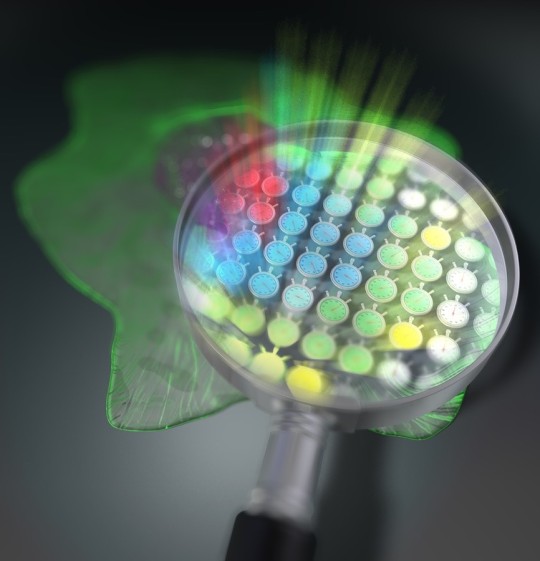
Image: 2D arrangement of 44,400 light stopwatches enables scan-less fluorescence lifetime imaging. Credit: Tokushima University.
One of the main pillars of their method is the use of an optical frequency comb as the excitation light for the sample. An optical frequency comb is essentially a light signal composed of the sum of many discrete optical frequencies with a constant spacing in between them. The word “comb” in this context refers to how the signal looks when plotted against optical frequency: a dense cluster of equidistant “spikes” rising from the optical frequency axis and resembling a hair comb. Using special optical equipment, a pair of excitation frequency comb signals is decomposed into individual optical beat signals (dual-comb optical beats) with different intensity-modulation frequencies, each carrying a single modulation frequency, and irradiated on the target sample. The key here is that each light beam hits the sample on a spatially distinct location, creating a one-to-one correspondence between each point on the 2D surface of the sample (pixel) and each modulation frequency of the dual-comb optical beats.
Because of its fluorescence properties, the sample re-emits part of the captured radiation while still preserving the aforementioned frequency–position correspondence. The fluorescence emitted from the sample is then simply focused using a lens onto a high-speed single-point photodetector. Finally, the measured signal is mathematically transformed into the frequency domain, and the fluorescence lifetime at each “pixel” is easily calculated from the relative phase delay that exists between the excitation signal at that modulation frequency versus the one measured.

Image: Principle of operation. Credit: Suana Science YMY.
Thanks to its superior speed and high spatial resolution, the microscopy method developed in this study will make it easier to exploit the advantages of fluorescence lifetime measurements. “Because our technique does not require scanning, a simultaneous measurement over the entire sample is guaranteed in each shot,” remarks Prof. Yasui, “This will be helpful in life sciences where dynamic observations of living cells are needed.” In addition to providing deeper insight into biological processes, this new approach could be used for simultaneous imaging of multiple samples for antigen testing, which is already being used for the diagnosis of COVID-19.
Perhaps most importantly, this study showcases how optical frequency combs, which were only being used as “frequency rulers,” can find a place in microscopy techniques to push the envelope in life sciences. It holds promise for the development of novel therapeutic options to treat intractable diseases and enhance life expectancy, thereby benefitting the whole of humanity.
--
Source: Tokushima University
Full study: “Full-field fluorescence lifetime dual-comb microscopy using spectral mapping and frequency multiplexing of dual-comb optical beats”, Science Advances.
http://dx.doi.org/10.1126/sciadv.abd2102
Read Also
Scientists develop technique improving the resolution of cryo-electron microscopy
1 note
·
View note
Photo

https://wa.me/5511941431076 #teratecsp #microscopio #microscope #microscopemaintenance #microscopia #mecanicadeprecisao #qualidade #precisao #solucoesemprecisao #manutencaodemicroscopio #apoieaspequenasempresas #contratos #manutencaopreventiva #excelencia #optica #optics #compredopequeno #compromisso #comprometimento @teratecsp [email protected] www.manutencaodemicroscopio.com.br (em Teratec Soluções em Precisão Ltda) https://www.instagram.com/p/CCatl9TAzSW/?igshid=1vrq4t403ua5l
#teratecsp#microscopio#microscope#microscopemaintenance#microscopia#mecanicadeprecisao#qualidade#precisao#solucoesemprecisao#manutencaodemicroscopio#apoieaspequenasempresas#contratos#manutencaopreventiva#excelencia#optica#optics#compredopequeno#compromisso#comprometimento
3 notes
·
View notes
Photo
Guiding Sight
Your eyes look at the world but it's your brain that sees it. This is thanks to your optic nerve, which connects to nerve cells in the backs of your eyes (retina) to deliver their messages to your brain for interpretation. Development of the optic nerve involves the gene Dcc. Researchers now probe what Dcc is doing here using confocal microscopy (pictured) to image nerve cells in the eyes. In mutant mice lacking Dcc in their eyes, projections from nerve cells within the retina failed to connect up to the optic nerve (bottom) as occurs in normal mouse eyes (top, left). Instead, projections stalled or went in the opposite direction, similar to mutant mice lacking the protein, netrin, which is known to guide nerve cells (top, right). Dcc is therefore vital in properly wiring up the eyes to the brain.
Written by Lux Fatimathas
Image adapted from work by Robin J. Vigouroux and colleagues
Institut de la Vision, Sorbonne Universités, UPMC Univ Paris 06, INSERM, CNRS, Paris, France
Image originally published with a Creative Commons Attribution 4.0 International (CC BY 4.0)
Published in eLife, February 2020
You can also follow BPoD on Instagram, Twitter and Facebook
6 notes
·
View notes
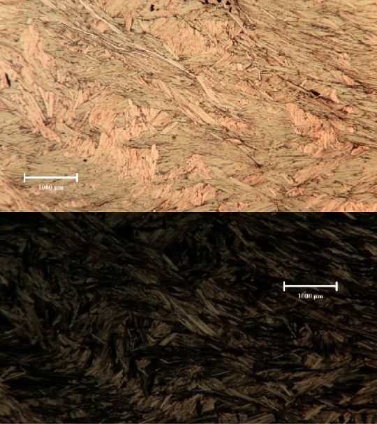
Text


Biotite under Polarized and Cross polarized light.
•Pleochroic (Lt. Brown to dark brown)
•1 direction of cleavage
•Parallel extinction
Found in granite along with Hornblende
#geology#minerals#crystallography#petrology#petrography#thin section#optics#biotite#science#microscope#hornblende#quartz#feldspar
94 notes
·
View notes
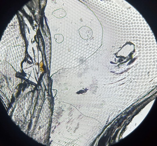
Photo

Working on polishing my samples for reflective light microscopy. This sample is a pyroxenite from Hogen Camp Mine in the Hudson Highlands region of New York. The area was once a lead producer of iron ore and magnetite for the east coast.
#gif#geochemistry#geologist#petrology#optical mineralogy#microscope#mineralogy#magma#minerals#plume#pretty#physics#geology#new york#nyc#pyroxene#pyroxenite#iron#iron ore#chemistry#crystals#magnetite#mining#east coast#optmin
384 notes
·
View notes