
Statistics
We looked inside some of the posts by ambadyscan and here's what we found interesting.
Average Info
Notes Per Post
1
Likes Per Post
1
Reblog Per Post
0
Reply Per Post
0
Time Between Posts
7 days
Number of Posts By Type
Text
17
Last Seen Tumblr Blogs


Fun Fact
BuzzFeed published a report claiming that Tumblr was utilized as a distribution channel for Russian agents to influence American voting habits during the 2016 presidential election in Feb 2018.
Text
Make the Horoscope of your Child by the NT Scan

A good radiologist or a foetal medicine specialist can be an expert horoscope maker most of time better than an astrologer because he or she has a better proved scientific approach.
HOW it is done you may wonder?
NT scan
NT scan is the key for him or her to make the horoscope. NT means Nuchael Translucency. It is the thickness of fluid behind the neck in the subcutaneous plane. NT scan is done between 11 weeks and 13 + 6 weeks (not 10 weeks 6 days or at 14 weeks) when the CRI- (total length from crown to rump) is between 45mm and 84mm.
The astrologer needs the correct time of birth and the child’s gender. Here the Doctor needs 5 things to make the horoscope chart.
The CRL
NT measurement
Nasal bone present or absent — all these obtained from scanning the fetus
From the maternal blood, he or she collects the levels of PAPP-A and
Free Beta HCG
He or she feeds all these to a computer which is loaded with special software which gives the result as follows
(a)Low risk (b) Intermediate risk (c) High risk
If the result is low risk, we will give a pass mark and allow the pregnancy to continue. No need to worry. This baby will have a more or less a normal life and will live up to death like any of us with all the pangs and happiness of life.
If the result is intermediate risk, we will give one more chance to the pregnancy by advising NIPT – a blood test for mother. If NIPT is negative, they are like the group mentioned above but if it is positive they fall into high risk category.
The high risk category— It is in this group, unfortunately the horoscope predictions will be bad for these children.
What to do with the high risk group?
To draw a “Thalakury” and predict the future instead of “Star Positions” we make it with four main factors.
Adenini
Quaninei
Cystinom and
Thyminym which the scientist call nucleotides
For more accurate reading of the Grahanila position we add three components of the nucleotides
(a) Phosphate group (b) A sugar molecule and (c) A nitrogen base
How is this data collected for the horoscope?
We do CVS (chorionic villus Sampling) before 14 weeks of pregnancy or amniocentesis after 16 weeks of pregnancy. These samples contain fetal cells. We culture it outside and multiply the cells. Then we analyse the chromosome and DNA by which we get all the data to draw the Grahanila.
Predictions
Will the parent get a full term baby or will the pregnancy end up in intra uterine fetal death?
If the DNA test comes as Edwards syndrome — trisomy 18 (Extra chromosome 18); PATAVU Syndrome (Trisomy 13); TURNER Syndrome (Monosomy X) ; TRIPLOIDY Hydatid mole ; Osteogenesis imperfect type Il, severe case of congenital diaphragmatic hernia; SMITH LEMLI — OPITS Syndrome
In the above conditions one can predict that most probably the pregnancy will end up in IUD.
By scanning you can decide whether it is a male or female child (We do not reveal this to the patient, as this is against Indian law)
If it is a male child will it have IUD?
X linked recessive disorders (DUCHENNE MUSCULAR DYSTOPHY; Haemophilia A and B ; X linked Hydrocephalus ; X linked severe combined immino deficiency ( SCID); X linked ICHTHYOSIS (severe form of a skin disease), X linked lympho proliferative syndrome ; Menkes disease; X linked MYOTUBULAR MYOPATHY; HUNTER SYNDROME; ORNITHINE TRANSCARBAMYLASE Deficiency – All usually will lead to IUD.
If it is a female fetus will it live up to term?
TURNER SYNDROME (MONOSOMY X); REIT Syndrome; Congenital adrenal hyperplasia; LEIGH syndrome; GOLTS Syndrome, severe form of cystic fibrosis etc. may end up in IUD
In the horoscope we can predict this “BOY” may not reach adulthood — like the story of MARKENDAYAN in Hindu mythology or we can predict the “GIRL” may not reach adulthood.
Also like “SARPADOSHAM” we can indicate this female child will potentially pass the disease to their offsprings.
0 notes
Text
Importance of Genetic Counselling Before and During Pregnancy

Genetic counselling is a vital component of prenatal care, offering expectant parents’ insights into the genetic health of their unborn child. This specialized form of counselling provides information about how genetic conditions might affect a baby, helping parents make informed decisions about their pregnancy. With advancements in medical technology, genetic counselling has become increasingly important in identifying potential risks and ensuring the health and well-being of both mother and baby.
Why Genetic Counselling Matters
Early Detection of Genetic Disorders:
Genetic counselling can help identify the risk of inherited genetic disorders such as Down syndrome, cystic fibrosis, and sickle cell anaemia. Early detection allows for better preparation and management of any potential health issues.
Informed Decision-Making:
Expectant parents can make informed decisions about their pregnancy and the future care of their child. This might include considering additional testing, exploring treatment options, or even preparing for the possibility of raising a child with special needs.
Personalized Care Plans:
Genetic counsellors can create personalized care plans tailored to the specific genetic risks identified. This ensures that both the mother and baby receive the appropriate medical care and support throughout the pregnancy.
Emotional Support:
The process of genetic counselling provides emotional support to parents. Understanding potential genetic risks can be overwhelming, and having a counsellor to discuss these concerns can be reassuring and help alleviate anxiety.
When to Seek Genetic Counselling
Before Pregnancy:
Couples planning to conceive may choose to undergo genetic counselling to understand their risk of passing on genetic conditions. This is particularly important for those with a family history of genetic disorders or those belonging to ethnic groups with a higher prevalence of certain genetic conditions.
During Pregnancy:
Genetic counselling is also crucial during pregnancy, especially if prenatal screening tests indicate a potential genetic abnormality. This allows for further diagnostic testing and timely intervention if necessary.
Types of Genetic Testing
Carrier Screening:
Carrier screening tests can identify if prospective parents carry genes for specific inherited disorders. This is typically done before or early in pregnancy.
Prenatal Screening:
Non-invasive prenatal testing (NIPT) and ultrasound scans can screen for chromosomal abnormalities such as Down syndrome, trisomy 18, and trisomy 13.
Diagnostic Testing:
If a screening test indicates a potential issue, diagnostic tests such as amniocentesis or chorionic villus sampling (CVS) can provide more definitive information about the foetus’s genetic health.
The Role of Genetic Counsellors
Genetic counsellors are healthcare professionals with specialized training in medical genetics and counselling. They play a crucial role in guiding expectant parents through the complex process of genetic testing and interpretation of results. Their responsibilities include:
Risk Assessment:
Genetic counsellors assess the risk of genetic disorders based on family history, ethnic background, and other factors.
Education:
They educate parents about genetic conditions, the implications of test results, and available options.
Support:
Counsellors provide emotional support and guidance, helping parents navigate the challenges and decisions that come with genetic testing.
Coordination of Care:
They work closely with obstetricians, paediatricians, and other healthcare providers to ensure a comprehensive care plan for both mother and baby.
The Impact of Genetic Counselling
The impact of genetic counselling extends beyond the immediate pregnancy. It can have long-term benefits for the family, including:
Preparation:
Parents can prepare for the birth of a child with special needs, ensuring they have the necessary resources and support systems in place.
Future Family Planning:
Understanding genetic risks can inform future family planning decisions, helping parents make choices about having more children.
Health Awareness:
Families gain a deeper understanding of their genetic health, which can be valuable for the health and well-being of future generations.
Partnering with Ambady Scan Centre
At Ambady Scan, the best scanning center in Kerala, we understand the importance of genetic counselling in ensuring a healthy start for mother and baby. Our team of experienced genetic counsellors are dedicated to providing comprehensive genetic services, from pre-conception counselling to prenatal screening and diagnostic testing.
We are committed to supporting families through every step of their journey, offering the highest quality care and guidance to help them make informed decisions about their pregnancy and their future. Contact Ambady Scan Centre for compassionate, personalized care that prioritizes the health and well-being of both you and your baby.
0 notes
Text
Screening for Chromosomal Abnormalities during Pregnancy

Introduction
When expecting a new addition to the family, ensuring the health of the baby is a top priority for parents. One important aspect of prenatal care is screening for chromosomal abnormalities. These screenings are designed to assess the risk of certain genetic disorders that can affect a baby’s development and overall health. Understanding the significance, types, and timing of these tests can help expecting parents make informed decisions and prepare for any potential health challenges their baby might face. This introduction into the world of prenatal screenings aims to clarify their necessity and process, helping parents navigate their journey with confidence and knowledge.
Importance of Screening for Chromosomal Abnormalities during Pregnancy
Significance of Prenatal Care
Prenatal care is not just about ensuring a healthy diet and regular check-ups; it’s a critical period for comprehensive screenings and tests to assess the health of both the mother and the fetus. Among these, screening for chromosomal abnormalities plays a pivotal role. These screenings help to identify potential health issues that could affect the baby’s development and lead to informed decisions about care and treatment. Early detection through these screens can prepare parents and healthcare providers to manage health conditions that could impact the baby’s life quality.
Risks of Chromosomal Abnormalities in Pregnancy
Chromosomal abnormalities occur when there is a defect in one or more chromosomes. The most well-known of these is Down syndrome, but there are many others like Trisomy 18 and Trisomy 13, which can significantly impact a baby’s development both physically and mentally. The risks associated with these disorders can vary; they include developmental delays, congenital disabilities, and, in some severe cases, stillbirth. The risks tend to increase with maternal age, particularly in mothers over the age of 35. Screening helps in assessing these risk factors early in the pregnancy, which is crucial for planning and management.
Common Screening Tests for Chromosomal Abnormalities
First Trimester Screening
First trimester screening is typically conducted between the 11th and 13th weeks of pregnancy. This screening involves a blood test and an ultrasound. The blood test measures the levels of certain substances in the mother’s blood that are associated with chromosomal conditions in the fetus. The ultrasound examines the nuchal translucency, which is a fluid-filled space at the back of the baby’s neck. Increased fluid can be a sign of Down syndrome, Trisomy 18, or heart defects. Together, these tests provide a risk assessment that helps in determining whether further diagnostic testing is needed.
Second Trimester Screening
Known as the “quad screen,” second trimester screening is conducted between the 16th and 18th weeks of pregnancy. This test measures four substances in the mother’s blood: AFP, hCG, Estriol, and Inhibin-A. Abnormal levels of these substances can suggest the risk of certain chromosomal abnormalities such as Down syndrome and Trisomy 18, as well as neural tube defects like spina bifida. This screening does not confirm the presence of these conditions but identifies if there is a heightened risk, requiring additional, more definitive tests.
Non-Invasive Prenatal Testing (NIPT)
Non-Invasive Prenatal Testing (NIPT) is a relatively new screening test that can be done as early as the 10th week of pregnancy. It involves taking a blood sample from the mother to examine fragments of fetal DNA that are present in the maternal bloodstream. NIPT is highly effective in detecting several major chromosomal abnormalities, including Down syndrome, Trisomy 13, and Trisomy 18. Due to its non-invasive nature and high level of accuracy in these areas, NIPT is increasingly becoming a popular choice among expectant parents. It offers not only peace of mind but also a safer way for early detection compared to invasive tests, which carry risks of miscarriage.
Benefits of Early Detection and Diagnosis
Understanding potential chromosomal abnormalities early on during pregnancy can have numerous benefits for both the mother and the developing baby. Early detection provides crucial insights into the health of the fetus and can guide further medical and personal decisions throughout the pregnancy.
Early Intervention and Treatment Options
Identifying chromosomal abnormalities early in pregnancy allows healthcare providers to offer specific interventions that may mitigate some complications. For instance, certain conditions like congenital heart defects, which can be associated with chromosomal abnormalities, can be managed better if detected early. Health professionals can monitor the baby’s development more closely, prepare for interventions soon after birth, and sometimes even suggest treatments before the baby is born. Early detection also opens the door to connect with specialists and hospitals equipped to handle specific conditions right from the start, ensuring the best possible care for both mother and baby.
Emotional and Psychological Preparation for Parents
Receiving news about potential chromosomal abnormalities during a pregnancy can be emotionally challenging. However, early detection gives parents valuable time to psychologically and emotionally prepare for the arrival of a child who may have special needs. It allows for additional time to research the condition, understand what it entails, and realistically set expectations. Support groups and counseling services can also be accessed sooner, providing much-needed emotional support and guidance through the pregnancy journey. This preparatory phase is crucial for building a supportive environment for the child and family.
Making Informed Decisions
The journey of prenatal screenings and tests provides critical information that can influence the decisions made by expectant parents. From understanding the scope and implications of test results to deciding on further diagnostic procedures, parents are better equipped to make informed decisions when they have comprehensive and timely information.
Understanding Screening Results
Navigating the results of prenatal screenings for chromosomal abnormalities can be complex. These tests typically assess the risk of the fetus having certain conditions but do not confirm the presence of abnormalities. Health professionals usually convey results as a probability or risk level, such as a 1 in 100 chance. Understanding what these numbers mean can significantly affect a parent’s decision-making process. For clarity, it is key that the healthcare provider explains the results comprehensively, discussing what they may imply about the baby’s health and what further steps might be advisable.
Follow-up Diagnostic Tests
If screening tests show a high risk of chromosomal abnormalities, follow-up diagnostic tests may be recommended. These tests, such as amniocentesis or chorionic villus sampling (CVS), are more invasive but provide conclusive results about whether a chromosomal abnormality exists. Decision-making at this stage is critical — parents must consider potential risks and benefits. Some may choose to proceed to understand definitively if an abnormality is present, while others may opt out due to the potential risks involved with these procedures. The choice can profoundly impact the approach towards the remaining period of pregnancy, labor, and postnatal care, underscoring the importance of clear, informed discussions with healthcare providers.
Each step in this process, from initial screening to potential diagnostic follow-ups, plays a pivotal role in how parents manage their pregnancy and prepare for their child’s future, making these early assessments incredibly significant.
Advocating for Prenatal Health

The journey through pregnancy is a remarkable period that brims with the promise of new life. However, it’s also a time when the health of both mother and baby should be monitored with great attention and care. Advocating for robust prenatal health is crucial to ensuring that the pregnancy progresses safely and healthily. This encompasses regular medical check-ups, effective communication with healthcare professionals, and adhering to prescribed prenatal routines.
Importance of Regular Check-ups
Regular check-ups during pregnancy serve as a critical tool for monitoring the health and development of the fetus, as well as the well-being of the mother. These visits typically involve a variety of tests and screenings designed to detect any potential issues early on, such as chromosomal abnormalities, which could affect the baby’s development. Routine check-ups enable doctors to:
– Track the growth and development of the fetus
– Monitor the health status of the mother, including blood pressure and weight
– Provide important updates on nutritional needs and physical activity
– Administer necessary vaccinations to protect both mother and child
One critical aspect of these check-ups is screening for chromosomal abnormalities. These screenings are usually non-invasive and provide valuable insights into the genetic health of the fetus. Early detection through these tests allows for a broader range of options and preparedness in managing any conditions that may be discovered.
Communicating with Healthcare Providers
Effective communication with healthcare providers is another pillar of strong prenatal care. Expectant mothers should feel empowered to discuss any concerns or symptoms they experience without hesitation. Open communication can lead to:
– Tailored health advice that aligns with individual health histories and needs
– Clarification and reassurance regarding the normal processes and symptoms of pregnancy
– Swift action and intervention if any unusual signs or complications are identified.
Patients should feel encouraged to ask questions about the screenings, including what each test involves, what the results could indicate, and the subsequent steps following different outcomes. Understanding the purpose and outcomes of tests like those for chromosomal abnormalities not only eases anxieties but also helps in making informed decisions. This dialogue ensures that the pathways of care are clear, inclusive, and aligned with the best interests of both the mother and the developing baby.
Conclusion – Ensuring a Healthy Start for Mother and Baby
Prenatal screening for chromosomal abnormalities is a crucial step toward ensuring the health and well-being of both mother and baby. These screenings help in early detection, which can provide families with valuable information to make informed decisions and prepare for any necessary medical care. Remember, while the prospect of screening can be daunting, the knowledge gained allows for better preparation and peace of mind during what should be a joyous journey. At Ambady Scan, the best scanning center in Kerala, we are dedicated to offering the highest quality prenatal screening services to support you every step of the way.
0 notes
Text
The Importance of Routine Antenatal Scans: A Comprehensive Guide for Expectant Mothers

Antenatal scans are a crucial aspect of prenatal care for expectant mothers, providing valuable insights into the health and development of the baby. At Ambady Scan Centre, we understand the importance of routine antenatal scans in monitoring the progress of pregnancy and identifying any potential complications early on. This comprehensive guide will walk you through the significance of routine antenatal scans, the different types of ultrasound scans available, and what to expect during your appointments at Ambady Scan Centre. Prioritizing your health and the health of your baby is paramount, and routine antenatal scans play a key role in ensuring a safe and healthy pregnancy journey.
What are routine antenatal scans and why are they important?
Routine antenatal scans are medical imaging procedures conducted during pregnancy to monitor the growth and development of the foetus, as well as to assess the overall health of both the mother and the baby. These scans are vital in detecting any potential issues or abnormalities early on, allowing for timely intervention and management. Regular monitoring through antenatal scans helps healthcare providers to ensure the well-being of both the expectant mother and her unborn child throughout the pregnancy journey. By prioritizing these routine scans, expectant mothers can proactively safeguard their health and that of their baby, leading to a smoother and healthier pregnancy experience.
Benefits of routine antenatal scans for expectant mothers and babies
Regular antenatal scans offer numerous benefits for both expectant mothers and their babies. These scans provide crucial insights into the baby’s growth and development, enabling early detection of any potential complications or abnormalities. Prompt identification of such issues allows healthcare providers to deliver appropriate care and interventions, ensuring the optimal health and well-being of both the mother and the baby. Additionally, routine scans offer expectant mothers the reassurance of monitoring the progress of their pregnancy, fostering a sense of comfort and security during this transformative period. By prioritizing regular antenatal scans, expectant mothers can actively participate in safeguarding their health and that of their precious bundle of joy.
When should you schedule your routine antenatal scans?
It is recommended to schedule your routine antenatal scans as early as possible in your pregnancy. Typically, the first scan, known as the dating scan, is ideally conducted between 8 to 14 weeks gestation to accurately determine your due date and assess the viability of the pregnancy. Subsequent scans may be scheduled at different intervals throughout your pregnancy to monitor the baby’s growth and development consistently. Discuss the recommended scan schedule with your doctor to ensure that you receive timely and comprehensive antenatal care. Remember, regular scans play a vital role in ensuring a healthy and successful pregnancy journey for both you and your baby.
What to expect during a routine Antenatal Scan?
During a routine antenatal scan, you can expect a qualified doctor to perform the scan using an ultrasound machine. The procedure is non-invasive and painless, involving the application of gel on your abdomen and the use of a transducer to capture images of your baby in the womb. You may also have the opportunity to see your baby’s movements and hear the reassuring sound of their heartbeat. The scan provides valuable information about your baby’s growth, position, and overall well-being, allowing your doctor to address any concerns promptly. Stay tuned to learn more about interpreting scan results and the significance of regular monitoring during your antenatal care journey.
Risks and limitations of routine Antenatal Scans
While routine antenatal scans are crucial for monitoring your baby’s development, it’s essential to understand that they may also carry some risks and limitations. For instance, some scans may not detect all abnormalities or issues present in the baby, leading to a false sense of security. Additionally, frequent exposure to ultrasound waves during scans may raise concerns about its impact on the developing foetus, although research indicates that the benefits generally outweigh the risks. Discuss this with your doctor and make informed decisions about the frequency and necessity of scans during your pregnancy
How to prepare for your Antenatal Scan?
Preparing for your routine antenatal scan can help ensure that the procedure goes smoothly and that you obtain accurate results. Begin by scheduling your appointment at a reputable healthcare facility that specializes in prenatal care and ultrasound imaging.
It’s advisable to wear loose clothing that can easily be removed or lifted to expose your abdomen during the scan.
Additionally, drink plenty of water beforehand as a full bladder can provide clearer images of your baby during the scan.
Lastly, bring any relevant medical records or previous scan reports to assist the healthcare provider in evaluating your baby’s development accurately.
Planning ahead and following these preparation tips can optimize the effectiveness of your routine antenatal scan.
Conclusion: The importance of routine Antenatal scans for a Healthy pregnancy.
In conclusion, routine antenatal scans play a vital role in monitoring the progress of your pregnancy and ensuring the well-being of both you and your baby. These scans provide valuable insights into your baby’s growth and development, as well as help of expert doctors detect any potential issues early on.
By following the essential preparation tips outlined above and attending your scheduled antenatal scans, you are actively contributing to the success of your pregnancy journey. Remember, early detection and monitoring can lead to timely interventions if needed, ultimately leading to better outcomes for you and your little one. Prioritize your routine antenatal scans for a healthy and happy pregnancy!
Schedule an appointment with us and free yourself from any worries during pregnancy.
0 notes
Text
Nuchal Translucency (NT) Screening during Pregnancy and its Importance

Pregnancy is a beautiful journey, filled with anticipation and joy. However, it also requires careful monitoring and regular check-ups to ensure the health and well-being of both the mother and the developing baby. One critical scan that should never be overlooked during pregnancy is the Nuchal Translucency (NT) scan. At Ambady Scan Centre, we prioritize the health of expectant mothers and their babies by providing comprehensive and reliable NT scans, helping to detect potential issues early and ensuring the best possible outcomes.
What is an NT Scan?
An NT scan is a specialized ultrasound performed between the 11th and 14th weeks of pregnancy. It measures the thickness of the nuchal translucency, a fluid-filled space at the back of the baby’s neck. This measurement, along with the mother’s age and other factors, helps assess the risk of chromosomal abnormalities namely Down syndrome (trisomy 21), Edwards syndrome (trisomy 18), and Patau syndrome (trisomy 13).
The Importance of an NT Scan
Early Detection of Chromosomal Abnormalities: The NT scan is one of the earliest and most reliable methods to screen for chromosomal abnormalities. Detecting these conditions early allows parents to make informed decisions about further testing and management options.
Reassurance for Parents: A normal NT scan can provide significant reassurance to expectant parents. Knowing that the initial screening results are within normal ranges can reduce anxiety and help parents enjoy the pregnancy journey more fully.
Guidance for Further Testing: If the NT scan indicates a higher risk of abnormalities, it provides a clear signal for further diagnostic tests such as chorionic villus sampling (CVS) or amniocentesis.
Monitoring Baby’s Development: The NT scan also helps in assessing the overall development of the baby. It can reveal other structural anomalies that might require closer monitoring or intervention later in the pregnancy.
Planning and Preparation: For conditions that may require special medical attention or surgical intervention after birth, an early diagnosis allows parents and healthcare providers to plan and prepare adequately. This can improve the baby’s prognosis and quality of life.
Ambady Scan Centre: Your Partner in Prenatal Care
At Ambady Scan Centre, we understand the profound importance of prenatal care and the pivotal role of the NT scan. Our team of experienced sonographers and foetal medicine specialists is dedicated to providing expectant mothers with accurate and detailed NT scans in a compassionate and supportive environment.
Why Choose Ambady Scan Centre?
Advanced Technology: We use state-of-the-art ultrasound equipment to ensure precise and high-quality imaging. Our advanced technology allows for the accurate measurement of nuchal translucency and detailed visualization of the baby’s development.
Expertise and Experience: Our team comprises highly trained and experienced professionals who specialize in prenatal diagnostics. Their expertise ensures that you receive the most accurate assessment and guidance during your pregnancy.
Comprehensive Care: Beyond the NT scan, we offer a full range of prenatal diagnostic services, including detailed anomaly scans, Doppler studies, and 3D/4D ultrasounds. Our comprehensive care approach ensures that all aspects of your baby’s development are closely monitored.
Personalized Attention: We believe in providing personalized care tailored to each expectant mother’s unique needs. Our compassionate staff takes the time to explain procedures, discuss results, and address any concerns you may have.
Convenient and Comfortable Environment: Located in a convenient and accessible area, Ambady Scan Centre is designed to provide a comfortable and welcoming environment. We strive to make your visit as stress-free and pleasant as possible.
Conclusion
The NT scan is a crucial component of prenatal care, offering invaluable insights into the health and development of your baby. Skipping NT scan mean missing out on early detection of potential issues, which could impact the management and outcome of your pregnancy. At Ambady Scan Centre, we are committed to supporting expectant mothers through this important journey with our expert care and advanced diagnostic services.
Choosing Ambady Scan Centre for your NT scan ensures that you receive the highest standard of prenatal care. Our dedicated team is here to guide you every step of the way, providing the reassurance and support you need for a healthy and happy pregnancy. Don’t skip your NT scan—schedule your appointment with Ambady Scan Centre today and take a proactive step towards ensuring the best possible start for your baby.
0 notes
Text
Make the horoscope of your child by the NT Scan.

A good radiologist or a fetal medicine specialist can be an expert horoscope maker, often better than an astrologer because he or she has a better scientifically proven approach.
How is it done, you may wonder?
NT Scan
The NT scan is the key for him or her to make the horoscope. NT means Nuchal Translucency. It is the thickness of fluid behind the neck in the subcutaneous plane. The NT scan is done between 11 weeks and 13 + 6 weeks (not 10 weeks, 6 days, or at 14 weeks) when the CRL (total length from crown to rump) is between 45mm and 84mm. The astrologer needs the correct time of birth and the child’s gender. Here the Doctor needs 5 things to make the horoscope chart:
(1) The CRL
(2) NT measurement
(3) Nasal bone present or absent – all obtained from scanning the fetus
(4) From the maternal blood, he collects the levels of PAPP-A and
(5) Free Beta HCG
He feeds all these to a computer loaded with special software which gives the result as follows:
Low risk (b) Intermediate risk (c) High risk
If the result is low risk, we will give a pass mark and allow the pregnancy to continue. No need to worry. This baby will have a more or less normal life and will live up to death like any of us with all the pangs and happiness of life.
If the result is intermediate risk, we will give one more chance to the pregnancy by advising NIPT – a blood test for the mother. If NIPT is negative, they are like the group mentioned above but if it is positive they fall into high-risk category.
The high-risk category – Unfortunately, the horoscope predictions will be bad for these children.
What to do with the high-risk group?
To draw a “Thalakury” and predict the future instead of “Star Positions” we make it with four main factors:
Adenine (2) Guanine (3) Cytosine and (4) Thymine, which the scientists call nucleotides.
For a more accurate reading of the Grahanila position, we add three components of the nucleotides:
Phosphate group (b) A sugar molecule and (c) A nitrogen base
How is this data collected for the horoscope?
We do CVS (chorionic villus Sampling) before 14 weeks of pregnancy or amniocentesis after 16 weeks of pregnancy. These samples contain fetal cells. We culture them outside and multiply the cells. Then, we analyze the chromosomes and DNA by which we get all the data to draw the Grahanila.
Predictions
Will the parent get a full-term baby or will the pregnancy end up in intra-uterine fetal death?
If the DNA test comes back as Edwards syndrome – trisomy 18 (Extra chromosome 18); PATAU Syndrome (Trisomy 13); TURNER Syndrome (Monosomy X); TRIPLOIDY Hydatid mole; Osteogenesis imperfect type II, severe case of congenital diaphragmatic hernia; SMITH LEMLI – OPITS Syndrome.
In all the above conditions one can predict that most probably the pregnancy will end up in IUD.
By scanning, you can decide whether it is a male or female child (you are not expected to reveal the patient).
If it is a male child, will it have IUD?
X-linked recessive disorders (DUCHENNE MUSCULAR DYSTROPHY; Hemophilia A and B; X-linked Hydrocephalus; X-linked severe combined immunodeficiency (SCID); X-linked ICHTHYOSIS (severe form of a skin disease), X-linked lymphoproliferative syndrome; Menkes disease; X-linked MYOTUBULAR MYOPATHY; HUNTER SYNDROME; ORNITHINE TRANSCARBAMYLASE Deficiency – All usually will lead to IUD.
If it is a female fetus, will it live up to term?
TURNER SYNDROME (MONOSOMY X); RETT Syndrome; Congenital adrenal hyperplasia; LEIGH syndrome; GOLTS Syndrome, severe form of cystic fibrosis, etc. may end up in IUD.
In the horoscope, we can predict that this “BOY” may not reach adulthood – like the story of MARKENDAYAN or we can predict the “GIRL” may not reach adulthood.
Also, like “Sarapadosham,” we can indicate that this female child will potentially pass the disease to their offspring.
The takeaway lesson is that the NT scan is a must. You can predict many things about the future of your baby by just adding a blood test (Double marker or NIPT) along with the scan. Many couples spend lakhs of rupees to celebrate their marriage, but we see them stingy to spend some money for a blood test. So the conclusion in a sentence is DON’T MISS 11 to 14 WEEKS scan followed by a Double marker / NIPT test of the mother.
0 notes
Text
Pregnancy and Birth – First-trimester scan (NT Scan)

The phase of pregnancy brims with enthusiasm, expectation, and a plethora of medical evaluations to safeguard the vitality and welfare of both the mother and the growing infant. Amidst these crucial antenatal assessments lies the inaugural detailed first trimester scan, commonly known as the Nuchal Translucency (NT) scan. It plays a pivotal role in evaluating the initial phases of embryonic evolution and pinpointing potential hazards for chromosomal irregularities. In this editorial piece, we shall delve into the significance of the NT scan, and the rationale behind its indispensable role in prenatal healthcare.
The NT scan is typically performed between 11 and 14 weeks of pregnancy. This ultrasound test measures the thickness of the nuchal translucency, which is the fluid-filled space at the back of the baby’s neck. During this period, all foetuses’ have some fluid in this area, but increased thickness can be an early indicator of chromosomal conditions such as Down syndrome, Edwards syndrome, and Patau syndrome.
Why is the NT scan important?
Importance of NT scan takes place in common scenarios like:
Early detection of chromosomal abnormalities: The primary purpose of the NT scan is to assess the risk of chromosomal abnormalities. When combined with a blood test that measures levels of pregnancy-associated plasma protein-A (PAPP-A) and human chorionic gonadotropin (hCG), the accuracy of detecting potential issues is significantly enhanced.
Reassurance for expectant parents: For many parents, the first trimester is a time of uncertainty. The NT scan provides valuable information that can offer reassurance about the baby’s development and help in making informed decisions about further testing and care.
Guidance for further testing: If the NT scan results indicate an increased risk of chromosomal abnormalities, doctors may recommend additional tests, such as chorionic villus sampling (CVS) or amniocentesis. These tests help to send samples to confirm the presence of specific genetic conditions.
Monitoring foetal development: Beyond screening for chromosomal abnormalities, the NT scan allows doctors to observe the baby’s overall development. This includes checking the baby’s heartbeat, and the development of the limbs and organs.
What to expect during an NT Scan
The NT scan is a simple scan that involves the following steps:
Preparation: There is typically no special preparation required for an NT scan. However, you may be asked to have a full bladder to provide a clearer view during the ultrasound if done trans abdominally.
The Procedure: The ultrasound technician or sonographer will apply a gel to your abdomen and a transducer is used to obtain images of the baby. The transducer emits sound waves that create detailed images on a monitor which is safe in pregnancy.
Measuring the Nuchal Translucency: The thickness of the nuchal translucency is measured. This measurement, along with the results of the accompanying blood test, will help calculate the risk of chromosomal abnormalities for that particular fetus.
Duration: The entire process typically takes about 20 to 30 minutes if the baby is in a favourable position.
Results and Follow-up: After the scan, the results are usually discussed with you by your doctors. If the scan indicates an increased risk, further testing options and next steps shall be discussed.
The Emotional Aspect
It is natural for expectant parents to feel a mix of emotions leading up to the NT scan. Anxiety and concern about potential outcomes are common. It is important to remember that most pregnancies proceed normally and that the NT scan is a tool to ensure the best possible care for you and your baby. Communicating openly with your doctor about any concerns can help alleviate stress and provide a clearer understanding of the process.
Conclusion
The first-trimester NT scan is a crucial part of prenatal care that provides invaluable information about the early development of the foetus and potential risks for chromosomal abnormalities. This non-invasive test offers reassurance to expectant parents and guides doctors providers in making informed decisions about further testing and care.
For those in Thrissur, the Ambady Scan Centre is a trusted facility offering comprehensive prenatal screening services, including the NT scan. With state-of-the-art technology and experienced sonographers, Ambady Scan Centre ensures that expectant parents receive accurate, compassionate, and timely care. Their commitment to excellence makes them a reliable partner in your pregnancy journey, helping you take the best possible care of your growing family.
Schedule a consultation with us and clarify your concerns before the scan.
0 notes
Text
How Breast Ultrasound helps in the early detection of Breast Health

Breast health is a crucial aspect of overall well-being for women, and early detection of any abnormalities can significantly enhance treatment outcomes. One of the pivotal tools in this early detection arsenal is the breast ultrasound. This non-invasive imaging technique plays a complementary role to mammography and physical examinations, providing detailed insights into breast tissue health. In this blog, we will explore how breast ultrasound works, its advantages, and its critical role in early detection of breast conditions.
Understanding breast ultrasound
Breast ultrasound scan, also known as sonography, uses high-frequency sound waves to produce images of the internal structures of the breast. The process involves a handheld device called a transducer that emits sound waves into the breast tissue. These waves bounce back to create real-time images on a computer screen, allowing radiologists to examine the breast’s internal architecture.
Unlike X-rays used in mammography, ultrasound does not use ionizing radiation, making it a safer option, especially for pregnant women or those who need frequent imaging due to high-risk factors.
Advantages of breast ultrasound
Differentiating Cysts from Solid Masses: One of the primary advantages of breast ultrasound is its ability to distinguish between fluid-filled cysts and solid masses. While mammograms can detect lumps, they cannot always determine their nature. Ultrasound provides clarity, helping to decide whether further investigation or intervention is necessary.
Guiding Biopsies: Ultrasound is often used to guide needle biopsies. When a suspicious area is detected, a radiologist can use ultrasound to precisely guide the needle to the target area, ensuring an accurate sample collection. This increases the likelihood of a definitive diagnosis and appropriate treatment planning.
Evaluating Palpable Lumps: When a woman or her doctor feels a lump during a physical examination, ultrasound can provide a quick and detailed evaluation. It helps in assessing the characteristics of the lump and determining whether it warrants further testing or can be monitored over time.
Dense Breast Tissue: Mammograms can be less effective in women with dense breast tissue because both dense tissue and tumours appear white on X-ray images, making it difficult to detect abnormalities. Ultrasound, however, can provide a clearer picture, as it is not affected by breast density.
Monitoring Known Conditions: For women with previously diagnosed breast conditions, ultrasound is a valuable tool for monitoring changes over time. It helps in tracking the size and characteristics of cysts or fibroadenomas, ensuring any significant changes are promptly addressed.
To evaluate pain in lactating breast
When breast abscess is doubted where the X Ray mammogram will be highly painful.
The role of Ultrasound in early detection
Early detection is paramount in the fight against breast cancer. The survival rates for breast cancer significantly improve when the disease is caught in its early stages. Breast ultrasound contributes to early detection in several ways:
Complementing Mammography: While mammography remains the gold standard for breast cancer screening, ultrasound serves as an excellent adjunct tool. It helps in cases where mammograms are inconclusive or when additional imaging is needed to further investigate abnormalities.
Screening High-Risk Patients: For women with a high risk of breast cancer, such as those with a family history or genetic predispositions, ultrasound provides additional reassurance. Regular ultrasound screenings can help in the early detection of changes that might not yet be apparent on a mammogram.
Evaluating Symptoms: When symptoms like pain, nipple discharge, or skin changes are present, ultrasound helps in evaluating these symptoms in detail. Early detection of the underlying cause can lead to timely and effective treatment
Conclusion
Breast ultrasound scan is an indispensable tool in the early detection and management of breast health. Its ability to provide detailed images, differentiate between different types of masses, guide biopsies, and effectively evaluate dense breast tissue makes it a critical component of comprehensive breast care.
For women in Thrissur, the Ambady Scan Centre stands out as a leading facility for breast ultrasound and other diagnostic services. With state-of-the-art equipment and a team of experienced radiologists, Ambady Scan Centre is dedicated to providing accurate and reliable imaging solutions. Their commitment to early detection and personalized care ensures that women receive the best possible support for their breast health.
By leveraging advanced imaging techniques like breast ultrasound and other ultrasound scans Ambady Scan Centre plays a vital role in helping women take proactive steps towards maintaining their breast health and overall well-being. Whether for routine screenings or more detailed evaluations, their expertise and compassionate care make a significant difference in the lives of their patients.
Our scanning procedures are highly affordable and we offer exceptional services to meet all your diagnostic requirements.
If you want to know more about various ultrasound scans, Book an appointment with us
0 notes
Text
Doppler Scan in the Third Trimester: Everything You Should Know

A Doppler scan is a specialized ultrasound technique used to evaluate blood flow within the umbilical cord and fetal vessels. This diagnostic tool operates on the principle of the Doppler effect, which involves the change in frequency of sound waves reflected by moving objects. In the context of prenatal care, the Doppler ultrasound device emits high-frequency sound waves towards the maternal abdomen. These waves penetrate the tissues and bounce off circulating red blood cells within the umbilical cord and fetal vessels. The Doppler ultrasound machine then detects the reflected sound waves and analyzes the changes in their frequency. By assessing the velocity and direction of blood flow, doctors can glean valuable insights into the circulatory dynamics of both mother and baby. Doppler scans provide information on various parameters, including the resistance index (RI), pulsatility index (PI), and systolic/diastolic ratios. These indices reflect the impedance to blood flow within the placenta and fetal circulation, offering crucial clues about fetal well-being.
One of the primary objectives of Doppler scans in the third trimester is to assess placental function and fetal growth. The placenta plays a pivotal role in facilitating nutrient and oxygen exchange between the maternal and fetal circulations. Any compromise in placental function, such as reduced blood flow or abnormal vascular resistance, can adversely affect fetal development. Doppler ultrasound enables doctors to evaluate placental perfusion and detect signs of placental insufficiency, which may manifest as abnormalities in umbilical artery Doppler waveforms.
Moreover, Doppler scans provide valuable information about fetal cardiovascular health. By examining blood flow patterns in key vessels such as the umbilical artery, middle cerebral artery, and Ductus Venosus, clinicians can identify conditions such as foetal growth restriction (FGR), intrauterine growth restriction (IUGR), and fetal anemia. Abnormal Doppler findings, such as elevated umbilical artery resistance or reversed end-diastolic flow, may indicate fetal hypoxia or hemodynamic compromise, necessitating further evaluation and management.
In addition to assessing placental and fetal circulation, Doppler scans are instrumental in monitoring pregnancies complicated by maternal medical conditions. Women with pre-existing conditions such as hypertension, diabetes, or autoimmune disorders are at increased risk of obstetric complications, including preeclampsia, gestational diabetes, and fetal macrosomia. Doppler ultrasound helps clinicians identify early signs of maternal-fetal compromise, allowing for timely intervention and risk stratification. Doppler scans serve as a valuable adjunct to standard prenatal care, providing essential information about placental function, fetal growth, and cardiovascular health. By integrating Doppler ultrasound into routine antenatal monitoring, doctors can detect potential complications early and implement appropriate interventions to optimize maternal and fetal outcomes. However, it’s important to interpret Doppler findings in conjunction with clinical history, physical examination, and other diagnostic tests to ensure comprehensive care for expectant mothers and their babies.
Indications and Timing of Doppler Scan
Doppler scans are typically performed during the third trimester of pregnancy, usually between weeks 24 and 34. doctors may recommend a Doppler scan if there are concerns about fetal growth, maternal hypertension, diabetes, or other medical conditions that could affect placental function. Additionally, Doppler scans may be indicated in cases of reduced fetal movements, maternal age over 40, or a history of pregnancy complications.
Procedure and Interpretation
Like in any USS, during a Doppler scan, a gel is applied to the mother’s abdomen, and a transducer is moved gently to obtain images of the umbilical cord and fetal blood vessels. The Doppler waveform patterns are then analyzed to assess blood flow velocity, resistance, and pulsatility indices. Deviations from normal patterns may indicate potential issues such as fetal distress or placental dysfunction, prompting further monitoring or interventions.
Benefits and Limitations
Doppler scans offer several benefits, including early detection of fetal compromise, facilitating timely interventions to optimize outcomes. However, it’s important to acknowledge the limitations of Doppler ultrasound, such as its inability to predict all adverse outcomes or its reliance on operator skill and interpretation.
Doppler scans in the third trimester play a crucial role in monitoring fetal well-being and identifying potential complications. By understanding the procedure, indications, and implications of Doppler ultrasound, expectant parents can actively participate in their prenatal care and make informed decisions regarding their baby’s health. With advances in medical technology and ongoing research, Doppler scans continue to evolve as a valuable tool in obstetric practice, contributing to improved outcomes for mothers and babies alike.
Conclusion
Ambady Scan Centre, located in Thrissur, stands as a trailblazer in maternal and foetal care, playing a vital role in monitoring pregnancies. Our wide array of scans, offering unparalleled early detection and detailed visualization, significantly contribute to the ongoing evaluation of the developing foetus. Employing state-of-the-art imaging technologies and advanced facilities, Ambady Scan Centre embraces a holistic approach to caring for expectant mothers facing genetic disorder risks. With a proud legacy spanning more than 25 years, the centre excels in genetic screening and remains steadfast in promoting interdisciplinary cooperation.
Get the best Doppler scan in Thrissur from Ambady Scan Centre, providing the best treatment services with experienced and skilled doctors. Book an appointment with us to know more about the scanning procedures and treatments for each individuals.
0 notes
Text
Is an Anomaly scan safe for babies?

Anomaly scans, also known as mid-pregnancy ultrasound scans, are a routine part of prenatal care for expectant mothers. These scans are crucial for assessing the development of the foetus and detecting any potential abnormalities. However, there are often concerns among parents about the safety of these scans for their babies. In this comprehensive guide, we will delve into the safety aspects of anomaly scans, debunk common myths, and highlight the numerous benefits they offer.
UNDERSTANDING ANOMALY SCANS:
Anomaly scans are typically performed between 18 to 22 weeks of pregnancy and are considered safe for both the mother and the baby. During the scan, a trained sonographer uses ultrasound technology to examine the foetus in detail, focusing on various anatomical structures and organs. The purpose of the scan is to check for any signs of structural abnormalities or developmental issues that may affect the baby’s health.
SAFETY & Benefits OF ULTRASOUND TECHNOLOGY:
Ultrasound technology, which is used during anomaly scans, has been extensively studied and deemed safe for use during pregnancy. Unlike X-rays, ultrasound does not involve ionizing radiation, making it harmless to the developing foetus. Instead, it relies on high-frequency sound waves to create images of the foetus in the womb. These sound waves penetrate the skin and bounce back to the transducer, which then creates a visual representation of the foetus on a screen.
The benefits of anomaly scans are manifold. These scans provide invaluable information about the baby’s growth and development, allowing healthcare providers to detect any abnormalities early on. Early detection enables timely interventions and appropriate management strategies, which can significantly improve outcomes for both the mother and the baby.
DEBUNKING MYTHS:
Despite the overwhelming evidence supporting the safety and efficacy of anomaly scans, there are several myths and misconceptions surrounding their use. One common myth is that ultrasound scans can cause autism in children. However, numerous scientific studies have debunked this claim, finding no causal link between ultrasound exposure during pregnancy and the development of autism.
Another myth is that ultrasound scans can harm the baby’s hearing. Again, research has shown that the sound levels used during ultrasound scans are well below the threshold for causing any auditory damage. Additionally, the sound waves produced by ultrasound are not directed towards the baby’s ears but rather towards the body tissues, further minimizing any potential risk.
BENEFITS OF ANOMALY SCANS:
Apart from detecting structural abnormalities, such as heart defects, neural tube defects, and limb deformities, anomaly scans offer a range of other benefits for expectant mothers and their babies. These include:
Reassurance for parents: For many parents, the anomaly scan provides peace of mind and reassurance about the health and well-being of their baby. Seeing the baby’s movements and hearing the heartbeat can be a profoundly emotional experience.
Bonding with the baby: Anomaly scans provide an opportunity for parents to bond with their unborn baby and form a deeper connection. Seeing the baby’s features and movements on the ultrasound screen can strengthen the parent-child bond and enhance the overall pregnancy experience.
Planning for the future: In cases where an anomaly is detected, anomaly scans allow parents and doctors to develop a comprehensive care plan for the baby. This may involve consultations with specialists, genetic testing, and early intervention programs to ensure the best possible outcome for the baby.
CONCLUSION
Anomaly scans are a safe and essential component of prenatal care, offering numerous benefits for both expectant mothers and their babies. Despite some common myths and misconceptions, the overwhelming consensus among doctors is that anomaly scans pose minimal risk to the baby and provide invaluable information about the baby’s development. By debunking myths and highlighting the benefits of anomaly scans, we hope to empower expectant parents to make informed decisions about their prenatal care and ensure the health and well-being of their babies.
Ambady Scan, the best scanning center located in Thrissur, stands as a pioneer in foetal and maternal care, playing a pivotal role in monitoring pregnancies. Our extensive array of ultrasound scans, offering unparalleled early detection and detailed visualization, significantly contributes to the ongoing assessment of foetal development.
Employing advanced imaging technologies and state-of-the-art facilities, Ambady Scan Centre embraces a comprehensive approach to managing expectant mothers at risk of genetic disorders. With a rich legacy spanning over 25 years, our centre excels in genetic screening and remains steadfast in promoting interdisciplinary collaboration.
Schedule a consultation today with a premier institution dedicated to the health and well-being of expectant mothers.
0 notes
Text
Early Pregnancy Scans After Miscarriage

Experiencing a miscarriage can be a deeply emotional and challenging event. For many women and their partners, the journey to conceive again is often accompanied by a mixture of hope, anxiety, and cautious optimism. Early pregnancy scans play a crucial role in providing reassurance and monitoring the health of subsequent pregnancies after a miscarriage. These scans help in detecting potential issues early and ensuring that both mother and baby receive the best possible care from the outset.
The Role of Early Pregnancy Scans
Early pregnancy scans are typically performed between 6 to 12 weeks of pregnancy. These ultrasounds are vital for several reasons, especially for women who have previously experienced a miscarriage:
Confirming Viability: The primary purpose of an early scan is to confirm the viability of the pregnancy. This includes checking for the presence of a fetal heartbeat, which is usually detectable by around six weeks of gestation. Seeing the heartbeat can provide significant reassurance to parents after a previous loss.
2. Dating the Pregnancy: Accurately dating the pregnancy is essential for monitoring development and planning further care. Early scans help determine the gestational age by measuring the length of the foetus, ensuring that growth is on track according to expected timelines.
Detecting Multiple Pregnancies: Early scans can identify if a woman is carrying more than one baby. Detecting twin or multiple pregnancies early on is crucial for appropriate medical planning and monitoring, as these pregnancies often require additional care.
Assessing the Pregnancy Location: It is essential to ensure that the pregnancy is developing inside the uterus and not an ectopic pregnancy, which occurs outside the uterus and can be life-threatening if not treated promptly.
Identifying Potential Issues: Early scans can detect signs of potential complications, such as subchorionic haemorrhage (a common cause of first-trimester bleeding) or other abnormalities. Early detection allows for timely intervention and management.
Occasionally cause for abortion due to congenital abnormalities of uterus like Septate uterus, Bicornuate uterus etc may be detected.
Emotional Reassurance
For parents who have previously experienced a miscarriage, the emotional reassurance provided by early scans cannot be understated. The period before the first scan can be fraught with anxiety and fear of recurrence. Seeing their baby’s heartbeat and knowing that the pregnancy is progressing normally can provide immense comfort and help reduce stress.
What to Expect During an Early Pregnancy Scan
An early pregnancy scan is a straightforward and non-invasive procedure. Here’s what typically happens during the scan:
Preparation: You might be asked to drink water before the scan to ensure that your bladder is full, which helps create clearer images.
The Procedure: The scan can be performed transabdominally (over the abdomen) or transvaginally (internally). A transvaginal scan is often preferred in very early pregnancy as it provides clearer images.
Observation and Measurement: The sonographer will measure the fetal node to determine gestational age, check for a heartbeat, and assess the overall development of the pregnancy.
Duration: The scan usually takes about 15-30 minutes. During this time, the sonographer will explain the images and findings.
Follow-Up: Depending on the results, your healthcare provider will discuss the next steps, which may include scheduling further scans or other prenatal tests.
Coping with Anxiety and Fears
It is natural to feel anxious about early scans after a miscarriage. Here are some tips to help manage these feelings:
Talk to Your Healthcare Provider: Discuss your fears and concerns with your doctor. They can provide information, reassurance, and support.
Seek Support: Consider joining a support group for women who have experienced miscarriage. Sharing your feelings with others who understand can be very comforting.
Focus on Self-Care: Prioritize your well-being by engaging in activities that relax and soothe you, such as gentle exercise, meditation, or spending time with loved ones.
Conclusion
Early pregnancy scans are a crucial part of prenatal care, especially for women who have experienced a miscarriage. These scans provide essential information about the health and viability of the pregnancy, offering reassurance and guiding further care. If you are navigating a pregnancy after a miscarriage, regular early scans can help ensure both your health and your baby’s development are closely monitored.
For those in Thrissur, Ambady Scan Centre, the best scanning centre in Kerala is a trusted facility offering comprehensive early pregnancy scans and other diagnostic services. With state-of-the-art technology and experienced sonographers, Ambady Scan Centre provides accurate, compassionate, and timely care. Our commitment to excellence ensures that expectant mothers receive the support and reassurance they need during this critical time.
Whether for routine checks or specialized care, Ambady Scan Centre is dedicated to helping you achieve a healthy and successful pregnancy. Schedule a consultation with us to know more about various ultrasound scans and procedures available with us.
0 notes
Text
Diagnosis of Ectopic Pregnancy with Ultrasound

Ectopic pregnancy is a potentially life-threatening condition where a fertilized egg implants outside the uterus, commonly in a fallopian tube. Early diagnosis and treatment are crucial to preventing complications. Ultrasound imaging plays a pivotal role in diagnosing ectopic pregnancies, offering a non-invasive, accurate, and reliable method to ensure timely medical intervention.
Understanding Ectopic Pregnancy
An ectopic pregnancy occurs when the fertilized egg attaches itself in a location other than the uterine cavity. The most common site is the fallopian tube, but it can also occur very rarely in the ovary, abdominal cavity, or cervix. Symptoms of ectopic pregnancy include:
– Sharp or stabbing pain in the abdomen or pelvis
– Vaginal bleeding
– Weakness, dizziness, or fainting
– Gastrointestinal symptoms
Risk factors for ectopic pregnancy include previous ectopic pregnancies, pelvic inflammatory disease, tubal surgery, certain fertility treatments, and smoking. Early detection through ultrasound is vital to prevent severe complications, including rupture of the fallopian tube and internal bleeding.
The Role of Ultrasound in Diagnosis
Ultrasound is the gold standard for diagnosing ectopic pregnancies due to its safety, accuracy, and non-invasive nature. There are two main types of ultrasounds used:
Transvaginal Ultrasound
Transvaginal ultrasound involves inserting a probe into the vagina, providing a clear and detailed view of the reproductive organs. It is particularly effective in early pregnancy (5-6 weeks gestation) and can detect an ectopic pregnancy before symptoms become severe. This method allows for a close-up view of the uterus, fallopian tubes, and ovaries, aiding in the identification of any abnormal implantation sites.
Transabdominal Ultrasound
Transabdominal ultrasound uses a probe placed on the abdomen. While less detailed than transvaginal ultrasound, it is beneficial for providing a broader view of the pelvic region and can be used in conjunction with transvaginal ultrasound for comprehensive assessment.
Diagnostic Criteria and Process
When performing an ultrasound to diagnose an ectopic pregnancy, several key findings and criteria are considered:
Absence of Intrauterine Pregnancy
A primary indicator of ectopic pregnancy is the absence of an intrauterine gestational sac when one should be visible based on the patient’s gestational age. In normal pregnancies, a gestational sac is typically visible by transvaginal ultrasound at around 5 weeks gestation.
Identification of an Adnexal Mass
An adnexal mass, located near the ovary or fallopian tube, may indicate an ectopic pregnancy. The presence of a gestational sac with or without a yolk sac or fetal pole within the adnexal mass strengthens the suspicion of ectopic pregnancy.
Free Fluid in the Pelvic Cavity
Free fluid in the pelvis, particularly if it is blood, suggests a ruptured ectopic pregnancy. This is a critical finding, as it indicates an emergency requiring immediate intervention.
Cardiac Activity
Detecting fetal cardiac activity within an adnexal mass confirms the diagnosis of an ectopic pregnancy. However, this is less commonly observed since many ectopic pregnancies are diagnosed before such development.
Advantages of Ultrasound in Ectopic Pregnancy Diagnosis
Non-Invasive and Safe
Ultrasound is a non-invasive diagnostic tool that poses no radiation risk, making it safe for both the mother and the developing embryo.
High Accuracy
Ultrasound offers high sensitivity and specificity in detecting ectopic pregnancies, especially with the use of transvaginal ultrasound.
Real-Time Imaging
Ultrasound provides real-time imaging, allowing for immediate assessment and decision-making regarding further management.
Early Detection
Ultrasound can detect ectopic pregnancies at an early stage, significantly reducing the risk of complications associated with delayed diagnosis.
Management and Treatment
Upon confirmation of an ectopic pregnancy through ultrasound, prompt medical or surgical intervention is necessary. Treatment options include:
Medical Management
Methotrexate, a medication that stops cell growth, can be administered to terminate the ectopic pregnancy if detected early and the patient is stable.
Surgical Management
In cases where the ectopic pregnancy has ruptured or is causing significant symptoms, surgical intervention is required. This may involve laparoscopy to remove the ectopic pregnancy or, in severe cases, removal of the affected fallopian tube.
Conclusion
The use of ultrasound in diagnosing ectopic pregnancy is indispensable. It provides a safe, accurate, and timely method to detect this potentially life-threatening condition. At Ambady Scan Centre, the best scan centre in Thrissur, Kerala our commitment to utilizing advanced ultrasound technology ensures that we provide the highest standard of care to our patients. Early diagnosis through ultrasound not only saves lives but also preserves fertility and promotes better health outcomes for women.
At Ambady Scan Centre, we encourage all women who suspect they might be pregnant, especially those with risk factors for ectopic pregnancy, to seek early medical consultation and ultrasound evaluation. Your health and safety are our top priorities, and timely diagnosis can make a significant difference.
Schedule a consultation at Ambady Scan Centre for comprehensive and compassionate care, where we drive the force of medical excellence forward, ensuring you receive the best possible care every step of the way.
0 notes
Text
Ultrasound for Common Urologic Conditions in Men

Men’s health is frequently affected by urologic conditions, which are sometimes marginalized or disregarded. However, the advent of ultrasound imaging has provided a valuable means of diagnosing and addressing various urologic issues in men. This comprehensive guide endeavors to examine the pivotal role played by ultrasound in assessing prevalent urologic conditions, ranging from benign prostatic hyperplasia (BPH) to kidney stones and beyond. Urologic conditions can significantly impact men’s health and well-being, often causing discomfort and affecting their overall quality of life. Despite this, discussions surrounding these issues are frequently lacking, leading to a lack of awareness and understanding. Fortunately, ultrasound imaging offers a non-invasive and cost-effective method for diagnosing and managing these conditions, providing valuable insights into the underlying causes and guiding treatment strategies.
Throughout this guide, we will explore how ultrasound imaging is used to evaluate common urologic conditions in men. From assessing the size and function of the prostate gland in cases of BPH to detecting and monitoring the presence of kidney stones, ultrasound plays a crucial role in diagnosing these conditions and guiding appropriate interventions. By shedding light on the importance of ultrasound in urologic care, we aim to empower men to take charge of their health and seek timely medical attention when needed.
UNDERSTANDING ULTRASOUND IN UROLOGY:
Ultrasound imaging, alternatively referred to as sonography, employs high-frequency sound waves to generate live images of the internal structures within the body. In urology, ultrasound is commonly used to evaluate the kidneys, bladder, prostate, and surrounding tissues. Unlike other imaging modalities such as computed tomography (CT), ultrasound does not involve ionizing radiation, making it a safe and non-invasive option for patients prostate effective.
ASSESSING BENIGN PROSTATIC HYPERPLASIA (BPH):
Benign prostatic hyperplasia, or BPH, is a common condition in men above 55 years old, characterized by the enlargement of the prostate gland, which can lead to urinary symptoms such as frequent urination, urgency, and incomplete emptying of the bladder. Transrectal ultrasound (TRUS) is often used to assess the size and shape of the prostate gland and to identify any abnormalities such as nodules or cysts. Additionally, TRUS-guided prostate biopsies may be performed to evaluate for prostate cancer in patients with elevated prostate-specific antigen (PSA) levels or abnormal findings on digital rectal examination (DRE). Assessing the quantity of urine after passing the urine is an integrated part of the exam to assess the present capacity to completely empty the bladder.
DIAGNOSING KIDNEY STONES:
Kidney stones, or renal calculi, are solid mostly calcium containing masses that form in the kidneys and can cause severe pain and discomfort as they pass through the urinary tract. Ultrasonography is a valuable tool for detecting kidney stones, as it can visualize the presence, size, and location of stones within the kidneys or ureters. Additionally, ultrasound can assess for signs of hydronephrosis, a condition characterized by the dilation of the renal pelvis and calyces due to obstruction from a kidney stone.
EVALUATING BLADDER FUNCTION:
Ultrasound imaging is also used to evaluate bladder function and assess for conditions such as urinary retention, bladder outlet obstruction, and bladder wall thickening. A bladder ultrasound, or bladder scan, measures the amount of urine left in the bladder after urination, helping to identify issues such as incomplete bladder emptying or overactive bladder.
DETECTING TESTICULAR ABNORMALITIES:
Testicular ultrasound is instrumental infertility evaluating various conditions affecting the testicles, including testicular masses, varicoceles, and hydroceles. It can visualize the size, shape, and vascularity of the testicles and identify any abnormalities such as tumors, cysts, or inflammation. Testicular ultrasound is often performed in cases of scrotal pain, swelling, or palpable masses to determine the underlying cause and guide further management.
MEDICAL RENAL DISEASES:
W.S.S of kidney is an integral part to assess the level of damage to kidney.
BEYOND DIAGNOSIS: MONITORING AND TREATMENT:
In addition to diagnosing urologic conditions, ultrasound imaging plays a crucial role in monitoring disease progression and guiding treatment decisions. For example, serial ultrasound examinations may be used to monitor the growth of kidney stones or the response to medical therapy for BPH. Ultrasound-guided procedures, such as percutaneous nephrolithotomy (PCNL) for kidney stone removal or transrectal ultrasound-guided prostate biopsy, offer precise targeting and minimally invasive treatment options for patients.
From diagnosing benign prostatic hyperplasia and kidney stones to assessing bladder function and detecting testicular abnormalities, ultrasound provides detailed anatomical information without the need for ionizing radiation or invasive procedures. By leveraging the capabilities of ultrasound technology, healthcare providers can offer comprehensive care to men with urologic concerns, ultimately improving outcomes and enhancing quality of life. With the utilization of advanced imaging technologies and state-of-the-art facilities, Ambady Scan Centre employs a holistic approach to caring for expectant mothers who may be at risk of genetic disorders.
Boasting a wealth of experience spanning 25 years, the centre demonstrates excellence in genetic screening and maintains a strong dedication to interdisciplinary collaboration. As the pioneering Ambady Fetal Maternal Care Center, we prioritize optimal care for both mothers and babies throughout the entirety of pregnancy. Ambady Scan Centre continues to lead as a premier institution committed to the health and well-being of expectant mothers.
Schedule a consultation with us to know more about various ultrasound scans.
0 notes
Text
Amniocentesis: A Major Footstep During Pregnancy

Embarking on the journey of pregnancy is a remarkable chapter in a woman’s life, marked by various milestones and crucial moments. One such significant step is Amniocentesis, a procedure that plays a pivotal role in ensuring the well-being of both the mother and the developing foetus. In this comprehensive guide, we’ll delve into the importance of Amniocentesis, the different types of scans available throughout pregnancy, focusing on early pregnancy scans, and the essential second and third-trimester scans.
What is Amniocentesis?
Amniocentesis is a diagnostic procedure conducted during pregnancy to gather valuable information about the developing foetus. It involves the extraction of a small amount of amniotic fluid surrounding the baby in the uterus. This fluid contains essential cells that can be analyzed to detect genetic abnormalities and chromosomal disorders. While the procedure carries a certain level of risk, the wealth of information it provides is invaluable for expectant parents in making informed decisions about their pregnancy.
Amniocentesis is an invaluable tool, providing essential genetic information that can influence critical decisions regarding pregnancy. It serves as a sentinel, identifying potential genetic or chromosomal abnormalities with a level of accuracy that contributes to informed decision-making. The knowledge gained through this procedure allows expectant parents to prepare emotionally, psychologically, and practically for the journey ahead. Whether it confirms a healthy progression or reveals challenges, amniocentesis equips individuals with the understanding needed to make choices aligned with their values and preferences. The procedure itself, though surrounded by apprehension, is conducted with meticulous precision and guided by advanced ultrasound technology. The collaboration between healthcare providers and expectant parents, including thorough counselling sessions, ensures that the decision to undergo amniocentesis is well-informed. The careful consideration of potential risks, such as the minimal risk of miscarriage, infection, or Rh sensitization, underscores the commitment to safety in this diagnostic process.
The Procedure
Preparation:
Before the procedure, a detailed ultrasound is performed to determine the fetus’s position and locate a suitable site for the amniocentesis needle insertion.
Needle Insertion:
Under ultrasound guidance, a thin needle is gently inserted through the mother’s abdominal wall and into the amniotic sac. A small amount of amniotic fluid, which harbors foetal cells, is then carefully withdrawn.
Post-Procedure Care:
Following the procedure, it’s common for expectant mothers to experience mild cramping or discomfort. Rest and post-procedural monitoring ensure a smooth recovery.
Purpose of Amniocentesis
Genetic Testing:
The primary objective of amniocentesis is to assess the genetic health of the fetus. This includes the detection of chromosomal abnormalities such as Down syndrome, Trisomy 18, and Trisomy 13.
Neural Tube Defects:
Amniocentesis can also provide information about neural tube defects, such as spina bifida, aiding in early intervention and management.
Considerations and Risks
Informed Decision-Making:
Before opting for amniocentesis, expectant parents are provided with thorough counseling to understand the procedure’s purpose, potential findings, and the associated risks.
Miscarriage Risk:
While amniocentesis is generally considered safe, there is a small risk of miscarriage associated with the procedure. This risk is often discussed with parents to help them make an informed decision based on their individual circumstances.
Results and Counseling:
Once the results are available, genetic counselors play a crucial role in interpreting the findings and guiding parents through the potential implications and further decisions.
Advancements in Prenatal Diagnosis
Technological Innovations:
Advances in genetic testing technologies have refined the precision of amniocentesis, offering more comprehensive insights into the fetal genome.
Early Detection and Intervention:
The early detection of genetic abnormalities allows for proactive medical interventions and personalized care plans tailored to the specific needs of the unborn child.
Beyond clinical, the emotional and psychological aspects of amniocentesis cannot be overlooked. The waiting period for results may evoke anxiety, emphasizing the need for holistic support systems. Genetic counsellors play a pivotal role in addressing these emotional considerations, providing a supportive space for expectant parents to express their concerns and navigate the uncertainties associated with genetic testing. Amniocentesis contributes to the paradigm shift in prenatal care. It embodies the essence of personalized medicine, tailoring healthcare decisions to the unique genetic makeup of the individual. This transformative approach fosters a deeper understanding of the intricacies of foetal development and underscores the evolving nature of prenatal care. As we acknowledge the gravity of the decisions made during pregnancy, particularly those involving diagnostic procedures like amniocentesis, it is crucial to recognize the empowerment that comes with knowledge. Informed decisions pave the way for a more proactive and personalized approach to healthcare. With each amniocentesis, a narrative unfolds—a narrative that embraces the complexities of life, the resilience of the human spirit, and the profound journey of parenthood.
Ambady Scan Centre, situated in Thrissur, is a pioneer foetal and maternal care centre, playing a crucial role in monitoring pregnancies. Our diverse range of scans, providing unmatched early detection and intricate visualization, contribute significantly to the continuous assessment of the developing foetus. Utilizing advanced imaging technologies and cutting-edge facilities, Ambady Scan Centre adopts a comprehensive approach to managing expectant mothers at risk of genetic disorders. With a proud history spanning over 25 years, the centre excels in genetic screening and remains committed to fostering multidisciplinary collaboration. Ambady Scan Center continues to be a leading institution dedicated to the health and well-being of expectant mothers.
0 notes
Text
Discover the Perfect Timing for a Fetal Echo—Uncover all the details!

WHAT IS A FETAL ECHOCARDIOGRAM?
A Fetal echocardiogram, commonly referred to as a fetal echo, is a specialized ultrasound examination that focuses on the detailed imaging of the developing fetal heart. This non-invasive and painless procedure plays a crucial role in assessing the structure and function of the baby’s heart while still in the womb.
When should I schedule FETAL ECHOCARDIOGRAM?
The timing of a fetal echo is a critical consideration for expectant parents. Typically, doctors recommend scheduling a foetal echo between the 18th and 24th weeks of pregnancy. This window allows for a comprehensive assessment of the foetal heart during a crucial stage of development. The 18th week marks a balance between detailed imaging and providing enough time for medical interventions if abnormalities are detected. Early detection is essential for prompt medical intervention and to plan appropriate postnatal and treatment plans, which can significantly improve the prognosis for the baby.
REASONS FOR SCHEDULING A FETAL ECHO!
Family history of congenital heart disease
If there is a history of heart anomalies in the family, a fetal echo may be done to assess the baby’s heart for potential inherited conditions.
Abnormalities detected in routine ultrasounds
If routine prenatal ultrasounds indicate anomalies or if there are concerns about the baby’s heart, a fetal echo becomes a valuable diagnostic tool.
Maternal Health Risks
Certain maternal health conditions, such as diabetes or lupus, can increase the risk of congenital heart defects in the baby. In such cases, a foetal echo may be recommended.
Exposure to medications or infections
If the mother has been exposed to certain medications or infections during pregnancy, there may be an increased risk of heart abnormalities in the baby, warranting a foetal echo.
THE PROCEDURE!
During a foetal echo, a skilled sonographer uses ultrasound technology to create detailed images of the foetal heart. The procedure involves placing a gel on the mother’s abdomen and using a transducer to emit sound waves. These waves bounce off the foetal heart structures, and the returning echoes are translated into visual images on a monitor.
The examination covers various aspects of cardiac development, including the four chambers of the heart, the heart valves, and the blood vessels. It aims to identify any structural abnormalities, irregularities in blood flow, or functional issues that may affect the baby’s health.
One of the primary objectives of a foetal echo is to assess the heart’s rhythm and the presence of congenital heart defects. Congenital heart defects are conditions present at birth that affect the heart’s structure and function. They can range from minor issues that may not require
intervention to more serious conditions that could pose a threat to the baby’s well-being.
BENEFITS!
The early detection of congenital heart defects through a foetal echo offers several benefits. It allows for timely medical interventions, which can range from monitoring the condition throughout pregnancy to planning for specialized care after birth. Early intervention can significantly improve the prognosis for the baby, offering them the best possible start in life. In pregnancy, knowledge is empowerment. Scheduling a foetal echocardiogram at the right time is a proactive step toward ensuring the health and well-being of your baby. By understanding the significance of a foetal echo and the optimal timing for this examination, expectant parents can take informed steps to safeguard their child’s heart health. In the world of prenatal care, a foetal echo is not just an image; it’s a window into the beating heart of the future—a heart that deserves the best possible care from the very beginning.
The information gathered through fetal echo not only aids in identifying heart-related issues but also facilitates comprehensive prenatal care. Early awareness enables medical teams to coordinate care effectively, ensuring the well-being of both the mother and the unborn child. Fetal echocardiogram facts stand as a vital component of prenatal care, contributing to healthier outcomes for newborns with congenital heart conditions and providing a foundation for informed decision-making throughout the pregnancy journey.
Find expert guidance and unwavering support at Ambady Scan Centre, Thrissur, the best pregnancy scanning center. The legacy of 25 years is built on cutting-edge technology, experienced doctors, and a commitment to multidisciplinary care. We have empowered expectant mothers with advanced imaging, early detection, and continuous foetal monitoring. From the first glimpse of your developing baby to comprehensive genetic screening, we provide the detailed information and support you need for a positive outcome.
0 notes
Text
What is Genetic Counseling?

Genetic counseling is a specialized field that combines medical knowledge, counselling skills, and expertise in the field of genetics to help individuals and families navigate the complex world of inherited conditions and genetic disorders. This form of counseling provides support, information, and guidance to those who may be at risk of or affected by genetic conditions, helping them make informed decisions about their health and family planning. The primary goal is to help people understand the genetic factors influencing their health or the health of their fetus and to make informed choices based on this knowledge.
AIMS OF GENETIC COUNSELING
Genetic counseling caters to a diverse range of individuals facing various genetic health concerns. Couples planning a family seek counselling to assess the risk of passing on genetic conditions and make informed decisions about family planning. Individuals with a personal or family history of genetic disorders turn to genetic counseling to understand their risk and explore available preventive measures or treatment options. Couples experiencing recurrent pregnancy loss or infertility may seek guidance to uncover potential genetic factors contributing to these challenges. Those with a family history of cancer use genetic counselling to assess their risk, guiding decisions about cancer screening, risk reduction strategies, or preventive surgeries. Women of advanced maternal age seek counseling to navigate the increased risk of chromosomal abnormalities in pregnancies, making decisions about prenatal testing and interventions. Additionally, individuals may proactively seek genetic counseling for preventive health measures, such as assessing the risk of hereditary cardiovascular disorders or neurodegenerative diseases, informing lifestyle choices, and developing early intervention strategies. Genetic counseling offers a tailored approach to a spectrum of genetic concerns, providing individuals with the knowledge and support needed to make informed decisions about their health and family planning.
Prenatal Genetic Counseling:
Prenatal genetic counseling is a crucial service that supports expecting parents in understanding and managing the potential genetic implications associated with pregnancy. During this process, geneticists assess the risk of genetic conditions that could affect the unborn child. By thoroughly reviewing the parents’ medical histories and family backgrounds, they provide valuable insights into the likelihood of specific genetic disorders. This information empowers parents to be informed and make decisions about whether to undergo prenatal testing, which can detect genetic abnormalities. Additionally, the counseling sessions delve into pregnancy management strategies, ensuring that parents are equipped with the knowledge needed to navigate potential challenges and make choices aligned with their values and preferences.
Cancer Genetic Counseling:
Individuals with a family history of certain cancers often turn to cancer genetic counseling to gain a comprehensive understanding of their cancer risk. Genetic counselors meticulously analyze family pedigrees to identify patterns that may indicate a hereditary predisposition to cancer. This information serves as a foundation for informed decision-making regarding surveillance, preventive measures, and available treatment options. Through counseling, individuals can explore the possibility of genetic testing to identify specific genetic mutations associated with increased cancer risk. This proactive approach allows for personalized and targeted interventions, such as increased screening or risk-reducing surgeries, ultimately empowering individuals to take control of their health and well-being.
Pediatric Genetic Counseling:
Pediatric genetic counseling focuses on children with genetic conditions, providing invaluable support to parents in understanding their child’s diagnosis and choosing the available treatment options. These counseling sessions address the emotional aspects of receiving a genetic diagnosis for a child and guide parents through the potential implications for the child’s future health and family planning. By offering a clear and compassionate explanation of the condition as well as available therapeutic interventions, pediatric genetic counseling enables parents to make informed decisions regarding their child’s healthcare journey.
Adult Genetic Counseling:
Addressing genetic concerns in adults, this form of counseling is broad-ranging, encompassing conditions such as neurodegenerative disorders, cardiovascular connective tissue issues, and other hereditary health challenges. Adult genetic counseling involves a comprehensive assessment of an individual’s medical and family history to identify potential genetic factors that may influence health outcomes. Counselors provide information about the genetic basis of specific conditions, assess the individual’s risk, and discuss available management strategies and treatment options. This empowers adults to make informed decisions about their health, potentially allowing for early detection and intervention in the presence of hereditary conditions.
THE IMPACT
Genetic counseling is a dynamic and evolving field that bridges the gap between genetics and individual health. It offers a compassionate and informed approach to addressing genetic concerns, ultimately enhancing the quality of life for those navigating the complexities of inherited conditions. Whether assisting with family planning, guiding individuals through a genetic diagnosis, or offering support in the face of uncertainty, genetic counseling is a valuable resource for individuals seeking to understand and manage their genetic health.
By providing education, emotional support, and practical guidance, geneticists play a crucial role in improving the overall well-being of their clients. Moreover, the data gathered through counseling sessions and genetic testing contributes to a better understanding of genetic conditions, their prevalence, and the effectiveness of interventions.
Ambady Scan Centre stands as a premier pregnancy scanning center in Thrissur, excelling in the monitoring of genetic disorders. Leveraging advanced imaging technologies and cutting-edge facilities, we specialize in early detection, meticulous visualization, and ongoing assessment of fetal development. With a rich history spanning over 25 years, Ambady Scan Centre has earned a reputation as a reliable source of comprehensive care for expectant mothers navigating heightened risks. Our proficiency in genetic screening and unwavering commitment to multidisciplinary collaboration contribute to our distinguished status. Committed to safeguarding the health and well-being of both mother and baby throughout pregnancy, Ambady Scan Center is synonymous with excellence in prenatal care.
0 notes
Text
Understanding Genetic Disorder Through Ultrasound
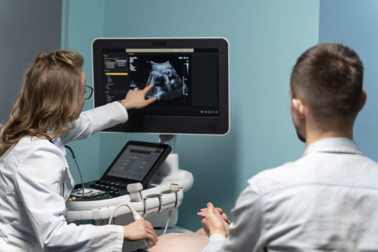
Pregnancy, a miraculous journey, unveils its wonders through an ultrasound scan. This non-invasive procedure uses sound waves to create images, offering a glimpse into the womb’s secret world. Typically performed during different trimesters, ultrasound tracks fetal development and ensures a healthy progression. The emotional resonance of seeing your unborn child on the screen enhances the parental bond, turning anticipation into a tangible connection. As the ultrasound captures the rhythmic heartbeat and tiny movements, it becomes a cherished window into the evolving miracle of life, providing reassurance and joy to expectant parents on their path to welcoming a new family member.
While ultrasound scans primarily monitor fetal growth and anatomy, they also play a crucial role in identifying pregnancies at risk for certain genetic disorders. Advanced ultrasound technology enables the detection of physical anomalies or markers associated with conditions like Down syndrome or neural tube defects. By examining features such as facial structure, heart abnormalities, and limb development, doctors can offer insights into potential genetic disorders. While not definitive, these scans contribute valuable information, guiding further diagnostic tests and facilitating informed decision-making for expectant parents. Early detection empowers medical teams to provide specialized care, enhancing the overall management of such affected pregnancies.
Everything you need to know
Ultrasound scans can provide valuable information about fetal development and help identify certain genetic disorder or structural anomalies. However, it’s important to note that ultrasound alone cannot definitively diagnose genetic disorders; additional tests may be required for confirmation.
Some conditions that might be observed or suspected during an ultrasound include the following:
Down syndrome (Trisomy 21): An ultrasound may reveal facial features such as a flattened nasal bridge and small nose, along with potential heart defects. These markers prompt further testing for confirmation. While ultrasound can indicate potential risks, a definitive diagnosis requires genetic testing.
Edwards syndrome (Trisomy 18): Ultrasound findings might include heart and kidney abnormalities, clenched hands, and a small jaw. However, these signs are not exclusive to Trisomy 18, necessitating genetic testing for an accurate diagnosis. The severity of these anomalies can vary, influencing decisions about the pregnancy.
Patau syndrome (Trisomy 13): Ultrasound may detect brain and facial abnormalities, as well as heart defects. However, like other trisomies, genetic testing is crucial for confirmation. Early detection allows for informed decisions regarding medical management and potential interventions.
Turner syndrome: Ultrasound may reveal structural heart defects, kidney abnormalities, or cystic hygromas. However, a definitive diagnosis involves genetic testing. Early identification facilitates appropriate medical care and counseling for families.
Cystic fibrosis: While ultrasound may indicate echogenicity changes in the fetal bowel, genetic testing is necessary for a conclusive diagnosis. This information can guide prenatal and postnatal care strategies for affected infants.
Neural tube defects (e.g., Spina Bifida): Ultrasound may identify spinal cord or brain abnormalities, prompting further evaluation, including fetal neurosonography.
Trisomy 16 Mosaicism: Ultrasound findings may include growth restriction and structural abnormalities. Confirmation through genetic testing is essential to determining the extent of mosaicism and its potential impact on fetal development.
Muscular dystrophy: Ultrasound may detect reduced fetal movements or abnormal muscle development. However, genetic testing is necessary to confirm the specific type of muscular dystrophy and its implications for the child’s health. Early diagnosis enables proactive medical planning and support for affected families.
The integration of ultrasound technology in prenatal care has proven invaluable for comprehending potential genetic disorders. By providing detailed insights into fetal development and detecting subtle anomalies, ultrasound serves as a vital tool for guiding expectant parents and doctors. While it offers valuable clues, it’s important to recognize that ultrasound alone is not a definitive diagnostic tool for genetic disorders; confirmatory testing is often required. Nonetheless, the ability to visualize and understand aspects of fetal health through ultrasound enhances our capacity for early intervention and informed decision-making, fostering a proactive approach to managing genetic concerns during pregnancy. As technology advances, the synergy between ultrasound and genetic testing continues to shape a more comprehensive understanding of the intricate journey from conception to childbirth.
Conclusion
Ambady Scan Centre Thrissur, the best pregnancy scanning centre plays an important role in overseeing pregnancies with a focus on genetic disorders. We also provide different types of scans which provide unparalleled early detection, detailed visualization, and ongoing assessment of the developing fetus.
Utilizing advanced imaging technologies and cutting-edge facilities, Ambady Scan Centre adopts a comprehensive approach to managing expectant mothers at risk of genetic disorders. With a wealth of experience spanning 25 years, the centre excels in genetic screening and upholds a commitment to multidisciplinary collaboration.
Ambady Fetal Maternal Care Center is the pioneer in ensuring optimal care for both mother and baby throughout the pregnancy. Ambady Scan Center remains a leading institution dedicated to the health and well-being of expectant mothers.
1 note
·
View note