
Don't wanna be here? Send us removal request.
Statistics
We looked inside some of the posts by conocimientoglobal and here's what we found interesting.
Average Info
Notes Per Post
106K
Likes Per Post
61K
Reblog Per Post
45K
Reply Per Post
20
Time Between Posts
2 months
Number of Posts By Type
Text
11
Photo
6
Last Seen Tumblr Blogs


Fun Fact
Total funding amounts to $125.3M.
Text
Energia solar: agora é a melhor hora de investir, diz o especialista Amon Mendes
Amon Mendes Franco de Sousa, especialista em energia solar, revela como essa tecnologia está revolucionando o setor energético
A cada dia, mais lares e empresas brasileiras passam a adotar energia solar como principal fonte de abastecimento, aproveitando-se do sol abundante que brilha sobre o país. E segundo Amon Mendes Franco de Sousa, CEO da Soreli Sol Energias, líder em soluções de energia solar no oeste do Paraná, o momento é mais do que propício para aqueles que estão considerando dar o salto para o futuro com a adoção da tecnologia da energia fotovoltaica .
Em entrevista exclusiva, na sede da Soreli Sol Energias em Foz do Iguaçu, Amon explicou sobre o panorama atual do setor e como a energia solar se tornou um investimento ainda mais atrativo nos últimos anos. “Vimos nos últimos tempos uma queda significativa nos preços das placas solares. Essa tendência é resultado de avanços tecnológicos, aumento da escala de produção e maior competição no mercado nacional e internacional”, disse Amon Mendes Franco de Sousa.
6 notes
·
View notes
Text

A shell around the star Tiaki (β Gruis) // Marcel Drechsler
Note: the image above is a combination of optical and ultraviolet light.
The name Tiaki comes from the Tuamotoan word Tiaki meaning "to care, guard, or protect"
383 notes
·
View notes
Text

"Space Settlement" by John Berkey, 1988.
382 notes
·
View notes
Text
New data from NASA’s James Webb Space Telescope has revealed that K2-18 b, an exoplanet nearly nine times the size of Earth, could be an example of a theorized class of planets with hydrogen-rich atmospheres, vast liquid oceans and the potential to support life. Observations taken by the telescope detected the presence of carbon-bearing molecules such as methane and carbon-dioxide, according to a NASA press release published on Monday. Astronomers also picked up signs of a molecule called dimethyl sulfide (DMS) – which, on Earth, is only produced by living things. K2-18 b is an exoplanet orbiting a dwarf star roughly 120 light years away from Earth in the constellation of Leo. It is categorized as a sub-Neptune, a class of planets that are between the size of Earth and Neptune and common in the universe, although not in our solar system. Scientists have known about K2-18 b for a while, with initial observations taken by NASA’s Hubble Space Telescope, but these new observations taken with the James Webb Telescope have provided details that astronomers had previously only wondered about. "Our findings underscore the importance of considering diverse habitable environments in the search for life elsewhere," Nikku Madhusudhan, an astronomer at the University of Cambridge and lead author of a paper announcing these results, said in the release. "Traditionally, the search for life on exoplanets has focused primarily on smaller rocky planets, but the larger Hycean worlds are significantly more conducive to atmospheric observations."
Continue Reading
463 notes
·
View notes
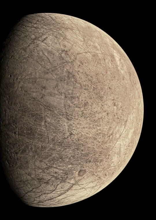
Text
NASA has released new images of Jupiter's moon Europa captured by the Juno spacecraft.
4K notes
·
View notes
Text
[OC] Most Frequently Found Geologic Age of Fossil in Each US State
3 notes
·
View notes
Photo

Pyrrole chemistry: Good things come in threes
A new approach leads to the long-awaited formation of rings made of three pyrroles, which could be used to produce compounds with a host of interesting properties, and explains why they have not been observed before.
Hokkaido University chemists have answered a long-standing question: Why do pyrrole monomers form ring-shaped compounds comprising four but never three pyrroles? Their findings, published in the Journal of the American Chemical Society, could enable the development of new compounds with unique properties.
Pyrrole is an aromatic organic compound made of carbon, hydrogen and nitrogen atoms. When four pyrrole molecules combine, they form ring-shaped tetrapyrrole macrocycles, which are found in nature in many forms—including heme and chlorophyll—and play critical biological roles in oxygen transport, light-harvesting and electron transport. Scientists have found ways to fabricate synthetic tetrapyrrole macrocycles for use as catalysts, photosensitizers and optoelectronic materials. But they have long wondered why they never see tripyrrolic macrocycles.
Read more.
27 notes
·
View notes
Photo

Withdrawal From Psychostimulants Restructures Functional Architecture of Brain
Withdrawal from psychostimulants including methamphetamines, cocaine, and nicotine, produced restructuring of brain regions and major increases in functional connectivity, a new mouse study reveals.
44 notes
·
View notes
Photo

Towards more energy-efficient 2D semiconductor devices
According to researchers from the Singapore University of Technology and Design (SUTD), a recently discovered family of two-dimensional (2D) semiconductors could pave the way for high-performance and energy-efficient electronics. Their findings, published in npj 2D Materials and Applications, may lead to the fabrication of semiconductor devices applicable in mainstream electronics and optoelectronics—and even potentially replace silicon-based device technology altogether.
In the quest of miniaturizing electronic devices, one well-known trend is Moore’s law, which describes how the number of components in the integrated circuits of computers doubles every two years. This trend is possible thanks to the ever-decreasing size of transistors, some of which are so small that millions of them can be crammed onto a chip the size of a fingernail. But as this trend continues, engineers are starting to grapple with the inherent material limitations of silicon-based device technology.
“Due to the quantum tunneling effect, shrinking a silicon-based transistor too small will lead to highly uncontrollable device behaviors,” said SUTD Assistant Professor Ang Yee Sin, who led the study. “People are now looking for new materials beyond the ‘silicon era’, and 2D semiconductors are a promising candidate.”
Read more.
49 notes
·
View notes
Text

Food for Thought: New Maps Reveal How Brains are Kept Nourished
Our brains are non-stop consumers. A labyrinth of blood vessels, stacked end-to-end comparable in length to the distance from San Diego to Berkeley, ensures a continuous flow of oxygen and sugar to keep our brains functioning at peak levels.
But how does this intricate system ensure that more active parts of the brain receive enough nourishment versus less demanding areas? That’s a century-old problem in neuroscience that scientists at the University of California San Diego have helped answer in a newly published study.
Studying the brains of mice, a team of researchers led by Xiang Ji, David Kleinfeld and their colleagues has deciphered the question of brain energy consumption and blood vessel density through newly developed maps that detail brain wiring to a resolution finer than a millionth of a meter, or one-hundredth of the thickness of a human hair.
A result of work at the crossroads of biology and physics, the new maps provide novel insights into these “microvessels” and their various functions in blood supply chains. The techniques and technologies underlying the results are described March 2 in the journal Neuron.
“We developed an experimental and computational pipeline to label, image and reconstruct the microvascular system in whole mouse brains with unprecedented completeness and precision,” said Kleinfeld, a professor in the UC San Diego Department of Physics (Division of Physical Sciences) and Section of Neurobiology (Division of Biological Sciences). Kleinfeld says the effort was akin to reverse engineering nature. “This allowed Xiang to carry out sophisticated calculations that not just related brain energy use to vessel density, but also predicted a tipping point between the loss of brain capillaries and a sudden drop in brain health.”
Questions surrounding how blood vessels carry nourishment to active and less active regions were posed as a general issue in physiology as far back as 1920. By the 1980s, a technology known as autoradiography, the predecessor of modern-day positron emission tomography (PET), allowed scientists to measure the distribution of sugar metabolism across the mouse brain.
To fully grasp and solve the problem, Ji, Kleinfeld and their colleagues at the Howard Hughes Medical Institute’s Janelia Research Campus and UC San Diego Jacobs School of Engineering filled 99.9 percent of the vessels in the mouse brain—a count of nearly 6.5 million—with a dye-labeled gel. They then imaged the full extent of the brain with sub-micrometer precision. This resulted in fifteen trillion voxels, or individual volumetric elements, per brain, that were transformed into a digital vascular network that could be analyzed with the tools of data science.
With their new maps in hand, the researchers determined that the concentration of oxygen is roughly the same in every region of the brain. But they found that small blood vessels are the key components that compensate for varying energy requirements. For example, white matter tracts, which transfer nerve impulses across the two brain hemispheres and to the spinal cord, are regions of low energy needs. The researchers identified lower levels of blood vessels there. By contrast, brain regions that coordinate the perception of sound use three times more energy and, they discovered, were found with a much greater level of blood vessel density.
“In the era of increasing complexities being unraveled in biological systems, it is fascinating to observe the emergence of shared simple and quantitative design rules underlaying the seemingly complicated networks across mammalian brains,” said Ji, a graduate student in physics.
Up next, the researchers hope to drill down into the finer aspects of their new maps to determine the detailed patterns of blood flow into and out from the entire brain. They will also pursue the largely uncharted relationship between the brain and the immune system.
117 notes
·
View notes

Text
Hydrodynamic Spin Lattices

Droplets bouncing on a fluid bath display some strikingly quantum-like behaviors thanks to the interactions between a drop and its guiding surface wave. Here, researchers use submerged wells beneath the drop to confine each droplet into a space where it bounces in a clockwise or anticlockwise trajectory. (Image and research credit: P. Saenz et al.; via Nature; submitted by Kam-Yung Soh) Read the full article
173 notes
·
View notes
Text
Candy Clouds Mid-Storm

There’s nothing quite like a towering storm cloud to showcase nature’s power. This gorgeous photo by Laura Rowe shows pastel clouds over West Texas in the middle of a thunderstorm. (Image credit: L. Rowe; via Colossal) Read the full article
2K notes
·
View notes
Photo

New EU ‘right to repair’ laws require technology to last for a decade - New devices will also have to come with repair manuals and be made in such a way that they can be dismantled using conventional tools https://ift.tt/2OerAfz
121 notes
·
View notes
Photo

(Image caption: Synchronized neurons strengthen emotional memories. In the image, maps show synchronized neurons during memory creation (left) and memory retrieval (right). Credit: René Hen / Columbia University Irving Medical Center)
Why Are Memories Attached to Emotions So Strong?
Memories linked with strong emotions often become seared in the brain.
Most people can remember where they were on 9/11, or what the weather was like on the day their first child was born. Memories about world events on Sept, 10, or lunch last Tuesday, have long been erased.
Why are memories attached to emotions so strong?
“It makes sense we don’t remember everything,” says René Hen, PhD, professor of psychiatry and neuroscience at Columbia University Vagelos College of Physicians and Surgeons. “We have limited brain power. We only need to remember what’s important for our future well-being.”
Fear, in this context, is not just a momentary feeling but a learning experience critical to our survival. When a new situation makes us fearful, the brain records the details in our neurons to help us avoid similar situations in the future or use appropriate caution.
What’s still a mystery is why these memories, recorded by the brain’s hippocampus, become so strong.
To find out, Hen and Jessica Jimenez, an MD/PhD student at Columbia, placed mice into new, frightening environments and recorded the activity of hippocampal neurons that reach out to the brain’s fear center (the amygdala). The neurons’ activity was also recorded a day later when the mice tried to retrieve memories of the experience.
Unsurprisingly, neurons that respond to the frightening environment send that information to the brain’s fear center.
“What was surprising was that these neurons were synchronized when the mouse later recalled the memory,” Hen says.
“We saw that it’s the synchrony that is critical to establish the fear memory, and the greater the synchrony, the stronger the memory,” Jimenez adds. “These are the types of mechanisms that explain why you remember salient events.”
How and when synchronization occurs is still unknown, but the answer could reveal the inner workings of the brain that create lifelong memories and lead to new treatments for posttraumatic stress disorder.
“In people with PTSD, many similar events remind them of the original frightening situation,” Hen says, “and it’s possible that synchronization of their neurons has become too strong. We’re really trying to dig into the mechanisms of how emotional memories form to find better treatments for people with PTSD and memory disorders in general.”
174 notes
·
View notes