
frenovo
FRENOVO
Frenovo Biotech is committed to providing consistent high-quality raw materials and practical technical solutions for global researchers and manufacturers in the life science and in vitro diagnostic field, especially the rapid diagnostic field. Through years of efforts on numerous exploratory experiments and mass production practice, we understand profoundly that to make a world-class high-quality diagnostic test kit, choosing the proper equipment, components and reagent materials are the fundamental factors. https://www.frenovobio.com
8 posts
Don't wanna be here? Send us removal request.
Last Seen Blogs

induskart1
Induskart Engitech

onenakedfarmer
1NF / One Naked Farmer


obatantiubangreenanglecia-blog
Obat Anti Uban Green Angelica

cuvepaciw
Untitled
Text
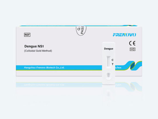
Dengue NS1 Antigen Rapid Test
Dengue NS1 Ag rapid test is an in-vitro immunochromatographic, one-step assay designed for the qualitative determination of dengue virus NS1 antigen in human serum, plasma, or whole blood for the diagnosis of early acute dengue infection. This test device contains a membrane strip, which is pre-coated with anti-dengue NS1 Ag capture on the test band region. The anti-dengue NS1 Ag-colloidal gold conjugate and serum, plasma or whole blood sample move along the membrane chromatographically to the test region and forms a visible line as the antibody-antigen-antibody gold particle complex forms.
Summary and Explanation of Dengue NS1 Antigen Rapid Test
Dengue is an acute vial disease, which is transmitted by Aedes aegypti mosquitoes. Dengue is characterized clinically by biphasic fever, rash and hematopoietic depression, and by constitutional symptoms such as malaise, arthralgia, myalgia and headache. Infrequently, more severe disease is seen, manifested by hemorrhage fever which may progress to lethal shock. It isendemic in the tropics and subtropics, worldwide, where an estimated 100,000,000 cases occur annually. It has been estimated that about 50 to 100 million cases of Dengue Fever (DF) occur every year with about 250,000 to 500,000 cases, of Dengue Hemorrhagic Fever (DHF).
During 2002, more than 30 Latin American countries reported over 10,000,000 (DF) cases with large number of DHF cases. This has been followed by extensive epidemic of DHF in in several parts of India during 2003 through 2005. In the Americas, the reported incidence has more than tripled from 1996 to 2002. The incidence of Dengue outbreak has been reported in Hawaii, and in Laredo Texas. The potential for the virus to cause a severe disease has also resulted in the inclusion of this pathogen on the CDC "Category A" list for potential biological warfare and bioterrorism agents. Dengue NS1 (non-structural) protein is a multimeric secreted protein that is believed to play a role in viral RNA replication. It is strongly immunogenic eliciting antibodies with complement fixing activity. NS1 antigen can be detected in circulating blood during acute Dengue infection. The Dengue NS1 RapidDip Test detects NS1 antigen in serum samples following infection.
Principle of Dengue NS1 Antigen Rapid Test
The Dengue NS1 Rapid Test is a qualitative, membrane based immunoassay for the detection of NS1 antigen in human serum.
The rapid test membrane is pre-coated with an NS1 specific antibody on the test line region and utilizes a separate control to assure assay flow and performance. During testing, the test sample is added directly to the sample region and the test is placed into a well containing 3 drops of buffer. The buffer and serum mix and interact with NS1-specific monoclonal antibodies conjugated to gold nanoparticles. The solution migrates upward on the membrane (via capillary action) to react with the anti-NS1 antibody on the membrane. If NS1 antigen is present, a red line will appear at the test line.
The red line at the control region should always appear if the assay is performed correctly. The presence of this red line verifies that proper flow has occurred and catastrophic failure of the conjugate has not occurred. The entire procedure takes approximately 30 minutes.
0 notes
Text
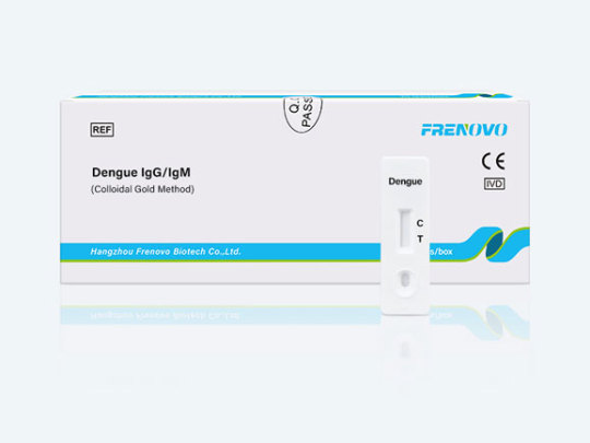
Dengue IgG/IgM Rapid Test
Intended Use
The dengue fever ELISA test is a qualitative enzyme immunoassay for the detection of antibodies to dengue, in samples of human serum or plasma. This test is intended to be performed by trained medical technologists only.
Assay Principle
The microwells are coated with purified dengue virus antigen from cell-cultured type 1-4 dengue. During the first incubation with the diluted patients sera, any antibodies which are reactive with the antigen will bind to the coated wells. After washing to remove the rest of the sample, the Enzyme Conjugate is added. If antibodies have been bound to the wells, the Enzyme Conjugate will then bind to these antibodies. After another series of washes, a chromogen (tetramethylbenzidine or TMB) is added. If the Enzyme Conjugate is present, the peroxidase will catalyze a reaction that consumes the peroxide and turns the chromogen from clear to blue. The addition of the Stop Solution ends the reaction and turns the blue color to a bright yellow color. The reaction may then be read visually or with an ELISA reader.
Intended Use of Dengue IgG/IgM Rapid Test
Dengue Fever Rapid IgG/IgM is an immunochromatographic assay designed for the qualitative detection and differentiation of specific IgM and IgG antibodies to dengue virus in human serum or plasma. It is intended to be used as in vitro diagnostic of dengue fever. The test provides a differential detection of anti-dengue IgM and anti-dengue-IgG antibodies and can be used for the presumptive distinction between a primary and secondary dengue infection. The results obtained should not be the sole determinant for clinical decision.
Summary And Explanation of Dengue IgG/IgM Rapid Test
Dengue virus, a virus belonging to the Flavavirus group of viruses, is one of the most significant mosquito-borne diseases in the world in terms of morbidity and mortality. Transmitted principally by the mosquito types Aedes aegypti and Aedes albopictus, the virus is found commonly throughout the tropic and sub-tropic regions of the world. There are four known serotypes of dengue. Symptoms of dengue fever include high fever, headache, muscle pain and skin rash. The complications often associated with this infection are dengue hemorrhagic fever or dengue shock syndrome.
Principle of Dengue IgG/IgM Rapid Test
The Dengue Fever Rapid IgG/IgM is an indirect solid-phase immunochromatographic assay. Serum or plasma samples may be used with this test.
When a specimen is added to the test device, IgG and IgM antibodies in the specimen sample, if present, will react with particles coated with dengue envelope proteins to form a complex. As this complex migrates along the length of the cellulose nitrate membrane, the anti-dengue IgG or IgM antibody particle complex is captured by the relevant IgG and/or IgM test bands located in the test device window causing a pale to dark pink purplish band to form at the IgG or IgM region of the test device window.
The intensity of the bands will vary depending upon the amount of antibody present in the sample. The appearance of any color in a specific test region (IgG or IgM) should be considered as positive for that particular antibody type (IgG or IgM). A pink-purplish procedural control band should always develop in the test device window to indicate that the test has been performed properly.
0 notes
Text
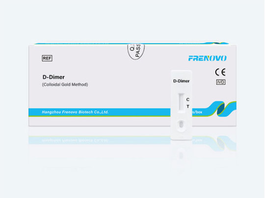
D-Dimer Antibody Rapid Test
D-dimer assays use mono- or polyspecific antibodies against D-dimer to provide quantitative or qualitative data on the concentration of D-dimer in whole blood or plasma. D-dimer is the product of lysis of cross-linked fibrin and the levels of D-dimer are increased in patients with acute VTE. However, the test is nonspecific because the level of D-dimer can be increased in a variety of other conditions, including malignancy, inflammatory conditions, and infections. Therefore the D-dimer assay is most useful as a tool to rule out suspected DVT.
What Happens During a D-dimer Test?
A health care professional will take a blood sample from a vein in your arm, using a small needle. After the needle is inserted, a small amount of blood will be collected into a test tube or vial. You may feel a little sting when the needle goes in or out. This usually takes less than five minutes.
Are There Any Risks to a D-dimer Test?
There is very little risk to having a blood test. You may have slight pain or bruising at the spot where the needle was put in, but most symptoms go away quickly.
0 notes
Text
D-Dimer Antibody
dimer test is often used to diagnose disseminated intravascular coagulation (DIC), deep vein thrombosis (DVT), pulmonary embolism, myocardial infarction, and cerebral infarction. This index can be used not only for the diagnosis of thrombotic diseases but also for the dose monitoring and efficacy observation of thrombolytic drugs.
Specification of D-Dimer Antibody
Description Type Item NO Source Application Recommend to use for
D-Dimer-1A4 mAb GHMA096-1 mouse EIA, LF, Fluorescent Labelling/Coating
D-Dimer-4E6 mAb GHMA096-2 mouse EIA, LF, Fluorescent Labelling/Coating
0 notes
Text
Cytomegalovirus (CMV) lgG/IgM Rapid Test
CMV infections are widespread and usually asymptomatic; however, severe infections can be seen in newborns and immunocompromised individuals. This virus may also persist as a latent or chronic infection.
Introduction of Cytomegalovirus (CMV) lgG/IgM Rapid Test
Cytomegalovirus (CMV) infections are widespread and usually asymptomatic; however, the virus may persist as a latent or chronic infection. The relatively frequent incidence and severe disease in newborns and immunosuppressed individuals clearly establishes this agent as an important human pathogen. CMV infection can be classified as Congenital (Acquired before birth), Perinatal (Acquired at birth) and Postnatal (Acquired after birth). The prognosis for congenitally infected infants who are asymptomatic at birth must be guarded. Ten to 25% may subsequently develop hearing loss. Five to 10% may exhibit various degrees of mental retardation and central nervous system motor disorders. Surveys show the incidence of congenital CMV infection to be from 0.5 to 2.5 %. Consequently, a careful documentation of the long-term effects of intrauterine infection is important.
Although the age at which CMV infection is acquired varies with socioeconomic condition, only about 10-15% children in the United States are seropositive. By the age 35 however, about 50% of the population is seropositive. The majority of individuals contracting postnatal CMV infections remain asymptomatic. A small percentage of individuals will develop a negative heterophile-antibody infectious mononucleosis syndrome. In immunocompromised patients CMV infections happen frequently, often from reactivation of latent infection, and may life-threatening. Antibody of the IgM class is produced during the first 2-3 weeks of infection with CMV and exists only transiently in most patients. Serologic procedures which measure the presence of IgM and IgG antibodies help discriminate between primary and recurrent infections since IgM antibodies are rarely found in recurrent infections.
Principle of Cytomegalovirus (CMV) lgG/IgM Rapid Test
CMV IgG/IgM test device has 3 pre-coated lines, "G" (CMV IgG Test Line), "M" (CMV IgM Test Line), and "C" (Control Line) on the surface of the membrane. All three lines in result window are not visible before applying any samples. The "Control Line" is used for procedural control. Control line should always appear if the test procedure is performed properly and the test reagents of control line are working. A purple "G" and "M" lines will be visible in the result window if there are enough IgG and/or IgM antibodies to CMV in the sample. If IgG and/or IgM antibodies to Cytomegalo viurs are not present in the sample, there is no color appearance in "G" and/or "M".
0 notes
Text
C-reactive Protein (Semiquantitative) Antibody Rapid Test
C-reactive protein (CRP) is an ancient highly conserved molecule and a member of the pentraxin family of proteins. CRP is secreted by the liver in response to a variety of inflammatory cytokines. Levels of CRP increase very rapidly in response to trauma, inflammation, and infection and decrease just as rapidly with the resolution of the condition. Thus, the measurement of CRP is widely used to monitor various inflammatory states. CRP binds to damaged tissue, to nuclear antigens and to certain pathogenic organisms in a calcium-dependent manner.
The function of CRP is felt to be related to its role in the innate immune system. Similar to immunoglobulin (Ig)G, it activates complement, binds to Fc receptors and acts as an opsonin for various pathogens. Interaction of CRP with Fc receptors leads to the generation of pro-inflammatory cytokines that enhance the inflammatory response. Unlike IgG, which specifically recognizes distinct antigenic epitopes, CRP recognizes altered self and foreign molecules based on pattern recognition. Thus, CRP is though to act as a surveillance molecule for altered self and certain pathogens. This recognition provides early defense and leads to a proinflammatory signal and activation of the humoural, adaptive immune system.
Principle of CRP Test
The C-Reactive Protein test is based on the principle of the latex agglutination. When latex particles complexed human anti-CRP are mixed with a patient’s serum containing C reactive proteins, an visible agglutination reaction will take place within 2 minutes.
Uses of CRP Test
CRP may be used to detect or monitor significant inflammation in an individual who is suspected of having an acute condition, such as serious bacterial infection like sepsis, a fungal infection and Pelvic inflammatory disease (PID).
The CRP test is useful in monitoring people with chronic inflammatory conditions to detect flare-ups and/or to determine if treatment is effective. Some examples include Inflammatory bowel disease, some forms of arthritis and Autoimmune diseases, such as lupus or vasculitis.
The determination of the CRP-level is useful to monitor the therapy.
It is done to check for infection after surgery. CRP levels normally rise within 2 to 6 hours of surgery and then go down by the third day after surgery. If CRP levels stay elevated 3 days after surgery, an infection may be present.
0 notes
Text
Fertility Rapid Test
Fertility testing involves both partners. While we may think of pregnancy as happening in the woman's body, conception takes two.
According to the American Society of Reproductive Medicine (ASRM), 25% of all infertile cases have one or more contributing infertility factors. For instance, the ASRM reports that 40% of infertility cases are due to male factor infertility while 25% of female infertility is due to abnormal ovulation.Not every fertility test will be done for every case. The more invasive fertility tests, like diagnostic laparoscopy, are only done when symptoms or other tests point in that direction, or when a cause for infertility cannot be found otherwise.
Types of Fertility Rapid Test
Project Sample Format
HCG Pregnancy Antigen Rapid Test Urine Strip / Device / Uncut Sheet
LH Ovulation Antigen Rapid Test Urine Strip / Device / Uncut Sheet

1 note
·
View note
Text
COVID-19 Test Products
Early testing and detection is very important
As some people infected with COVID-19 only have mild symptoms, members of the public are advised to seek medical advice promptly when feeling unwell (even if having very mild symptoms only) and get tested under the instructions of a healthcare worker, so that appropriate diagnosis and management can be given at the earliest possible time.
Types of COVID-19 Test Products
Project Sample Format
COVID-19 Ag Test Nasal Cassette/Strip/Uncut Sheet
COVID-19 IgG/IGM Test WB/S/P Cassette/Strip/Uncut Sheet
COVID-19 Ag Saliva Test Saliva Cassette/Strip/Uncut Sheet
COVID-19 Neutralization Antibody Test WB/S/P Cassette/Strip/Uncut Sheet

1 note
·
View note