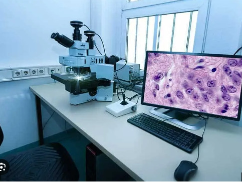
Text

immunohistochemistry cancer|IHC-PRS
#immunohistochemistry cancer#pathology slides#ihcstaining#digital pathology#immunohistochemical staining
0 notes
Text

digital pathology scanner|IHC-PRS
#digital pathology scanners#ihcstaining#immunohistochemistry protocol#digital slide scanners#immunohistochemistry antibodies
0 notes
Text

Digital pathology-ihc-prs
#Digital pathology#digital pathology scanner#pathology slides#ihcstaining#immunohistochemistry cancer
0 notes
Text

immunohistochemistry protocol-ihc-prs
#immunohistochemistry protocol#ihc staining#pathology slides#digital slide scanners#digital pathology scanner
0 notes
Text
Unlocking the Power of Immunohistochemical Staining: A Deep Dive into Pathology Slides
Immunohistochemical staining (IHC) is a crucial technique in the field of pathology, offering invaluable insights through pathology slides. This method allows for the detection and visualization of specific proteins within tissue samples, providing a detailed understanding of disease mechanisms.

IHC involves using antibodies to target specific antigens in tissue sections. When these antibodies bind to their targets, they produce a detectable signal, usually visualized under a microscope. Pathology slides treated with Immunohistochemical Staining can reveal the presence, localization, and concentration of proteins, which is critical for accurate diagnosis and treatment planning.
The application of IHC-PRS (Immunohistochemical Staining - Pathology Slide) is particularly significant in oncology, where it helps identify tumor markers and guide therapeutic decisions. By analyzing pathology slides with IHC, pathologists can differentiate between various types of cancers, assess their aggressiveness, and predict patient outcomes.

Moreover, immunohistochemical staining is not limited to oncology; it is also instrumental in research and diagnostics across various medical fields. It provides a deeper understanding of cellular processes and disease pathology, aiding in the development of targeted therapies.
In summary, immunohistochemical staining of Pathology Slides, or IHC-PRS, is an indispensable tool in modern medicine. Its ability to provide precise information at the molecular level enhances diagnostic accuracy and contributes to more effective patient management. Embracing IHC-PRS in pathology ensures a more detailed and informed approach to diagnosing and treating diseases.
#immunohistochemical staining#pathology slides#immunohistochemistry protocol#digital pathology scanner#ihc staining
0 notes
Text

digital slide scanners - IHC-PRS
Landing Med's portable cytology and histology digital pathology scanners boost diagnostic efficiency and confidence for early-stage tumor diagnosis.
0 notes
Text

immunohistochemical staining|IHC-PRS
#immunohistochemical staining#ihc staining#digital pathology scanner#digital pathology#immunohistochemistry antibodies
0 notes
Text

Digital pathology-ihc-prs
#digital pathology#pathology slides#digital slide scanners#ihc staining#immunohistochemical staining
0 notes
Text

digital pathology scanner|IHC-PRS
#digital pathology scanners#immunohistochemistry protocol#ihc staining#digital slide scanners#immunohistochemical staining
0 notes
Text

immunohistochemistry cancer|IHC-PRS
#immunohistochemistry cancer#pathology slides#digital pathology scanner#ihc staining#digital pathology
0 notes
Text

Digital pathology scanner - IHC-PRS
Landing Med's portable cytology and histology digital pathology scanners boost diagnostic efficiency and confidence for early-stage tumor diagnosis.
0 notes
Text
The Role of Immunohistochemistry in Cancer Diagnosis: A Step-by-Step Protocol
Immunohistochemistry (IHC) is an essential technique in cancer diagnosis, providing crucial insights into the molecular characteristics of tumors. This powerful tool helps pathologists identify specific antigens in tissue samples, offering precise information that guides treatment decisions.
Immunohistochemistry Protocol: A Detailed Overview
The IHC protocol begins with the preparation of tissue sections, typically from a biopsy. These sections are carefully mounted on glass slides and fixed to preserve the cellular structure. Following fixation, the tissue is treated with a blocking solution to prevent non-specific binding of antibodies, ensuring that the staining is accurate and specific.

Next, the primary antibody is applied. This antibody is designed to bind specifically to the antigen of interest, such as a cancer-related protein. After incubation, a secondary antibody, which is linked to a detectable marker (often an enzyme or a fluorescent dye), is added. This secondary antibody binds to the primary antibody, amplifying the signal.
The final step in the immunohistochemistry protocol involves the visualization of the antigen-antibody complex. This is typically achieved through the application of a substrate that reacts with the marker on the secondary antibody, producing a visible color change in the tissue. Pathologists then examine the stained slides under a microscope, identifying the presence and intensity of the antigen.

Immunohistochemistry in Cancer Diagnosis
In cancer diagnosis, immunohistochemistry is invaluable. It allows for the detection of specific cancer markers, such as HER2 in breast cancer or PD-L1 in lung cancer. These markers provide vital information about the type of cancer and its likely behavior, enabling tailored treatment strategies.
By following a meticulous immunohistochemistry protocol, pathologists can deliver accurate and detailed cancer diagnoses, significantly improving patient outcomes.
0 notes
Text

immunohistochemistry cancer|IHC-PRS
#immunohistochemistry cancer#pathology slides#ihcstaining#digital pathology#immunohistochemistry antibodies
0 notes
Text

digital pathology scanner|IHC-PRS
0 notes
Text

Digital pathology-ihc-prs
#digital pathology#pathology slides#ihcstaining#immunohistochemistry protocol#immunohistochemistry cancer
0 notes
Text

immunohistochemistry protocol-ihc-prs
0 notes
Text

Immunohistochemistry procedure - IHC-PRS
We provide all primary antibodies for your immunohistochemistry procedure requirements at 50% of your purchase cost, with maintaining equal or superior affinity
0 notes