Journal of Dairy & Veterinary sciences is a part of the Juniper publishers which publishes subject-specific articles in the field of Dairy and Veterinary Sciences
Don't wanna be here? Send us removal request.
Statistics
We looked inside some of the posts by juniperpublishers-jdvs and here's what we found interesting.
Average Info
Notes Per Post
2
Likes Per Post
2
Reblog Per Post
0
Reply Per Post
0
Time Between Posts
6 days
Number of Posts By Type
Text
17
Last Seen Tumblr Blogs


Fun Fact
Tumblr.com is the 103rd most visited website in the world.
Text
Effect of Melatonin and Vitamin E on SOD-Cu / Zn Levels and Membrane Permeability During Weed Semen Conservation

Abstract
In species such as the hair, oxidative stress is related to sperm damage during sperm conservation, which affects its fecundity. This research aimed to determine the effect of the addition of melatonin and vitamin E on the levels of Dismutasa-Cu / Zn Superoxide (SOD-Cu / Zn) during the conservation of diluted bristle semen. Seminal maestras of 7 to 18 months old verracos were collected. The optimal parameters are diluted and divided into aliquots: control (with diluent), control with vehicle (diluent and ethanol) and semen with melatonin (1.25 mM) and vitamin E (1 mM. at 16 °C, if enzyme activity (SOD-Cu / Zn), with commercial kit (Cayman®-Chemical Company Ann Arbor, MI, USA) and Hypo-osmotic Test (HOST +), to evaluate the integrity of Plasma membrane Results were analyzed by ANOVA, with SPSS 15.0 statistical package for Windows with p≤0.05 If SOD-Cu / Zn was observed in melatonin-based women, but significantly (p <0.05) higher rates and increased percentage of HOST + sperm, the conservation day with respect to the different groups Melatonin has an antioxidant effect on the conservation of seminal hair at 16 °C with an increase in enzymatic activity of CuZnSOD, which improve the seminal quality to increase the of the plasma membrane (HOST +) in sperm cells.
Keywords: Semen; Conservation; Cerdo; Radicales libre
Introduction
The cryopreservation of semen is one of the most important procedures in the development of biotechnology for assisted reproduction [1], which allows for maximum distribution of germinal material of interest specimens, facilitating the development of genetic improvement programs [2]. However, during the 24-hour cooling of semen, there is an increase in concentrations of reactive oxygen species (ROS) affecting seminal quality (decreased sperm motility and loss of membrane permeability) events. which have been associated with the lipoperoxidation process [3] and antioxidant enzymes such as the Dismutasa Superoxide (SOD) in its three isoforms (SOD-Cu / Zn- the cytosolic SOD, the SOD-Mn, the mitochondrial SOD and the SOD-Cu / Extracellular or secreted zn) [4], have the protective effect during frozen or refrigerated semen storage [5-8], improving sperm motility and membrane integrity [9].
In this sense, in recent years there have been studies on the use of different antioxidants (melatonin, vitamin C and vitamin E) in seminal diluents to prevent deleterious effect of RL during sperm conservation [10,11]. On the basis of this background, the present research has aimed to determine the effect of adding melatonin and vitamin E on the activity of SOD-Cu / Zn during the conservation of diluted bristle semen.
Materials and Methods
Población y muestra: The study was carried out in UCLA (Barquisimeto, Venezuela). The 20 weeks of semen for the studio were taken from 7-18-month-old Landrace, Large White and Duroc verrace warts from the Porcine Inversion Breeders’ farm, located in the Yaritagua-Barquisimeto range, state Yaracuy-Venezuela. Breeding males selected as donors of semen and healthy animals, maintained in optimum health conditions, in a clean and balanced nutrition habitat, established in the Code of Ethics for the Life of the Bolivarian Republic of Venezuela (2010) in their second part chapter 3 [12]. Makes a big deal. The collection of the seminal seminal is realized by means of penean massage with the manguado in a suitable foal for such purpose. The ability to obtain experimental trials has been analyzed in the farm laboratory and those that meet the seminal selection criteria for artificial insemination programs have been diluted for conservation at 16 °C with commercial dilution for bristle semen (MR -A®), taking into account the volume and the optical density of the eyelid.
Handling of the Master
From each selected sample randomly, the first day (day 0) prepared 3 5ml aliquots, which were labeled control (diluent soil MR-A®), vehicle control (ethanol), and aliquot with melatonin (1.25 mM (Sigma®)) and 1 mM vitamin E (Sigma-Aldrich), added at a ratio of 20μl per ml of diluted semen
Dismutasa Superoxide Enzyme (SOD)
For the determination of the enzyme Dismutas-Cu/Zn Superoxide enzyme (SOD-Cu / Zn) using a commercial kit (Cayman®-Chemical Company Ann Arbor, MI, USA) For the preparation of the enzyme If 1.5 ml of diluted semen were taken into an Eppendorf® tube and 2 ml of lysis buffer (20mM tris base, 1 mM EDTA, 210mM mannitol and 70 mM sucrose at pH 7) were added. 2) Chilled by a large grain and mixed in a Thermoline® Model 376000 vortex for minutes to centrifuge at 1500 X g for 10 minutes at 4 °C in the microcentrifuge. The supernatant obtained was used to quantify SOD-CuZn by means of Sunrise model ELISA, Tecan® model 0,3360093, Austria) at 450nm
Hypo-osmotic test (HOST +)
To carry out the preparation, a hypoosmotic solution (100mosmol/L) with 490 mg sodium citrate and 900 mg fructose per 100mL distilled water is prepared. From this solution 100μL were taken for each 25μL of diluted semen seed and incubated at 37 °C for 30 minutes. If those sperm are considered positive in those which are observed any degree of helical torsion of the glue (“swelling”), counting 100 sperm for each month.
Results
In order to evaluate the SOD-Cu/Zn in melatonin monuments it is observed significantly (p <0.05) the highest conservation day with respect to the different groups. This difference was maintained only on day 1 (p <0.05) with the control group and vitamin E, the latter being the one presenting the poorest enzymatic activity (Figure 1 & Graphic 1). SOD-Cu/Zn levels in diluted semen of 7-19-month-old bristles, day 0 and 1 of preservation at 16 °C with melatonin (1.25 mM) and vitamin E (1mM). *Significant differences in semen with melatonin with respect to other controls, vehicle and vitamin E day 0, and significant differences with other controls and with vitamin E day 1 of conservation. ANOVA (p <0.05).
Hypo-osmotic test (HOST +) As far as the integrity of the membrane (HOST +) is concerned, the melatonin changes present a significant increase in the HOST + sperm count with respect to all groups, the conservation day; it differs from conservation day 1 in a group with vitamin E, which shows poorer HOST + percentages, even because of the lack of control (Figure 2). Figure 2 Membrane integrity (positive HOST) in diluted semen of 7-19-month-old hairs, day 0 and 1 of preservation at 16 °C with melatonin (1.25 mM) and vitamin E (1mM). *Significant differences in semen with melatonin and conservation day 0 with respect to control, vehicle and vitamin E, and conservation day 1 with protein by vitamin E. ANOVA (p <0.05).
Discussion
In this investigation the semen of melatonin semen presents the greatest enzymatic activity SOD-Cu / Zn during the conservation. Similar results will be found in semen of poles and connection to add melatonin during conservation, significantly increasing the activity of endogenous enzymes (peroxidase glutathione (GSHPx), Catalase (CAT) and SOD) in post-thawing ( P <0.05) [13,14] and the activity of MnSOD, CuZnSOD and catalyzing correlate significantly with various sperm parameters as progressive movement [15], which underlies their possible role in the etiology of various sperm anomalies [16].
In the case of vitamin E, it has been reported to increase the activity of SOD during conservation with liquid stem semen [17]. There are further investigations into the fact that vitamin E addition has been studied in the preservation of semen, however it has been considered that CuZnSOD activity, despite this, can be used as a predictor of lifetime. in particular, [18] that CuZnSOD is the antioxidant enzyme most affected during the process of seminal cryopreservation [19]. It is therefore important to look for mechanisms to maintain the proper levels of these antioxidant enzymes. On the basis of these results, it can be inferred that melatonin is capable of neutralizing the superoxide radical in semen, which results in the greater enzymatic activity of SODCu / Zn, in addition to vitamin E in the effective display of it. that decreases the activity of the SOD-Cu / Zn, including values by the control group, could be related to the increase in mitochondrial superoxide radical production, specifically at the rate of electron carrier, such as it is the principal source of reactive oxygen species (ROS) [20] associated with alterations of the hierro-SH residues [21] based on the complete II of the electron carrier, altering the mitochondrial structure and functioning [22,23].
Evaluating plasma membrane integrity as a parameter of seminal quality, as being responsible for morphological and functional integrity of sperm [24], changes with melatonin in this investigation will reveal that my membrane integrity protection (HOST +) significant increase in HOST + sperm count, with respect to all conservation groups. This difference was maintained by day 1 of conservation with a group of women with vitamin E, a result that coincides with other investigations into the working of logs [25] and buffalo [26] and shells [13]. In the case of vitamin E the results were similar to those obtained in sheep by Gómez and col. [27] with HOST + percentage included due to other controls. This result is given by the melatonin antioxidant effect and vitamin E prooxidant effect found in the study. Alteration of membrane integrity is related to oxidative stress, disruption of SOD activity [28] causes excess of RL that interact with membrane lipids at the level of unsaturated carbonate [29] , process called lipoperoxidation, which has as a result deep changes affecting the organization and function of the sperm membrane [30,31] what it could take to the cell phone [32].
Conclusion
Melatonin has an antioxidant effect during the preservation of bristle semen at 16 °C with an increase in CuZnSOD enzymatic activity, which increases seminal quality and increases plasma membrane integrity (HOST +) in sperm
To Know More About Journal of Dairy & Veterinary sciences
Please click on: https://juniperpublishers.com/jdvs/index.php
For more Open Access Journals in Juniper Publishers
please click on: https://juniperpublishers.com/index.php
#veterinary science#Veterinary Anatomy#Veterinary Virology#Juniper Publishers#open access publishers
0 notes
Text
Welfare In Dairy Cows - Evaluation Indicators

Abstract
Sustainability, animal welfare, environmental and climate concerns and awareness of social responsibility towards the community have increased consumers interest in knowing how, where and by whom food is produced and handled on its way from the farm to the table. This constitutes a business opportunity for farmers as a growing number of consumers want to buy food, produced locally or regionally directly or under farm certification schemes whereby acceptable animal welfare conditions play often an important role. Farming systems for dairy cows, including housing and management conditions, are important factors affecting their health and other aspects of their welfare, partly through housing and equipment and partly through management and handling practices.
Importance of European Food Safety Authority (EFSA)
The panel on Animal Health and Animal Welfare (AHAW) is a scientific advice on all aspects of animal diseases and welfare of food producing-animals during breeding, rearing, transportation and slaughter. Analysis of the impact that the conditions and treatment of animals can have on both animal and human health. Animal welfare is an important part of EFSA’s remit. The safety of the food chain is indirectly affected by the welfare of animals, particularly those farmed for food production, due to the close links between animal welfare, animal health and food-borne diseases [1,2].
The AHAW Panel has published five scientific opinions and a scientific report on the overall effects of the most relevant farming systems on the welfare of dairy cows and related diseases, assessing the potential impacts of housing, feeding, management and genetic selection [3]. Due to the wealth of data, the experts subdivided the risk assessments into four areas: a. Metabolic and reproductive disorders. b. Udder disorders. c. Leg and locomotion problems. d. Behavioural disorders, fear and pain. Following a request from the European Commission, the AHAW Panel was asked to deliver the first scientific opinion on the welfare of dairy cows, in july 2009 considering whether current farming and husbandry systems comply with the requirements of and welfare of dairy cows from the pathological, zootechnical, physiological and behavioural points of view [4-6]. Later in January 2012 published a new scientific report on welfare in dairy cows, which was one of the most important to increase the monitoring of welfare in the farms: the use of animal-based measures to assess welfare of dairy cows. Other international organisations have also issued recommendations and guidelines concerning animal welfare, such as the World Organisation for Animal Health (OIE) and the Council of Europe. The EU is a signatory to the European Convention for the protection of animals kept for farming purposes, adopted by the Council of Europe [7].
Indicators of Welfare in Dairy Cows
The study of useful variables for evaluating animal welfare in dairy herds has increased considerably in the last years, and a number of indicators are now available which are well documented for being included in animal welfare protocols. However, the protocols that have been proposed and applied until now are costly and difficult to implement, and are starting to be evaluated. There is consensus in the reliability of measurements based directly on the animal as useful indicators, such as body condition, foot diseases, mastitis and other more general indicators, such as infertility and mortality rates obtained from records of dairy farms that can be studied under our production conditions [8].
In order to identify appropriate indicators which address the most important animal welfare problems known from practice, its very important have selected indicators for assessing animal welfare with regard to reliability, validity and practicability. In animal welfare legislation as well as agricultural practice, mainly resource- and management-based animal welfare indicators have been used so far. These describe the conditions, e. g. the space available and the management, which are expected to safeguard animal welfare. However, such resource- and managementbased indicators only allow indirect conclusions to be drawn on how well the animals may fare under these conditions. With the animal-based indicators required in the self-assessment system, on the other hand, the behaviour/health of the animals is recorded directly, so that direct conclusions about their welfare can be drawn. EFSA assessed the welfare risks related to the most commonly used and specialised dairy cows farming systems, integrating the use of animal-based measures to assess their consequences (Table 1).
The livestock keeper should benefit from the on-farm selfassessment. To be able to use the results effectively for planning and implementing improvement measures, the single indicators should not just be assessed, but instead be integrated into more comprehensive management aids. The sets of indicators should therefore also be applied as a whole as far as possible, as with each missing indicator, the informative content is reduced and at the same time the risk increases that animal welfare problems are not recognised. The use of behavioral indicators, health and management in the evaluation of welfare in cows milk is a very valuable tool, considering the difficulty of using physiological and immunological indicators, or drawbacks, such as costs.
To Know More About Journal of Dairy & Veterinary sciences
Please click on: https://juniperpublishers.com/jdvs/index.php
For more Open Access Journals in Juniper Publishers
please click on: https://juniperpublishers.com/index.php
0 notes
Text
Enteritis: Still a Problem in Dairy Calves
Abstract
The neonatal phase of calves is a phase that needs extra care due to newborns’ vulnerability. Enteritis - an inflammation of the intestinal mucosa, resulting mainly in diarrhea - stands out among the conditions that affect animals in this period. Enteritis are responsible for huge losses in cattle breeding, especially in the early stages of rearing. Besides the losses caused by mortality, there are also expenses with veterinarians, treatments and decreased performance of the animal throughout its productive life. The present study aimed to perform a review of diarrhea in newborn calves.
Keywords: Neonatal diarrhea; Infectious agents; Dairy cattle
Abbrevations: ETEC: E. coli enterotoxigenic; EHEC: E. coli enterohemorragic; BVDV: Bovine Viral Diarrhea Virus
Introduction
The neonatal period in cattle - that goes from birth to 28 days of age - is especially important from a health point of view, since approximately 75% of losses in young calves occur in this phase [1], and the first week of life is considered the most critical phase, with 50% of losses. Therefore, maintaining the health of calves is highly related to the hygiene of the place where they live, as they are extremely sensitive to environmental pathogens [2]. Lorenz [3] report that there are several measures to maintain calf health from birth to weaning, including the provision of good quality colostrum in adequate quantity in the first hours after birth and the need to emphasize the prevention of diseases of the gastrointestinal tract and respiratory system. Among the main conditions that cause loss in the early stages of calves development are pneumonia, malformations, central nervous system diseases, and enteritis [4]. Enteritis is clinically mainly manifested by diarrhea and stands out due to its high mortality rate [2,3,5,6], since it is commonly difficult to recover because it is almost always accompanied by malnutrition [7].
Diarrhea is a complex multifactorial disease involving animal, environmental, nutritional, and infectious agents and it is a major cause of mortality, morbidity, and economic loss in cattle worldwide [8], because the treatment of affected calves is slow and impacts on growth, weight gain to weaning and loss of genetic potential of recovered animals [9]. Due its clinical and economic importance and due the preventive measures are often neglected, it is necessary an approach on this subject, to broaden the knowledge and to promote a better conduct regarding the prevention, diagnosis and treatment of the affected animals. Therefore, the present study aimed to review diarrhea in newborn calves.
Diarrhea in Newborn Ruminants
Newborn calf diarrhea is a disease of great impact on the economic viability of cattle herds worldwide [10] (Table 1). The economic impact caused by this condition is significant, although many new intervention strategies, such as vaccine development drug development and herd management, have been developed and implemented to minimize it [2]. In this sense, the veterinarian needs to assess the status of immunoglobulins in calves, feeding, shelter, environmental disinfection, hygiene and sanitary management, to prevent neonatal deaths caused by the disease [11]. The processes involved in the pathophysiology of diarrhea are related to intestinal secretion/ hypersecretion, nutrient bad absorption and digestion, osmolarity, abnormal intestinal motility, increased hydrostatic pressure, and gastrointestinal inflammation [12-21], which may occur singly or, more commonly, by the combination of two or more factors of these mechanisms [22,23].
Secretory diarrheas occur due to abnormal stimuli to the intestinal mucosa crypts that may be caused by the action of enterotoxins and/ or the action of inflammation mediators such as prostaglandins, causing an imbalance in physiological processes, like secretion and intestinal resorption, with consequent diarrhea [24]. Diarrhea is typically profuse without blood or effort, and signs in affected calves include depression, weakness, and sometimes shock and death secondary to hypovolemia and mild acidemia [25]. The difference in osmolarity with increased concentration of solutes within the intestinal lumen, promotes greater absorption of water by the lumen, thus resulting in dehydration of the animal. Osmotic particles include poorly digested disaccharides and increased levels of D-lactate from bacterial fermentation of unabsorbed nutrients entering the colon. Reduced intestinal transit time can lead to poor digestion and malabsorption due to inadequate time for digestion and absorption of ingested food, impaired fluid resorption has a major impact on fluid balance [23].
When a calf has diarrhea, there is a huge loss of fluids and electrolytes from its body. Thus, the consequent dehydration and the appearance of metabolic acidosis are the main causes of death of these animals [26]. This happens partly because the evaluation of the animal is generally based only on clinical examination, and a more detailed approach to assessing the degree of electrolyte disturbance and acidosis through blood gas analysis is lacking or not [27]. Although this condition being common in rural properties, treatment is usually inadequate and / or insufficient, because the administration of antibiotics and anti-inflammatory drugs do not correct the hydroelectrolytic disorders and acid-base [28]. Therefore, in order for the recovering of the animal, these parameters must be measured and corrected quickly, enabling the return to homeostasis. The high frequency and persistence of calf neonatal diarrhea has attracted the interest of many researchers. The multifactorial etiology (bacteria, viruses and protozoa) influenced by nutritional and environmental factors, as well as difficulties in the precise diagnosis of the agent and the failure of treatment has required the adoption of prophylactic measures, such as cow hygiene, management and vaccination [8].
Diarrhea Infectious Agents
Diarrhea is a condition of complex multifactorial etiology, influenced by infectious, nutritional and environmental factors, as well as improper management practices. Causes include toxins, bacteria, protozoa, viruses, and management / environmental factors such as overfeeding, low temperature, poor hygiene, colostrum deprivation, and individual susceptibility of the animal [8]. Numerous infectious agents have been implicated in diarrhea of calves, such as Escherichia coli, Salmonella spp., Cryptosporidium spp., Rotavirus and coronavirus. Coinfection is commonly seen in diarrheal calves, although a single primary pathogen may be the cause in some cases. The non-infectious causes of origin are related to improper management and poor hygiene of the environment in which the animals are placed. The incidence of the disease may vary according to the geographical location of the farms, farm management practices and herd size [2]. Rotaviruses, coronaviruses and cryptosporides, the most commonly recognized enteric pathogens of calves, all produce intestinal villi atrophy, intestinal bacterial overgrowth, malabsorption, and osmotic diarrhea [25].
In general, infections caused by viruses and protozoans tend to damage the intestinal mucosa promoting alteration in intestinal absorption due to damage to intestinal cells, compromising the normal absorption of nutrients, fluids and electrolytes, without alteration in intestinal secretion [22]. Rotaviruses are the most common cause of diarrhea in newborn calves and are often involved in co-infections with other agents [11,23,25]. Clinical signs usually appear 1 to 3 days after infection lasting 5 to 9 days [23]. High environmental contamination, herds with high numbers of animals and management that favors the transmission of the agent, associated with an inexpressive immunization rate, provide favorable conditions for the spread of rotavirus in dairy herds in Brazil, justifying the prevalence and difficulty to control the infection and the spread of the virus [28]. The incidence of many etiological agents varies with the calf’s age (Table 2) and this is useful for establishing the probability of a particular agent being involved and it is generally impossible to establish a definitive field diagnosis [11].
Diarrhea may result from hypersecretion or decreased absorption. Enteropathogenic strains of E. coli are occasionally causing diarrhea in calves [29]. Enterotoxigenic E. coli, Salmonella spp, Campylobacter spp. and rotavirus cause diarrhea by secreting enterotoxins that stimulate increased intestinal secretions, while protozoa and enteric viruses cause epithelial destruction of the absorptive cell villi. Enterotoxigenic E. coli produces profuse watery diarrhea, mainly in calves older than 4 days of age and occasionally in older calves. The F5 antigen may produce a mild clinical syndrome characterized by diarrhea, dehydration and weakness in calves from 1 to 4 days of age with rapid course and may progress from healthy to decubitus and death from 6 to 12 hours [11]. Salmonella spp. is an important causative agent of diarrhea and septicemia in dairy calves and the depression caused in the animal is probably due in part to endotoxemia, not just dehydration and acidosis. Campylobacter jejuni and Campylobacter fecalis are believed to be of minor importance in calves and lambs [11].
Cryptosporidium is cited as the main agent of diarrhea in calves, not only as an opportunistic agent, but also as a primary agent. Preventive measures should be taken related to the management of cows at the time of giving birth, avoiding the agglomeration of animals and environmental contamination to reduce economic losses, and to avoid the risks to public health arising from infection [24]. The recognition of enteropathogens guides the adoption of effective prevention and control measures, besides alerting to public health reflexes, due to the zoonotic potential of several of these enteric pathogens [29,30].
Treatment
Physical examination of the diarrheal calf comprises the first step in establishing the therapeutic approach, requiring the determination of the presence of any intercurrent disease. Treatment of simple cases depends on the estimative of dehydration (Table 3), severity of acidosis, likelihood of concomitant infection, presence or absence of hypothermia and hypoglycemia [11]. The most common causes of death are dehydration and acidosis. Blood gas analysis will accurately determine the degree of metabolic acidosis [29] (Table 4). Therefore, the immediate goal in treating depressed calves is to restore them to physiological systemic status. The estimated severity of dehydration can be combined with estimates of diarrhea loss and maintenance of essential functions to manage total daily fluid requirement [11,29].
Abbreviations: pCO2, carbon dioxide pressure; pO2, oxygen pressure; HCO3-, plasma bicarbonate concentration; TCO2, total carbon dioxide in plasma; BE, base excess in the blood; StB, standard bicarbonate blood concentration; SatO2, blood oxygen saturation. Fonte: Lisbôa et al. [31]. Replacement may be administered intravenously or orally, reminding that for the latter one should be increased by 60 to 80% for partial fluid absorption [11,29]. If performed early in the disease, oral replacement can be highly effective and inexpensive. In animals with severely impaired intestinal motility, the intravenous way may be more effective in correcting hydroelectrolytic imbalances than oral administration [23]. Success of therapy is monitored based on clinical signs of calf and restoration of urination [11]. Another point to consider in chronically diarrheal calf is the need for nutritional support. When a samll quantity of milk or solid food is ingested, energyrich oral electrolytes may be used to maintain the body condition of the animal. Stop giving milk can reduce the severity of diarrhea and depression in severe diarrhea, because malabsorption exacerbates diarrhea by the osmotic effect of unabsorbed milk nutrients and also promotes bacterial proliferation and possibly poor fermentation generating organic acids. However, stop giving milk reduces weight gain [11].
Antibiotic use is frequent in the treatment of diarrhea, although few agents respond to antimicrobials, viral and parasitic agents are not directly sensitive to antibiotics. Their indiscriminate use promotes the selection of resistant strains and complicates future therapeutic efforts. However, they can attenuate clinical disease, decrease the release of pathogens to the environment and animal mortality [11,29]. Some treatment protocols include the use of anti-inflammatory drugs to help reduce the secretory effects of some agents [11]. The use of non-steroidal anti-inflammatory drugs (NSAIDs) should be restricted in dehydrated animals and administered only when the patient is sufficiently hydrated [23]. The use of probiotics, oligosaccharides and intestinal protectors is also cited, and the use of gastrointestinal motility modifiers is contraindicated, as the reduction in motility will lead to the accumulation of bacteria and pathogenic toxins [29].
Prevention
The principles of prevention are based on ensuring adequate colostral intake, specific help and nonspecific immunity, reduction of the possibility of introduction / dissemination of infectious agents [11]. Colostrum is important in preventing morbidity and mortality of diarrheal calves. Colostral antibody is responsible for the low incidence of rotavirus infections in calves under 4 days of age. Vaccination of pregnant cows is important to increase colostral immunity. Colostrum privation, lack of maternal instinct, and early separation of cow and calf are major causes of failure to transfer immunity in dairy calves [11]. Prophylactic measures include separating calves from each other with enough space to prevent contact and infection through contaminated feces and urine. All feeding facilities and equipment (buckets and bottles) must be maintained with strict hygiene conditions. There is not much difference between the patterns of disease development and the prevention of calf diarrhea according to each etiological agent. Knowledge of the causal pathogen (s) is important to accurately avaliate the current status of the affected property and to develop new interventions [2].
To Know More About Journal of Dairy & Veterinary sciences
Please click on: https://juniperpublishers.com/jdvs/index.php
For more Open Access Journals in Juniper Publishers
please click on: https://juniperpublishers.com/index.php
#wildlife management#wild life rehabilitation#wildlife diseases#dairy microbiology#Juniper Publishers#open access journals
0 notes
Text
The Effects of Freeze-Thaw Cycles and of Storage Time on the Stability of Proakap4 Polypeptide in Raw Sperm Samples: Implications for Semen Analysis Assessment in Breeding Activities
Abstract
Evaluation of the concentrations of the sperm macromolecule called proAKAP4, has been successfully introduced as a pertinent sperm parameter to assess sperm quality and high concentrations of proAKAP4 was shown to be highly correlated with sperm motility and fertility in large mammals. The sandwich ELISA kits known as Pig 4MID® Kits allowed the artificial insemination stations to monitor sperm quality more accurately with threshold values qualifying each ejaculate and animal. Introducing new methods and procedures are always challenging and sperm frozen collections have been suggested to standardize sperm assessment in daily routine. We thus have designed an experimental study to assess proAKAP4 stability and integrity in neat frozen ejaculates. Following baseline measurements, fresh ejaculates were aliquoted and stored at -20 °C for stability experiments up to 6 months and following up to 10 freeze-thawing cycles. ProAKAP4 concentrations were assayed at each time point using the Pig 4MID® Kit and western blot. Median or mean changes from baseline concentrations were evaluated statistically. We showed that the frozen storage conditions neither modified the total proAKAP4 concentrations nor changed the degradation rates of the proAKAP4 into mature AKAP4, that should in turn ensures signaling, capacitation and motility. This sperm parameter was shown then to be robust for semen quality analysis on fresh and on frozen neat semen. Taken together, proAKAP4 polypeptide can be considered as a highly stable analyte when kept frozen in raw semen up to the semen quality analysis using the Pig 4MID® Kit
Keywords: Boar; Proakap4; 4MID®; Fertility; Stability; Precursor; Freeze-thaw cycle; Storage; Semen processing
Abbrevations: AKAP4: A-kinase Anchor Protein 4; PKA: Protein Kinase A; CASA: Computer Assisted Semen Analysis
Introduction
ProAKAP4 concentrations are considered as a new sperm parameter that have been validated by field studies for sperm analysis assessment in large mammals [1-6]. Measurement of proAKAP4 concentrations was thus reported to generate pertinent information to guide the prognosis of sow fertility and prolificity in highly competitive breeding activities [1,4]. This quantitative approach of semen assessment is based on a sandwich ELISA method that allow to compare up to 87 semen simultaneously and is commercialized under the brand name of the 4MID® Kits (4BioDx, France). The Pig 4MID® Kits provide then a reliable and valuable figure reflecting the amount of proAKAP4 in pig ejaculates, with threshold values that are allowing a follow-up of the sperm quality inside and between pig breeding centers. Structurally, proAKAP4 is a precursor protein and will have to be converted by motile and alive spermatozoa in AKAP4 (A-kinase anchor protein 4) that in turn, coordinate the main transduction signals regulating sperm motility, capacitation and fertility [1,7-11]. ProAKAP4 concentrations has been reported to be correlated with total and progressive motility in stallion, in human and in bull [2,3,6,12,13]. Clearly the proAKAP4 concentrations is a reflect of the sperm motility giving a more objective figure compared to microscopic observations of the spermatozoa that are motile only at the time of analysis. In contrast, with the Pig 4MID® Kit, the more the proAKAP4 concentration is high in the ejaculate, the more the spermatozoa will be motile and efficient to go up to the site of fecundation. They have been evidences that spermatozoa with few or without proAKAP4 will be less motile or immotile and then infertile [14-18]. Therefore, we considered as essential to determine the stability and integrity of the full-length proAKAP4 in frozen storage conditions before the critical step of the sperm quality analysis. Data concerning the effects of freezing, thawing, and long-term storage effect on sperm proAKAP4 concentrations were not yet available in the literature. In this study, we aimed then to examine the analytical stability of proAKAP4 in fresh boar semen. We then assess the variations of proAKAP4 concentrations and proAKAP4 degradation rates in following freeze-thaw cycles and in long-term storage at minus 20 °C, in a final goal to improve operating procedures for semen analysis in swine breeding centers.
Materials and Methods
Sperm Preparation
Fresh boar sperm samples (Large White strain) were obtained from a boar stud and was first checked for total volume. They were then aliquoted into 1.5-mL polypropylene cryovials for the stability experiment. For stability assessment, the sampling of each ejaculate was then composed of 5 aliquots (2 for freezethaw cycle experiment and 3 for long-term storage experiment). Following baseline measurement (T0), they were all maintained frozen until analysis. The remaining boar ejaculates were either processed for the control quality experiment or for proAKAP4 expression controls. Samples stored at -20 °C were kept in a freezer equipped with a temperature recorder.
Freeze thaw Cycles and Long-term Storage Experiments
After 24 hours, 2 frozen sperm aliquots were thawed at room temperature until completely thawed, and then mixed properly with a micropipette before analysis (freeze-thaw 1). Samples were immediately re-frozen at -20 °C. This cycle was repeated for ten consecutive time points (T1, T2, T3, T4, T5, T6, T7, T8, T9, T10) to yield freeze- thaw processing. A group of 3 semen aliquots were stored at -20 °C for up to 1, 3 and 6 months, and then analyzed for stability at three-time intervals (T1M, T3M, T6M). As described below, the concentrations of proAKAP4 were assessed at each time point using the Pig 4MID® Kit (4BioDx, France). In parallel, proAKAP4 expressions and metabolism of the same aliquot were examined by western blotting. The results were compared with those obtained from the initial analysis of fresh samples. Median or mean changes from baseline (T0) concentrations were evaluated statistically.
Spermatozoa and Seminal Plasma Preparation from Boar Semen
A volume of 500μL of the remaining fresh semen was added in a 1.5mL Eppendorf tube and then centrifuged during 10 min at 2000rpm. The supernatant over the spermatozoa pellet was recovered with a 200μL micropipette and corresponded to the seminal plasma fraction. One volume of Tris Buffer (10 mM Tris HCl pH 6.8) with 2% SDS was added to the seminal plasma and sonicated at 22kHz (15 Watts) for 30 seconds. In parallel, 250μL of Tris Buffer with 2% SDS was added to the spermatozoa pellet, mix thoroughly with a vortex and then sonicated for 30 seconds (22 kHz, 15 Watts). Protein concentrations were determined using the Bradford’s method (BioRad, France). Then 50μL of the Tris-SDS sample was added to 1 volume of 2x concentrated NuPAGE LDS Sample Buffer (ThermoFisher, USA) and 10μL of NuPAGE Sample Reducing Agent (ThermoFisher, USA). Samples were vortexed and heated at 80 °C for 10 min.
Analysis of pig ProAKAP4 Expression and Metabolism by Western Blot
Equal protein concentrations of each sperm preparation were loaded on polyacrylamide gel (4-12% NuPage Precast Gels) and then transferred onto 0.45μm nitrocellulose membranes (G&E Healthcare, USA) using the Liquid Transfer System (Life Technologies, USA). Membranes were incubated overnight at 4 °C with the first antibody at a dilution of 1:4000 in 25 mM Tris-HCl (pH 8.0), 150 mM NaCl, 0.1% (v/v) Tween 20 (TNT Buffer), either with the clone 7E10, a monoclonal antibody anti-AKAP4 (4BioDx, 4BDX-1602, France), or with the clone 6F12, a monoclonal antibody anti-proAKAP4 (4BioDx, 4BDX-1701, France). After washing 3 times in TNT (10 min), each membrane was incubated with the secondary anti-mouse antibody coupled to horseradish peroxidase at 1:50000 diluted (Vector Laboratories, Burlingame, CA USA) and revealed with the ECL™ chemioluminescence kit (G&E Healthcare, USA). Images were acquired using the Image Quant™ LAS 4000 system (G&E Healthcare, USA).
The Pig 4MID® ProAKAP4 ELISA Assays
Thawed semen samples (respectively 50, 25 and 12μL) were mix with the Pig Lysis Buffer (450, 475 and 488μL) and then proceeded for ELISA quantification using the Pig 4MID® Kit (4VDX-18K2) according to the manufacturer’s instructions (4BioDx, France). Briefly, 100μL of semen lysates was then added to each well of the antibody-coated plate. A solution with conjugated proAKAP4 antibody was then added and after appropriate washing, the complexed sandwich was incubated with a substrate solution. The resulting color intensity was proportional to the amount of proAKAP4 present in each semen sample and could be measured by spectrophotometry at 450nm. A standard curve was determined in parallel for precise concentrations of proAKAP4 in the pig semen sample. Results of proAKAP4 concentrations were always expressed in ng/mL.
Statistical Analysis
Statistical analysis was achieved using Prism 8.2 GraphPad software (GraphPad Software, USA). D’Agostino and Pearson normality tests were performed to determine if the populations were following a Gaussian distribution and Pearson correlation coefficients were determined for each proAKAP4 concentration. The threshold for statistical significance was set to be p<0.05. In normally distributed groups, results were presented as mean ± standard deviation. The significant differences from T0 value were determined by a non-parametric paired samples t-test Mann Whitney U-test. Stabilities of proAKAP4 after freeze thaw cycles and after long term storage were assessed by the percentage change from T0 for paired groups (T0-T1, T0 -T2, etc. and T0 – T1M, T0 -T3M, etc.). Bias was calculated by the formula: [(CX - C1)/C1] × 100%, with C1: the mean or median of the T0 sample; and Cx: the mean or median of the experimented sample. For non- Gaussian groups, median variations from T0 were determined by non-parametric Friedman test and Wilcoxon signed rank test
Results
ProAKAP4 Expression in Boar Raw Semen
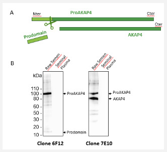
As observed previously from other mammals [1-3] proAKAP4 was only expressed in spermatozoa preparation and not in the seminal liquid as revealed with the monoclonal antibody (clone 6F12) against proAKAP4 (Figure 1). The proAKAP4 was cleaved in AKAP4 mature protein and the prodomain was released (Figure 1A). This cleavage and metabolism of the precursor proAKAP4 can also be followed by western blotting using specific monoclonal antibodies such as the clone 7E10 which recognized the C-terminus of both proAKAP4 and AKAP4 (Figure 1B). Therefore, in this initial T0 experiment, we observed the same amount of proAKAP4 and AKAP4 in the spermatozoa preparation sample of the fresh pig ejaculate. As expected, we confirmed that proAKAP4 is a spermatozoa specific protein expressed in the flagellum of pig spermatozoa
Stability of Boar proAKAP4 during Freeze-Thaw Cycles of the Same Aliquot
The concentration of proAKAP4 was measured in the ejaculate using the Pig 4MID® Kit as T0 value for the stability experiments. The initial mean concentration of ProAKAP4 was of 50.7 ± 1.3ng / mL, reflecting a high-quality semen [1]. After semen aliquots have been frozen and thawed up to ten times, there were no statistically significant differences in proAKAP4 concentrations as quantified using the Pig 4MID® Kit from T0 to T10 (Table 1).
All concentrations were in ng /mL and indicated as a mean± SD and median (interquatile ranges). Clearly, the proAKAP4 concentrations were not modified statistically after ten freezethaw cycles and the global percentage of variations was at 9.54%. Dilutions of the neat semen (half and quarter dilution factor) had no effect on the recovery of proAKAP4 concentrations as shown graphically on Figure 2. These dilutions highlighted the robustness of the Pig 4MID® Kit to quantify accurately the amount of the proAKAP4 polypeptide in neat pig semen. We checked then the expression and metabolism of proAKAP4 by western blotting (Figure 3). None of proAKAP4 and AKAP4 expressions or metabolisms were altered by the freeze-thaw cycles. Neither the integrity of proAKAP4 or AKAP4 was shown to be altered along the 10 freeze-thawing cycles and proAKAP4 was not further converted into AKAP4 showing that proAKAP4 and AKAP4 processing were not modified by freeze thawing cycles. The proAKAP4 was therefore considered as a very stable analyte when kept frozen in raw semen until we performed the Pig 4MID® Kit analysis
Stability of the Frozen proAKAP4 Polypeptide in longterm Storage Conditions
They were no significant variation in proAKAP4 concentrations as measured with the Pig 4MID® Kit for fresh pig sperm when stored until 6 months at -20 °C (Table 2). No variations were obtained when stored at - 80 °C (data not shown). Statistical significances were evaluated as described in the Materials and methods section. Our results showed that total proAKAP4 concentrations were clearly stable up to six months of storage at -20 °C with the variation in proAKAP4 concentration always below 5%. The western-blot analysis displayed no degradation of the sample stored at -20 °C from up to 6 months highlighting the robustness of the protein when kept frozen in raw semen (data not shown).
Intra-assay and Inter-Assay of the Pig 4MID® Kit
We further assess the robustness of the Pig 4MID® Kit by evaluation of the intra-assay and inter-assay CV’s on the Pig 4MID® Kit with neat pig semen as in the design of our study. These intra-assay and inter-assay CV’s were performed with two different ejaculates of the same animal (Table 3). Inter-assay variation was assessed from 10 determinations (with 2 aliquots each day) on ten consecutive study days, and intra-assay variation was calculated from eight sequential determinations obtained from the first day of the study period
Discussion
This study examined the storage effects and repeated freezethaw cycles on pig proAKAP4 sperm protein integrity in preanalytical conditions (meaning before the 4MID® Kit procedures) to evaluate the robustness of this new parameter in daily routine of semen analysis for swine breeding activities. We clearly show that proAKAP4 polypeptide is highly stable when frozen at minus 20 °C, for a long time period (up to 6 months) and will not be altered by multiple freeze-thaw cycles in neat semen. These data are of importance as they highlighted for the first time, that specimens of one ejaculate can be aliquoted and kept at minus 20 °C until their analysis and shipped from AI stations to central laboratories without loss of proAKAP4 integrity.
The reason of this stability could be due to the localization and the inherent functionality of the proAKAP4 itself. As shown on Figure 1, the proAKAP4 is a sperm specific protein that is neither found on the membrane nor released in the seminal plasma. The proAKAP4 polypeptide is inside the spermatozoa, more precisely in the fibrous sheath of the principle piece of the flagellum [19-22] and will need to be released from the fibrous sheath to be further quantified using the 4MID® assay. ProAKAP4 has been shown to be strictly localized to the principal piece of the flagellum and not in other spermatozoa compartments [20-21], tightly anchored to the fibrous sheath, along the longitudinal columns and ribs of the sperm tail [2,3,20-21].
According to the Pig 4MID® assay procedure, the proAKAP4 has then first to be extracted from the spermatozoa. Proteins markers described in sera or in seminal fluids [1,23] are frequently reported to suffer from the shear stress induced in buffered solutions and from long-term storage conditions. In contrast of what we reported with sperm proAKAP4, proteins in buffer solution can be fragile and they may even acquire conformations susceptible to degradation during frozen and post-thawed conditions. Clearly, proAKAP4 concentrations appears to be stable as long as the polypeptide is maintained in neat semen within the spermatozoa flagellum, with the fibrous sheath bringing stability for proAKAP4 integrity. The maintenance of proAKAP4 as a fulllength precursor is then important for the aliquot processed for the initial quality assessment of the ejaculate and at further steps, for the quality control during dose processing in AI stations. High proAKAP4 concentrations in the ejaculate and then in doses, will ensure to have enough motile and functional spermatozoa populations in the hours post the artificial insemination
The total amount of proAKAP4 per spermatozoa is fully synthetized within the testis and before ejaculation. Therefore, an aliquot of the ejaculate could be frozen immediately after semen collection in boar studs as this will represent the exact picture of the long-term motility of the spermatozoa. Freezing of an aliquot of ejaculate at collection point will then facilitate the analysis of semen (related to the proAKAP4 concentration) and favors also transport of such aliquot up to external laboratories. Our results clearly showed that degradation rates of the proAKAP4 were not impacted by frozen storage conditions of the aliquot and are in favor of such collection for delocalized sperm quality assessments. Furthermore, proAKAP4 stability when stored in aliquots in sperm frozen collections, will allow to better take in account technical and logistical constraints such as i) delays in shipping frozen aliquot when in need to analyze hypofertile animal; ii) being less dependent of any power cut or voltage fluctuations of the low-cost freezers; or the use of frost-free freezer that goes through numerous defrost cycles, as may happen in small breeding centers.
In boar stud, the storage of frozen aliquoted samples could also be convenient to process all the semen in the same time to compare ejaculates of different animals at the end of collection time. The dose semen processing will then not be impacted as the 4MID® analysis will be completely run in 2 hours. The amount of proAKAP4 as a read out of sperm quality should add marketing values for AI stations by ensuring high quality semen. In swine industry, there is also a real interest to identify the best male and then to follow up the sperm production during exploitation. Boars are usually kept from 6 to 9 months in the AI stations. That will be of importance to have a stable parameter to follow animal along all his career and keeping a safe measure of the initial quality of the first ejaculate after quarantine. In this context, the storage of frozen aliquoted samples may allow likewise to identify genetical traits of interest in a particular pig strains, such as fertility, death at birth or litter size, that may be related to proAKAP4 levels of expression [1,24-27].
Finally, keeping frozen aliquots of pig semen could allow to reanalyze the same samples stored to confirm previous results or to perform additional analysis, establishing new path for boar sperm preservation investigations. Better understanding proAKAP4 stability allows now to compare ejaculates at different collection points and compared to extended semen which is being shipped and used many days later. The storage capacity of extenders should be then further explored in relation with proAKAP4 consumption and degradation rates, during several days and in chilled conditions, when spermatozoa will stay alive.
Conclusion
One of current challenge of the swine industry is to standardize the semen processing procedures within boar studs. The proAKAP4 parameter have been initially introduced to facilitate the identification of ejaculates of inferior motility and quality, that were not identified by classical sperm parameters, and that could then be withheld before their release into the field. Having a stable sperm parameter such as proAKAP4 that can be kept stable as frozen up to the analysis time should be further interesting for quality check control and to follow up this parameter evolution during all the boar career. Taken together, the proAKAP4 parameter stability present then multiple advantages in favor of harmonizing sperm quality assessment between laboratory and AI centers.
To Know More About Journal of Dairy & Veterinary sciences
Please click on: https://juniperpublishers.com/jdvs/index.php
For more Open Access Journals in Juniper Publishers
please click on: https://juniperpublishers.com/index.php
#veterinary pathology#veterinary pharmacology#Veterinary Toxicology#Veterinary sonography#Juniper Publishers#open access journals
0 notes
Text
Stem Cell Therapy: A Reparative Approach in Veterinary

Abstract
Stem cell therapy is a growing field in veterinary medicine and has created a lot of hope for developing treatments for diseases that otherwise cannot be treated by traditional medical approaches. Clinical studies in animals show promising results indicating that stem cells may facilitate tissue repair and improve quality of life in pets, cats and horses. In this mini review, summarizes the current status of stem cell therapies in veterinary medicine describing the procedures used for isolation, characterization, administration of cells and treatment of various diseases that affect dogs, cats and horses.
Keywords: Mesenchymal Stem Cells, Veterinary Medicine, Cell Therapy
Introduction
The field of stem cell research has attracted many investigatories in the past several years. Although stem cells have been known for some time, the biology of stem cells and their manipulation for therapeutic purposes have become the subject of intense reserach only in the last decade. At present, is clearly that every tissue and organ in this body has its own reservoir of stem cells, namely mesenchymal stem cells, that provide the homeostatic maintenance of the body. The aging process no doubt reflects a numerical or funcional degradation in these stem cells. Mesenchymal stem cells have emerged in the literature as cells with marked potencial in the realm of immunomodulatory and reparative medicine. Originally isolated form bone marrow, the mesenchymal stem cells can be found in numerous tissues including skin, adipose tissue, synovial membrane, umbilical cord blood, dental pulp, lung, as well as from fetal/neonatal tissues. For veterinary medicine, adipose tissue, umbilical cord blood and bone marrow are commonly used [1]. Mesenchymal stem cells can be characterized by their spindle-shaped, long and flattened cells exhibiting a fibroblastic morphology, ability to adherence, high proliferative capacity and miltilineages differentiation potential able to regenerate all the mature cells in the tissue from their origin along the lifespan of na individual. These cells must express > 95% of cell-surface markers such as cluster differentiation CD105, CD73, and CD90 as week as < 2% of expression of CD14, CD19, CD34, CD45, and HLA-DR. Although Mesenchymal stem cells can be expanded in vitro, they are capable to self-renew for limited time in vitro, and their lifespan can also vary from species to species. The aim of this mini-review is to present the use of stem cell therapy in animals and focuses on provide a guide for the therapeutic use in animals.
Discussion
Mesenchymal stem cells can be obtained from dog and cats using an aspiration needle such as a jamshidi, to collected form the femur, tibia or humeral head and from horses by the sternum and the tuber coxae [2,3]. In adipose tissue, mesenchymla stem cells can be obtained from inguinal, abdominal and thoracic wall fat in dogs and cats. In horses can be obtained from the superficial gluteal fascia [4]. Dog, cat and horse umbilical cord blood was collected in a bag with anticoagulant [5]. All samples following delivery to the laboratory where mesenchymal stem cells will be isolated and characterized. Once mesenchymal stem cells are isolated and characterized, the method used for the administration of cells (direct or intravenous injection), depends the patient´s disease and condition. For the treatment of osteoarthritic joints, an intra-articular injection is used and for damaged tendons, ultrasound guidance permits direct implantation of mesenchymal stem cells. Although, delivery mechanisms that direct the maximum number of cells to the diseased area are essential, in some cases intravenous injection have been successfully used in the treatment of some disease as feline chronic gingivostomatitis. Each route bas advantages and disadvantages. The best route is the easiest to perform, less invasive and traumatic, has minimal side effects and enables a high survival rate of transplanted cells.
Animals in their course of life suffer from different diseases which are treated by different therapeutical approaches. The therapeutic application of stem cell technologies in veterinary medicine was first used to treat equine suspensory ligament desmitis that involved direct injection of bone marrow aspirate obtained from the sternum into an injured ligament [6]. Now, there are multiple disease conditions that are being treated with stem cells. Dogs with osteoarthritis that were treated with intraarticular injection of stem cells demonstrated statistically significant improvement in lameness, pain, and range of motion. This shows that stem cell therapy decreases patient discomfort and increases patient functional ability [7]. Dogs affected by keratoconjunctivitis sicca had stem cells implanted around the lacrimal glands that proved to be safe and effective with a significant improvement of tears production and in all ocular clinical signs associated with the disease [8]. Dogs, suffering from atopic dermatitis for at least 12 months, not responding to conventional therapy, received an intravenous dose of mesenchymal stem cells. A single systemic administration produces positive results in the remission of clinical signs of canine refractory atopic dermatitis without adverse events [9]. Cats diagnosed with feline chronic gingivostomatitis, not responded to conventional therapy, were recruited to the study. Each cat received a mesenchymal stem cells transfusion by Intravenous [10]. Cats affected by asthma were treated with mesenchymal stem cells that proved to be safe and effective with a significant reduction of airway inflammation, airway hyperresponsiveness and remodeling without adverse effects [11]. Cats with stable chronic kidney disease were received an intravenous dose of mesenchymal stem cells that proved to be safe and effective with adverse effects, clinical improvement, greater disposition, appetite and healthful weight gain [12].
Stem cell therapy for animal are using for the treatment of different diseases in valuable animals like horses, cats and dogs with multiple benefits. Because of this several companies like VetStem in the United States and CELLTROVET in Brazil were offering commercially stem cell therapy for pets and horses. For this reason, we can say that stem cell therapy for animal is not just research, it´s a reality.
Conclusion
Currently, the clinical application of stem cell therapy in veterinary medicine has been resulting in improved quality of life for dogs, cats and horses. Although studies related to the treatment of various diseases have yet to be performed, the future of this field will be promising.
To Know More About Journal of Dairy & Veterinary sciences
Please click on: https://juniperpublishers.com/jdvs/index.php
For more Open Access Journals in Juniper Publishers
please click on: https://juniperpublishers.com/index.php
#veterinary microbiology#veterinary obstetrics#veterinary pathology#veterinary pharmacology#Juniper Publishers#open access journals
0 notes
Text
Bromatological and Phythochemical Study of the Medicinal and Edible Plant Solanum Nigrum L. (Solanaceae) in Huambo Municipality - Angola

Abstract
In order to determine the phytochemical and bromatologic components of S. nigrum L., plant widely used as medicine and food for the population in the Huambo province, in Angola, young leaves were harvested in the morning period. They were subsequently put to dry in shade for 15 days, mashed in a traditional mortar and passed through a sieve to reduce them in finer powder. 500g of leaves powder were sent to National Center for Animal and Plant Health in Cuba (CENSA) for the determination of secondary metabolites and bromatologic analisis. Qualitative phytochemical analysis were performed using the Rondina and Coussio methodology (1969), the bromatologic trials by the Kjeldahl methods and thermogravimetric balance was used for dry matter and moisture calculation. Tannins, alkaloids, flavonoids, primary and secondary amines, leucoanthocyanidins, free phenols and triterpenes and or steroids were identified. In this study quinones rings lactónicos were not detected. 29.04% protein was found in dry matter, 16.35% of dry matter and 84.65% of humidity. This plant is considered a functional food for its phytochemical and bromatological composition.
Keywords: S. nigrum; Secondary metabolites; Primary metabolites; Medicinal; Eating plant
Introduction
The use of plants in the treatment of several diseases is a practice that was widely used by our ancestors, especially in times of lack of more advanced pharmaceuticals. The use of natural products with therapeutic properties is as old as human civilization and for a longtime animal, plant and mineral products were the main sources of medicines [1]. On the other hand, the rescue of traditional medicine as a way of replacing high cost and potentially toxic synthetic medicines with natural medicines, once again, is a trend that includes veterinary medicin [2,3]. There are medicinal and edible plants that constitute a nutritional and therapeutic source. Thus, it is essential to identify and study them in order to determine their phytochemical and bromatological composition, as well as the study of general and specific toxicity, to better ensure therapy and food safety in their consumption, either by rural and urban populations. The food is an essential factor in disease prevention and health improvement, it prevents and control various types of chronic untransmissible diseases, such as diabetes, hypertension, cancer, heart disease, among others. Several studies have been conducted to prove the beneficial properties of certain foods in the face of decreased resistance, imbalance of microbial flora, inflammatory bowel disease, atopic eczema, among other disorders. This way such foods are called functional [4].
Angola is located in the southern part of Africa and is one of the regions with the greatest diversity of flora in the world. It has more than 5000 species, however, there are few studies of this extensive vegetation, still little known as to its beneficial potential for the population [5]. Angolan medicinal plants have several therapeutic actions, many of them however are not described in traditional use [6]. Huambo province has a flora rich in medicinal plants and extensive knowledge of the use of traditional medicine, as well as a large culture of consuming wild vegetables with therapeutic properties, including Solanum nigrum. This plant species belongs to the Solanaceae family and is popularly known in Brazil as Erva-Mora and in Angola as losuwa. It is an annual herbaceous plant and belongs to the same genus as eggplant (Solanum melongena), potato (Solanum tuberosum) and tomato (Solanum lycopersicum). Thus, due to the lack of knowledge about its phytochemical and bromatological composition in the province of Huambo Angola, this study aims to conduct such a survey
Materials and Methods
Harvest and preparation of plant material
S. nigrum was harvested in Huambo municipality, located in the central plateau, belonging to the agricultural zone 24. The average temperature vary between 19ºC and 21ºC, the average annual precipitation vary from 1100 mm to 1400 mm and two seasons established according to the rainy season. and dry; the soils are of ferralitic characteristics [7]. The plant was identified at the Luanda National Botanic Center (CNB) where was deposited a copy under the code Hb74. Young leaves were harvested between March and April 2013, in the morning, following the techniques proposed by [8]. The leaves were dried in the shade for 15 days, crushed in a traditional mortar (pestle) and sieved to shrink the particles to fine powder.
Phythochemical and bromatological study
For the qualitative characterization of secondary metabolites, the [9] phytochemical filtration was used (Table 1) held at the biopharmaceutical department of the National Center for Animal and Plant Health in Cuba (CENSA). In the qualitative analysis the cross system was used and the presence or absence of secondary metabolites in the samples was specified according to [10]. To determine the plant dry matter and humidity, a thermogravimetric balance was used. The protein percentage was determined by the Kjeldahl method. This method is based on the digestion of sulfuric acid in the presence of a catalyst to convert the nitrogen of the organic compounds into ammonia nitrogen. Ammonia is released by the addition of distilled sodium hydroxide and recovered in a boric acid solution.
Quiz
A survey was conducted to survey the population’s knowledge regarding the use of the plant as a medicine and food. This was based on obtaining the knowledge of the population most directly involved with the sale of this plant. For this, semi structured forms were used as proposed by [11], plus free questions and informal conversations investigating the use of S. nigrum for the consumption and treatment of some diseases, considering the one proposed by [12]. The quiz consisted of two fundamental parts: 1) knowledge of the plant, its use as a food and / or medicine, including its therapeutic benefits, 2) parts of the plant used and form of preparation. One hundred and four people of both genders answered the questionnaire, most of them women who were selling the plant in formal and informal markets. The Microsoft Excel program was used for the percentage quantification of the results of the applied quiz.
Results
There is a high knowledge of the plant as food by the population, which justifies its consumption in rural communities mainly, 73% consume the plant as food, 18% have little knowledge about consumption and 9% did not know it as an edible plant. In turn, they affirm the existence of various forms of preparation of the plant for its consumption; it can be simply boiled and then boiled, added salt and consumed accompanied by funje (cornmeal dough or sweet potato) and also with rice. Another form of consumption after cooking would be to season it with vegetable oil, tomatoes, onions and other spices. About the knowledge of the plant as a medicine, it can also be observed in figure 1, that it is widely used in communities for the treatment of various health disorders. 56.7% use the plant as a medicine, 29.8% have little knowledge about its use as a medicine and 13.5% do not use it as a medicine. The S. nigrum questionnaire was associated with the treatment of various health disorders such as pain, inflammation, liver, gastric and skin diseases. The functional groups found are shown in (Table 2) that are involves in therapeutic effects of S. nigrum plant. Bromatological analysis of S. nigrum indicated 16.35% dry matter, 84.65% humidity and 29.04% protein in dry matter.
Discussion
In this study the results confirm that the people use this plans as feed the same [13], in their studies of edible wild plants in India, found similar ways of preparing S. nigrum cooked with rice and meat. In subcontinental India, 9500 wild plants are used as food, medicine, fuel, essence, fiber and other purposes by more than 53 million tribes belonging to 550 different communities [14]. The S. nigrum is part of a group of medicinal plants studied with action on the cardiovascular system and that, besides this activity, would also have action on diabetes control and high blood lipid content [15]. Studies by [16] and [17] refer to the use of S. nigrum as a drug, presenting antibacterial, antifungal, antiinflammatory, anticancer, antioxidant, antipyretic and cytotoxic activity. Therefore, in Huambo, this plant is known as both food and medicine, that is, it is used for nutritional and therapeutic purposes, effectively proving to be a functional food. As can be seen, secondary metabolites with relevant therapeutic properties were detected in this study. They may be related to the pharmacological effects to which the investigated population referred, such as the use of this plant as an analgesic, in inflammatory processes and in cases of typhoid fever. Thus, primary and secondary amines, free phenols and alkaloids are abundantly present.
Qualitative phytochemical filtering studies performed by [18] in S. nigrum, revealed the presence of tannins, alkaloids, flavonoids, saponins and proteins, which shows the components that give the plant nutritional and therapeutic properties. Alkaloids are given pharmacological activities on the central nervous system as a depressant or as a stimulant, act at the level of the autonomic nervous system as sympathomimetic, sympatholytic, anticholinergic and parasympathomimetic. Antiparasitic, analgesic, antimalarial, anxiolytic and antihypertensive activity are also described [19]. Although tannins are not as abundant, this constituent is extremely important from a therapeutic point of view, as it has antioxidant, anthelmintic, astringent, healing, antidiarrheal action, among others. [20] state that there are several studies that detected anthelmintic action in legumes being the same attributed mainly to the presence of condensed tannins. Tannins are also attributed to antiseptic and antimicrobial action (antibacterial and antifungal), as well as reports of regenerative and healing action in wounds or burns [21].
Leucoanthocyanidin, flavonoids and triterpenes were notably found in this study. These secondary metabolites have various therapeutic properties, including analgesic, anti-inflammatory, antibacterial, anticancer, antioxidant action and Flavonoids present as biological properties, decreased blood capillary permeability and increased resistance. They are also antiinflammatory, antiallergic, hepatoprotective, antispasmodic, antioxidant and are free radical scavengers [19,21]. Flavonoids have become important dietary compounds with promising therapeutic potential. Epidemiological reports and evidence suggest that flavonoid-rich diets, such as quercetin, have effects on the prevention and treatment of cardiovascular disease, cancer, and kidney and liver failure [22]. Triterpenes are compounds with antiviral, antibacterial, anticancer and antifungal activity [23]. This plant has a bitter taste, which may be related to the occurrence of hydrophobic alkaloids and amino acids, such as valine, leucine, isoleucine, phenylalanine, tyrosine and tryptophan [24].
According this study bromatologically is rich. Dry matter is the water-free part of food, which means that it is where all or most of the nutrients are concentrated. This high amount of protein demonstrates its high nutritional value for the population, associated with its high availability, ease of culture, low cost, annual growth, and the presence of metabolites with functional properties. According to [25], vegetable proteins are a source of nutrients of great interest due to their variety, availability and cost. The functional properties and nutritional benefits of each protein group can be explored [26] describe for proteins functions such as tissue formation (connective tissue, muscle, blood, keratin, among others), constitution of enzymes and support to the immune system. The amount of protein detected in the plant under study is of great interest in the food field and its participation in the human diet should be better explored and encouraged [27]. This edible and therapeutic plant reveals a range of phytochemicals and an enviable amount of protein that gives it great importance in feed as well as in the treatment of various diseases in both humans and animals. Its routine use is facilitated by its availability, low cost and ease of cultivation [28].
Conclusion
S. nigrun is well known and used as medicine and food by the population of Huambo Angola. Studies have also indicated that this plant is a functional feed as it has in its composition secondary metabolites of therapeutic relevance and a large amount of protein in the dry matter.
To Know More About Journal of Dairy & Veterinary sciences
Please click on: https://juniperpublishers.com/jdvs/index.php
For more Open Access Journals in Juniper Publishers
please click on: https://juniperpublishers.com/index.php
0 notes
Text
Coagulase-Negative Staphylococci (CNS) as Emerging Mastitis Pathogens

Introduction
Mastitis caused by Coagulase-negative Staphylococci (CNS) usually remains subclinical or mildly clinical [1], however it was reported that CNS mastitis could be aggravated to severe clinical cases [2], but most CNS mastitis cases are chronic [3] based on their intramammary persistence for lactation milk exceeding periods, even extended to the upcoming ones [4]. CNS mastitis is a hidden but serious threat to dairy herd including further horizontal transmission to lactating cows and vertical to suckling calves because of environmental origin of most CNS and hidden subclinical nature [5]. CNS mastitis hazards aren’t exclusive to the dairy herds, but also extended to public health due to possible horizontal transmission of resistance genes (Soares et al., 2012) to other human pathogens or direct transmission to humans because of shared zoonotic virulent CNS species between animal and humans [6]. Pathogenicity of CNS is generally amplified by two parameters: invasiveness (capability to permeate the protective barriers and to spread) and toxicity (ability to produce enzymes and toxins). CNS are capable of producing enzymes instead of coagulase enable the invasion of host tissues and spread of the inflammatory process (e.g. lipase, fibrinolysin, urease). Moreover, they were found capable of producing proteolytic enzymes, exotoxins and haemolysins, which facilitate the uptake of iron [7]. Besides other various virulence constituents protecting CNS from local and systematic host immunity actions [8].
Antimicrobial therapy is still an important component in any CNS mastitis control or prophylaxis actions. But, with the indiscriminate use of antimicrobials and emerging of multidrug resistant CNS, desired results are no longer obtained [9]. Antimicrobial resistance in CNS and other mastitis pathogens has been a worldwide concern during the past decades and it has also brought increasing attention to the use of antimicrobials in animal agriculture and its potential impact on public health [10]. The contribution of agricultural antimicrobial use to development and spread of resistance to human pathogens, however, remains under investigation and debate [11]. Mechanism of CNS resistance to antimicrobials including genotypic detection of resistance genes have been investigated for long time to update knowledge that may help in CNS control programs [12]. For example, mecA-encoded alternative penicillin binding protein, PBP2a, causing reduced binding to β-lactams antibiotics [13]. β-lactamases encoded by blaZ gene. Also, antimicrobials inactivating enzymes, efflux pumps and protective methylation of the antibiotic’s ribosomal target site help resistance to other common antimicrobials used in dairy medicine as tetracyclines, aminoglycosides and macrolides [14].
Ability to form biofilm is a very important virulence constituent, enabling CNS to be organized in multilayered cell clusters embedded in a matrix of extracellular polysaccharide (slime) permitting persistence of CNS in udder tissue unaffected by antimicrobials and protected from host immunity [7,15]. Biofilms improve the ability of microorganisms to resist adverse factors and colonize the environment besides being mainly accused for repeated therapeutic failures as CNS isolates growing within biofilms are less susceptible to antimicrobials commonly used on dairy farms, including β-lactam members [15]. Therefore, biofilm-formation by CNS species could possibly impede antimicrobial therapy [16]. Biofilm formation in CNS also contributes to distinguish them as a main cause of persistent inta-mammary infection (IMI) which enables CNS to survive in the udder tissue from season to season as a constant source of infection [16,17]. Although biofilms do not appear to affect disease severity [18].
Increased antimicrobials resistance of bacteria causing mastitis including CNS is globally and hazardously increasingly. This what recently guided scientific attention to the plant kingdom members, extracts and essential oils (EOs) as cinnamon [19] and carvacrol [20] which might be a substitute cure once the synthetic chemical compounds are unable to perform their role [21].
To Know More About Journal of Dairy & Veterinary sciences
Please click on: https://juniperpublishers.com/jdvs/index.php
For more Open Access Journals in Juniper Publishers
please click on: https://juniperpublishers.com/index.php
#Animal Nutrition#Wild Life Medicine#Veterinary Virology#Veterinary Therapeutics#Juniper Publishers#juniper publishers open access
0 notes
Text
Biological Efficient Dairy Cows in Grazing Systems

Abstract
The availability of indicators that would help to avoid the overvaluation of one of the characteristics involved in the assessment of a good dairy cow over others that are important as well would also allow identifying the most adapted biotypes to the different environments existing at the place of the evaluation This article aims to discuss the need to use several productive and reproductive indicators when measuring the biological efficiency of a dairy cow in grazing systems. It was used retrospective data corresponding to the lactations of 300 primiparous and multiparous cows of the Holstein breed - American-Canadian biotype, along with records of their entire productive life, from their incorporation into the system until their sale or death. The animals were divided into two categories: pure cows (PC, n = 120) and cows in the breeding record (CBR, n = 180). It is observed that there are two different strategies in some variables that achieve the same biological efficiency, where the CBRs live longer, produce less and have smaller first delivery intervals. There are no significant differences in the milk index but in the fat index. It is concluded that the greater individual production does not guarantee a greater production at the end of the productive life of the cow, nor a greater productive efficiency when considering the time involved to produce a certain amount of liters. In grazing systems, the contribution of other variables included in the milk index - longevity, rearing efficiency and reproductive behavior - should be considered while searching for an aggregate indicator that tends to achieve greater productive efficiency.
Keywords: Dairy cows; Indicators; Efficiency; Grazing Systems
Introduction
The notable increase in the productive performance and size of these modern high-production cows has been made possible by the repeated and asymmetric use of a selection based exclusively on milk production. Although this process has been accompanied by changes in the nutritional area, these have not been sufficient to prevent vital function deterioration such as reproduction and survival. It becomes increasingly difficult to provide a non-limiting environment, being almost impossible during the initial phase of lactation [1]. The efficiency of a productive system is one of the most important factors from an economic and social point of view. And the most used modality to evaluate it is assessing indicators of biological and economic productivity. However, when producing in conditions where resources are scarce and expensive (grazing systems), not only products or outputs but also inputs should be considered when evaluating efficiency. In the particular case of high production dairy cows, the traits associated with biological efficiency or fitness (reproduction and longevity) have deteriorated despite their importance for the viability of the company [2,3].
Suggest that the sustainability of dairy systems depends, to a large extent, on the availability of a biotype adapted to the handling conditions and that it is capable of efficiently transforming food into good quality milk. This biotype must have a good reproductive performance, being the main goal of the system to maximize an efficient productive response per unit area [4]. The search for the maximization of the value of a single productive variable disregarding the remaining variables can alter the equilibrium and deteriorate the overall efficiency of productive systems [5]. Every open system processes the inputs received and generates outputs. In productive systems, the concept of efficiency refers to the most appropriate way to use resources with existing technology and products.
As a result of this positioning, it is considered that a production process is efficient if the maximum output is obtained with the lowest possible inputs [6]. In dairy production, the expression “maximize outputs” may have different connotations: maximize individual production during lactation or maximize production considering the entire life of the cow, reproductive success should be considered in the analysis. The amount of milk produced by a cow can be considered the most important indicator in intensive systems, even though, this indicator alone is not the most appropriate to make operational a complex variable like productive efficiency, when the goal is to make the most out of grazing systems. In these cases, it should be complemented, or even replaced, by other more aggregated indicators that constitute alternatives as a more comprehensive measure to assess the behavior of production in those systems in which pasture is the basic component of the diet.
The availability of indicators of this nature would help to avoid the overvaluation of one of the characteristics involved in the assessment of a good dairy cow over others that are important as well. It would also allow identifying the most adapted biotypes to the different environments existing at the place of the evaluation [7]. This article aims to discuss the need to use several productive and reproductive indicators when measuring the biological efficiency of a dairy cow in grazing systems.
What Is Efficiency
Efficiency is the relationship between an income and an expense, between an input and an output or between a resource and a product [8]. When measuring efficiency, it is necessary to specify exactly which elements are used to evaluate the result through a relation of its values. And to define the units used to measure the values of these elements. The concept of efficiency refers to a relationship between elements and that the circumstances in which the relationship is established have a high specificity. As a consequence, the term itself is very relative, and any value that can be considered as good or low is even more so [8].
How to Measure Efficiency
The advantages of grazing systems are sought within the framework of this approach, in which the cow is provided directly with the necessary input to meet its food requirements, without the need for transportation, processing or distribution of rations [9]. Although it is the most widespread modality, the amount of milk produced by a cow does not represent the most appropriate indicator to make operational a complex variable as productive efficiency. As such, it should be complemented or replaced by other more aggregated indicators creating alternatives for a more comprehensive production measurement to assess their performance in grazing systems. The availability of indicators of this nature would help to avoid the overvaluation of one of the characteristics involved in the assessment of a good dairy cow over others that are important as well. It would also allow identifying the most adapted biotypes to the different environments existing at the place of the evaluation [10].
It was used retrospective data corresponding to the lactations of 300 primiparous and multiparous cows of the Holstein breed - American-Canadian biotype, along with records of their entire productive life, from their incorporation into the system until their sale or death. The animals were divided into two categories: pure cows (PC, n = 120) and cows in the breeding record (CBR, n = 180). The existence of significant differences in the assessed time between groups was studied by applying variance analysis to a classification criterion. JMP 5.0 for Windows (JMP®, SAS Institute, 2003) was used for statistical analysis Table 1.
The following variables were analyzed: a. Number of births b. Milk production (PL by its initials in Spanish): liters produced per cow adjusted to 305 days c. Age at first birth (PPE by its initials in Spanish) in days d. Total Butyrose Fat production in kg: GB, ΣGBi where “i” are the kilograms produced in the j-th lactation e. Total milk production (liters) = pl, Σpli, where “i” are the liters produced in the j-th lactation f. Milk index (milk production per day of life) in liters = IL, by its initials in Spanish, (il: LT / e e: age in days at the end of the last lactation): il = pl / age [11] g. Fat Index (production of Butyrose fat per day of life) in kg = IG by its initials in Spanish, (ig: Total GB / e e: age in days at the end of the last lactation): ig = kg GB / age [10] h. First delivery interval - delivery in days (IPP by its initials in Spanish): Σipp, where “i” are the days between deliveries / number of deliveries [12].
Pure cows are pregnant at an age closer to optimal, showing a difference of 60 days with cows in the breeding record. They produce in their five lactations 6516 liters on average against the 5933 liters in six lactations of cows in the breeding record, achieving a total of 41767 liters in its life against the 39653 liters of pure cows. Additionally, the pure cow takes for each new delivery 51 days (total 306 days (6 deliveries * 51 days)) more than the cow in the breeding record. In conclusion, the advantage in days they have because of its first birth during its life is lost in a single IPP, as well as producing almost for a year in the less efficient part of the lactation curve.
It is observed that there are two different strategies in some variables that achieve the same biological efficiency, where the CBRs live longer, produce less and have smaller IPP. There are no significant differences in the milk index but in the fat index. Although considering each variable separately is relevant, when reference values are available and allow these cows to be positioned in particular in the framework for milk production of the Holstein breed, the characterization of the efficiency of a productive system requires a joint analysis of all of them instead of individual consideration [5].
Conclusion
It is concluded that the greater individual production does not guarantee a greater production at the end of the productive life of the cow (LT) nor a greater productive efficiency when considering the time involved to produce a certain amount of liters (il). In grazing systems, the contribution of other variables included in the milk index - longevity, rearing efficiency and reproductive behavior - should be considered while searching for an aggregate indicator that tends to achieve greater productive efficiency.
To Know More About Journal of Dairy & Veterinary sciences
Please click on: https://juniperpublishers.com/jdvs/index.php
For more Open Access Journals in Juniper Publishers
please click on: https://juniperpublishers.com/index.php
0 notes
Text
Heavy Metal Bioaccumulation by Cestode Parasites of Mustelus Schmitti (Chondrichthyes: Carcharhiniformes), from the Bahía Blanca Estuary, Argentina

Abstract
The environment of the Bahía Blanca estuary is considered a hot spot in terms of pollution. Bioindicators should have the ability to react relatively fast to certain pollutants and environmental disturbances. Therefore, an exploratory study was carried out determining and quantifying the concentrations of cadmium (Cd), chromium (Cr), copper (Cu), lead (Pb) and zinc (Zn) in the muscle and liver of Mustelus schmitti narrownose smooth-hound and were compared with the values obtained from their respective helminth assemblies. In most of the fishes analyzed, the concentration of heavy metals was higher in the infra communities of cestodes compared to the host. Our results position the cestodes as efficient sentinel species of pollution by bioaccumulating higher concentrations of heavy metals than the host tissues, thus behaving in excellent early warnings of environmental pollution, more real than quantifications in sediments, in water and fish
Keywords: Heavy metals; Bioaccumulation; Sentinels parasites
Introduction
The estuary of Bahía Blanca (39° 03′44 ″ S 62° 04′00 ″ W) is an adequate environment to develop pollution studies, considering that it is an area that includes urban centers, several industrial parks and deep-water ports. All the effluents are discharged with different degrees of pretreatment, so they generate different impacts on the ecosystem. In environmental monitoring to detect heavy metals, organisms are often used as bioindicators, which have the ability to react relatively fast to certain toxic products and environmental disturbances. Some of these organisms, such as parasites, may be highly sensitive to brief exposures, poorly detected in water, sediment or fish [1-5]. Our previous studies in the estuary have focused on evaluating the parasitism of fish in the time scale to be able to compare and analyze them as effect indicators altering some parasites population parameter such as prevalence and abundance or causing symptoms in their hosts in response to environmental disturbances [6-10]. The narrownose smooth-hound Mustelus schmitti Springer, 1939 is a resident fish of the estuary of Bahía Blanca and third in importance as a fishing resource. Based on the fact that some parasites, such as cestodes, have the ability to absorb and accumulate more chemicals than their host tissues [11].
The objective of the present study was to analyze whether a higher concentration of metals in the parasites respect to their host was applicable in the cestodes- narrownose smooth-hound model and to evaluate if these helminths possess ecotoxicological value and could be used in the study area as early bioindicators of anthropic impact. Therefore, an exploratory study was carried out in order to determine and quantify the cadmium (Cd), chromium (Cr), copper (Cu), lead (Pb) and zinc (Zn) concentrations in the muscle and liver of the narrownose smooth-hound and compared with the values obtained from their respective cestode assemblages (Dollfusiella sp., Orygmatobothrium schmittii, Calliobothrium australis and Symcallio sp.) The samples were analyzed by Inductive Coupling Plasma Atomic Emission Spectrometer (ICP-AES, LANAQUI-CERZOS-CONICET-UNS). The values were compared with the limit values allowed by the European Union for fish meat.
Results and Discussion
In most of the fishes analyzed, the concentration of heavy metals was higher in the infra communities of cestodes compared to each host. The parasites concentrated 270 times more Cadmium than the fish muscle. For this metal, the standards established in the liver and those of the parasites were exceeding the limits established by the European Union for muscle or liver. Chromium was bioconcentrated in the cestodes two times more than the muscle and six times more than the liver. Copper was accumulated with values 65 times more than muscle and up to four times more than in the liver. Lead had values 48 times more in helminths than both muscle and liver of fishes. For this metal in most dosages, the concentration measured in parasites exceeded the limit value established by the European Union. Zinc bioaccumulated in parasites seven times more than muscle and four times more than liver. Only in one case the Zinc concentration was three times higher in the liver than in parasites.
The environment of the Bahía Blanca estuary is considered a hot spot in terms of pollution and is included among the most eutrophic coastal ecosystems known [12]. Also, since many years it have been reported high concentrations of heavy metals and pesticides in water and sediments [13,14]. The combined effect of these pollutants, plus the sewage discharge, the industrial effluents of petrochemical origins, and the overheated water from a thermoelectric power station (620MW) all of them represent a growing threat to the environment. These increase in anthropogenic activity around estuaries, coupled with the persistence of heavy metals, their high toxicity, strong tendency to bioaccumulate, and non-degradability [15], usually affect water. As negative effects it could change the trofic web in the aquatic fauna, eliminate the spawning and larval recruitment sites and a potential decrease in diversity, affecting all the ecosystem [16,17]. That is why the need to choose of efficient bioindicators in the evaluation of the quality of the environment [18].
Conclusion
The estuary of Bahía Blanca is one of the most important in Argentina, having the main deep-water port system in the country. Although fish species can be used as efficient and useful bioindicators, our results position cestodes parasites as sentinel species of contamination for the fact that reported higher concentrations of heavy metals than their hosts. This would avoid possible underestimations in pollution levels by being quantified only in sediments, in water and in fish.
To Know More About Journal of Dairy & Veterinary sciences
Please click on: https://juniperpublishers.com/jdvs/index.php
For more Open Access Journals in Juniper Publishers
please click on: https://juniperpublishers.com/index.php
#veterinary microbiology#veterinary obstetrics#veterinary pathology#veterinary pharmacology#Juniper Publishers#open access journals
0 notes
Text
Significance of Feed Supplementation on Milk Yield and Milk Composition of Dairy Cow

Abstract
Milk composition and production are the interaction of many elements within the cow and her external environment. Composition of milk influenced by many factors such as genetic and breeds differences, stage of location, milking interval, seasonal variation, disease and nutrition. Nutrition is the major factor on both milk yield and composition. The three factors: Genetic makeup, nutrition and management decide the productivity of dairy cows. Improvement of genetic make up only contributes up to 30% to production, while the 70% is dependent on nutrition and management. Unfortunately, indigenous of tropical dairies are low milk producers because of the shortage of nutrition. Poor nutritive values of feeds lower the production capacity and fertility potential of dairies. If fed well, with supplementary feeds and under good management, more milk could be produced from them. So, supplementary feed with optimum dietary ration providing for dairy cows in good management improves the production level and good proportional composition of product with high nutritive value.
Keywords: Milk fat; Globule membrane; Volatile; Fatty acids; Nutrition; Dairy production
Abbrevations: Igs: Immune Globulins; MFGM: Milk Fat Globule Membrane; NFC: Non-Fiber Carbohydrate; NRC: Nutritional Requirement of Cattle; SNF: Solid Not Fat; TMR: Total Mixed Ratio; VFAs: Volatile Fatty Acids
Introduction
From agricultural activities, dairy production and its management is the one which is the interest of every country because of high nutritional value of milk and milk products and another purpose gained from them. And the feed they consume is not compete with human food and also, they convert feed which is not directly eaten by human being to products that human being can consume. That means, special ability of dairy cattle to transfer feed stuffs into edible food for humans and as much as 70% of their total feed intake is from non-human food. Food requirements of rapidly expanding human population is the other reason which initiates or give importance the development of dairy production
Milk composition and production are the interaction of many elements within the cow and her external environments Chemical composition of milk is variable and influenced by intrinsic factors like genetic and breed differences, stage of lactation, milking interval, seasonal variation, disease and nutrition. Protein content of milk is positively correlated within a population of dairy cattle; however, different breeds of cattle vary in average component levels. Holsteins have the lowest fat and protein content, while Jersey and Guernsey breeds have the highest. Because Holsteins produce more milk, they generally have a higher total yield of fat and protein than other breeds. There are many factors that can affect milk fat and protein, and many of them can be manipulated to enable you to achieve higher than average levels of milk components. Keep in mind that herds that are below breed average will have more opportunity to improve component levels. Herds that are already above average may have better success by focusing on increasing milk yield, which will increase the total amount of fat and protein production [1].
Stage of lactation affects milk protein and fat percentages very similarly. The highest amount of protein and fat in milk is found just after freshening, in colostrum. Levels drop to their lowest point between 25 and 50 days after calving and peak at 250 days as milk production begins to decrease. Age tends to cause both milk fat and protein to decline as the animal becomes older. Milk fat falls about 0.2% each year from the first to fifth lactation likely as a result of higher production and more udder infections. Protein decreases 0.02 to 0.05% each lactation as animals age.
Season dramatically affects milk fat and protein. The hot, humid months depress fat and protein content. There is a gradual increase of protein and fat in milk through the fall and peak levels occur in the colder months of winter. As temperatures increase through the spring, component levels are gradually decreased. These changes may be indicative of feed intake patterns, which are lower in summer due to changes in weather and temperature. Mastitis infections reduce fat and casein but increase blood protein content of milk. Of all the factors affecting milk composition, nutrition and feeding practices are most likely to cause problems; however, management changes made here are able to quickly and dramatically alter production of fat and protein other than genetics. Digestion of fiber in the rumen produces the volatile fatty acids (VFAs) acetate and butyrate. Butyrate provides energy for the rumen wall, and much of it is converted to betahydroxy butyrate in the rumen wall tissue. About half of the fat in milk is synthesized in the udder from acetate and betahydroxy butyrate. The other half of milk fat is transported from the pool of fatty acids circulating in the blood. These can originate from body fat mobilization, absorption from the diet, or from fats metabolized in the liver. Rumen microbes convert dietary protein into microbial protein, which is a primary source of essential amino acids for the cow. These amino acids are used by the mammary gland to synthesize milk proteins
Glucose is required to provide energy to support this protein synthesis. Glucose is either formed from the VFA propionate in the liver or absorbed directly from the small intestine. If too little propionate is absorbed from the rumen, the cow will have to breakdown amino acids and convert them to glucose (a process called gluconeogenesis); this can reduce the supply of amino acids available to make milk protein. In addition, some albumin and immunoglobulin protein are transferred directly to milk from the blood. The relative amounts of protein and energy that are available in the rumen at a given time is the major factor affecting rumen fermentation and therefore milk components. Any diet or management factors that affect rumen fermentation can change milk fat and protein levels. Consistently providing adequate energy and protein and balanced amounts of rapidly fermentable carbohydrate and effective fiber are keys to maintaining optimum levels of milk components.
The challenge in feeding for milk components is that high energy, low fiber diets that increase milk protein are likely to reduce fat levels. This may also be the case in some diets with rumen modifiers, such as Rumensin®; however, this product has other ways to affect the rumen that do not necessarily alter milk components. Any situation that causes cows to eat abnormally or limits feed intake may affect milk components. Examples include: overcrowding at feed bunks, housing heifers with older cows in facilities at or near full capacity, feeding rations that encourage sorting, feeding infrequently in a conventional system (non- TMR), failing to push feed up or feed TMR often enough, feeding protein feeds before energy feeds and feeding grain before forage in non- TMR systems. These conditions can create slug feeding (one or two meals per day versus 10 to 15) or allow cows to eat high grain meals part of the time and high forage meals the remainder of the day. Ensure that fresh feed is available 20 hours each day, spoiled feed is removed from bunks, and shade or cooling is provided during hot weather to help maintain normal intake and normal meal patterns. Poor ventilation or cow comfort also can depress milk fat and protein production by reducing intake. Finally, make ration changes gradually to allow rumen microorganisms time to adapt.
Any reduction in rumen microbial protein production from nutrition or feeding management imbalances will reduce milk protein by way of less microbial protein for the cow to digest and depress fat by limiting VFA production in the rumen. Proper body condition is essential so that high producing cows can draw on body stores of nutrients to support milk production. If body stores are minimal, yields of milk and milk components will suffer. On the other hand, excessive body condition increases the risk of metabolic problems and calving difficulty. Weight loss in early lactation can increase milk fat content for a short period of time. Both thin and fat cows tend to have low milk fat in later lactation. Protein can be depressed at calving if animals are overly obese or underweight. In addition, some research shows that underfeeding protein during the last three weeks before calving can depress milk protein [1]. In general, as energy intake or ration energy density increase and/or fiber decreases, milk fat content will be reduced, while protein is increased. In contrast, as ration fiber levels increase and/or energy is reduced, milk protein is depressed, and milk fat is increased. Lack of energy intake or lower ration digestibility may reduce milk protein by 0.1 to 0.4%. This reduction may result from underfeeding concentrates, low forage intake, poor quality forage, and failure to balance the ration for protein and minerals, or inadequately ground or prepared grains. Shifting rumen fermentation so that more propionic acid is produced is apt to increase milk protein and decrease fat content. However, excessive energy intake, such as overfeeding concentrate, may reduce milk fat content and increase milk protein. Normal protein levels can be expected when energy needs are being met for most of the cows. Often this is impossible to achieve with high producing animals.
A deficiency of crude protein in the ration may depress protein in milk; marginal deficiency could result in a reduction of 0.0 to 0.2%, while more severe restriction of diet crude protein would have greater impact. However, feeding excessive dietary protein does not increase milk protein, as most of the excess is excreted. Dietary protein has little effect on milk fat levels within normal ranges. Diet protein type also could affect milk protein levels. Use of non-protein nitrogen (NPN) compounds, like urea, as protein substitutes will reduce protein in milk by 0.1 to 0.3% if the NPN is a main provider of crude protein equivalent. Rations higher than recommended in soluble protein may lower milk protein by 0.1 to 0.2 points. NPN levels in milk will be increased by excessive protein or NPN intake, heavy feeding of ensiled forages, ensiled grains, immature pasture and lack of rumen undegradable protein in the diet. Balance rations for crude protein, rumen undegradable protein, rumen degradable protein, and soluble protein. For high producing cows, balancing for amino acids also may be required.
An increase in the intake of concentrates causes a decrease in fiber digestion and acetic acid production. This creates an increase of propionic acid production. Propionic acid production encourages a fattening metabolism that is in opposition to milk fat. Addition of buffers to some rations may help to prevent acidosis; this will not change milk protein but will increase milk fat content [2]. Animals that eat a substantial amount of concentrates or a low ratio of dietary forage to concentrate may develop acidosis even when buffers are added to the ration. The non-fiber carbohydrate (NFC) portion of the diet is highly digestible and can influence both fat and protein in milk. Excessive amounts of NFC can depress fiber digestibility, which reduces the production of acetate and leads to low milk fat (1% or more reduction). At the same time, greater propionate production allows higher milk protein levels of 0.2 to 0.3%. Generally, an NFC of 32 to 38% of ration dry matter is recommended to optimize production of milk fat and protein.
Balance rations for lactating cows to contain at least 40 to 45% of ration dry matter from forage. This may be altered by the level of corn silage in the ration and the level of high-fiber by-product feeds in the ration. Low forage intake can cause a major reduction in the fat content of milk due to low fiber levels. Several potential reasons for low forage intake are inadequate forage feeding, poor quality forage, and low neutral detergent fiber (NDF) content in forage that was cut too young or late in the fall. Although low forage (high energy) diets increase milk protein production, this strategy is not recommended. The low forage levels contribute to acidosis and laminitis; they do not promote good health for the rumen or the cow in the long run. Protein and fat content also can be changed due to the physical form of forage being fed. Much of this is related to ration sorting and failure to provide a consistent diet throughout the day. Coarsely chopped silage and dry hay are the most common causes of sorting. At the other extreme, very finely ground diets negatively affect rumen metabolism and depress fat and protein production. Monitor ration particle size to ensure that adequate effective fiber is provided, TMRs are mixed properly, rations are distributed evenly to all cows, and sorting is minimal [3].
Adding fat to the ration can affect milk component levels depending on the amount and source of fat. Fat is generally toxic to rumen microbes and may reduce fiber digestibility when fat from natural sources exceeds 5% of ration dry matter. If rumen inert or bypass fat is used, total fat content may safely reach 6 to 7%. At low levels of dietary fat, milk fat content could increase slightly or show no change at all. Milk fat is reduced at higher levels, especially with polyunsaturated oils. If fat or oil is rancid, milk fat content decreases even at low levels of consumption. Milk protein content may be decreased by 0.1 to 0.3% in high-fat diets. Generally, the objective is reviewing the significance of feed supplementation on milk yield and milk composition of dairy cow
Rate of Milk Secretion
The period following milk removal is characterized by low intra-alveolar pressure, which facilitate the transport of newly synthesized milk into the alveolar lumen. As secretion continues between milking’s, pressure is exerted on the secretory process by the alveolar luminal contents. When the luminal pressure exceeds the force of secretion as the alveolar enlargement reaches its limit. It is presumed that the distention pressure of the lumen exceeds the strength of the secretory mechanism needed to push the newly forced milk precursors by chemical feedback mechanism and or physical factors (e.g.,intra-mammary pressure [4].
The physical factors are a result of the distended alveoli partially displacing all other intra-mammary compartments, including the blood vessels. With restricted blood flow, less nutrients are available for milk production, less hormones are available to drive the mammary synthetic systems, removal of waste products of synthesis is reduced and less ox toxin is available to stimulate the myoepithelial cells. In dairy cows, average secretion rate begins to decline after ten hrs since the last milking and secretion stops after thirty five hrs .The pressure measured in the teat cistern increases in three phases. An initial rapid increase in the pressure caused by the movement of residual milk into the cistern from the alveoli and small ducts. The second, lower phase can be an accumulation of newly synthesized milk that is released into the duct system from the alveolar lumens as they begin to accumulate milk. The third phase is marked by the accelerated pressure increase and probably represent over filling of alveoli, ducts and gland cisterns [4].
Factors affecting Milk Yield and Milk Composition
Milk composition and production are the interaction of many elements within the cow and her external environments [5]. High milk yield of satisfactory composition is the most important factor ensuring high economic returns. If the composition of milk varies widely, its implication is that nutritive value and its availability as a raw material will also vary. Chemical composition of milk is variable and influenced by intrinsic factors like genetic and breed differences, stage of lactation, milking interval, seasonal variation, disease and nutrition [1].
Genetic and breed differences: Heritability is defined as the ratio of genetic variance to total phenotypic ratio. The concentrations % of the three major milk constituents are genetically controlled to a considerable extent. Heritability’s of fat, protein, and lactose contents average: 0.58, 0.49 and 0.5 respectively, while that of milk yield average is 0.27 [1]. The above Table1 indicate that there is a room to increase milk protein % by genetic selection without increasing fat % and that selection for high milk yield alone may reduce milk fat and protein %. Milk from Holstein cows has a lower milk fat % than milk from Jersey and Guernsey. droplets also differ among breeds. Holstein has smallest fat droplet while Guernsey and Jersey Brown swiss has the largest. Milk of Jersey cows also has a higher total solid than milk from other dairy cattle breeds. Differences in milk composition among individual with a breed are often larger than differences among breeds. Milk color also affected by breed type. For example, milk from Guernsey and Jersey is yellowish in color because if these breeds convert much less carotene (yellow pigment) to vitamin A than other breeds of dairy cow (Table 2).
Stage of lactation: Colostrum, the first mammary secretion after parturition differs greatly from normal milk. Cows colostrum contains more minerals, protein and less lactose than milk. Fat is usually higher in colostrum than in milk.Ca,Mg,P,and Cl are high in colostrum’s, whereas K is low. Fe is 10-17 times higher in colostrums than in milk. The high levels Fe are needed for the rapid increase in hemoglobin in the red blood cells of newborn calf. Colostrum contains ten times as much vitamin A and three times as much vitamin D as milk [6]. The most remarkable differences between colostrum and milk is the extremely high levels of Ig content of colostrum. Mammary secretion gradually changes from colostrum to normal milk within 3-5 postpartum [7]. From normal milk changes in composition occur during the first few days continue but at reduced rate for about five weeks of lactation. Fat and protein then rise gradually and may increase mare sharply near the end of lactation. Lactose decreases while mineral concentration increases slightly during that period.
Milking Interval: When milking is done at longer intervals the yield is also more with a corresponding smaller percentage of fat, whereas milk drawn at short intervals yield smaller quantities with higher amount of fat. The effect milking interval is mainly on fat percentage rather than the SNF [8]. The fat content of milk is usually lower in the morning than in the evening milking, because there is usually a much shortage interval between the morning and evening milking than between evening and morning. SNF content varies little even if the intervals between milking vary. Cows are usually milked at equal intervals (12-hrs interval for two times milking). Cows milked at unequal intervals produce less milk than those milked at equal intervals. The reduction in milk yield is more i8n high producing cows than in low producing ones. In complete milking for several consecutive days can permanently reduce milk yield for the entire lactation. Milking time for most cows is 5-6 minutes per cow [7].
Season of calving and seasonal variation: The effect of season of calving on milk yield is confounded by breed, the stage of lactation and climatic condition. Cows calving in the late fall to soring produce more milk (up to 8% more) than cows calving in the summery. This is likely due to an interaction between day light and ambient temperature in case of tropical areas. Seasonal differences have become less significant because of better feeding and management of dairy cow can overcome this effect. The seasonal variations in milk composition are commonly observed with dairy cattle in temperate regions. Milk fat and SNF percentages are highest in Winter and lowest in Summer. Milk fat and protein percentages are lower by 0.2-0.4 in summer than in winter. The effect of ambient temperature on milk yield is dependent up on the breed, for example, Holstein and the other larger breeds are more tolerant to lower temperature whereas the smaller breeds particularly the Jersey and Zebu are more tolerant to high temperature. Milk production declines when environment temperature exceeds 27 degree Celsius. The reduction in milk yield is largely due to drop in feed intake. High temperature affect high producing cows more than low producers and it is particularly harmful during the peak of lactation.
Disease: The main disease affect milk yield and milk composition of dairy cows is mastitis. It impairs the ability of secretory tissue synthesize milk composition and destroys the secretory tissues and consequently lowering milk yield. A decrease in milk production persists after the disappearance of the clinical signs of mastitis due to a destruction in the secretory tissues [9]. Infection of udder (mastitis) greatly influences milk composition. Concentration of fat, SNF, lactose, casein, beta-lacto globulin and alfa-lactalbumin are lowered and concentrations of blood of blood serum albumin, Igs, sodium, chloride are increased [10]. In severe mastitis, the casein content may be below the normal limit of 78 % of total protein and chloride content may be rise above the normal maximum level of 0.12 %. Mastitis is also responsible for differences observed in milk composition from different quarters of the udder
Nutrition: Nutrition has also a major effect on both milk yield milk composition. According to O’Connor [10], under feeding reduces the amount milk production, the fat, protein and SNF, contents of milk. As a general rule it is believed in that any ration of diet that increases milk production, usually reduces the fat percentage of milk and fat content is influenced more by roughages (fiber) intake and SNF content can fall if the cow fed a low energy diet, but it is not greatly influenced by protein deficiency, unless the deficiency is acute. Of all milk components, milk fat is the most influenced by dietary manipulations. Most of changes in milk composition due to dietary manipulation are related to changes in ruminal acetate: propionate ratio. Several nutrition factors can influence milk composition. These includes plan of nutrition, forage concentrate ratio, forage quality (e.g., particle size), level and type of dietary fat. In plan of nutrition, under feeding dairy cows reduces lactose percentage and increases fat percentage. Feeding imbalance rations (e.g., low energy: protein ratio) may reduce milk fat and protein percentages. In case of forage concentrate, as the proportion of the concentrate in the ratio increases (above 50-60 % of ration), milk fat % tends decline. This is mainly because of the lower ruminal production of acetate and butyrate (precursors of milk fatty acid synthesis in the mammary gland) associated with feeding high concentrate diets. The extent of milk fat depression is influenced by other feeding practices such as frequency of feeding and feeding system. Feeding cows less frequently especially if the concentrates are fed separately from the forage results in a reduced ruminal acetate: propionate ratio which in turn can result in reduced milk fat % will be less where total mix rations are fed and or if feed is offered three or more times daily
Forage particle size (forage processing), feeding finely chopped forages has a negative impact on milk fat % and may cause milk fat depression syndrome (drop of milk fat % below 3 %). Cows fed finely chopped forages spend less time to chewing and therefore, will produce less saliva. Ruminal PH will drop as less saliva is produced to buffer the acid production in the rumen. As the ruminal PH drops below 6, the activity of cellulolytic bacteria is reduced and so it is the production of acetic acid and butyric acid (precursors of short chain fatty acid synthesis in mammary gland). In case of level of starch in the ration, as the level of starch in the ration increases, the level of acetate produced in the rumen is decreased while that of propionate is increased. This may cause a reduction in milk fat %. Dietary Fat Corporation or oil in dairy cow ration can substantially alter the profile of milk fatty acids. The effect of supplemented fat in milk fat % depends on the type of supplement of fat. Feeding poly unsaturated fat (susceptible bio hydrogenation in the rumen) such as vegetable oils may reduce milk fat % whereas feeding protected fat tend to increase milk fat %. Changes in dietary protein levels have minimum effects on milk fat content. When the protein content of the diet is limiting, increased dietary protein may increase milk fat content through increases in roughage intake (Table 3).
Nutritional Requirement of Dairy Cow
Feed serves many different purposes, including the following Maintenance: The normal activities of staying alive breathing, blood circulation, digestive process, etc. all requires nutrient. This maintenance is not for extra function like production unless extra feed is provided for cell function [11]. Reproduction: Pregnancy and delivery make demands on the dam which have to be met from her feed, if it is not to lose weight. The fetus increases in size quickly during the last two to three months of gestation, drawing on the body reserves of the dam. Lactation: Producing milk either for one or two offspring or for human consumption requires high levels of energy and protein and good access to protein and good access to water.
Factors Influencing Nutritional Requirement of Dairy Cow
Nutritional requirement of dairy cow influenced by many factors like stage of production, condition of the environment, size of the cow and the like. Stage of production: One of the most challenging aspects of dairy cow nutrition is that their requirements change during the course of a year based on stage of pregnancy and lactation NRC [12]. Weather: Cold weather greatly increases the nutritional requirement. Therefore, during cold weather, the cow’s diet may need to be supplemented to allow for the additional requirement dairy perform optimally in their “their monaural zone” where temperatures are either too hot or too cold. When the ambient temperature, which includes wind, humidity, solar radiation and air temperature, is outside of that zone, dairy performance is depressed [13]. The most common situation dairy man face is an ambient temperature below the lower critical temperature or the lower range of the thermo neutral zone. Tit should be pointed out that in cases simply feeding more of a low-quality feed stuff will not meet these additional requirements, in which case the energy density of the diet must be increased by either feeding a highquality forage or by adding a high energy supplement. Size: As cows size increases, the nutritional requirement for energy and protein increases. This should be expected because the larger cow is the more energy and protein it takes to maintain normal body functions.
Priorities and strategies for feed Resources Development
The feed value of forage that form the basis for ruminant feeding is a functional of its nutrient content and digestibility, its palatability (which determines its consumption level) and the associative effects of the other feeds [14]. Interplay of these factors determines the effective utilization or feed value of the material. Strategies for ensuring adequate nutrition of animal includes the following like: matching dairy production system to available resources, selection of crops and cropping systems that will maximize biomass production, and developing the simple techniques to optimize the use of different components of crops for different end purpose, making more efficient and wide spread use of agricultural and industrial by products as source of dairy feed, and also conserving feeds when it is available for drought season. From these strategies, increasing feed availability with production system of dairy number is through increasing off take of animals through sale (destocking). The amount of feed available to the remaining animals will increase in the process [14] (Table 4).
Types of Supplementary Feeds
Supplementary feed is any stuff added to the total diet of the animal to increase the nutritive value of the feed and to increase content of single nutrient or compound nutrient. These supplementary feeds includes protein supplement (legumes, oil seed cause, meat meal, fish meal), mineral supplements (salt (Na), limestone (ca), bone meal (ca and p), and others), vitamin supplement (natural and synthetic) and energy supplement (fat and carbohydrate like concentrate feed those the high amount of energy and low fiber content and high digestibility with high protein content [15]. Protein supplement: Conditions under which milk production can be increased by feeding protein supplements are well defined, although it is not possible to estimate liters of milk per kg of supplement with great accuracy. Results from feeding trials in Australia indicate that milk responses from protein supplements can be up to 1.5 liter per kg supplement than from equal weights of cereal grains. Usually the responses are much lower when energy is first limiting. In most cases milk production from tropical pastures is limited primarily by energy. When energy is limiting, protein supplements gives similar milk responses equal amount of cereal grains and surplus nitrogen is converted to ammonia and excreted as urea [16] However, as energy supply from cereal grains is increased, the protein content of the diet becomes limiting for milk production. Protein supplement then allow increases in milk yield with only small changes in milk composition. The conditions where protein supplements give greater milk responses than cereal grains are determined by stage of lactation, Genetic potential, forage quality degradability of the protein supplement, substitution rate.
Energy supplement: In order to improve milk production levels, energy input such as concentrate feeds have to be considered essential for any enterprise, even for those based on dual purpose systems, since reduced intake of energy by dairy cows consuming low quality forages is the principal cause of low milk production .Traditionally, energy supplements are based on cereal grains that include barley, sorghum, wheat, cats, maize, and etc, Molasses is a very popular energy source for cattle grazing tropical pastures. Agro- industrial by products are fed as supplement to roughagebased diets, particularly in dairy production system for milking. Concentrates rich in energy mixture or adulteration with other depends on the quality of the basal roughage and the level of production. Agro industrial by products can be utilized by mixing of two or more of the ingredients to make concentrate at home or using a single in gradient. They have special value in feeding cattle mainly in urban and pre urban dairy production systems as well as in situation where the productive potential of the animals is relatively high ad require high nutrient supply. These by products are rich in energy and protein contents or both, they have low fiber content, high digestibility and energy values compared to with the other class of feeds .To prevent the effect of heavy concentrate feeding on low forage, concentrate ratios can be mitigated by splitting up the concentrate allowance in to several smaller meals spread more evenly over the twenty four hours. By this means, digestive up sets are avoided, protein is more efficiently waltzed, and lactation partition is more normal. Rig milk fat is improved [17].
Mineral supplement: In providing proper nutrition to dairy cows, the dairy man needs to consider minerals in addition to protein, energy, water, and vitamins. Even through minerals are needed only in small amounts, they are very important for optimum reproduction, immune function, and optimal milk production. Minerals are divided in to two groups by the amount needed of each. Macro minerals are required in larger amounts, while micro minerals are required in smaller amounts. The micro minerals required includes calcium, phosphorus, magnesium, potassium, sodium, chloride and sulfur. The micro minerals required includes Iron, cobalt, copper, manganese, zinc, Iodine, and selenium cows get some of the micro minerals and micro minerals from the feeds they eat. However, minerals must be added to the ration in order to meet the requirements, because, the forages and grain do not provide adequate amounts. If these minerals are not, supplemented, problems may occur. For instance, selenium deficiency can cause retained placentas [18].
Several items must be taken into consideration when buying mineral supplement. First, the supplement must contain all the macro minerals and micro minerals that are deficient in the ration. Also, the supplement must contain the appropriate amounts of each mineral to be effective. The in gradients with supply the semimetals should also be considered because some a lower bio availability than others. Bio availability is the ability of the cow to digest and utilize the minerals provided. If the bio availability of the cow is low, then the amount of the mineral fed must be increased, so the cow will get an adequate amount. For example, copper oxide has very low bio availability. Copper sulfate is a better source of copper [19]. The best way to feed the mineral supplement is by force feeding rather than free choice. When minerals are supplemented free choice, the cow does not eat to meet her mineral requirement needs. Force feeding refers to mixing the mineral supplement with the grain mix or the total mixed ration. This ensures that the cow gets enough of each mineral to meet her requirements. It is equally important not to just dump a lot of mineral supplement with the grain mix or total mixed ration because too much of certain minerals will cause toxicity problems or inhibit the functioning ability of other minerals. Thus, the forages fed to the cows should first be analyzed for their mineral content, if it is not already known. Next, the ration should be balanced so that all of the mineral requirements are met. Then the deficiency can be identified and corrected by feeding the correct mineral supplement
Specific Disadvantage of Heavy Concentrate Feeding in Early Lactation
Even though cows should be fed heavily with concentrates in the first few weeks of lactation, to encourage high peak yield, there are some specific problems, some of which are dealt with more heavy concentrate feeding in early lactation. These includes ailments such as ketosis, abomasal displacement, laminitis, and mal partition syndrome involving low fat milk and reduced lactation efficiency [20].
Processing Concentrate Feeds
It is generally accepted that some processing of cereal grains is required before cattle can effectively utilize the energy and nutrient content of concentrate feeds. While increasing the degree of processing improves utilization, it may also lead to digestive problems when high levels of grain are fed and may accentuate fat depression in milk. The type and extent of processing required depends on number of factors including the grain type, the proportion of grain in the diet, palatability, and the risk of developing digestive problems. If a whole untreated grain is fed, large proportion of it can pass undigested in the faces. The minimum level of processing required to ensure efficient grain digestion is cracking the seed coat t expose the endosperm. This must be achieved by mechanical or chemical treatments as dairy cattle have only delimited ability to chew small cereal grains. The main nutritional significance of the seed coat is the extent to which it dilutes the amount of starch in the diet [21].
The second level of processing involves grinding and rolling, to reduce particle size which in turn determines the surface area, which is exposed to microbial and digestive enzymes. This ultimately influences the number of starch granules freed from the protein and no starch carbohydrate matrix of the endosperm [22]. When starch granules are tightly held with in endosperm matrix, it may be necessary to use gelatinization and or hydration (i.e. high temperature with or without water) to disrupt the granules. Conversely, grinding or milling can produce extremely fine particles which can be rapidly fermented digested and can reduce the palatability of the grain if excessively dusty.
The third method of processing is steam flaking. With this treatment, the whole grain is heated with steam for 10-40 minutes and subsequently rolled to varying degree [22]. This breaks the seed coat and endosperm, although the whole grain remains as one. This process gelatins much of the starch making it more susceptible to enzymatic attack. Grains such as barley, whet and oats, which have a naturally high fermentation and intestinal digestion when ground or dry rolled, are not affected as much by steam flaking rolled grain. At present time, practical problems such as risks in handing NaoH as well as corrosion concerns restrict use of this processing method. For this reason, alternatives such as ammonia treatment might be more practical.
The fourth method is polluting which is common commercial process where small particles are combined into large particle by means of a mechanical process in combination with moisture, heat and pressure. It is believed in that concentrate polluting decrease waste, reduces dust, minimizes spoilage [23], improves feed efficiency and provides a means for uniform distribution of protein and minerals. There are several potential advantages of feeding pellets over meal or a loose ix like Balanced proportion of protein, minerals, vitamins, and buffers can be in corporate in to the pellets, the higher the level of concentrate feeding, the greater the live hood that nutrient balancing will be necessary [24], risk of excessive un palatable and toxic substances associated with supplements for example urea are avoided by careful blending of ingredients, pellets usually are loss dusty than mechanically processed grains. Therefore, it appears that relatively small change in the processing of concentrate can have a substantial influence on the degradation characteristics of the concentrate and can alter the yield of milk components significantly. Polluted for mutations, when compared to textured concentrates, tend to improve degradability, lower rumen PH, increase milk and protein yield and can depress milk fat yield and percentage, without affecting intake [25-27].
Summary and Conclusion
Milk composition and production are the interaction of many elements within the crow and her external environments. Composition of milk influenced by many factors like genetic and breed differences, stage location, milking interval, seasonal variation, disease and nutrition from these factors, nutrition is the major factor on both milk yield and milk composition. Under feeding reduces the amount milk production, fat, protein and SNF contents of milk. Of all milk components, milk fat is the most influenced by dietary manipulations. Several nutritional factors can influence milk composition. These includes: - plan of nutrition, forage concentrate ratio, forage quality (like particle size, level and type of dietary fat). Major components of milk are water lactose, lipids, proteins, salts, minerals and vitamins. These components arising from several factors including breed, individuality of the animal, stage of location, health of the animal, (especially mastitis) and nutritional status.
Dairy cows use nutrition for purpose of maintenance, reproduction, location (production) and etc. while factors influencing nutritional requirement of dairy cow are stage of lactation, condition of the environment size of the cow and the like. Strategies for ensuring adequate nutrition of dairy cows are matching dairy production system to available resources, selection of crops and cropping system that will maximize biomass production, and developing the simple techniques to optimize are use of different components of crops residues, making more efficient and wide spread use of agricultural and industrial by products as source of elating feed, conserving feeds when it is available for drought seasons when saucily of feed is happen and using grazing system for pastures for avoiding wastage of resources. Supplementary feed is any feed stuff added to the total died of the animal to increase the nutritive value of the feed and to increase content of a single nutrient or compound nutrient. These types of supplementary feeds are protein, energy supplement (carbohydrate and fat), mineral, and vitamin. It is believed in that some processing of supplementary feeds is required before cattle can effectively utilize it. But, type and the extent of processing depends on number of factors like supplementary feed type, the proportion of supplementary diet, palatability and etc. the methods and system of processing includes creaking, grinding, steam flaking, polluting and etc
Recommendation
In tropical areas except commercial dairy farm, state farm, and farms follow modern method of keeping dairy cattle, others like farmers dairy cow fail under shortage of nutrition, poor management, and the production and product obtained from these dairy cows are less. This under feeding susceptible the dairies for disease and lose of the animal also there. To reduce some extent degree of these problems, the following activities should be done. a. Supplementary feeds should be supplied. b. Management should be considered as major activity of keeping. c. Having more dairy cows without enough feed available should be reduced to be profitable from optimum number of head of dairies under good management. d. Feeds should be conserved when it is available for the period shortage of feed is occurring. e. Ways of providing supplementary feeds should be proper to avoid extra problem happen with in the cow of improper ration
To Know More About Journal of Dairy & Veterinary sciences
Please click on: https://juniperpublishers.com/jdvs/index.php
For more Open Access Journals in Juniper Publishers
please click on: https://juniperpublishers.com/index.php
#avian diseases#Dairy Diseases#dairy farms#veterinary microbiology#Juniper Publishers#open access journals
0 notes
Text
Causes of Carcass Condemnation and its Associated Financial Losses in Slaughtered Pigs at the Kumasi Abattoir Company Limited

Abstract
The study was conducted from August 2017 to December 2017 at the Kumasi Abattoir Company Limited to investigate the causes of carcass condemnation in slaughtered pigs and their associated financial losses. Out of the 1221 pigs examined, 212(17.4%) were affected with pathological conditions. The five most frequent pathological conditions were: Lung congestion (7.9%), Ascariasis (2.7%), Cysticercosis (2.4%), Liver congestion (1.1%) and Liver abscess (0. 9%). Pathological conditions were more prevalent (57%) in female pigs than in males (43%). Cross bred pigs were more (81%) affected by pathological conditions than the local Ashanti black pig (19%). The overall carcass condemnation rate was 1.7% with Cysticercosis being the major cause. The average condemnation rate of visceral organs was 1.8 %. Annually, a total of One hundred thousand, six hundred and twenty-eight cedis, seventy-two Pesewas (GHc100, 628.72) was lost due to condemnation of carcasses and organs at the Kumasi Abattoir. Butchers, pig owners and other stakeholders should be educated on pre-slaughter management and handling of animals
Keywords: Pigs; Cysticercosis; Lung congestion; Ascarioisis
Abbrevations: MOFA: Ministry of Food and Agriculture; GHc: Ghana cedis; GDP: Gross Domestic Product
Introduction
Agriculture employs about 60% of the working population and contributes to 21.5% of Ghana’s Gross Domestic Product (GDP) [1]. The livestock population in Ghana is estimated to be 682,000 pigs, 1,657,000 cattle, 4,335,000 sheep and 6,044,000 goats [2]. Swine production is an important economic activity in Ghana due to the high prolificacy and high feed conversion rates of pigs. Pigs are reared under intensive conditions on many farms in the Greater Accra, Ashanti, Brong Ahafo and Volta regions of Ghana [3]. Unfortunately, there is poor documentation on swine diseases in the country. The availability of this information will be invaluable for designing disease control mechanisms. The abattoir has been recognized as a reliable source of information on livestock diseases. This study therefore seeks to investigate the causes of carcass condemnation in pigs slaughtered at the Kumasi abattoir and assess the associated financial losses.
Materials and Methods
The study was conducted from August to December 2017 at the hog unit of the Kumasi Abattoir Company Limited (KACL). The study population were pigs brought for daily slaughter at the hog line during the peak hours of 6 am and 10 am. The abattoir was visited thrice in a week during the study period. Ante mortem inspection was conducted on pigs at the lairage. The pigs passed for slaughter were led to the hog line for stunning and slaughtering. Postmortem inspection was conducted on them by trained veterinary personnel [4]. After inspection, the number of carcasses passed for human consumption and number of partial or totally condemned carcasses were recorded. The reason/s for carcass condemnation was noted for each case and the financial losses associated with carcass condemnations were estimated as described by [5] and modified as follows: Annual Financial loss due to total carcass condemnation = average slaughter rate* condemnation rate of carcass* prevailing market price of average carcass.
Annual Financial loss due to viscera’s condemnation = average slaughter rate *condemnation rate of viscera * prevailing market price of viscera. The total financial losses due to condemnation was obtained from the sum of the losses due to whole carcass and viscera condemnation respectively. Data obtained were analyzed using descriptive statistics
Results and Discussion
The proportion and causes of carcass condemnation are presented in Table 1. Out of the 1221 pigs examined, 21 (1.72%) carcasses were totally condemned, while 191(15.64%) were partially condemned. Partial condemnation included the rejection of diseased offal’s as well as trimmings on the carcass itself. In this study, total condemnation of carcasses was due solely to generalized cysticercosis. The low rate of total carcass condemnation was similar to reports by [6] at a commercial slaughterhouse in Brazil. The prevalence of porcine cysticercosis at this abattoir had previously been reported by [7]. Cysticercosis is an emerging agricultural and public health problem in Africa [8,9]. The risk factors for cysticercosis in developing countries have been identified as general poverty, free ranging of pigs, poor sanitary conditions and home slaughter of pigs without inspection [9,10].
The rate of partial carcass condemnation of 15.64% in this study was lower than (25.44%) recorded at the Addis Ababa Municipal Abattoir [10]. This could be due to difference in location, environmental conditions and slaughtering methods used. The top five causes of partial carcass condemnation in descending order were as follows: lung congestion (7.94%), Ascariosis (2.70%), liver congestion (1.06%), liver abscess (0.90%) and localized cysticercosis (0.65%). These findings are similar to reports by [11] that the major causes of partial carcass condemnation in pigs slaughtered at a municipal abattoir in Addis Ababa, Ethiopia, were hydatidosis, cysticercosis, fasciolosis, liver cirrhosis, pneumonia and abscesses. On the other hand, these findings contradict a report in Northern Portugal by [12]. They concluded that Osteomyelitis (38.5%), Granulomatous lymphadenitis (22.7%), pleurisy/ pneumonia (21.2%), abscesses (8.4 %) and peritonitis (2.6%) were the main causes of carcass and organ condemnation
The financial losses associated with carcass condemnation were calculated as: described by [5] For this study, partial carcass condemnation included carcass trimming and rejected offal’s. At this abattoir, the offal’s were considered as a lot and as priced as such. The annual slaughter of pigs as taken from the abattoir records was 9805. Annually, a total of One hundred and six thousand, five hundred and fifty-four cedis, eighty-six pesewas (GHc 106,554.86) was lost due to carcass condemnation at this abattoir (Table 2). At an exchange rate of GHc4.90= 1 US dollar, this was equivalent to 21, 745.89 United States dollars
Conclusion and Recommendations
Porcine cysticercosis was the main cause of total carcass condemnation. The annual financial loss associated with carcass condemnation amounted to One hundred and six thousand, five hundred and fifty-four cedis, eighty-six pesewas (GHc 106,554.86) or 21, 745.89 United States Dollars. It is recommended that Farmers should be educated on intensive pig rearing. The butchers, pre-slaughter animal handlers should be educated on pre-slaughter management and handling of animals.
To Know More About Journal of Dairy & Veterinary sciences
Please click on: https://juniperpublishers.com/jdvs/index.php
For more Open Access Journals in Juniper Publishers
please click on: https://juniperpublishers.com/index.php
#Milk Born Diseases#metabolic studies#livestock management#laboratory animal medicine#Juniper Publishers#open access journals
0 notes
Text
C-Reactive Protein in Veterinary Practice
Abstract
Animal body reacts to all kinds of injuries and stress to keep up the homeostasis mechanism of the body. This homeostasis achieved either by or nonspecific mechanism. The nonspecific innate resistance of the body like cytological and cytokine reactions including fever, leukocytosis etc. is known as acute phase response. In this response, there will be increase or decrease of serum concentration of proteins. These proteins are known as acute phase proteins. Measurements of serum concentration of these acute phase proteins are found to be useful in assessment of health status and prediction of diseases of the man and animals. The serum concentration of these acute phase proteins returns to base levels when the triggering factor is no longer present. The acute phase response is now considered to be a dynamic process involving systemic and metabolic changes providing an early nonspecific defence mechanism against insult before specific immunity is achieved. Use of one of the acute phase proteins, C-reactive protein as biomarkers for animal disease diagnosis and health status assessment has got high potential in modern veterinary practice is discussed in this review
Keywords: Acute injury; C - reactive protein; Acute phase proteins; Veterinary practice
Abbrevations: CRP: C Reactive Protein; Hp: Haptoglobin; AGP: Acid Glycoprotein; SAA: Serum Amyloid A
Introduction
The acute phase reaction encompasses all the phenomena which take place in animals following tissue damage and is particularly associated with inflammation from whatever cause. During the acute phase reaction, the body mounts a multifactorial response to remove and replace damaged tissue and one of the mechanisms involved is the production and secretion by the liver of a number of ‘acute phase proteins’ [1]. The concentrations of these proteins increase during the reaction are called as Positive acute phase proteins (APPs) such as C reactive protein (CRP), Serum Amyloid A (SAA), Haptoglobin (Hp), Ceruloplasmin, α2- Macroglobulin, α1 Acid Glycoprotein(AGP), Fibrinogen and Complement (C3,C4) while those of others, including albumin, Transferrin, Transthyretin and Retinol-binding protein decrease as the liver switches production of protein towards the synthesis of the proteins required to deal with the damage; is called as negative APPs [2] (Figure 1).
Biological functions of C-reactive Protein
The protein was named the C-reactive protein because of its ability to bind pneumococcal C-polysaccharide. The presence of CRP has also been described in human patients during acute infections caused by acute lobar pneumonia, active rheumatic fever and bacteraemia caused by “colon bacillus”. Among the biological functions described in the literature are Complement activation and opsonisation [3,4] Modulation of monocytes and macrophages, cytokine production [5] Binding of chromatin [6] Prevention of tissue migration of neutrophils
CRP in Bovines
During the early stages of infection, the serum concentration of CRP increases [3]. This increase has been described to be evident before an elevated rectal temperature is observed. Even though increased concentrations of bovine CRP during naturally occurring infections and a correlation with herd health status have been reported, CRP is generally not considered an acute phase protein in cattle. As stress increases to a critical point, the liver rapidly synthesizes large amounts of CRP and releases it into the blood to provide immediate protection against stress [7]. Diseases in a dairy herd elevated the serum CRP level. The serum CRP level was also correlated with milk production. The greater the milk production, the higher the level of serum CRP. Diseases, especially acute infections, induced much higher levels of CRP production than stress or lactation. Also showing that plasma C-reactive protein concentration is related to different kind of stress (poor health, high lactation, blood collection). Strong correlation was observed in cows after delivery (0-1 month) between fibrinogen and CRP values [8]. Obtained results suggest that not only inflammations but also physiological factors such as pregnancy, delivery and/or state of lactation may have a significant impact on APPs values in the blood plasma of dairy cows. It would be worth in the future to check whether there is a relationship assessing the animal health status obtained using acute phase proteins method relatively to other indicators, such as milk yield, length of lactation or others. Morimatsu [9] also discovered Elevation of bovine serum C-reactive protein by lactation when compared to other Acute Phase protein such as serum amyloid P component levels (Figure 2).
CRP in Swine
In the pigs, as in the dogs and humans, C-reactive protein (CRP) is the prototypical acute phase protein with major diagnostic value. CRP concentrations are useful for evaluating the health status of a swine herd, but not for the health status of an individual animal or the differentiation of diseases. Serum haptoglobin (HP) concentration is better than serum CRP concentration as an indicator of inflammatory reactions in pigs, and HP is an important marker for swine health status [10]. On the other side, pigs undergone experimental study had CRP serum concentration below 22μg/ml (mean 18.64 ± 2.59). Twenty-four hour after coinfection with swine influenza virus (H1N1) and Pasteurella multocida, the mean concentration of CRP reached 62.85 ± 35.55μg/ml. Significant difference were noticed as compared to control animals [11]. The presence of elevated serum Hp and CRP concentrations in apparently healthy pigs at slaughter could provide important information to a veterinary inspector about the presence of sub-clinical lesions that could lead to condemnations or a decrease in the quality of carcasses [12].
CRP in Canines
In canines CRP is the major APP used as marker for systemic inflammation / infection. Normally the level of CRP is less than 1.5 mg/ dL or even lower than 0.5 mg/dl. The normal range may be 0.08 to 2.26 mg/dl [13]. The level rises within 4 to 6 hrs after onset of inflammation / infection. Serum CRP level above 3.5 mg/dl, indicates presence of systemic inflammation. Level above 5 mg/dl is a strong evidence of systemic inflammation. Strong correlation was observed between CRP and animal’s temperature and Total Leukocyte counts of canine patients naturally infected with Leptosporosis [14]. CRP levels could be used to monitor early responses to antibiotic treatment and might alert veterinarians to the need for further evaluation or additional treatment. Serum CRP concentrations provide useful information about the severity of inflammation inside the Urinary Bladder. These correlations suggest that CRP concentrations can represent a safe, convenient, and alternative method for evaluating the status of bacterial cystitis [15]. Significantly high CRP values were observed in cases like Lympoma, pyometra, panniculitis, acute pancreatitis, polyarthritis, leptospirosis, babesiosis, parvo viral enteritis, glomerulonephritis, immune mediated disease and malignant neoplasia [16]. Rise in CRP may not be observed in local tumours like leiomyosarcoma, upper respiratory tract infection, diabetes, neurological problems involving intracranial disorders. Since the CRP concentration did not increase in patients with intervertebral disk protrusion, it might be useful in distinguishing arthritis from spinal / brain diseases in patients with lameness. Thus, CRP is a nonspecific inflammatory marker, it could facilitate the diagnosis by indicating the presence and the extent of inflammation
CRP in Elephants
The reference interval for CRP reported herein for Asian elephants (1.3–12.8 mg/l) is like CRP intervals reported in harbor seals (Phoca vitulina) and bottlenose dolphins (Tursiops truncatus). When compare to the CRP, SAA is demonstrated to be the most responsive major APP in elephants [17]. This agrees with previous reports where SAA elevations were noted consistently in elephants with Elephant endotheliotropic herpesvirus (EEHV) and in 2 captive elephants with inflammatory lesions. Further studies are needed to address the reactivity of the CRP reagents with elephant proteins and to consider the use of either elephantspecific reagents or non–antibody-based assays
CRP in Chicken
The birds positive for E.Coli, Pasturella multocida and Staphylococcus aureus infections as well as histomoniasis and adjuvant injection found to be positive for CRP. CRP also positive in clinically normal population. These birds on post-mortem, had lesions consistent with chronic respiratory disease, which is highlighting the use of CRP as a potential biomarker for nonclinical disease [18]. CRP did not rise in chickens as quickly as it does in humans, whereby CRP was detectable 36-48 hours post infection in chickens, compared to 16-18 hours in humans. A more recent study investigated CRP serum concentrations using a human CRP kit and found CRP concentrations to increase
CRP in Equines
A high serum concentration was also found in horses with pneumonia, enteritis, arthritis and after castration. Increased plasma concentration has been observed in carbohydrate induced laminitis. Increased serum concentration of CRP has been found in horses suffering from aseptic inflammation induced by intramuscular turpentine injections [3].
To Know More About Journal of Dairy & Veterinary sciences
Please click on: https://juniperpublishers.com/jdvs/index.php
For more Open Access Journals in Juniper Publishers
please click on: https://juniperpublishers.com/index.php
#wild life rehabilitation#Veterinary sonography#Veterinary Therapeutics#Veterinary Virology#Juniper Publishers#open access journals
0 notes
Text
Veterinary Drug Residue: The Risk, Public Health Significance and its Management

Abstract
Veterinary drugs are any substances applied to or administered to animals for their therapeutic, prophylactic and diagnostic purposes or modification of physiological function or behavior. They are used throughout the world and more than half of all medicines are prescribed, dispensed or sold improperly. In Ethiopia, also different studies revealed the improper utilization of drugs is common. The use of veterinary drugs in food-producing animals has the potential to generate residues in animal derived products and poses a health hazard to the consumer. The most likely reason for drug residues might be due to improper drug usage and failure to keep the withdrawal period. The residual amount ingested is in small amounts and not necessarily toxic. The major public health significances of drug residue are development of antimicrobial drug resistance, hypersensitivity reaction, carcinogenicity, mutagenicity, teratogenicity, and disruption of intestinal normal flora. The aim of this paper is to review about risk of occurrence of veterinary drug residue, public health effects and management. Even though, veterinary drugs have a great importance in treating, preventing and diagnosing diseases, it has major public health hazards. To avoid this it is important to use these drugs rationally, the safety levels of food must be strictly observed, drug products should be used in accordance with the labeled directions and public awareness should be created on the public health significance of drug residue.
Keywords: Antimicrobial; Drug; Residue; Risk; Veterinary drug
Abbrevations: ABZ: Albendazole; ADI: Acceptable Daily Intake; AMR: Antimicrobial Resistance; APCI: Atmospheric Pressure Chemical Ionization; APEC: Asian-Pacific Economic Cooperation; BZDs: Benzimidazoles; CFR: Code of Federal Regulation; DES: Diethylstilbestrol; DNA: Deoxyribonucleic acid; EC: European Community; EFSA: European Food Safety Authority; ELDU: Extra-label drug use; ELISA: Enzyme linked-immunosorbent assay; ELU: Extra-label use; ESI: Electrospray Ionization; EU: European Union; FAO: Food and Agricultural Organization; FDA: FOOD and Drug Administration; FDACVM: Food and Drug administration Center for Veterinary Medicine; FEB: Febantel; HPLC: High-performance liquid chromatography; IgE: Immunoglobulin E; LC-MS/ MS: Liquid chromatography-mass spectrometry/mass spectrometry; MBZ: Mebendazole; MRL: Maximum Residue Level; NAP: National Academies Press; NOEL: No observed effect level; RNA: Ribonucleic acid; UEMOA: West African Economic and Monetary Union; WHO: World health organization
Introduction
Veterinary drug” means any substance or mixture of substances which is used, or is manufactured, sold or represented as suitable for use, in (1) the diagnosis, treatment, mitigation or prevention of disease or abnormal physical or mental state or the symptoms thereof in an animal; or (2) restoring, correcting or modifying any physical, mental or organic function in an animal [1]. The use of veterinary drugs in livestock production is inevitable as they are essential for treatment of diseases (therapeutic), prevention of diseases (prophylaxis), modification of physiological functions (such as tranquilizers, anesthetic drugs), improvement of growth and productivity (growth promoters) as well as for ensuring food safety [2]. The veterinary drugs are used throughout the world and they comprise a broad variety of classes of chemical compounds including vaccines, antimicrobials, antiparasitics and β-agonists [3]. Antimicrobials are the most important and most frequently used group of veterinary drugs [4]. Antimicrobials are medicine (natural, synthetic or semi-synthetic origin) that inhibits the growth of or destroys microorganisms when applied at low concentrations without causing host damage [5]. Among the antimicrobials that are commonly used in livestock production are tetracyclines, amprolium, penicillin, streptomycin, sulphonamides, tylosin, aminoglycosides, β-lactams, macrolides and lincosamides, quinolones and sulfonamides [6]. While that of antiparasitic agents include anthelmintics or coccidiostats, stilbenes, amphenicols, nitrofurans, nitroimidazoles, carbamates, pyrethroids and sedatives [5].
A residue, defined in the simplest terms, results when a drug or pesticide is deliberately applied to a food-producing animal or plant. Residues of veterinary drugs include the parent compounds and/or their metabolites in any edible portion of the animal product and include residues of associated impurities of the veterinary drug concerned [7]. Residual amounts of antimicrobials or their toxic metabolites found in meat, organs or other products such as milk and egg of food producing animals is called veterinary drug residues [8]. Consumption of such food products poses a major health risk due to the failure of treatment following the development of resistant microorganisms [9]. Many livestock producers treat their animals by themselves. Even if they use the same drugs as veterinarians, they have little understanding of the conditions and quantities to administer or the waiting periods. The uncontrolled use of anti-infectious agents can lead to residues in animal products, especially when users fail to respect waiting periods. The risks of residues in foodstuffs of animal origin could be reflected into several forms [10]. The immediate effect of antimicrobial residue is allergenicity and toxicity in human through the food chain [11]. The long-term health adverse effects such as increased likelihood include disruption of normal human flora in the intestine (microbiological effects), carcinogenicity, and teratogenicity [12]. Other drug residue problems are the development of antibiotic-resistant microbes and drug misuse [13]. The objective of this paper is to review: The risk of occurrence of veterinary drug residue, public health effects and management (Figure 1).
Historical Background
A whole series of known or new foodborne biological and chemical hazards are threatening health [14]. In the European Union (EU), following a string of health crises, the food safety mechanism has evolved towards a risk analysis approach. This shift to the concept of ‘farm to fork’ risk management [15] led to the establishment of food safety agencies at the European level. The risks of residues from veterinary medicinal products used in livestock production were taken on board in the 1980s, most notably through European harmonization of the regulations on medicinal products for veterinary use. Over the past decade, the EU has improved its regulatory framework to better supervise, assess, monitor and control food production under the ‘Food Law’. More recently, the use of anti-infective in livestock and its contribution to the development of antimicrobial resistance has attracted considerable attention [16].
In Africa - particularly West Africa - only microbial pathogens, pesticide residues and aflatoxins have been the subject of measures to protect the safety of food for human consumption. These hazards were perceived as the greatest threat to public health. In April 2007, the eight UEMOA countries (Benin, Burkina- Faso, Cote d’Ivoire, Guinea-Bissau, Mali, Niger, Senegal and Togo) adopted regulation 07/2007/CM/UEMOA concerning plant, animal and food safety in the UEMOA area [17]. More recently, in 2010 and 2011, two training sessions were held in Benin to familiarize these countries with the theoretical framework for health risk analysis [18]. Yet, there have been very few studies on antimicrobial residues affecting food safety [19]. However, in developing countries, failure to respect waiting periods [20] leads to high exposure to antimicrobial residues [21].
Veterinary Drug and their Use in Food Animals
Drug in animals can be used as therapeutic, prophylactic and growth promotion. Therapeutic use refers to the treatment of established infections whereas prophylaxis is the use of drugs either in individual or groups to prevent the development of infections. Growth promoters (GPs) are any antimicrobial agents administered at low or sub therapeutic dose to destroy or inhibits growth of microbe which reduce the yield of food animals. The use of antimicrobials as feed supplements can promote the growth of food animals and also enhance feed efficiency. The uses of GPs are resulting in meat of better quality with less fat and increased protein contests [22]. The use of drugs in food animals is fundamental to animal health and well-being and to the economics of the industry. There are five major classes of drugs used in food animals: (1) topical antiseptics, bactericides, and fungicides used to treat surface skin or hoof infections, cuts, and abrasions; (2) ionophores, which alter rumen microorganisms to provide more favorable and efficient energy substrates from bacterial conversion of feed and to impart some degree of protection against some parasites; (3) steroid anabolic growth promoters (for meat production) and peptide production enhancers (bovine somatotropin for increased milk production in dairy cows); (4) antiparasite drugs; and (5) antibiotics as used to control overt and occult diseases, and to promote growth [23] (Figure 2).
Authorized Veterinary Antimicrobials
The medicinal products containing antimicrobials authorized for veterinary use are those that have passed the marketing authorization process of the competent national authority. After an evaluation of the scientific data proving the efficacy of the product and its safety for humans, animals and the environment the Competent Authority authorizes its importation, distribution and use. No medicinal product may be marketed unless it has first been authorized by the Competent Authority. However, there are huge shortcomings in the implementation because the technical evaluation of a marketing application is limited to an administrative procedure alone especially in most African countries [24].
Prohibited Veterinary Antimicrobials
Prohibited antimicrobials are substances for which it is not possible to determine the Maximum Residue Level (MRL). Chloramphenicol, dimetridazole, ipronidazole, nitroimidazoles, furazolidone, nitrofurazone, and fluoroquinolones are prohibited for extra-label use in food-producing animals [24]. Chloramphenicol is a broad-spectrum antimicrobial against Gram-positive and Gram-negative bacteria. It was not possible to determine an MRL based on the available data. The inability to set a threshold value and shortcomings in the marketing authorization application led to chloramphenicol being classified in 1994 as a prohibited substance for use in food-producing animals. Dapsone, which is used to treat leprosy in humans, is not authorized for use in food-producing animals in Europe because of insufficient toxicology data, making it impossible to determine the acceptable daily intake (ADI) [25]. In the year 1995 European Union (EU) prohibited the use of nitrofurans for the treatment of bacterial diseases in livestock production, due to concerns about the carcinogenicity of their residues in edible tissue [26]. In subsequent years Australia, USA, Philippines, Thailand and Brazil also prohibited the use of nitrofurans in food animals [27] (Table 1).
Origin of Residue
Veterinary drugs are generally used in farm animals for therapeutic and prophylactic purposes and they include a large number of different types of compounds which can be administered in the feed or in the drinking water. The great majority of residues found in edible tissues of animals have their source at the farm of origin. In some cases, the residues may proceed from contaminated animal feedstuffs. By far the most common cause of residues is the failure to observe the proper withholding period following treatment [28].
Risk of Drug Residue for the Public Health
Human health risk can result from the presence of residues of veterinary drugs and/or their metabolites in edible organs and tissues of treated animals, in particular residues in concentrations exceeding the MRL established by Council Regulation 2377/90 [29]. Occurrences of veterinary drug residues pose the broad range of health consequences in the consumers. The residues of antibacterial may present pharmacological, toxicological, microbiological and immunopathological health risks for humans [30].
Anthelmintics, such as benzimidazoles and probenzimidazoles, are veterinary drugs used against endoparasites for the prevention of animal infestations caused by nematodes, cestodes and trematodes in food producing animals. Among the most popular benzimidazoles are Albendazole (ABZ) and Mebendazole (MBZ) [31]. Benzimidazoles (BZDs) such as albendazole (ABZ), fenbendazole and thiabendazole are a kind of broad-spectrum veterinary drugs for prevention and treatment of helminthic parasites in domestic animals. When BZD drugs were fed to domestic animals, they were metabolized and then converted into other compounds in vivo. Thus, these BZDs and their metabolites can be left inedible animal foods or exist in the environment for a period of time. The harmful BZDs and their metabolites residues in some foods lead to a series of toxic effects such as congenic malformations, teratogenicity, diarrhea, pulmonary edemas, polyploidy, and necrotic lymphoadenopathy [32]. Febantel (FEB) is a probenzimidazole with which is further metabolized in vivo to Fenbendazole, a benzimidazole anthelmintic also [31] (Table 2).
Risk Factors for Drug Residue Occurrence
Disease status: The disease status of an animal can affect the pharmacokinetics of drugs administered, which can influence the potential for residues. This can occur either when the disease affects the metabolic system (and consequently drug metabolism), or when the presence of infection and/or inflammation causes the drug to accumulate in affected tissues. For example, cattle with acutely inflamed mastitis quarters, apramycin penetrates these areas of the body, and concentrations of the drug have been observed at ten times over the level recorded from cows without mastitis [33].
Extra-label drug use: Extra-label drug use (ELDU) refers to the use of an approved drug in a manner that is not in accordance with the approved label directions. It occurs when a drug only approved for human use is used in animals, when a drug approved for one species of animal is used in another, when a drug is used to treat a condition for which it was not approved, or the use of drugs at levels in excess of recommended dosages. For instances, the use of enrofloxacin solution as a topical ear medication (Only approved for use as an injection) are the common ELDU in veterinary medicine [34].
Improper Withdrawal Time: Improper withdrawal time is another risk factor; the withdrawal time is the time required for the residue of toxicological concern to reach safe concentration as defined by tolerance. Based on the drug product, dosage form, and route of administration it may vary from few hours to days or weeks. It is the interval from the time an animal is removed from medication until permitted time of slaughter for the production of safe foodstuffs.
Safety Evaluation and Detection Methods of Drug Residues
Safety Evaluation
Acceptable daily intake: Acceptable daily intake (ADI) is the amount of substance that can be ingested daily over a lifetime without appreciable health risk. The evaluation of the safety of residues is based on the determination of the ADI on which in turn maximum residues limits (MRL) is based. The ADI is determined by consecutive estimate of a safe ingestion level by the human population on the lowest no effect level of toxicological safety studies [35]. If the drug is not a carcinogen, the no observed effect level (NOEL) of the most sensitive effect in the most sensitive species divided by a safety factor is used to determine an ADI for drug residues. The FDA will calculate the safe concentration for each edible tissue using the ADI, the weight in kg of an average adult (60 kg), and the amount of the product eaten per day in grams as follows; Safe concentration = [ADI (μg/kg/day) x 60 kg] /[Grams consumed/ day].
Maximum residue level: A tolerance level (or maximum residue levels, MRLs) is the maximum allowable level or concentration of a chemical in feed or food at a specified time of slaughter or harvesting, processing, storage and marketing up to the time of consumption by animal or human [36]. The MRL in various foodstuffs (muscle, liver, kidney, fat, milk and eggs) is determined to minimize the risk of consumer exposure, considering dietary intake. Such considerations as food technology, good farming practices and the use of veterinary medicinal products may also be considered when setting the MRL [37].
Calculating Withdrawal Period: The withdrawal period is determined when the tolerance limit on the residue concentration is at or below the permissible concentration. Withdrawal times are determined in edible, target tissues. Most commonly, they are liver or kidneys as they are primary organs of elimination and typically display a residue for the longest time. During withdrawal studies, the target organ is determined and animals are sampled at various times after drug administration is stopped. For those drugs for which only a kidney or liver tolerances has been established, if a violative residue is found in the target organ, the whole carcass would need to be discarded. On the other hand, for the drugs for which a muscle tolerance has been established, even if a violative residue is found in the kidney or liver a violative residue is not found in the muscle, the carcass would not need to be discarded [38].
Detection Methods
Screening Test: Screening of food products from animal origin for the presence of antimicrobial residues started soon after the introduction of antibacterial therapy in veterinary medicine. Initially it mainly concerned process monitoring in the dairy industry to prevent problems in fermentative dairy production, but from the early 1970s regulatory residue screening in slaughter animals also became more commonly introduced. An efficient screening method needs to be low-cost and high-throughput, able to effectively identify potential noncompliant samples from a large set of negative samples [39].
Advantage of these methods is that they have a wide detection spectrum; they are simple to carry out and cheap; and can be used for the screening of a large number of samples; [40] Possibility of automatization; Reduced time to obtain the result; Good sensitivity and specificity and Detection capability with an error probability (b) < 5% [41]. This method includes a large variety of detection methods, ranging from physico-chemical analysis or immunological detection to microbiological method [42].
Immunological Detection
The immunological methods are based on the interaction of antigen-antibody which is very specific for a particular residue. The most usual technique consists in the enzyme linkedimmunosorbent assay (ELISA) and the detection system is usually based on enzyme-labeled reagents. There are different formats for antigen quantification like the double antibody or sandwich ELISA tests and direct competitive ELISA tests [43]. ELISA kits are allowing the analysis of a large number of samples per kit, do not require sophisticated instrumentation, the results are available in a few hours and are quite specific and sensitive. It has good performance for the analysis of antibiotic residues in meat like tylosin and tetracycline [44], chloramphenicol [45], nitroimidazoles [46] and sulphonamides [47] and also for sedatives [48].
Microbiological Detection
Microbial inhibitions assays are very cost-effective and they have the potential to cover the entire antibiotic spectrum within one test. There are two main test formats: the tube test and the (multi-) plate test. A tube (or vial, or ampoule) test consists of a growth medium inoculated with (spores of) a sensitive test bacterium, supplemented with a pH or redox indicator. At the appropriate temperature, the bacteria start to grow and produce acid, which will cause a color change. The presence of antimicrobial residues will prevent or delay bacterial growth, and thus is indicated by the absence or delay of the color change. This format is commonly applied in routine screening of milk, but it is also increasingly used for analysis of other matrices [49]. A plate test consists of a layer of inoculated nutrient agar, with samples applied on top of the layer, or in wells in the agar. Bacterial growth will turn the agar into an opaque layer, which yields a clear growth-inhibited area around the sample if it contains antimicrobial substances
Biosensors
Different types of biosensors have been developed in recent years as an alternative approach to screen veterinary drugs in meat. In general, these sensors usually contain an antibody as a recognition element that interacts with the analyte. The resulting biochemical signal is measured optically or converted into an electronic signal that is further processed in appropriate equipments [50]. Biosensors can be able to detect simultaneously multiple veterinary drugs residues in a sample at a time [51]. In general, these sensors are valid for control laboratories because they can detect multiple residues in one sample and can thus allow the analysis of a large number of residues and samples [52].
Identification and Confirmation
The next step after initial screening consists in the unambiguous identification and confirmation of the veterinary drug residues in foods of animal origin. The full procedure and the methodologies for confirmatory analysis are costly in time, equipment’s and chemicals. In addition, they require trained personnel with high expertise [53]. Different analytical techniques are available for such purpose. When the target analyte is clearly identified and quantified above the decision limit for a forbidden substance or exceeding the maximum residue limit (MRL) in the case of substances having a MRL, the sample is considered as noncompliant (unfit for human consumption). Identification is easier for a limited number of target analytes and matrices of constant composition [54]. Some examples of the available confirmatory methodologies are as follows: The use of HPLC-electrospray ionization (ESI) tandem mass spectrometry [55] or liquid chromatography-mass spectrometry with atmospheric pressure chemical ionisation (APCI) [56].
ESI technique facilitates the analysis of small to relatively large and hydrophobic to hydrophilic molecules and is thus very adequate for the analysis of veterinary drug residues [57] even though it is more sensible to matrix effects than APCI ionization [58]. ESI and APCI interfaces are the sources of choice to promote the ionization of antibiotics and both complement each other well with regards to polarity and molecular mass of analytes [59].
Public Health Significance of Veterinary Drug Residues
Short Term and Direct Effect
Drugs used in food animals can affect the public health because of their secretion in edible animal tissues in trace amounts usually called residues. For example, oxytetracycline [60] and enrofloxacin residues [61] have been found above the maximum residual level in chicken tissues. Similarly, diclofenac residues were reported to be the cause of vulture population decline in Pakistan [62].
Allergic Reactions: Drug hypersensitivity is defined as an immune mediated response to a drug agent in a sensitized patient, and drug allergy is restricted to a reaction mediated by IgE. An allergic or hypersensitive effect following administration of a drug (i.e., drug allergy is quite similar to that typified by allergic response to protein, carbohydrate, and lipid macromolecules. Allergic reactions to drugs may include anaphylaxis, serum sickness, cutaneous reaction, a delayed hypersensitivity response to drugs appear to be more commonly associated with the antibiotics, especially of penicillin [63]. Certain macrolides may also in exceptional be responsible for liver injuries, caused by a specific allergic response to macrolide modified hepatic cells [64
Long term and Indirect Effect
Mutagenic Effects: The term mutagen is used to describe chemical or physical agents that can cause a mutation in a DNA molecule or damage the genetic component of a cell or organisms. Several chemicals, including alkalizing agents and analogous of DNA bases, have been shown to elicit mutagenic activity [65] that may have adversely affected human fertility [66]. Carcinogenic Effects: The term carcinogenic refers to any substance or an agent capable of altering the genetic makeup of an organism so that they multiply and become rancorous while carcinogen refers to any substance that promotes carcinogenesis, the formation of cancer or having carcinogenic activity. Carcinogenic residues functions by covalently binding intracellular components including DNA, RNA, proteins, glycogen, phospholipids and glutathione [67]. The ban of Diethylstilbestrol (DES), a hormone-like compound used for food producing animals, was as a result its strong carcinogenic effect. Teratogenic Effect: The teratogen applies to chemical agents that produce a toxic effect on embryo or fetus during a critical phase of gestation. Of the anthelmintic, benzimidazole is embryo toxic and teratogenic when given during early stage of pregnancy because of the anthelminthic activity of the drug [67]. Disruption of Normal Intestinal Flora: The normal Intestinal Flora is essential to human health. Not only does the symbiosis exist to contribute to nutrient absorption [68] it also obstructs and inhibits pathogen invasion, as well as aids in the development and optimal functioning of the host immune system [69]. The bacteria that usually live in the intestine act as a barrier to prevent incoming pathogenic bacteria from becoming established and causing disease [70] by producing antimicrobial substances (such as bacteriocins), altering luminal pH, and directly competing against pathogens for nutrients. In addition, commensal bacteria promote angiogenesis and the development of the intestinal epithelium [71]. Antibiotics might reduce total numbers of these bacteria or selectively kill some important species when consumed in food which contain their residues [70]. Development of Antimicrobial Resistance: Indiscriminate use of veterinary drugs, mainly antimicrobials, anthelmintics, and acaricides in food animals also play a major role in the development of antimicrobial resistance (AMR) which has put the public health at risk [72]. This problem is further worsened by irrational use through free access to prescription drugs and their administration at sub-therapeutic concentrations for a long period of time. Such conditions favor the selection and spread of antimicrobial resistant strains in animals, environment and humans [73]. The consequences of antimicrobial resistance in bacteria causing human infections include increased number of infections, frequency of treatment failures and severity of infection, and finally increased costs to society associated with disease. Increased severity of infection includes prolonged duration of illness and increased frequency of bloodstream infections, hospitalization, and mortality [74].
The Extent of Drug Residue in Ethiopia
Globally, more than half of all medicines are prescribed, dispensed or sold improperly. This is more wasteful, expensive and dangerous, both to the health of the individual patient and to the population as a whole that magnifies the problem of misuse of anthelmintic agents [75]. In many African countries, antibiotics may be used indiscriminately for the treatment of bacterial diseases or they may be used as feed additives for domestic animals and birds [76]. The ongoing threat of antibiotic contamination is one of the biggest challenges to public health that is faced by the human population worldwide [77]. Such residues are spreading rapidly, irrespective of geographical, economical, or legal differences between countries.
In Ethiopia, as the study conducted from March 2016 to June 2016 in University of Gondar veterinary clinic revealed, anthelmintic drugs are quite commonly but improperly utilized in the clinic. Three group of anthelmintics namely benzimidazoles (Albendazole, fenbendazole, mebendazole and triclabendazole), imidazothiazole (tetramisole and levamisole) and macrocyclic lactone (Ivermectin) were used. Utilization of limited group of drugs for a long period may favor the development of resistance which is risk factor for drug residues [78]. Though the primary purpose of veterinary drugs is to safeguard the health and welfare of animals [79], 44.3% anthelmintics were prescribed irrationally to treat diseases that were tentatively diagnosed as nonparasitic cases and 92.1% of anthelmintics were utilized to treat diseases that were tentatively diagnosed without getting correct laboratory supported diagnosis. This may be due to inadequate recognition of the disease, unavailability of diagnostic aids for confirmatory tests, and absence of a right drug and to make the treatment broader anthelmintics can be given in combination with other drugs [78].
There also other study conducted in this country in 2007 indicated that the proportion of tetracycline levels in beef; the study focused on the Addis Ababa, Debre Zeit and Nazareth slaughterhouses. Out of the total 384 samples analyzed for tetracycline residue 71.3% had detectable oxytetracycline levels. Among the meat samples collected from the Addis Ababa, Debre Zeit and Nazareth slaughterhouses, 93.8%, 37.5% and 82.1% tested positive for oxytetracycline respectively. Agricultural pesticides are important chemicals that are used to mitigate crop damage or loss and improve productivity. However, pesticides may cause negative environmental and human health effects depending on their specific distribution and use [80]. Its residue has become a major food safety hazard; synergy toxic made it a much higher risk. The toxicity of organic phosphorus, organochlorine, carbamate and other pesticides is mainly manifested as neurotoxicity [81].
Ethiopia is confronted with a number of problems associated with unsafe handling of pesticide distribution and use. Most pesticides used in Ethiopia are imported by international manufacturing companies represented by local agents [82]. Currently, pesticide use practices are changing as a result of the government plan to intensify and diversify agriculture by promoting high value export crops such as flowers and vegetables. For instance, more than 212 types of pesticides with different active ingredients are being used to cultivate roses in Ethiopia. But also, small holders growing vegetables are facing challenges because they are usually resource-poor but also risk averse and under these conditions it is challenging to decide when, how, how much and which pesticide to apply among the hundreds available on the market [80].
Herbicides are widely used in agricultural crops to control weed. Their introduction in the food chain via the environment can be considered a risk for human health due to the toxicity of the most of these compounds. In addition, herbicides are relatively long-lived in the environment, and can be accumulated by means of food chain amplification. Due to their extensive use in cultivation of crops (e.g. soybean, wheat, maize) and relatively stable nature in environments, the residues of herbicides were frequently detected in soil, cereal grain and water. To ensure human food safety, the United State (US), and the European Union (EU) have set maximum residue limits (MRLs) for some herbicide residues in soybean, corn and wheat in the range 0.01-2mg/kg, depending on the particular grain matrix and herbicide, but without the MRL for most herbicides [83].
Use of synthetic acaricides is the primary method of tick control. Synthetic insecticides particularly organophosphates, carbamates, pyrethroids and neonicotinoids have been extensively used by farmers for protecting medicinal and aromatic plants. Consequently, toxic residue of pesticides in raw material posed serious concerns of risk to human health. Therefore, an integrated management including cultural practices, plant-derived products and biological control has been experimented on limited scale [84].
Management of Veterinary Drug Residue
Legislation and Regulations toward Drug Residue
The European Union has strictly regulated the use of veterinary drugs in food animal species. Some of these drugs can be permitted only in specific circumstances (therapeutic purposes) but under strict control and administration by a veterinarian [85]. The use of substances having hormonal or thyreostatic action as well as b-agonists is controlled by official inspection and analytical services following Commission Directive 96/23/EC on measures to monitor certain substances and residues in live animals and animal products. This Directive contributed to a sensible reduction in the number of growths promoting reported cases. However, laboratories in charge of residues control usually face a large number of samples with great varieties of residues to search in short periods of time making it rather difficult. The availability of simple and useful screening techniques is really necessary for an effective control [86].
Establishment of a legislative framework and of an institutional structure is the first step in the assessment and management of drug-related risk. From this point of view, according to pending European legislation the use of veterinary drugs must be based on risk evaluation. The risk due to the use of veterinary drugs is “any risk for animal or public health relating to the quality, safety and efficacy of the veterinary medicinal product and any risk of undesirable effect on the environment”. Risk management is a task of both private and public veterinary services that are involved in the prevention and control of all hazards arising from the use of veterinary drugs. A major tool for veterinarians to prevent and control drug-borne risk is “pharmacovigilance” [87]. Pharmacovigilance is the post-marketing surveillance of veterinary drug and vaccine safety used for prevention, diagnosis and therapy and consists of the report of any adverse effects of a drug by veterinarians, pharmacists, farmers and other health care professionals, in the improvement of knowledge about the pharmacological action of a drug and hence, in the evaluation of the risk/benefit balance of a drug [88].
The main tasks of pharmacovigilance can be summarized as follows: a. Control of clinical safety of veterinary medicinal products; b. Control of potential reaction in man linked to user safety; c. Evaluation of decreased efficacy or lack of expected activity of a veterinary medicinal product; d. Control of maximum residue levels (MRL) of veterinary drugs in food products of animal origin; e. Assessment of risks for the environment related to the use of veterinary drugs; f. Control of the development of drug resistance, with particular concern to antibiotic resistance [89].
Control and Preventive Measures
The control of parasitic helminths in domestic animals relies largely on the use of anthelmintic drugs. But inappropriate and indiscriminate use of anthelmintic leads to the emergence of anthelmintic resistance, treatment failure and increase in mortality and morbidity [90]. Most failures during anthelmintic therapy may occur when the parasite is unknown and anthelmintic drugs are administered empirically. To avoid these problems, it is important to apply confirmatory diagnosis and selection of the right anthelmintic [91]. Maximum Residue Limits (MRLs) in certain products of animal origin, including meat and milk have been established by the European Union. The need for more intensive residue controls becomes stronger considering several studies which indicate that benzimidazoles are not degraded after microwave and oven-baking, storage at -18 ℃ for three to eight months and after cooking. However, no major losses for residues of ABZ, MBZ or FBZ, after roasting of meat and liver (40min at 190 ℃) or shallow frying (muscle 8-12 min, liver 14-19min) in a domestic kitchen [91]. Consequently, conventional cooking hardly protects consumers against the ingestion of residues of anthelmintic veterinary drugs in these foods. Accordingly, the estimation of residues intake through certain food items consumption becomes a necessity to ensure that the Acceptable Daily Intakes (ADIs) of the drugs are not exceeded [92-95].
Irrational use of drugs in veterinary medicine as well as the need for control of their use becomes even bigger problem when used on food producing animals. In this case, there is the possibility that minimal quantities of drugs and their metabolites (residues) which remain in edible tissues or in animal products (meat, milk, eggs, honey) induce certain harmful effects in humans as potential consumers of such food [92]. When drugs are used to improve the productivity of food animals that are intended for human consumption, then there is possibility for producing adverse effects on humans. To prevent this risk, it is necessary to use drugs rationally, i.e., to use them only when they are really indicated, in the right way, at the right time, in the right dose and respecting withdrawal period. The residue control strategy is based on a twostep approach: (1) the detection of residues using sensitive tests with a low rate of false negatives; (2) followed by confirmation, requiring quantification against the MRL and identification with a low rate of false positives. Hence, the residue prevention strategy is based on preventing entry of violative residues in food of animal origin intended for human consumption by proper drug use guide developed for use by both veterinarians and food animal producers include the following: a. Herd health management; Drug residues are best avoided by implementing management practice and herd health program that keep animals healthy and producing efficiently. b. Use of approved drugs. c. Establishment of valid veterinarian-client-patient relationship; the use of prescription drug and the ELU necessitate a veterinary-client patient relationship. d. Proper drug administration and identification of treated animals; before administering or dispensing drugs one has to know the drugs approved for all classes of cattle on the farm and be familiar with approved dosage, route of administration, and withholding time. e. Proper maintenance of treatment records and identification of treated animals. f. Creating awareness of proper drug use, and methods to avoid marketing adulterated products principally educational, total residue avoidance program is based upon the objective of improving the livestock producer’s management and quality control of marketing animals with emphasis on avoidance of drug residues.
Conclusion and Recommendation
Although the veterinary drugs have played a great role in control and prevention of disease in animals and promote the growth of food animals, its use is associated with problems such as development of resistance and residue effects in food animals. These adverse effects are generally due to irrational use of drugs such as misuse, extensive use, failure to keep strict adherence of withdrawal and withholding time of drugs. The development of resistant microorganisms in animals and the presence of drug residue in food of animal origin have significant effect on public health. Globally, more than half of all medicines are prescribed, dispensed or sold improperly. Many livestock producers treat their animals by themselves. The uncontrolled use of anti-infectious agents can lead to residues in animal products. The risks of residues in foodstuffs of animal origin could be reflected into several forms of adverse effects. The great majority of residues found in edible tissues of animals originated in farms but, some cases may proceed from contaminated animal feedstuffs. By far the most common cause of residues is the failure to observe the proper withholding period following treatment. In general, when various types of veterinary drugs; antimicrobials, antiparasitic and β-agonists, food additives, Industrial and agricultural products; pesticide, acaricide, herbicides etc. are used in food producing animals intended for human consumption and in the environment indiscriminately and they pose a great public health effect. Therefore, strict control measures to promote rational veterinary drug use have crucial importance on global economy and public health. Based on above conclusions the following recommendations are forwarded: a. The government should regulate irrational and unauthorized use of drugs and, implement residue control strategy such as management practice and herd health program that keep animals healthy and producing efficiently to avoid drug residues, b. Improperly prescribed, dispensed and sold drug should be regulated, c. Proper maintenance of treatment records and identification of treated animals should be implemented, d. The withdrawal time should be appropriately protected, e. Creating awareness of farmers, consumers and health professionals about drug residues and its public health significance.
To Know More About Journal of Dairy & Veterinary sciences
Please click on: https://juniperpublishers.com/jdvs/index.php
For more Open Access Journals in Juniper Publishers
please click on: https://juniperpublishers.com/index.php
#veterinary pathology#veterinary surgery#Veterinary Therapeutics#Veterinary Virology#Juniper Publishers#open access journals
1 note
·
View note
Text
Production and Nutritive Value of Floating Bed Fodder (German and Dhal Grasses)
Abstract
Floating Beds (FB) were prepared in Jaintapur and Kanaighat and one in the Sylhet Agricultural University (SAU) campus of Sylhet region. German and Dhal grass were cultivated infloating beds. There was about 11Kg/sqm and 5.75 Kg/sqm of average fodder production per bed in German and Dhal grass respectively. German grass showed superiority in production and nutritional quality compared to Dhal and local grasses and considered as suitable fodder for FB cultivation. In vitro degradability (IVD) was higher in floating bed German grass than local grasses. By analyzing water quality of the wetland in which fodder bed was constructed it has been revealed that the Dissolved Oxygen (DO) level of the floating bed was at a range in which the fish population had a threat to survive. So, these abandoned ponds were utilized by constructing a floating bed for fodder production. There was a positive correlation between IVD and DO of water. Experimental bucket silage production was carried out for storing German grass and found the potentiality for preserving the grass without any nutrient loss for a long period. Presence of Lactobacillus spp. in silage lowered its pH during ensiled at anaerobic condition and helped to preserve the quality. Confirmation and screening of Lactobacillus spp. in silage was carried out by culturing the microorganisms in a selective De Man, Rogosa & Sharpe (MRS) media followed by different biochemical tests. The FB method of fodder cultivation can be helpful to survive on climate change vulnerability and to ensure sustainable livestock production in haor and low-lying areas.
Keywords: In vitro degradability; Water quality; Bucket silage; Lactobacillus
Abbrevations: LAB: Lactic Acid Bacteria; FBF: Floating Bed Fodder; IVD: In Vitro Degradation; BOD: Biological Oxygen Demand; DO: Dissolved Oxygen; MRS: De Man, Rogosa & Sharpe
Introduction
For sustainable livestock production, climate change acts as a major obstacle in a number of countries. Many countries in the different regions of the world are vulnerable to the impacts of global warming and climate change due to their geographic location, the dominance of floodplains, and low elevation from the sea, high population density, high levels of poverty and overwhelming dependence on nature, its resources and services. Every year different locations are being affected by flood during the rainy season at various locations. The scarcity of green grass increases as the grazing lands and pasture lands are submerged under water during a flood. Many parts of these countries are waterlogged for several months every year during monsoon and the poor peoples of that area suffer a lot mostly to fulfill their various needs. Many of the people have livestock which gives them food, daily income, and financial backup. During the rainy season, their livestock suffers mostly from a lack of food and waterborne diseases. As pasture lands become submerged under water, it limits the livestock to avail green grass. Alternative ways should be developed as sustainable livestock production cannot be achieved by feeding the livestock with low nutrients forages and insufficient feeds during the flood period. These techniques must be inexpensive and easily adopted by farmers so that they can easily produce good quality and sufficient amount of forage that can meet the need in case of flood or another emergency crisis. To ensure food security for flood-prone and water lodged areas, floating bed (FB) vegetable cultivation has been developed [1]. FB is an innovative technique that can be a good tool for ensuring livestock production when flood submerged the pastureland followed by a severe scarcity of grass to feed the livestock. So it is important to develop ideas that can help the farmers to afford quality and sufficient available forage when flood affect the grazing land. Floating bed (FB) for fodder production is a technique that can be a good tool for livestock production when flood submerged the pastureland followed by a severe scarcity of grass to feed the livestock [2]. Floating bed agriculture is a locally adopted production system in southern Bangladesh. Use of such cultivation practice allow the farmers to cultivate crops against the obstacle of a disaster like a flood, also it is cheap, easy and widely accepted by the local farmers and nowadays practiced in many parts of Bangladesh [2,3]. During monsoon, when farmers face the problem of feed shortage for their livestock, high nutritional fodder production on FB can be a good adaptation program. Islam et al. [2] reported thatGerman grass (Echinochloapolystachya) is suitable for FB as it is aquatic or semiaquatic fodder that has also good nutritional value.Dhal grass (Hymenachneamplexicaulis) could be another candidate suitable for FB as it is a wetland grass inhabiting margins of swamps, river floodplains, and drainage canals, mostly in water to about 2 m deep, occasionally extending into water 3-4 m deep. It can be grown for pasture in natural or artificially inundated pond areas [4].
Islam et al. [2] observed the production of German grass on the floatingbed. Water qualityand the seasoncan vary the production and degradability of these grasses that need to be checked. Again, maintaining green fodder availability round the year is a challenge in livestock farming. For proper livestock farming, it is desirable that surplus green grass to be preserved with a minimum loss of nutrients for supply during lean periods when the availability of organic fresh forage is negligible. For forage preservation, silage production may be a key component of high input systems. It has allowed farmers to intensify the productivity of the land and the productivity of the cows independently from each other. As silage making allows storage and preservation of feed resources for months, farmers can focus to maximize the yield of digestible nutrients (energy, protein, etc.), can maximize milk production per cow throughout the year. Fermentation in silage reduces harmful nitrates accumulated in plants during droughts [5]. Therefore, to ensure the feed security of livestock during the rainy season, two upazilaof Sylhet district in Bangladesh namely Kanaighat and Jaintapur were selected. There several floating beds were developed for cultivating fodder (German and Dhal grass) as well as silage production and subsequent research works were carried out for microbial and biochemical assessment of the forage
Materials and Methods
Selection of Study area and Contract Farmers
A survey was performed among the villagers from the different unions under Kanaighat and Jaintapurupazila, Sylhet, Bangladesh. A prepared questionnaire was used to evaluate the socioeconomic status of the villagers. Farmers who have a minimum of five cattle, have a pond nearby and interested to improve their animal management system to develop their cattle stock were selected
Training to the Farmers
A day-long training program was organized at Jingabari and Darbast union Parishad complex of Kanaighat and Jaintapurupazila respectively. All Farmers, selected according to survey, gathered together in the training program.
Preparation of Floating bed
Seven floating beds in Jaintapur, four in Kanaighat and one at Sylhet Agricultural University (SAU) campus, were prepared according to Islam et al. [2]. The size of each floating bed was nearly about 18.5sqm (200 square feet) but the shape of each floating bed was varied with the shape of the pond on which a bed was floated. Material required for a floating bed were bamboo, plastic net, banana plant, soil, cow dung, water hyacinth, rope, and knife. A bamboo frame was prepared and covered with a plastic net. Four pieces of mature banana plants were fixed below the bamboo frame for primary floating management of the bamboo frame. In some beds, empty plastic water bottles were used as an alternativeto the banana trees to float the bed for a long time. Water hyacinths were stocked on the floating bamboo frame with around one feet height to make the first layer of the floating bed. Then the top layer of floating bed about 3 inches was prepared with soil and cow dung.
Fodder Plantation on the Floating bed
German and Dhal grass was found as suitable for FB fodder cultivation [2,6]. Cuttings of German and Dhal grasses were prepared. Each cutting contained three complete internodes with four nodes. The cuttings were planted alternatively on row by row. The distance of one row to another was about 0.25m. German grasses were planted on four floating beds and Dhal grasses were planted on three floating beds at Jaintapur. Among the fourfloating bed with German grass, two were used with a plastic bottle instead of Banana plant. The floating beds at Jaintapur were constructed from July to September when there was flood water available in this area. In Kanaighat only German grass was planted. Among the four-floating bed, two were constructed in October, that is late rainy season and two were constructed in December, i.e. in the winter and dry season to check the production difference in the different season. One floating bed was constructed at SAU campus with German grass in the rainy season.
Care and Management of Floating Bed Fodder Cultivation
There was regular check-up of the bamboo frames that was supporting the structure of the floating bed. The beds were always kept enough away from the pond bank to secure the beds from cattle attack.
Determination of Water Quality of the Floating Bed
The water sample was collected from the ponds where the floating bed was constructed. Two 300ml biochemical oxygen demand (BOD) bottle was filled with the sample water. One bottle was kept for measuring dissolve oxygen (DO) and another one was incubated at room temperature in a dark place for five days. One ml of manganese sulfate was added into the bottle by pipetting. One ml of freshly prepared solution of potassium hydroxide and potassium iodidewas then added. The bottle was shaken to mix the reagents and allowed to stay for five minutes. Light yellow colored precipitate produced to indicate the presence of dissolved oxygen. One ml of concentrated sulphuric acid was added into the BOD bottle and shaken vigorously to mix it well and waited for twenty minutes. After that, the hundred ml of water from BOD bottle was taken into a conical flask and 5-6 drops of the freshly prepared starch solution were added. The solution was then titrated by using 0.025 N sodium thiosulfate drop by drop from the burette. The color changed from blue to coller less indicates the titration point. The initial and final burette reading was then calculated to measure the DO of water. After 5 days, the incubated bottle was then tested by the discussed procedure to measure the DO of the incubated BOD bottle. The difference between the DO of the first day and DO after 5 days indicate the BOD of the water sample [7].
Sample Collection and Fodder Production Evaluation
The first harvest/cut was carried out according to table 1 after plantation. Samples from floating bed among the farmers of Kanaighat and Jaintapurupazila were collected for the study of present research work for production, microbial and nutritional evaluation. Similarly, three local grasses namely Durba, Binna, and Katu, grown naturally in Kanaighat and Jaintapur area, were collected. All the German and Dhal grasses were harvested by cutting at the fourth node (between fourth and fifth internodes) from the base/root. For local grasses, only the Aerial part was collected. Fodders produced on 1sqm space were taken and weighed to evaluate production performance. Spring balance of 20kg was used for this purpose. Then the collected fodders were shifted to Biochemistry laboratory of SAU for nutritional analysis.
Nutritional Evaluation of the Fodders
Sample Preparation
The fodders from the first cut of the floating beds were collected for the analysis. For preparation, the whole grass sample was cut in pieces of less than one cm size with a knife. After taking samples for dry matter and ash test, rest of the samples were dried at 105 °C for overnight, grinded with blender machine and kept separately in an airtight sample bottle. Eleven samples of floating bed from Jaintapur and Kanaighat region, one from SAU campus and three local grasses from the Jaintapur and Kanaighat area, a total of 15 samples were processed. From each of the samples, with three replication cycle, fodders were prepared for proximate analysis and in vitro trial.
Proximate Analysis
The proximate analysis including dry matter (DM), Ash, crude protein (CP), and ether extract (EE) of the fodder samples was performed according to Islam et al. & Khan [2,8].
In vitro Evaluation of the Fodders
Collection of rumen fluid from Cattle Rumen fluid (RF) was collected from healthy slaughtered cattle. The RF was then transported in the insulated flasks under anaerobic conditions to the laboratory that was preheated at 39 °C with water. The RF was strained through a porous cloth into the pre-warmed McDougall buffer at a 1:4 ratio to prepare the inoculums [9]. The flasks were then screw capped and kept at 39 °C in a water bath until used.
Measurement of In Vitro Degradability of Fodders by Rumen Fluid
In Vitro degradability (IVD) was performed according toKhan and Chaudhry [9]. For the 24-hour IVD test, 0.3g of the sample was shifted to a 50ml falcon tube and 30ml of buffered inoculum was poured on it. The tube was screw capped and mixed by updown movement and incubated 24 hours at water bath. Sample from each tube was filtered by a suction pump and the filter paper with residue was dried in an oven and the dry matter was checked and calculated.
Silage Production from Fodder
A technique called ‘Bucket Silage’ was used for the production of silage using German grass collected from a floating bed. A freshly new bucket (25L) was used for storing the silage. About 24kg chopped fodder material was uniformly mixed with one kg molasses
The grass materials were chopped to a short length (1-3cm) and filled into a bucket and sealed tightly to make the bucket airtight to maintain anaerobic condition for fermentation. The bucket was then kept for 28 days for the ensiling the fodder
Screening and Confirmation of Lactic Acid Bacteria (LAB)from Silage
Culturing in MRS Media
Microorganisms from silage were first cultured in nutrients broth and after 24 hours of incubation microbes from the nutrients broth, was cultured in lactobacillus specific MRS media. After 3 days of the inoculation whitish round culture were appeared, these were then subsequently subcultured
Biochemical tests for Confirmation of LAB from Silage
For the confirmation of the presence of Lactobacillus spp. gram Staining, catalase test, oxidase test, indole test, methyl red (MR) test, voges–proskauer (VP) test and carbohydrate fermentation test were performed
Statistical Analysis
Microsoft Excel was used for statistical analysis. Dissolved oxygen (DO) was compared with in vitro degradability (IVD), in data analysis following correlation. Single factor ANOVA was used to measure significant variation among the various samples.
Result
Fodder Production from Floating Bed
The production of fodder from the floating beds significantly (P<0.05) differed from each other and also differed from floating bed in SAU campus (Table 1). Production of floating bed German grass was higher compared to the floating bed Dhal grass and local grasses (Durba, Katu. Binna). The maturity of grass took longer time (90 days) in Kanaighat which were planted in winter. The picture of mature grass in a floating bed is given in Figure 1
Determination of Water Quality
By analyzing water quality of the wetland in which fodder bed was constructed it has been revealed that the DO level of the floating bed was at a range in which the fish population had a threat to survive. So, these abandoned pondswere utilized by constructing a floating bed for fodder production. Water sample of floating bed quality had revealed in Table 2.
Proximate Analysis and in Vitrodegradabilityof Forages
Though the difference was not significant, the DM was lowest in German grass from Jaintapur region than other grasses. After 24 hours, IVD was higher in some of the German grass of floating bed field (FBF) and in the floating bed Sylhet Agricultural University campus (FBS) than local grasses (Table 3). There was a higher value of IVD of silage than German and local grasses (Table 3).
Production of Silage
After 28 days of ensiling in airtight bucket the quality of good silage (Figure 2) was ensured by the characteristics such as the appearance of the silage was greenish brown, absent of mold, there was a smell of lactic and acetic acid (like dahi and vinegar) indicating good quality silage, When the silage was squeezed, the silage breaks slowly into pieces, indicated good quality. In vitro degradability study showed that silage also made the fodder more digestible In vitro degradability study showed that silage also made the fodder more digestible (Table 3).
Screening of Lactobacillus spp.
After 2 days of incubation on MRS media bacterial culture appeared as small, white creamy, colonies (Figure 3) indicating the presence of Lactobacillus spp. The result of the biochemical test (Table 3) confirmed the presence of Lactobacillus spp. grown on De Man, Rogosa & Sharpe media (MRS) agar media
Discussion
The variation in fodder production was due to the dissimilarity in size, care taken by the farmers and seasonal variation at which floating beds were constructed. The floating beds in Kanaighat region were constructed at the beginning of winter which reduced the production comparing to the floating beds at SAU and Jaintapur region. So, to get a better result in fodder production from floating bed, current research suggested that floating bed should be constructed and fodder harvesting should be performed within the rainy season or the summer. To maintain the structural integrity of the floating bed it is suggested that plastic bottles should be used as an alternative to the banana tree to float the bed properly for a prolongedperiod of time since it was found that the banana tree was susceptible to rotting. Local grasses had a slightly higher average DM comparing to the floating bed fodder (P<0.05) (Table 3) as local grasses were grown naturally in soil. DM of German grass that was produced in winter or dry season was higher. Among the FBF there were lower IVD in which fodder was grown and harvested during winter but was a higher IVD value in fodder which was harvested at rainy season (Table 1).
In silage, fodder was fermented and the cellulosic materials slightly breakdown through lactic acid producing bacteria and that makes fodder easily digestible [10]. The present study revealed that there was a positive correlation between IVD and DO of water (r=0.746) that had the floating bed i.e. the fodder sample from floating bed had a less IVD that had a low DO value. So, the fodder sample from FBS had highest IVD among the floating bed samples as it had a maximum DO value. The quality silage can be a good alternative for the storing fodder materials during the lean period to increase feed security for livestock. Proximate analysis study revealed that it had higher nutritional value than normal grasses. Molecular biology techniques can be carried out to identify the specific strain of Lactobacillus present in the silage in future research work. So, it can be used as a probiotic to convert low-quality forage materials into a nutrient rich one [11].
Conclusion
Farmers of Kanaighat and Jaintapur were very much interested after watching the innovative technique of fodder production in floating bed. It was a completely new practice for them. As this technique didn’t require any chemical fertilizers and construction required only locally available materials it was very much cost effective and easy to develop for the poor farmers. So, farmers can adopt floating bed for producing fodder even in the flooded period and silage during the lean period to preserve feeds as a tool for sustainable livestock production. Future research can be carried out on the influence of these floating bed fodders on growth and milk production performance in the ruminant
Acknowledgment
Acknowledgment to National Agricultural Technology Transfer Program-Phase II Project (NATP-2), Project Implementation Unit (PIU), Bangladesh Agricultural Research Council (BARC) for funding the research
To Know More About Journal of Dairy & Veterinary sciences
Please click on: https://juniperpublishers.com/jdvs/index.php
For more Open Access Journals in Juniper Publishers
please click on: https://juniperpublishers.com/index.php
#veterinary pharmacology#Veterinary sonography#veterinary surgery#Juniper Publishers#open access journals
0 notes
Text
Morphometric Description of the Hoof IN Pura Raza Chilena Horses

Abstract
In the present study we analyzed 55 Pura Raza Chilena horses, from them, 37 males and 18 females clinically healthy, ranging between 5 and 15 years of age, in training for the competition. The objective of this study was to describe the hoof region by means of a morphometric analysis of the angle of the hoof, length of heels and shafts of the frog, obtaining reference parameters that can be used in this particular breed. For the measurement of the angle of the hoof, we used a podogoniometer and for measurement of the length of heels and shafts of the frog a caliper was used. As outcome, it was observed that the angle of the hoof presented an average of 55.88° in the thoracic limbs and 55.46° in the pelvic limbs, without statistically significant differences. The females presented an angle of 54.15° and the males 56.40°, observing significant differences. The results indicate that the heels of the pelvic limbs are shorter compared to the thoracic limbs, and comparing females and males, it was observed that the females presented heels significantly shorter than the males. In the measurements of the frog, neither differences between thoracic and pelvic members were observed, nor between males and females. The present study provides reference values for this breed that must be taken into consideration when selecting an animal in the evaluation of the pastern and at the time of the fitting, as well as for proper estimation of frequency of certain pathologies associated with this anatomical region.
Keywords: Horses; Pura raza chilena; Hoof angle; Heels; Frog; Morphometric analysis
Introduction
The hoof corresponds to the cornified epidermal tissue that protects the distal end of the equine finger region. It consists of three regions: wall, sole and frog [1]. The wall covers the dorsal and lateral region of the finger. Its surface presents epidermal ridges that go parallel with the coronary margin and indicate variations in the growth of hoof. There are also parallel grooves that extend from one edge to another and indicate the direction of the internal corneal laminae. Also, the sole forms most of the basal surface of the hoof and is attached to the solar margin of the wall by a horny substance called “white line”. This portion indicates the transition of the superficial corneal tissue with the sensitive chorion, which irrigates the hoof [2]. The frog is constituted by a wedge-shaped horn that occupies the angle limited by the bars and the sole, has a cranial vertex and a base towards the caudal region. Externally it is divided by the central groove of the frog [3]. The frog and the sole do not contact the ground at the moment of the passage, only the solar margin of the wall contacts the ground by means of the horseshoe [1]. Meanwhile, the heels are at the level of both sides of the base of the frog, where it joins the wall, are even structures that provide the caudal support to the hoof and generate in relation to the cranial edge of the wall, the angulation and perpendicularity of the limbs, and from this, the angle of the hoof [4].
The angle of the hoof corresponds to the conjunction of the wall with the ground [5], describing that the angle of the pelvic limbs is 5° higher than in the case of the thoracic limbs [3]. The value on the angle of the hoof are described in a general for the domestic equine (Equus caballus), with some variations according to different authors. Some studies indicate values of 53° to 58° in the thoracic limbs, and 55° to 60° in the pelvic limbs [6]. Other authors indicate that the normal inclination varies from 45° to 50° in the thoracic limbs and from 50° to 55° in the pelvic limbs, where the angle is slightly more vertical [1]. Evidently, these measurements are not absolute and their increase or decrease have relation with the conformation of the hoof [7]. Thus, it is mentioned that the angle in Brazilian Creole horses varies from 54.9°± 0.81 to 57.7°± 0.68 [8]. In addition, researchers indicate that this angle shows considerable differences between juvenile domestic equines and mature adult equines [9]. Likewise, German breed horses shows no considerable difference between the contralateral members (left and right) of the same animal in relation to the angle of the hull [10].
It is important to indicate that the hoof angle has important effects on the different structures of the hand/foot, such as modifying the support shape, modifying the tendon tensions of the deep digital flexor and superficial digital flexor muscles, modifying the inclination of the axis of the proximal phalanx, can favor the formation of heels tucked when the angle is less than 53° occurring faster in hoof with angles less than 45° [11]. The reduction of the angle increases the tension on the tendon of the deep digital flexor muscle and the ligaments of the navicular bone, making the equine more susceptible to suffering from navicular syndrome and distensions in the superficial digital flexor tendon, also increasing the time of takeoff of the hoof in thoracic and pelvic limbs [12]. It is described that extremely high angles of 60° or more, produce excessive bending of the crown joint, in addition to arthritis of the crown joint, extensor process injuries, osteitis pedal and further distension of the suspensory ligament and tendon of superficial digital flexor muscle [2].
The anatomy and rusticity of Pura Raza Chilena horses, allow them to adapt to various uses and activities such as of work and sport, showing an increase interest in other countries to reproduce this horse and use it as a breeder. In Chile, Pura Raza Chilena horses have economic and cultural relevance due to its use in the national sport, called Rodeo [13]. Despite the above, there is little specific information on this breed published to date, so the morphometric parameters of the hoof region are still unknown and assumed angles are described for other breeds with different morphologies, performing different activities with subsequent particular biomechanics. Therefore, there is a need to acquire knowledge about the anatomy and morphometry of the hoof in the Pura Raza Chilena horses, this because a morphological alteration could be correlated with an indication of different pathological states of the foot region.
Materials and Methods
Chilean Pura Raza Chilena horses breeding sites were located in the Maule Region of Chile. The equine total used in this study were 55 animals, which were divided into two subgroups: one composed of 18 females and two composed of 37 males. The inclusion criteria were horses registered in the National Society of Agriculture of Chile. Whole and castrated males were included. With no alterations detected to the musculoskeletal examination and with more than three fittings in the season. Weight between 300-450Kg. equivalent to body condition 3. Age ranging between 5 to 15 years belonging to competition horses of high training level (more than 5 days in the week). For the morphometric analysis, the hoof was cleaned for subsequently measurement of the existing angle between the hoof wall and the ground by using a podogoniometer. In addition to the above, a measurement of the length of the medial and lateral heels of the right and left thoracic limbs and right and left pelvic limbs was performed as an indicator of the medial-lateral balance of the hoof. This was done by measuring the existing length between the transition of the skin and the hoof, until the end of each heel with the use of a caliper. Finally, a measurement of the frog on its transverse and longitudinal shaft was made by using a caliper on all limbs. The sample size was determined by the formula of Cochran [14]. With a confidence range of 95%. The morphometric results obtained for each subgroup were expressed by means and standard deviation for each variable. The results between the subgroups were compared by ANOVA (p <0.05).
Results
In relation to the variable angle of the hoof, the thoracic limbs presented an average of 55.8º and the pelvic limbs of 55.4°, without statistical differences (Figure 1 & Table 1). The length of the heels in the thoracic limb was 4.4 cm on average and in the pelvic limbs 4.0cm, without statistical differences (Figure 2 & Table 1). The longitudinal shaft of the frog presented an average of 8.8cm in the thoracic limbs and 8.2cm in the pelvic limbs (Figure 3 & Table 1). Finally, the transverse shaft of the frog presented 2.1 cm in the thoracic limbs and 2.2 cm in the pelvic limbs (Figure 3 & Table1). The shafts of the frog showed statistically significant differences between the thoracic and pelvic limbs (Table 1). In addition, the analysis performed between subgroups 1 and 2, showed significant differences in the angle of the hoof, where males presented higher angles than females also showing significant differences on the length of the heels where males have longer heeled than the females (Table 1). In the morphometric variables corresponding to the frog shafts, no statistical difference was observed by sex (Table 1). The results obtained in the present study indicate that there is no significant difference between the medial and lateral heels of the right and left sides of the thoracic and pelvic limbs (Figure 4).
Discussion
The Chilean government has recognized the Chilean horse as a pure breed of the species Equus caballus, with morphological and functional characteristics that distinguish it from other criollo breeds in the world [15], it is also classified as a unique specimen in Latin America, both for its rusticity and for its morphological and functional characteristics [16]. In relation to the angulation of the hoof, it is indicated that the angle between the thoracic and pelvic limbs differs by 5°, being in the thoracic members of 50° and in the pelvic members of 55°. However, these specifications are not described regarding race or sex [1]. It is indicated that the Arabian horse has an angle of approximately 45°, although the pelvic limbs tend to have somewhat less inclination than the thoracic limb [17]. The results of the present study differ from the foregoing, since the angulation of the hoof in Pura Raza Chilena horses is 55° both in the thoracic limbs and pelvic limbs. This result of angulation between members is relevant to consider specially when performing barefoot and fitting processes. The latter is of vital importance since most of the pathologies are caused by badly wounds. Some studies [18,19] indicate that the training applied to Pura Raza Chilena race frequently does not renew in a timely manner, also finding narrow fittings in heels and horseshoes that coincide with the edge of the wall. The training therefore is a procedure that can favor the appearance of pathologies such as encasement, navicular disease and atrophy of the frog. This would indicate that the fitting and the anatomical conformation of the hoof are important factors to consider and, in this way, prevent pathologies that affect the region of the hand or foot of the animals [20].
The angle of the hoof is indicated as correct when the hoof and the angulation and perpendicularity of the limbs are aligned, that is, the dorsal surface of the hoof is parallel to an imaginary line or axis that passes through the center of the proximal phalanx and the fitting process is fundamental to achieve a palmar/plantar and medial/ lateral balance of the foot [2]. Investigations warn that when an angle is arbitrarily imposed on the hoof it could generate undesirable effects on the different structures of the hand/foot, such as modifying the shape of the hull support, when the angles are low they can cause a support of the tip in first place, which is not healthy or natural; the tensions of the tendons of the deep digital flexor and superficial digital flexor can be modified as well as modify the inclination of the axis of the proximal phalanx; an elevation of the angle decreases the concussions of the limb. Also it is indicated that the angle of the hoof modifies the circulation, low angles produce blood congestion in the heels and increases the pressure in the navicular bone. Finally, the angle controls the distribution of weight between the clamps and the heels, the decrease of the same causes that the heels must support more weight [11]. Therefore, according to the results of this study, there should be no predisposition to any of the pathologies mentioned above, since the hoof angle is within the parameters described for other races.
It was also observed that the angle in males is greater than that of females, which may be due to the size of the sample, however, it should be considered as a reference value and should continue its study in order to know whether the difference responds to a characteristic of the race in particular considering this dimorphism as normal. In this regard, in draft horses there are antecedents indicating that the hoof angle can show differences between females and males, being the angles for the females of 52.28° in the thoracic limb and 55.0° pelvic limbs, while for the males is 52.80° in thoracic limbs and 57.10° in pelvic limbs [21].
In reference to the length of the heels, in a study carried out at the Universidad Austral de Chile [22], 319 Chilean horses were evaluated, obtaining an average of 5.56 cm for the thoracic limbs and 5.71cm for the pelvic members. Another study indicates that the heels are longer in the pelvic limbs, but without specifying races [23]. This differs from what was observed in this work, where the length of the heels of the thoracic limb was greater than in the pelvic limbs, this may be due to an excessive barefoot of the heels in the pelvic limbs carried in horses in order to favor the animal slide on these members during sudden stops, what in the Rodeo is called “leg entry test” thereby also leading to sudden changes of direction typical on this sport. Some horse breeders believe that the animal will have better propulsion by leaving the tip of the hoof longer in the pelvic limbs [24]. Therefore, we believe that differences in the length of the heels could happen mainly due to the hardware of the animals.
It is indicated that a medial/lateral imbalance of the heels can lead to an application of disproportionate forces on the wall, chronic alteration and fracture on the heels, navicular syndrome and chronic synovitis of the metacarpophalangeal joint [2]. In the present study, an analysis of each member was performed to observe the balance between the medial and lateral heels, resulting in no statistically significant differences on this variable, indicating that there should be no predisposition to present the aforementioned pathologies. The foregoing should be considered in the semiology and medical analysis of the hoof on sport horses and our values can serve as reference for this particular breed.
In relation to the frog, it is indicated that the thoracic member should be large and well developed with a good cleft, be elastic, smooth and divide the plant into two almost equal halves, the apex should point to the center of the toe. In most horses the apex should end at 2 or 3cm behind the tip of the hoof. An unequal size of the two halves of the frog may indicate a broad or narrow base conformation [12]. In a study carried by Universidad Austral de Chile [22], an average of the longitudinal shaft of the frog of 7.39 cm was observed in the thoracic limbs and 7.51cm in the pelvic limbs. On the other hand, there are data that indicate that the frog is more developed in the thoracic than in the pelvic limbs, without specifying whether they correspond to the longitudinal or transverse shafts of the frog [23]. According to our results we observed a significant difference both in the longitudinal and transverse shafts of the frog between the thoracic and pelvic limbs of the Pura Raza Chilena horses, indicating that it is longer and narrower in the thoracic than in the pelvic limbs. This conformation could be related to an excessive barefoot of the heels in the pelvic limbs, so the frog would be wider and shorter when in greater contact with the ground than the thoracic limbs. Consequently, it cannot be ignored that this conformation can also be a characteristic of the race and should be considered as a reference value and for further related studies in the future.
Conclusion
In the present study, we demonstrate the existing of morphometric differences in the length of the heels and the frog shafts between thoracic and pelvic limbs in Pura Raza Chilena horses here analyzed. The heels are shorter in the pelvic limbs compared with the thoracic limbs. Likewise, the frog is longer and narrower in the thoracic than in the pelvic limbs. Moreover, the hoof angle was 55° in both, thoracic and pelvic limbs, situation that differs from other equine breeds described. Finally, males and females showed significant differences in the hoof angle as well as in the length of the heels. In resume, the present study provides reference values for the hoof angle, frog shafts and heel length of thoracic and pelvic limbs of Pura Raza Chilena horses. These reference values should be considered in the selection of animals, on angulation and perpendicularity of the limb’s evaluation, during the training as well as serving as an indicator of predisposition to certain foot pathologies.
To Know More About Journal of Dairy & Veterinary sciences
Please click on: https://juniperpublishers.com/jdvs/index.php
For more Open Access Journals in Juniper Publishers
please click on: https://juniperpublishers.com/index.php
#wildlife management#wild life rehabilitation#wildlife diseases#Dairy Microbiology#Juniper Publishers#open access journals
0 notes
Text
Zootoxins as a Health Problem in Animals and People

Abstract
Poisons are mixtures of enzymes with useful functions for the evolution and survival of each of the species that produce them. Among the poisons produced by the same family, cross antigenic reactions can occur, among various species of animals such as scorpions, toads, snakes, etc. [1,2]. The effect of zootoxins in animals and people the symptoms are similar: local pain, erythema, itching, edema, burning pain, redness, with whitish area in the center, change of color to purple, hemorrhage in the area, formation of vesicles, formation of vesicles, formation of pustules, necrotic scab, deep ulceration [3-5]. In Mexico, bites by Arthropods represent a serious problem, including scorpions of the Centruroides genus and spiders of the genera Loxosceles and Latrodectus, [6-8] and their severity. is represented by the cases reported in 2018, Poison intoxication of animals 2,680 men and 2,486 women in 2017 and 22, 166 men and 23, 446 women give a cumulative in 2017 of 50 518. Regarding intoxication of Scorpion poisoning, added 22 166 men and 23 446 women with an accumulated in 2018 of 50 518 cases [9].
Introduction
The poisonous animals are those that possess a poison gland and inoculate it by means of structures adapted to inoculate such as bite, sting or contact that lacerate the skin and tissues and are frequent worldwide since they are important in the health sector of great impact [5,10]. Toxic or poison any substance external to the body, that affects the vital phenomena when it comes into contact with the organic surface or when it enters the body through an appropriate route, aided by the chemical properties of the substance. Tay & Louis [11,12]. Toxin poisons produced by living beings, plants (phytotoxins), animals (zootoxins), bacteria (exotoxins and endotoxins), fungi (mycotoxins) [7,13].
There are two toxic, the so-called general or systemic effect whose action is on a system an example is to be blocked oxygen, all cells of organisms are affected; there are also toxins that act on a specific organ, and are called (white organs) or tissues (white tissues) [6,7], white organ to the hematopoietic system. Vemon “poison of the animals that introduce it by means of a specialized apparatus (example: poison of bees, wasps, scorpions [13-15]. Poisons are a mixture of biologically and pharmacologically specialized compound enzymes: proteins and polypeptides with toxic and enzymatic activity; they can be proteolytic, coagulant, hemolytic and neurotoxic, with useful functions for the evolution and survival of each of the species that produce those [16]. But when being deposited in other organisms their results are fatal, an example is the Betatoxina present in the scorpions, responsible for altering the permeability of the ion channels, with greater effect in the potassium when acting on the membranes of neural, muscular and ganglion cells, releasing chemical mediators, such as acetyl choline and adrenaline, causing continuous depolarization [17,18]. Among the poisons produced by the same family, cross-antigenic reactions can occur, as well as among different species of animals such as scorpions, toads, snakes, etc. [19].
Epidemiology of Poisonous Animals
Between 1979 and 1994 a total of 11,272 deaths occurred in children under 15 years of age in the Mexican Republic. 6,300 were due to poisoning and toxic reactions caused by poisonous plants and animals. From these 73% were scorpion stings. Pérez G. in 2018 mentioned that an average of 350 thousand cases of poisoning by animals of poison are registered per year, so that alacrism and loxoscelism become a health problem. The SINAVE / DGE / Salud, recorded intoxications by poison of animals; 2,680 men and 2,486 women in 2017 and 22, 166 men and 23, 446 women give an accumulated in 2017 of 50 518. With regard to poisoning by scorpion sting, they added 22 166 men and 23 446 women with a cumulative in 2018 of 50 518 cases.
Geographic and Age Distribution
In the Mexican Republic, the highest incidence of cases of poisoned animals occurs in the Northeast and Northwest, with the number increasing during the summer. 25% occurs in adolescents (11 to 20 years) with a predominance of males (greater than 66%), 70% of cases occur in the lower limbs, below the knee [16]. The environment in which the life of the animal species unfolds is abundant in toxic substances or potentially poisonous substances that can cause poisoning when the appropriate circumstances exist [15]. It must be demonstrated that there has been exposure to the substance to support the diagnosis [12,20].
Effect of Poisonous Secretions
The main effect is neurotoxic because it acts on the calcium channels of the neurons, causing incomplete activation of the same, presenting repetitive discharges in the axons. Exposure to zootoxins occurs through five routes, such as:
a. Digestive tract: dogs and cats play and consume rodenticide dead rodents, toads, wasps, scorpions, etc.
b. Pulmonary route: If the toxic substances are in gaseous, solid or liquid state, exposure occurs by inhalation, passes to the lungs and to the blood circulation.
c. Skin: When the skin is inflamed it is vulnerable to the action of toxic agents.
d. Transcutaneous route: Exposure in this way is through bites, bites: insects or animals, when injecting their poison directly.
e. Parenteral route: allergic reactions of each animal [12,20].
Clinical Case
Symptoms are related to quantity, power (younger snakes are more aggressive, and their venom is more concentrated) and the nature of the venom and snake species [21]. Clinical severity is related to the site of inoculation: face or trunk; if the deposit is in the blood vessel; the size of the snake involved because the larger ones inoculate more poison and the age of the affected person (child or elderly) [22,23]. The clinical case ranges from local pain, erythema, pruritus, edema, burning pain, redness, with whitish area in the center, color change to purple, hemorrhage in the area, formation of vesicles, formation of pustules, formation of pustules, eschar necrotic, deep ulceration [24]; Poisonous arthropods in Mexico, such as scorpions of the genus Centruroides and spiders of the genera Loxosceles and Latrodectus, are a major public health problem. Cases of more than 200,000 accidents per year have been reported due to scorpion stings and 3,000 to 5,000 per sting of spider [25,26].
Toads
They possess two voluminous glands called “parotid” located on both sides of the neck in a post orbital position [27,28]. The whole body of the toads presents ovoid glands, produces a watery and whitish venom irritating to predators, accumulates in a central cavity, and is excreted through a duct by the action of surrounding muscle fibers that when compressed is expelled [29]. The glands of Bufo paracnemis are called, paracnemis, located in the posterior area of the tibia [29]. They have a neutral fraction constituted by the derivatives of perhidrociclopentanofenantreno to the free state (cholesterol and buraginas) and conjugates with the suberilargina (bufotoxinas), that are responsible of the cardiotonic action of the poisons of toads; 2) A basic traction formed by substances of strong vasopressor action (adrenalin) and by a homogeneous group of triptamine derivatives (bufetenin, bufotanidina, dehidrobufotenina, bufothionina, to a less degree [24,30].
The poison contains budienols, bufotenins, bufotoxins, catecholamines: adrenaline and noradrenaline; and non-cardiac sterols [31]. Bufodienoles-bufofagins: are cardioactive steroids synthesized by the parotid gland from cholesterol, with action similar to digitalis, cardiac glycosides, possess a steroid nucleus, with a lactone ring on its carbon 17, selective activity on the heart, at a of carbon 3 produces glycosidic bonds with physical properties of solubility and liposolubility. The potency and union with plasmatic proteins, elimination and duration of the effect. Bufotoxin: is a component that is formed as a result of the union of bufofagins with an arginine molecule. Its action is observed enzymatically by inhibiting the ATPase of the Na + -k + pump of the cardiac muscle fiber, blocking the activity in the Na channels, raising the intracellular Ca ++ concentration, causing an increase in the contraction of the heart and a reduction in heart rate [29,32].
Signs
The toxicity of toad venom of the genus Bufo varies according to the species, some are more poisonous than others, eg. B. marinus is more poisonous than B. vulgaris [33]. The signs range from mild: irritation oral mucosa salivation; moderate: irritation oral mucosa, salivation depression and weakness, ataxia (walking in a circle), irregular heartbeat, defecation and urination [34] Severe: irritation oral mucosa salivation, abdominal pain, depression and position in sternal decubitus, seizure irregularity heart rate, pulmonary edema, cyanosis, dyspnea and death. The cats that lick or bite the skin of the toads present tialismo, convulsions, blindness and death, in addition all the toads have bad flavor, and the signs depend on the absorbed quantity. The companion intoxicated animals by this poison must be taken care of immediately.
Treatment
Wash the mouth with water for 5-10 minutes to reduce the greatest amount of poison, preventing it from crossing the mucous membrane of the mouth and induce vomiting if the animal is conscious. If you have hyperthermia, it is advisable to take a bath with cold water. Check the heart rate and make an electrocardiogram when being in treatment to check that the treatment is working. In case of suffering extreme pain, provide anesthetics to reduce the severity of symptoms. If it is necessary to give artificial respiration. Patients may remain with the signs for 2 or 3 days: with dizziness, temporary blindness due to the effect of atropine. Dexamethasone, fluid therapy with 5% dextrose with B complex and 2cc ascorbic acid can be administered of each, in the serum (even after 12 hours) to protect the liver and improve diuresis to excrete the toxic. Control Mix activated charcoal with water (5grams per 20cc), giving one teaspoon per kilogram of body weight orally taking care not to give more than the patient is able to swallow, because it can cause broncho aspiration. Thirty minutes later, give sodium sulfate (glauber salt, Mangxiao), one teaspoon per 5 kg of body weight, or milk of magnesia, one teaspoon per 2.5Kgs orally.
NOTE: If these agents are not available, protect the intestinal mucosa by orally supplying milk, egg whites, vegetable oil mixing well, and put an enema of warm water 10 to 15ml.
Frogs
Poisonous frogs (arrow or dart) are distributed in tropical America, are diurnal and mainly of terrestrial habits [35,36] located in the superfamily Dendrobatoidea, which is subdivided into the families Aromobatidae and Dendrobatidae [37].
Litoria phyllochroa
They present two types of glands: mucous and poisonous. The mucous glands secrete a colorless and liquid mucus and its function is to prevent drying of your skin and maintain ionic balance. Poisonous glands produce irritating or poisonous substances [38].
Composition of the Poison
The skin of amphibians is made up of two layers called epidermis and dermis. They have two types of mucosa glands and granular, serous or poisonous glands. The frogs Epipedobates and Phyllobates secrete a poison formed by alkaloids and toxic compounds similar to nicotine, morphine and cocaine [39,40]. There are also similar effects to those produced by steroids. The poison is given by its food, among which are simple biogenic amines, peptides, steroids and alkaloids, having similarity with the ant venoms [38,41]. The urine is used as a defense, and a drop is enough to blind the attacker, and even to kill.
Signs
When the predator comes into contact with the frogs, for example if dogs and cats or children play with frogs, the contact with their skin starts the intoxication with a progressive paralysis until it affects the diaphragmatic motor capacity; enters through the pores of the skin, passes into the circulation: vasoconstriction, with effect on the Central Nervous System (CNS) altering the transmission of nerve impulses, modifying the regulation of cation exchange, such as Na, K and Ca. there is a sustained contraction [42]. When there is a contact with the mucous membrane of the mouth, the signs are: irritation, numbness of the oral mucosa, sialorrhea, later, respiratory depression, dyspnea, ataxia, arrhythmia, intense headache, ascending numbness, defecation and involuntary urination, abdominal pain, convulsion, edema pulmonary, cyanosis and death in a matter of seconds [43] (Figure 1).
To prevent fibrillation that leads to death, cardiac activity should be monitored using vasoconstrictors and antihistamines intravenously, plus the use of activated charcoal in the first 30 minutes, to absorb the toxin accompanied by water. Magnesia milk (adults 30ml, children 15ml) one hour after administering the activated charcoal Field. The supply of cholinergic agents is also recommended: neostigmine at an adult dose of .5mg IM and at pediatric doses of .02mg/kg / IV or .04mg/kg. IM.
Prevention
Do not play, do not touch frogs with bright colors, because the toxins they secrete are more toxic [44]. Avoid direct contact with the frogs of the family Dendrobatidae
Centipede
The feet of centipedes are long, and those of millipedes are short. Diplopods and Pauropoda: With jaws; first pair of fused maxillae originating a gnatoquilario; they do not present the second pair of maxillae. Symphyla: The jaws, first pair of separate maxillae and second pair of maxillae fused to form a lip [45]. In the segments of its body, they have two legs in each, they also have a pleural membrane on the sides, responsible for the exchange of gases, except for Scutigeromorpha presents in the dorsal area. In the terminal segment, they have a pair of legs, which they use to defend themselves and to attract the opposite sex. In this segment, the sexual organs are also located externally in Scutigeromorpha, Lithobiomorpha and some Geophilomorpha, so that it is very easy to distinguish males from females [46]. The Scolopendromorpha does not possess external sex organs, and the difference of sexes is through its size, since females are longer and wider than males [47]. Although the word “centipedes” means “a hundred legs” and the word “millipedes” means “a thousand legs”, the centipedes have at least 30 legs, only very few have more than a hundred legs. There is not a type of millipede with more than 400 legs [47].
Poison Composition
Centipede zootoxins compete with those of spiders to be the most toxic because their bite can paralyze prey up to 15 times larger than them. When the toxin enters the body, if the dose is high (spooky toxin) they close the movement of the potassium ions through the cell membrane and this is different in other poisonous animals, because they simultaneously block the muscles, the respiratory system, the cardiovascular and the nervous. The blood flow stops and the heart suffers a stoppage at the same time that the rest of the muscles are paralyzed, which shows that it is a very toxic toxin [48]. The poison is a limpid liquid (pure), homogeneous, transparent and acid pH with enzymes such as endopeptidase, exopeptidase (carboxypeptidase, an extra and isoenzymes of acidic and alkaline phosphatases [49,50]. With electrophoretic analysis of toxic fraction, two toxic components of 32.6 and 23 kDa were found with 11 amino acids on the amino terminal side of each of the components containing two acid toxins [49]. Its flattened head hides the three buccal pieces behind and ventrally in the chilopoda is the first pair of legs, thick and transformed for predation in a pair of forceps called forcicles that contain the poisonous glands, which are used to inject the venom during the bite.
Symptom
The centipede attacks with a pair of fangs that is behind the head, on its first pair of legs. Within each tusk there is a gland that secretes a potent neurotoxin. When the centipede bites, its fangs penetrate the body of the prey, the muscles surrounding the glands contract and the venom is expelled through a channel that ends near the tip of the tusk [51]. The prey is quickly paralyzed, it does not escape anymore. The toxin is not deadly, but the bite is painful, and the poison can redden and numb the affected area [52-55]. Pain in the area of the bite, inflammation, redness in the area of the bite, inflammation of the lymph nodes, although it is rare, numbness at the site of the bite. There may be pain in the body and in the area of the bite, inflammation with the possibility of spreading to the lymph nodes [56].
On the skin of the zone involved redness in the area of the sting and numbness. People who are allergic to centipede venom may have respiratory distress, tachycardia, throat inflammation [57,58]. There are few fatal accidents by centipede´s bite, the symptoms can be varied and range from light, local from intense pain due to serotonin and burning sensation to inflammation and subcutaneous hemorrhages, with superficial necrosis and can last between 1 to 3 weeks.
Serious symptoms include generalized alterations: anxiety, headache, dizzy, nausea, vomiting, cardiac and respiratory dysrhythmias, lymphangitis, stupor, paralysis, contractures and in extreme cases death [59,60]. Rhabdomyolysis (destruction of muscle cells and the consequent release of intracellular content into the bloodstream) and acute renal failure as a result of a giant centipede bite have been described. Gomez have identified a toxin called protein S, 60 kDa acid and thermolabile and with cardiotoxic effect, the components of the non-protein venom are biogenic amines serotonins with vasoconstrictor effect and histamine, vasodilator), polysaccharides and lipids ( Phospholipids, cholesterol, free fatty acids, triglycerides, esters of cholesterol and squalene, three lipoproteins. Solopendra´s venom has effects on blood coagulation by intensively activating the fibrinolysis system [61-65].
Clinical case / Treatment
Clinical case: Mrs. Josefina Alvarez A., patient of 60 years of age, was asleep in her bed, at two o’clock in the morning, she felt a bite on the back of her left hand, at the base of the index finger, as it passed. the time the pain became more and more intense, to the degree of being unbearable, she cried out in pain, went to the emergency room of the nearest hospital, she was given a buscapan®, ice alternated with hot water compresses in the area of the bite, and pain did not decrease, ended up applying morphine, naproxen and local xylocaine to 10%, was hospitalized for 4 days with the application of tranquilizers and analgesics, after this period the hospital discharged her, but without recovery of mobility and strength of the affected arm, when the effect passed of the analgesic the pain appeared again. After 2 years of this encounter with the centipede, she did not regain the function of her hand, she did not have the strength to raise a cup with that hand and sporadically reappeared the pain [66-68].
It is important to consider: Age, weight and condition of the patient, identification of the centipede if possible, time of the bite. It is advisable to collect the centipede carefully and take it if you must go to the hospital. It is not advisable to use alcohol when washing the bitten area, use plenty of water and soap. In case of entering the toxin into the eyes wash with enough water, apply ice wrapped with a cloth in the place of the bite with intervals of ten minutes, repeating the process. If there are circulatory problems, decrease the time of application of the ice to prevent possible damage to the skin and monitor the patient for 48 hours. Treatment at home: Place ice (wrapped in a cloth) at the site of the bite for 10 minutes and then remove it for another 10 minutes, as many times as necessary. But if the patient presents circulatory problems, decrease the time of application of ice to prevent possible skin cell death.
Prevention
Avoid contact with the aggressor, do not lift stones with your hands or feet, use a mosquito net if you sleep in the field and if you rest on top of us do not reject it with your hands but with some object.
To Know More About Journal of Dairy & Veterinary sciences
Please click on: https://juniperpublishers.com/jdvs/index.php
For more Open Access Journals in Juniper Publishers
please click on: https://juniperpublishers.com/index.php
#wild life rehabilitation#Dairy Microbiology#Dairy Processes#dairy equipment#Juniper Publishers#open access journals
0 notes
Text
Effect of Vaccination to IBR with Live and Dead Viruses, about the Premise Rates in Mixed Holstein Heifers

Abstract
The present study was conducted to determine whether vaccination with bovine herpes virus type 1 (HVB-1) as prophylaxis for infectious bovine rhinotracheitis has an effect on pregnancy rates in mixed Holstein heifers in the Ecuadorian Amazon region. Sixty heifers divided into three groups of 20 animals each were used: no immunogen, live virus vaccine and killed virus vaccine. The diagnosis of pregnancy was determined (45 days post-dissemination) by trans-rectal ultrasonography. For the management of the experiment, all groups were subjected to a protocol of synchronization with prosastagens and estradiol. Significant statistical differences were observed in the percentages of pregnancy, being higher in the control treatment (29.9%) respectively followed by treatment with live virus (19.5%) and treatment with dead virus with (19.2%). where there were statistically different values in the three treatments under study. Inoculation with bovine herpes virus type 1 has effects on pregnancy rates in mixed Holstein heifers.
Keywords: Progesterone; IBR; Ultrasound; Live; Dead Vaccines
Introduction
Infectious bovine rhinotracheitis (IBR) is an infectious and contagious viral pathology. The virus can remain dormant within the nerve ganglia and reactivated by various situations that provoke stress such as transportation, delivery and treatments with glucocorticoids causing a reduction in reproductive efficiency producing necrotic lesions in follicular, luteal, embryonic, neonatal, weight loss, and lactic acid production [1]. In the cattle breeding context of our country, the lack of control programs and prophylaxis for viral diseases becomes a predisposing environment for the incidence and prevalence of diseases such as IBR, since bovine herpes virus is a disease that attacks the tract respiratory disease characterized by rhinitis, tracheitis and fever, with abortion being the most serious direct consequence from an economic point of view. HBV-1 also causes infectious pustular vulvovaginitis, balanoposthitis, conjunctivitis; occasionally it has been associated with metritis, endometritis, mastitis, epididymitis, dermatitis, enteritis and encephalomyelitis. The objective of the present investigation is to evaluate the effect of vaccination to IBR with live and dead virus, on pregnancy rates in Holstein heifer mestizas.
Materials and Methods
The work was carried out in the Santa Clara Canton Province of Pastaza in the Ecuadorian Amazon, 60 mixed Holstein heifers were used, the animals were sexually mature (checked by sonography) and clinically healthy, weighing at least 350 kg, aged between 18 and 24 months, who have lived for at least one year in the tropics, with a body condition between 2.75 and 3.5, also vaccinated for foot-and-mouth disease, rabies and anthrax. Three groups were formed: without control group (T) 20 heifers, vaccinated with live cattle (VV) 20 heifers and vaccinated with killed virus (hiprabovis-4) (VM) 20 heifers. At this stage, the animals were submitted to a synchronization program, so that the heifers started homogeneously a new estrous cycle, within which the respective monitoring could be performed. For synchronization, we used: estradiol benzoate at a dose of 0.5 cc per animal, and an intravaginal CIDR slow-release progesterone device at day zero, seven days later the application of prostaglandin, withdrawal of the P4 implant, estradiol injection thus initiating a new estrous cycle, which was estimated that ovulation is approximately 60 hours post treatment where were artificially inseminated with conventional semen each of the groups under study and from there determine the times for subsequent studies for percentages of pregnancy. The vaccine was applied 60 hours post-treatment of synchronization and IATF, the respective vaccine prophylaxis was with live virus (cattle master) and died (hiprabovis-4), as explained previously in the scheme of the experiment. The diagnosis of gestation was performed at 45 days by ultrasonography on the same day to all groups under study. A completely randomized design (DCA) was used, the results were submitted to the homogeneity test, for each studied variable the arithmetic mean and the standard error (EE) were estimated. We tested whether there were significant differences between genotypes by applying variance analysis (ANOVA) to a classification criterion and multiple comparisons tests of Tukey-Kramer HSD (p≤0.05).
Result and Discussion
Table 1 shows the existence of significant differences (p <0.05), from the control group with reference to the percentages of pregnancy with treatments with live virus and dead virus. The results agree with Geiser [2], where six heifers inoculated with bovine infectious bovine rhinotracheitis virus showed low pregnancy rates and progesterone levels were low. In this study the values found were lower. Woodbine [3] performed work on heifers inoculated intravenously with infectious bovine rhinotracheitis virus at days 7, 14, 21, and 28, and sacrificed 13 to 15 days after inoculation and then examined reproductive tracts to detect cytopathological changes, virus and viral antigen, where heifers inoculated on days 7 and 14 had mild oophoritis characterized by foci of necrosis and accumulation of mononuclear cells in the corpus luteum, most of these heifers also had some necrotic follicles in at least one ovary, heifers inoculated at days 21 and 28 showed no lesions of the corpus luteum but necrotic follicles were numerous in both ovaries, the viral antigen was observed in all ovarian lesions and infectious virus was isolated from some affected tissues in uterus of all heifers inoculated at 21 and 28 days [4-7].
Conclusion
It is concluded that IBR prophylactic vaccines with live and dead virus could affect the anatomical and endocrinological characteristics of the corpus luteum and therefore their reproductive behavior, being reflected significantly in the pregnancy rates.
To Know More About Journal of Dairy & Veterinary sciences
Please click on: https://juniperpublishers.com/jdvs/index.php
For more Open Access Journals in Juniper Publishers
please click on: https://juniperpublishers.com/index.php
#health products#Harbin Veterinary#general veterinary medicine#food process equipment#Juniper Publishers#open access
1 note
·
View note