kairaaxh
Kai Raaxh
Hello! I'm Kai and I'm an aspiring paleontologist. I talk about nature and science. Check my carrd: https://tiasart.carrd.co/
686 posts
Don't wanna be here? Send us removal request.
Last Seen Blogs

fromsolariamoved
MOVED.

churchwright
RP Hub Blog

bigblacktittyzone
BIG BLACK TITTY QUEEN'S

multiverse-edge-slobs
Slobs at The Edge Of The Multiverse

mozquito13
Untitled
Photo
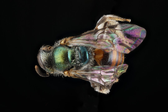
Lasioglossum eleutherense. Not your mother’s #Lasioglossum. This is a member of a group of primarily Caribbean Lasioglossum species that have gone beyond the dull colors of ordinary North American species to be all prettied up in bright colors. Oddly this particular species has a toehold in south south Florida, this one from Biscayne Bay. #bees #bee
57 notes
·
View notes
Link
It started with a mystery: How did molten salt breach its metal container? Understanding the behavior of molten salt, a proposed coolant for next-generation nuclear reactors and fusion power, is a question of critical safety for advanced energy production. The multi-institutional research team, co-led by Penn State, initially imaged a cross-section of the sealed container, finding no clear pathway for the salt appearing on the outside. The researchers then used electron tomography, a 3D imaging technique, to reveal the tiniest of connected passages linking two sides of the solid container. That finding only led to more questions for the team investigating the strange phenomenon.
They published the answers on Feb. 22 in Nature Communications.
“Corrosion, a ubiquitous failure mode of materials, is traditionally measured in three dimensions or two dimensions, but those theories were not sufficient to explain the phenomenon in this case,” said co-corresponding author Yang Yang, assistant professor of engineering science and mechanics and of nuclear engineering at Penn State. He is also affiliated with the National Center for Electron Microscopy at Lawrence Berkeley National Laboratory, as well as the Materials Research Institute at Penn State. “We found that this penetrating corrosion was so localized, it only existed in one dimension – like a wormhole.”
Read more.
25 notes
·
View notes
Photo
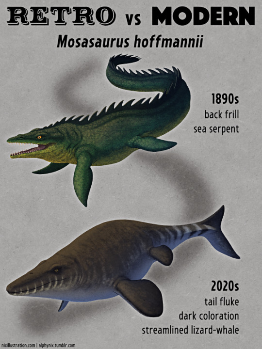
Retro vs Modern #07: Mosasaurus hoffmannii
The first scientifically documented mosasaur fossils were skulls discovered in the Netherlands during the 1760s and 1770s, but these remains were initially interpreted as belonging to a fish, crocodile, or whale. In the late 1790s their resemblance to monitor lizards was noted, and the fossils were soon recognized as belonging to giant marine reptiles unlike any known living species – a revolutionary concept at the time, and influential in the early development of ideas about extinction.
In the 1820s Mosasaurus hoffmannii was the first species officially described. For several decades it was thought to be a giant amphibious lizard with either webbed feet or flipper-like legs, with one of the earliest popular reconstructions being the 1850s Crystal Palace statue.
By the 1870s more complete fossil discoveries in North America had revealed the paddle-like flippers and fully aquatic nature of mosasaurs. Skin impressions showed overlapping keeled diamond-shaped scales resembling those of rattlesnakes, but proportionally much smaller compared to their body size.
1890s
Then, in the late 1890s, one mosasaur specimen was interpreted as having a mane-like “fringe” of soft tissue along its back.
Only a few years later this was realized to be a mistake, actually being preserved tracheal cartilage, but it was too late. The idea had already caught on in artistic depictions and quickly became a paleoart meme, with mosasaurs frequently portrayed with elaborate frills for the majority of the next century.
2020s
Early arguments about whether mosasaurs’ closest relatives were monitor lizards or snakes had settled down by the 1920s, with the consensus at the time being monitor lizards, and the first half of the 20th century saw little mosasaur research beyond the naming of a few new species. Much like the ichthyosaurs and plesiosaurs it was only really in the wake of the Dinosaur Renaissance that interest in these marine reptiles and their paleobiology really began to pick up again.
Rather than sea-serpent-like creatures we now recognize that mosasaurs actually looked more like lizards converging on whales or ichthyosaurs, with smooth streamlined bodies and vertical tail flukes. The size and shape of their scales varied across different parts of their bodies, parts of their bodies had dark coloration (likely with a countershaded pattern), and they probably had forked tongues.
They had a higher metabolic rate than most modern lizards, and may even have been warm-blooded. They probably also gave birth to live young, although a recently-discovered fossil soft-shelled egg found in Antarctica has been suggested to have come from a large mosasaur.
The debate about their evolutionary relationships has been reignited, too, with some recent studies once again supporting a very close relationship to snakes – although there’s currently no clear consensus.
Our modern view of Mosasaurus hoffmannii is a large chunky mosasaur that grew to at least 11m long (~36’). It lived during the end of the Cretaceous period, about 70-66 million years ago, and inhabited a wide range of climates across much of the ancient Atlantic Ocean and various connected shallow seaways, with fossils known from Europe, Africa, and North and South America.
Its long jaws had a powerful bite force and it seems to have been a more visual hunter than some other mosasaurs, with relatively large eyes and a less well-developed sense of smell. It was one of the largest marine animals of its time and was probably a generalist apex predator, feeding on a wide variety of prey such as fish, ammonites, and other marine reptiles.
———
Nix Illustration | Tumblr | Twitter | Patreon
1K notes
·
View notes
Photo

Retro vs Modern #05: Ichthyosaurus communis
Fossilized ichthyosaur bones have been found for centuries, but were initially misidentified as being the remains of fish, dolphins, and crocodiles. More complete skeletons began to be discovered in the early 19th century – particularly by pioneering paleontologist Mary Anning – and Ichthyosaurus communis was one of the first species of these ancient “fish lizards” to be scientifically recognized.
1830s-1870s
Early reconstructions of ichthyosaurs in the 1830s depicted flippered crocodile-like animals with long straight eel-like tails and strangely shrinkwrapped features, showing the sclerotic rings of their eyes and the internal bones of their flippers as highly visible externally. They were also frequently portrayed as being amphibious, hauling themselves out of the water to bask.
By the late 1830s impressions of smooth scaleless skin had been found, and specimens with tail-tips that were always “broken” in the exact same place were interpreted as evidence of the presence of some sort of paddle-like tail fin. The 1850s Crystal Palace Ichthyosaurus statues show this slightly updated version, along with a low dorsal ridge on their backs reminiscent of a beluga whale.
2020s
From the 1880s onwards the discovery of exceptional ichthyosaur specimens preserving whole body outlines revealed a fully aquatic streamlined shape, a triangular dorsal fin, and a crescent-shaped vertical tail fluke. Numerous examples of fossilized pregnant females also showed that ichthyosaurs gave live birth rather than laying eggs.
This highly dolphin-like version of ichthyosaurs quickly caught on and became the standard depiction into the early 20th century, frequently showing them as highly active animals – swimming in groups, chasing fish and ammonites, and leaping dramatically out of the water like their modern cetacean counterparts. While we don’t actually know if they were social or acrobatic like dolphins, it was still a surprising and refreshing contrast to the increasingly lumpy and sluggish depictions of non-avian dinosaurs that were happening around the same time.
Actual further paleontological study on ichthyosaurs was scarce for decades, however, with a general attitude that the group was already scientifically “complete” and there wasn’t much new or interesting left to learn about them anymore. It wasn’t until the late 20th century that they began to have their own “ichthyosaur renaissance” alongside the dinosaurs, with a sharp rise in research in the last few decades bringing us a lot of new information about their diversity and biology.
Ichthyosaurus communis was just one of several species in the Ichthyosaurus genus, living during the Early Jurassic, about 196-183 million years ago, in the shallow tropical seas of what is now Europe. About 3.3m long (~11’), it was adapted for high-speed long-distance swimming like a modern tuna, and it probably had a large keeled peduncle on the sides of its tail.
Bone structure and isotope analysis show that ichthyosaurs were all warm-blooded. One exceptional specimen also preserves an insulating layer of cetacean-like blubber, along with some evidence of its coloration: overall darker on the top and lighter on the underside in a countershaded pattern.
(I’ve given this reconstruction some speculative disruptive camouflage, too.)
Some of the preserved pigmentation has enough microscopic detail to show what appear to be branched melanophore cells associated with the ability to change color – suggesting that ichthyosaurs may have been able to actively darken and lighten their coloration like some modern lizards.
———
Nix Illustration | Tumblr | Twitter | Patreon
3K notes
·
View notes
Photo

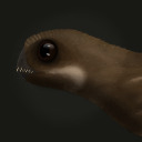
Pram Bug
Phronima sedentaria
The Pram Bug is a deep sea amphipod that is located between 200 to 1000m in the ocean. It has a translucent exoskeleton and can see primarily blue light. It is also is contained in a hollowed out barrel that is used for protection and to house babies. The image above is a female pram bug carrying its young.
Photo Credit:https://ocean.si.edu/ocean-life/invertebrates/phronima-female-and-young
406 notes
·
View notes
Text
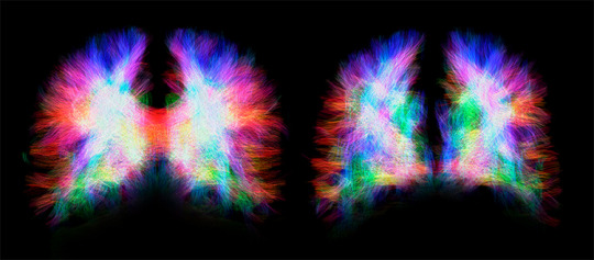
(Image caption: Neuronal fibres in a healthy brain (left) and a brain with agenesis of the corpus callosum (right). In the healthy brain, the two hemispheres are connected by the corpus callosum fibers, shown in red. These fibres are absent in the brain with corpus callosum agenesis. Credit: ©UNIGE/SIFFREDI)
A malformation illustrates the incredible plasticity of the brain
One in 4,000 people is born without a corpus callosum, a brain structure consisting of neural fibres that are used to transfer information from one hemisphere to the other. A quarter of these individuals do not have any symptoms, while the remainder either have low intelligence quotients or suffer from severe cognitive disorders. In a study published in the journal Cerebral Cortex, neuroscientists from the University of Geneva (UNIGE) discovered that when the neuronal fibres that act as a bridge between the hemispheres are missing, the brain reorganises itself and creates an impressive number of connections inside each hemisphere. These create more intra-hemispheric connections than in a healthy brain, indicating that plasticity mechanisms are involved. It is thought that these mechanisms enable the brain to compensate for the losses by recreating connections to other brain regions using alternative neural pathways.
The corpus callosum develops in utero between the tenth and twentieth week of gestation. Agenesis of the corpus callosum is a congenital brain malformation in which this brain structure fails to develop, resulting in one out of 4,000 babies born without a corpus callosum. When it is missing, nothing replaces this structure measuring about ten centimetres, with the exception of cerebrospinal fluid. This means that the information transmitted from one hemisphere to the other can no longer be conveyed by the neuronal projections from the corpus callosum. «Their role in a healthy brain,» begins Vanessa Siffredi, a researcher in UNIGE’s Faculty of Medicine, «is to ensure the functioning of various cognitive and sensorimotor functions». Surprisingly, 25% of people with this malformation have no visible signs; 50% have average intelligence quotients and learning difficulties; and the remaining 25% suffer from severe cognitive disorders.
Mysterious fibres
The scientific literature shows that, in the absence of the corpus callosum, certain fibres designed to serve as a bridge between the hemispheres, known as Probst bundles, bypass the absent brain area and curl up inside each hemisphere. «The back-up zones vary from one individual to another. And we don’t understand their functions,» explains the neuroscientist. The UNIGE scientists – working in collaboration with their colleagues at the University of Melbourne – set out to understand this variability and to examine the role of the fibres. Using MRI brain imaging, they studied the anatomical and functional links between different brain regions of approximately 20 Australian children aged 8 to 17 suffering from agenesis of the corpus callosum.
A salutary role
This approach first made it possible to observe the physical relationships between the different regions of the brain, i.e. their structural links. In children with corpus callosum agenesis, the neural fibres inside each hemisphere are greater in number and of higher quality than in healthy brains. Furthermore, the UNIGE scientists succeeded in determining the correlations between the activity of different brain regions and their functional links. «If two regions are active together, it means they are communicating with each other,» explains Dr Siffredi. The data shows that intra and inter-hemispheric functional connectivity of brains without the corpus callosum are comparable to those of healthy brains. «Remarkably, communication between the two hemispheres is maintained. We think that plasticity mechanisms, such as the strengthening of structural bonds within each hemisphere, compensated for the lack of neuronal fibres between hemispheres. New connections are created and the signals can be re-routed so that communication is preserved between the two hemispheres.»
Predicting cognitive impairment
The Geneva neuroscientists likewise observed a correlation between the increase in intra-hemispheric connections and cognitive skills. This information is very interesting for clinical work since, as agenesis is currently detected by means of ultrasound during pregnancy, it is often proposed that a pregnancy be terminated. «In the not-too-distant future, we could imagine using MRI imaging to predict whether the malformation observed by ultrasound runs the risk of being associated with cognitive impairment or not, and so better inform future parents», concludes Dr Siffredi.
156 notes
·
View notes
Photo
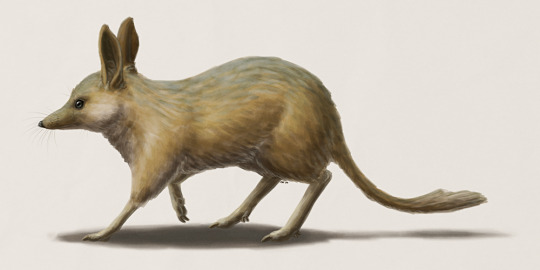
The pig-footed bandicoot (Chaeropus ecaudatus) was a small Australian marsupial which became extinct during the 20th century. Despite its name it wasn’t actually a bandicoot, being considered a sister group to the true bandicoots and bilbies within the Peramelemorphia.
Its most unusual features were its limbs, resembling those of ungulates more than other marsupials. The forefeet had two hoof-like toes similar to a deer or pig, giving it its common name, and the hindfeet had a single enlarged toe with a heavy claw. Two fused vestigial toes were also present on the ankle, but these were used for grooming rather than locomotion. Descriptions of how it moved around are contradictory, with the only known scientific account claiming it moved like a limping horse dragging its hind legs, while Aboriginal people familiar with the animals reported that they could move at high speed in a smooth galloping sprint.
Although once widespread across much of Australia, the species was rare in numbers even before the arrival of Europeans on the continent. It disappeared from some areas of its ancestral range before the most destructive invasive species – foxes and rabbits – had even arrived there, and its extinction may in fact have been due to habitat change. The end of many thousands of years of Aboriginal burning and land management combined with the introduction of sheep and cattle farming may simply have been too much for the already sparse pig-footed bandicoots.
The last confirmed specimen was found in 1901, and the last remnants of the species hung on the deserts of Western Australia until the 1950s. The distantly related lesser bilby suffered a similar fate, but recent conservation efforts are fighting to bring the greater bilby back from the brink – and it’s also a much more appropriate Australian Easter mascot than the highly destructive rabbit.
64 notes
·
View notes
Text

THIS is the bear cave painting i was talking about, the line weight, the proportions, the fine details around the face, and the fact that this all had to be drawn from memory, idk man, it’s incredible to me. if i could meet one person from history it’d be the person that painted this bear 30,000 years ago
169K notes
·
View notes
Text
"For a eukaryote to be classified as a ciliate, it really needs to meet one important criteria: it needs to have those short little hairs called cilia. And with such lax requirements, it's not surprising that ciliates can take on so many different forms. You've got the long-necked lacrymaria olor. The trumpet-y stentor coeruleus. The springy floral of the vorticella. They may look very different from one another, but they're all decorated with cilia that help them move or eat."
Journey to the Microcosmos- Suctorians: The Ugly Duckling of Ciliates
Images Originally Captured by Jam's Germs
Suctorian 400x, Stentor coeruleus 200x, Cilitae 600x, Lacrymaria olor 200x, Ciliate 400x, Vorticella 200x
79 notes
·
View notes
Photo

Oligocene Whale Had Both Teeth and Baleen, New Research Shows
http://www.sci-news.com/paleontology/aetiocetus-weltoni-09690.html
73 notes
·
View notes
Text
"Meanwhile, there's no sign of cilia on this suctorian. And there's none on this one either. They're hairless, practically bald by microbe standards... except for a few tentacles on their head. And there's some other weird traits that distinguishes suctorians from other ciliates. So why consider them a ciliate? Well, we'll get to that."
Journey to the Microcosmos- Suctorians: The Ugly Duckling of Ciliates
Images Originally Captured by Jam's Germs
Suctorian 630x, Suctorian 400x, Suctorian 630x, Suctorian 630x
44 notes
·
View notes
Photo
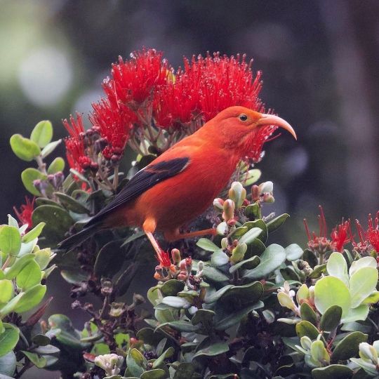
Meet the ʻIʻiwi (Drepanis coccinea)! It lives in tropical forests across the Hawaiian islands, where it sips on nectar and plays an important role in pollination. Its skinny decurved bill is specialized for fitting into and reaching nectar from tube-shaped flowers. 🌺
Photo: Gregory “Slobirdr” Smith, CC BY-SA 2.0, flickr
https://www.instagram.com/p/CPUk3x8grFA/?utm_medium=tumblr
404 notes
·
View notes
Photo

Bacteria Colonize Our Gut During and After Birth, New Study Shows
http://www.sci-news.com/biology/gut-microbial-colonization-09665.html
22 notes
·
View notes
Text
This is a coral appre-sea-ocean post
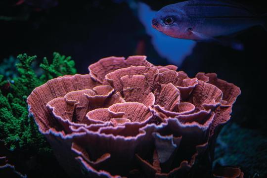
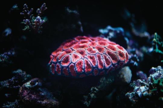
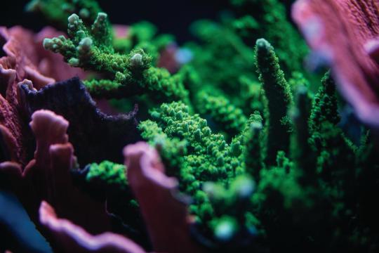
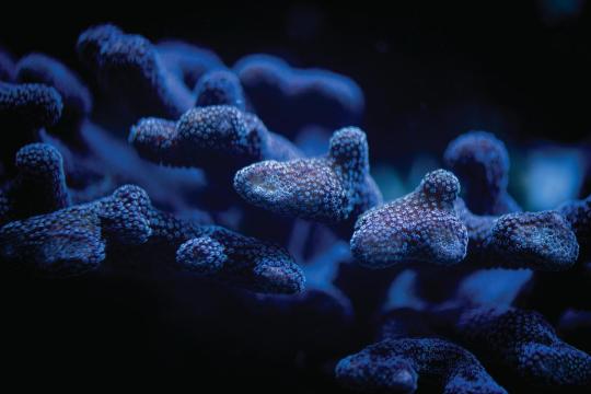
Shoutout to all you cnidariengineers banding together with your symbiotic photosynthesizers—you know who you are!
1K notes
·
View notes

Photo
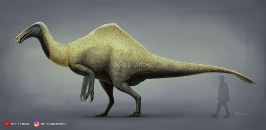
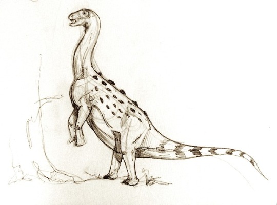
DEINOCHEIRUS
New reconstruction 2021. Available at Redbubble.com/people/mariolanzas for prints and more merch
instagram @mariolanzarensis
Youtube Channel
118 notes
·
View notes