Don't wanna be here? Send us removal request.
Statistics
We looked inside some of the posts by mbbshelp and here's what we found interesting.
Average Info
Notes Per Post
1
Likes Per Post
1
Reblog Per Post
0
Reply Per Post
0
Time Between Posts
24 days
Number of Posts By Type
Photo
17
Last Seen Tumblr Blogs


Fun Fact
Tumblr.com rank in the US is 25.
Photo

Anesthesia 2020 PDF (PG&FMGE both PDF) – S. Singh MBBS Help is No. 1 website for Handwritten Note. Why should you buy this pdf? Latest 2020 Note…
0 notes
Photo

Geographical ulcer branches of dendritic ulcer enlarges and combine to form 'amoeboid' or 'geographical configuration. Steroid use in dendritic ulcer also lead to geographical ulcer #Ophthalmology #geographicalulcer #amoeboidulcer #HSV https://www.instagram.com/p/B50eR96BUW5/?igshid=10wewkzbgd1qg
0 notes
Photo

Dendritic corneal ulcer (after fluorescein staining) seen in HSV. Herpetic simplex keratitis is a form of keratitis caused by recurrent herpes simplex virus (HSV) infection in the cornea. https://www.instagram.com/p/B5yB_JXBMAa/?igshid=1x2h6mg4ke40f
0 notes
Photo

Fungal keratitis refers to a corneal infection caused by fungi. One type of fungus that can infect the cornea is Fusarium. When Fusarium infects the cornea, the eye disease is referred to as Fusarium keratitis. https://www.instagram.com/p/B5sTGpSh7Bn/?igshid=17akowi6cl91b
0 notes
Photo
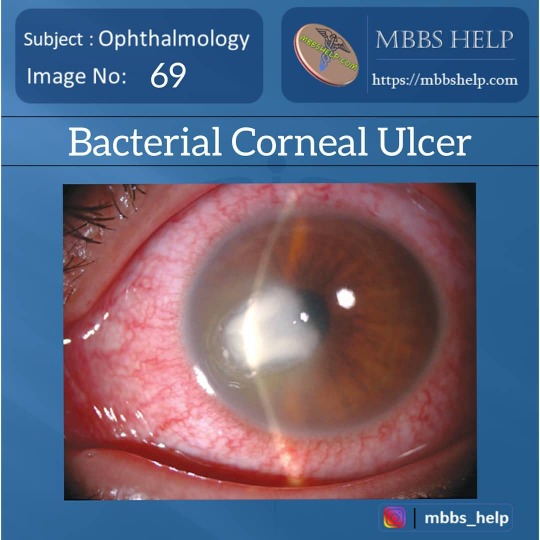
Bacterial corneal ulcers may follow a break in the corneal epithelium, thereby providing an entry for bacteria. The traumatic episode may be minor, such as a minute abrasion from a small foreign body, or may result from such causes as tear insufficiency, malnutrition, or contact lens use. Increased use of soft contact lenses in recent years has led to a dramatic rise in the occurrence of corneal ulcers, particularly due to Pseudomonas aeruginosa. Bacteria that can penetrate through intact corneal epithelium include Corynebacterium diphtheriae, Haemophilus influenzae, Neisseria gonorrhoea, Neisseria meningitidis, and Listeria species. https://www.instagram.com/p/B5rp651BTXV/?igshid=yvkq2qgcujxb
0 notes
Photo
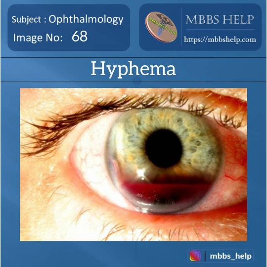
Hyphema is blood in the front (anterior) chamber of the eye. It may appear as a reddish tinge, or it may appear as a small pool of blood at the bottom of the iris or in the cornea. https://www.instagram.com/p/B5qO24hB41h/?igshid=12337htrr0kvq
0 notes
Photo

Subconjunctival hemorrhage, is bleeding from a small blood vessel in the outer layer of the eye (the conjunctiva) into the space between the conjunctiva and the sclera. It results in a red spot in the white of the eye. https://www.instagram.com/p/B5qN7xfhjNd/?igshid=1tckppler18in
0 notes
Photo
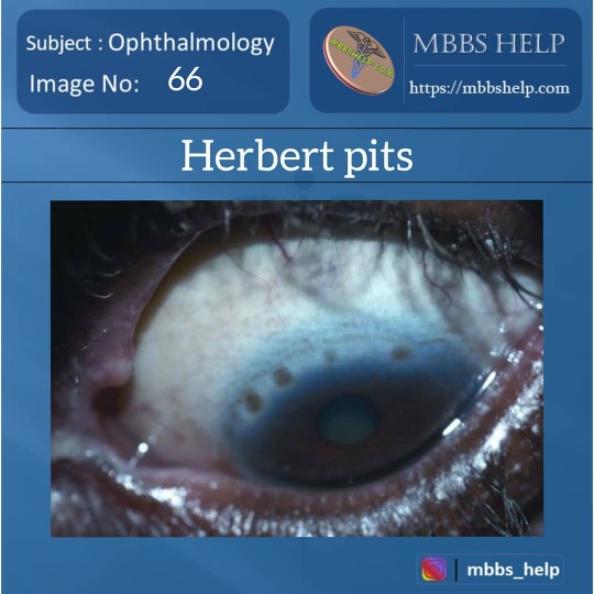
Trachoma exhibiting Herbert pits of the superior limbus (round to oval, pigmented areas within pannus). https://www.instagram.com/p/B5qM1WjBx44/?igshid=s2pc1unocoy
0 notes
Photo

Arlt's line is a thick band of scar tissue in the conjunctiva of the eye, near the lid margin, that is associated with eye infections. Arlt's line is a characteristic finding of trachoma, an infection of the eye caused by Chlamydia trachomatis. The line runs horizontally, parallel to eyelid, and is found at the junction of the anterior one third and posterior two thirds of the conjunctiva. The line is named after the Austrian ophthalmologist Carl Ferdinand von Arlt. https://www.instagram.com/p/B5qLuiNBqJ-/?igshid=hzn34c64twii
0 notes
Photo

BOILED SAGO GRAIN appearances seen in upper palpebral conjunctiva TRACHOMA Trachoma, an infection of the eye caused by Chlamydia trachomatis. Subepithelial aggregation of lymphocytes, multinucleated giant cells with necrosis. https://www.instagram.com/p/B5ncVHChQwS/?igshid=19mrca5esrkdh
0 notes
Photo

BOILED SAGO GRAIN appearances seen in upper palpebral conjunctiva TRACHOMA Trachoma, an infection of the eye caused by Chlamydia trachomatis. Subepithelial aggregation of lymphocytes, multinucleated giant cells with necrosis. https://www.instagram.com/p/B5ncEehBg6j/?igshid=1ng5cjqt72s4m
0 notes
Photo
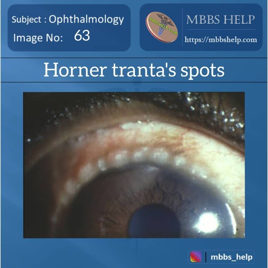
Horner-Tranta's spots seen in Vernal Keratoconjunctivitis. Horner-Tranta's spots/dots (raised, white accumulations of eosinophils at the limbus), gelatinous limbal follicles, and copious amounts of mucoid discharge. https://www.instagram.com/p/B5naEq3hBL9/?igshid=lgusmf1fn8rj
0 notes
Photo
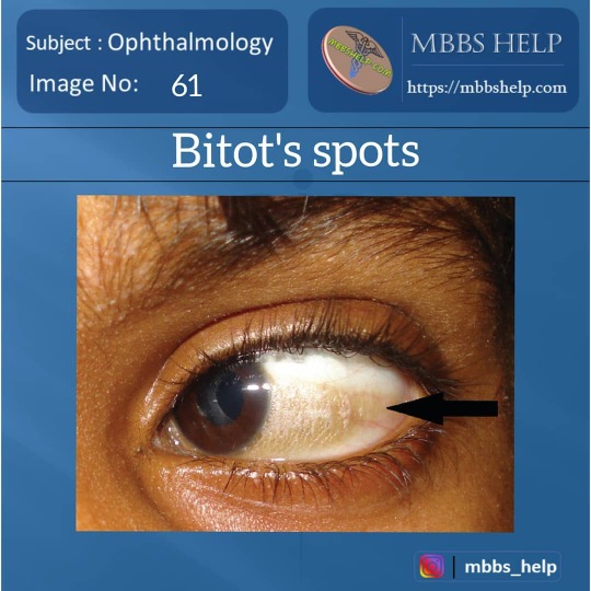
A BITOT'S SPOT is a sharply defined, gray or white lesion involving the exposed portion of the bulbar conjunctiva. Frequently bilateral, it is triangular in shape with its base at the temporal limbus and its apex extending toward the lateral canthus. The elevated, dry, lusterless surface of the Bitot's spot is distinctively frothy or foamy in appearance. The lesion is due to a keratinization of the conjunctival epithelium and is considered to be one of the classical features of vitamin A deficiency. As such, the Bitot's spot may be accompanied by night blindness or be followed by extensive xerosis of the conjunctiva, xerosis of the cornea, and keratomalacia. https://www.instagram.com/p/B5mwD7UBb4o/?igshid=1c4tq74vycqqv
1 note
·
View note
Photo

Microbiology PDF – S. Panwar MBBS Help is No. 1 website for Handwritten Note. Why should you buy this pdf? Latest 2019 Note…
0 notes
Photo
Physiology PDF – V. Nalgirkar MBBS Help is No. 1 website for Handwritten Note. Why should you buy this pdf? Latest 2019 Note…
0 notes
Photo

Obs and gynae PDF – P. Vij MBBS Help is No. 1 website for Handwritten Note. Why should you buy this pdf? Latest 2019 Note…
0 notes