
Statistics
We looked inside some of the posts by relianceonscience and here's what we found interesting.
Average Info
Notes Per Post
25K
Likes Per Post
16K
Reblog Per Post
9K
Reply Per Post
49
Time Between Posts
16 days
Number of Posts By Type
Photo
6
Note
1
Link
7
Text
2
Video
1
Last Seen Tumblr Blogs


Fun Fact
Tumblr has 411 employees.

Photo

New research find that the naked mole rat’s chances of dying do not increase over time.
The naked mole rat has been boggling the minds of scientists for many years now. What it lacks in conventual cuteness it makes up for with some superpower-like qualities. It can survive for 18 minutes without oxygen. It’s practically immune to cancer. But perhaps it’s most notable characteristic is that the naked mole rat can live longer than any animal its size…up to 30 years or more.

And now scientists have discovered one more thing about this rodent’s abnormally long life—its chances of dying don’t increase over time. For most mammals, the rate of mortality increases along with age once the animal reaches adulthood (referred to as the Gompertz-Makeham law of mortality). For example, in humans, our risk of dying roughly doubles every year after turning 30. For naked mole rats, death is random.

“We’ve got as much chance of finding a one-year-old [mole rat] that has died as finding a 25-year-old that has died,” says Dr. Rochelle Buffenstein, Senior Principal Investigator at Calico. She joins Ira to discuss what she discovered digging into data on the naked mole rat. Listen to the interview with Buffenstein here.

Photos by Science Friday and Shutterstock.
717 notes
·
View notes
Note
This is amazing!
What kind of frogs do you think are the weirdest/coolest frogs?
Oh, this might need a really long answer as there are such a lot of cool and weird frogs!
Personally, I have a weakness for animals that look chubby and grumpy, so I love Rain frogs (Breviceps sp.)…

(x)

(x)

(x)
…and Horned frogs (Ceratophrys sp.)

(x)
…they have also a great colour variance:


(x)(x)
Glass frogs (Centrolenidae) are translucent…

…and a lot of them have absolutely amazing eyes:

Mossy frogs (Theloderma corticale) live reallly up to their name…

…as does the Malayan leaf frog (Megophrys nasuta)…

…the Pinocchio frog (Litoria sp. nov.)…

…and the Fringe leaf frog (Cruziohyla craspedopus)…

…so Hemiphractus fasciatus (a kind of Horned tree frog) might as well get the name Pyramid head frog :-)

Marsupial frogs (Gastrotheca and Flectonotus) can look quite bizarre when the egg poaches on their backs are occupied…

…whereas Surinam toads (Pipa) look kind of weird whether or not they have eggs and tadpoles implanted:


(x)
The Turtle frog (Myobatrachus gouldii) looks like a turtle that lost its shell :-)


I think I’ll have to stop for now (I could go on for quite a while: Colourful frogs! Poisonous frogs! Shovel-nosed frogs! Striped frogs! Aquatic frogs! Burrowing frogs! Tiny frogs! Giant frogs! Frogs frogs frogs frogs frogs!).
Don’t forget to take a look at the last post about the most bad-ass of all frogs: the Hairy or Horror frog (Trichobatrachus robustus), also known as the Wolverine of the amphibian world :-)

3K notes
·
View notes
Link
The gnashers inside your mouth may well have originated as fish scales, according to new research that found the same type of cells that human teeth have in the thorny scales of the little skate fish.
These neural crest cells were spotted using fluorescent markers to track cells inside the embryo of a little skate, one of the cartilaginous fish group – like sharks and rays – that have skeletons made entirely of cartilage rather than bone.
Continue Reading.
392 notes
·
View notes
Link
It may be possible, scientists say, to save many thousands of newborns in poor countries by giving them a simple probiotic — a strain of bacteria originally scooped out of the diaper of a healthy baby.
A large clinical trial in rural India has found that babies fed a special strain of Lactobacillus bacteria for just one week were 40 percent less likely to develop sepsis, a life-threatening bloodstream infection.
Sepsis kills 600,000 newborns a year, many of them in India and neighboring countries.
The treatment was so successful that an oversight panel stopped the trial early because it would have been unethical to keep giving a placebo to half the babies in the study.
Continue Reading.
140 notes
·
View notes
Photo

Katherine Johnson (b. 1918) is a physicist and mathematician who has made crucial contributions to several NASA missions, assuring their success with her highly accurate calculations. She worked with NASA for several decades, and helped advance the rights of both African-Americans and women.
She initially worked as a human computer, and later as an aerospace technologist. She calculated trajectories for missions such as the 1961 Mercury mission or the 1969 Apollo 11 flight. She was portrayed by Taraji P. Henson in the 2016 film Hidden Figures.
6K notes
·
View notes
Photo

What Bloodied This Teen’s Feet? Tiny Marine Flesh Eaters Had Scientists Stumped
The attack took all of half an hour.
That’s about how long Sam Kanizay says he spent relaxing waist-deep in the waters of Melbourne, Australia’s Brighton Beach on Saturday. The 16-year-old had been footsore from a round of soccer, he says, and the cold water felt pleasant as he absently listened to songs on his iPhone.
It was only when he finally waded out of the water that he discovered what had happened to him: Blood streamed from thousands of tiny pinpricks that littered his legs from the ankle down.
The Museums Victoria, a consortium of state-owned museums in Melbourne, declared Sunday they believe they had cracked the mystery. After testing the creatures captured by the father, marine biologist Genefor Walker-Smith identified them as lysianassid amphipods, “a type of scavenging crustacean.”
The little crustaceans, often known as “sea fleas,” are “naturally-occurring scavengers, which commonly bite but do not usually cause these kind of injuries,"
"It’s possible the amphipods contained an anti-coagulant, which would account for the inability to stop the flowing blood and that the very cold water may be the reason Sam didn’t feel the bites. The amphipods have no venomous properties and will not cause lasting damage.”
354 notes
·
View notes
Link
Researchers have developed a tiny chip that, when applied to an affected area, can regenerate and repair failing body functions by turning skin cells into other types of healing cells.
The technology could mean a breakthrough in the treatment of Alzheimer’s or Parkinson’s disease, as doctors could grow brain cells on a person’s own skin, harvest the cells and inject them into the brain using the chip.
It sounds like science fiction, but Dr. Chandan Sen, director of Ohio State’s Center for Regenerative Medicine & Cell Based Therapies says that the technology implies a huge realm of possibilities. “By using our novel nanochip technology, injured or compromised organs can be replaced,” Sen said in a press release. “We have shown that skin is a fertile land where we can grow the elements of any organ that is declining.”
The process, called Tissue Nanotransfection (TNT), uses a chip loaded with specific genetic code or certain proteins; a cargo that’s been designed for cell conversion. The chip is placed on the affected area and a small electrical current is applied, creating channels in the patient’s tissue. This allows DNA or RNA to be injected into those channels, where they take root and reprogram the affected cells to begin the healing process.
–
Submitted by @neena213.
543 notes
·
View notes
Text
Allergic to the Sun?: A Genetic Look into X-Linked Protoporphyria
Advancement in genetics have assisted doctors and scientists around the world in identifying the genetic causes behind some of the most puzzling disorders. These advancements have been most helpful with Mendelian disorders – those that are caused by a mutation in only one gene. While these disorders are rare, the results of a single mutation can be dramatic. I am going to focus on one in particular: X-Linked Protoporphyria (XLP). No, not the porphyria from which King George III allegedly suffered. This porphyria’s main symptom is photosensitivity, colloquially known as a sun allergy.
The name sun allergy is actually a misnomer. An allergy refers to an immune system response (ex. hay fever) while XLP is actually a chemically induced skin irritation (similar to a sun burn, pictured below) caused by an overabundance of a compound called a protoporphyrin. In XLP, this overabundance is caused by a single mutation in one gene. For those whose high school biology days are a bit too far away to remember, a mutation is an alteration in the DNA code. A gene is a portion of the DNA that gives the instructions for the creation of proteins. The protein affected by XLP is called ALAS2.
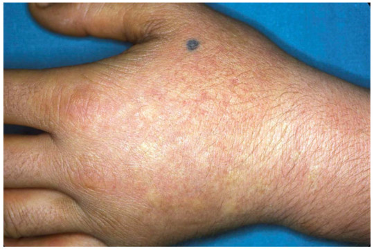
Acute photosensitivity reaction
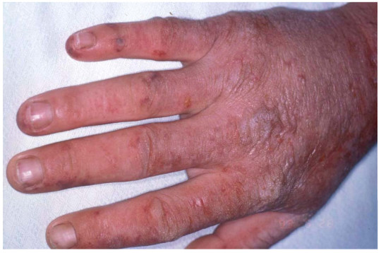
Another acute photosensitivity reaction
Symptoms of XLP show at infancy but vary in severity between males and females. The symptoms include photosensitivity (a painful skin reaction to sunlight), anemia, and low iron levels in the blood. Uncommonly, those affected can have liver damage which can eventually lead to the need for a liver transplant. Unfortunately, the number of people affected with XLP is hard to determine since it is so rare and very similar to another porphyria, Erythropoietic Protoporphyria. So far, a dozen or so families have been identified and there seems to be no correlation to geographic location or ethnicity. It affects everyone equally.
Knowing that other porphyrias are related to a mutation in the DNA, doctors aimed to find the mutation that caused XLP. This relied on many past studies, the largest of which was the Human Genome Project. It amassed a huge amount of information on what many of our genes do and where they are located.
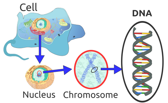
DNA in Human Cell

Human chromosomes, coloured by UCSC browser default colours
As you can see above, each human cell contains all 23 pairs of chromosomes within it’s nucleus. Each pair contains part of your DNA and it’s own unique genes. Which chromosome and gene would XLP be found in? Using previous studies, Whatley et al made an educated guess that is was in a gene called ALAS2 (5-aminolevulinic acid synthase), named after the protein it’s “blueprints” create. ALAS2 is located on the X chromosome, number 23. Females have two X chromosomes and males have one X and one Y, hence the name sex chromosomes. The fact that it is only on the X chromosome means that it is “linked” to that chromosome, so the mutation only shows up in offspring if the affected X chromosome is inherited. Daughters get one X chromosome from their father and one from their mother. Sons, however, get one Y chromosome from their father and one X chromosome from their mother. Depending upon which parent’s X chromosome is affected, and which one gets passed on, affects which children inherit the mutation.

X-Linked Dominant Inheritance
Since XLP doesn’t get passed to every child, the image above helps to show part of why XLP was difficult to identify. The first scientists to do so was a team lead by Sharon D. Whatley that published the find in 2008.
While everyone has an ALAS2 gene, as it is part of blood cell production, not everyone has the mutation. However, scientists know that the ALAS2 gene is found on chromosome X, so that’s where Whatley et al looked. Below is an X portion of chromosome 23 with the area containing the ALAS2 gene highlighted. It turns out that Whatley et al was right and further studies have identified other mutations within the ALAS2 gene that also cause XLP.
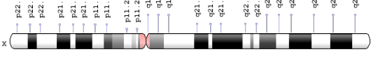
ALAS 2 Gene Location
After identifying the location of the mutation, scientists attempted to find why the mutation causes the multitude of symptoms. They started by looking at what the ALAS2 protein does. It had already been found that this protein is involved in your blood cell production, which is involved in creating protoporphyrins. It turns out that the ALAS2 mutations change the “blueprints” (DNA) enough to change the shape of the ALAS2 protein. Below is an example structure of non-mutated ALAS2 protein.

Crystal Structure of ALAS2 Protein from R. capsulatus
The change in shape, or conformation, causes the protein to do it’s work faster. Due to this, a chemical chain reaction occurs and eventually leads to too many protoporphyrins within a patient’s body. The main pathway involved is called the heme pathway, and as you may have guessed, it’s involved in blood cell production.
Since blood cell production and it’s related compounds are so important, there are many other diseases related to them, including the porphyria King George III was theorized to suffer from. Many of the other diseases and porphyrias have distinct symptoms, but one in particular, Erythropoietic Protoporphyria (EPP), is so similar to XLP that it is nearly impossible to tell them apart without a genetic test. In fact, many people diagnosed with EPP may have XLP but have never been tested. Currently, there are only 3 testing locations that can identify XLP through a genetic test: the Mayo Clinic in Rochester, MN, Icahn School of Medicine at Mount Sinai in New York, NY, and Odense University Hospital in Odense, Denmark.

Locations of XLP Testing Locations (edited from original)
Even with the identification of XLP as the cause of symptoms, doctors have not found a cure for it or an FDA-approved treatment. However, doctors have figured out numerous ways to manage the symptoms. The main method is sunlight avoidance which prevents the chemical reaction with porphyrins in a patient’s skin from occurring. There are many UV-protective clothes now, but staying inside is key. Some drugs have been shown to improve tolerance to sunlight, such as Lumitene or Cysteine and another drug has been shown to absorb some porphyrins, Cholestyramine. A new medication, Afamelanotide, is in clinical trials and may provide another option for sunlight protection.
None of these treatments would have been possible if the underlying mutation and subsequent chemicals had not been identified. Since the 90s and the Human Genome Project, technology has advanced rapidly. Sequencing, the method of reading DNA, used to cost millions. Now, it costs hundreds to thousands depending upon how much of the DNA you want to read. This decrease led to the birth of genomics, a discipline that uses genetics and molecular biology to identify the structure, function, and evolution of entire genomes (all of the genes within one person or species). So many different diseases and disorders have been identified, and it was mainly possible due to what is called open-source software. Doctors and scientists worked together by posting their findings on the internet, which are freely available for anyone to view. In sharing this knowledge, they were able to build upon each other’s work. This is exactly how Whatley and her team were able to identify the XLP mutation.
Identifying the genetic components of diseases and disorders is the first step in treating and curing them. The birth of genomics was a direct result of advancement in computer technologies. Without the internet, increased computing capabilities, and larger data storing methods, the human genome may not have been mapped. The faster our technology advances, the faster we can understand the role our genes play in our lives. This leads to identifying cures not only for rare disorders like XLP, but for more common diseases/disorders, such as cancer, Alzheimer’s, and Huntington’s.
References:
1. “X-Linked Protoporphyria – NORD (National Organization For Rare Disorders)”. NORD. N. p., 2013 & 2016. Web. 12 June 2017.
2. Whatley, Sharon D. et al. “C-Terminal Deletions in the ALAS2 Gene Lead to Gain of Function and Cause X-linked Dominant Protoporphyria Without Anemia or Iron Overload”. The American Journal of Human Genetics 83.3 (2008): 408-414. Web. 12 June 2017.
3. “Homologene – NCBI”. Ncbi.nlm.nih.gov N.p., 2017. Web. 12 June 2017.
4. “OMIM Entry – “300752 – PROTOPORPHYRIA, ERYTHROPOIETIC, X-LINKED; XLEPP”. Omim.org. McKusick, Victor A., and Kniffin, Cassandra L.,2017. Web. 12 June 2017.
5. Balwani M, Bloomer J, Desnick R; Porphyrias Consortium of the NIH-Sponsored Rare Diseases Clinical Research Network. X-Linked Protoporphyria. 2013 Feb 14. In: Pagon RA, Adam MP, Ardinger HH, et al., editors. GeneReviews® [Internet]. Seattle (WA): University of Washington, Seattle; 1993-2017. Web. 12 June 2017.
6. Balwani, Manisha et al. "Loss-Of-Function Ferrochelatase And Gain-Of-Function Erythroid-Specific 5-Aminolevulinate Synthase Mutations Causing Erythropoietic Protoporphyria And X-Linked Protoporphyria In North American Patients Reveal Novel Mutations And A High Prevalence Of X-Linked Protoporphyria.". Mol Med 19 (2013): 26-35. Web. 21 June 2017.
7. Fratz, Erica J. et al. "Human Erythroid 5-Aminolevulinate Synthase Mutations Associated With X-Linked Protoporphyria Disrupt The Conformational Equilibrium And Enhance Product Release". Biochemistry 54.36 (2015): 5617-5631. Web. 21 June 2017.
8. Fratz-Berilla, Erica J. et al. "Isoniazid Inhibits Human Erythroid 5-Aminolevulinate Synthase: Molecular Mechanism And Tolerance Study With Four X-Linked Protoporphyria Patients". Biochimica et Biophysica Acta (BBA) - Molecular Basis of Disease 1863.2 (2017): 428-439. Web. 21 June 2017.
9. Minder, Elisabeth I. "Afamelanotide, An Agonistic Analog Of Α-Melanocyte-Stimulating Hormone, In Dermal Phototoxicity Of Erythropoietic Protoporphyria". Expert Opinion on Investigational Drugs 19.12 (2010): 1591-1602. Web. 21 June 2017.
10. Minder, El., Schneider-Yin, X., Steurer, J., Bachmann, L.M. "A Systematic Review Of Treatment Options For Dermal Photosensitivity In Erythropoietic Protoporphyria". Cell Mol Biol (Noisy-le-grand) 55.1 (2009): 84-97. Web. 21 June 2017.
11. "ALAS2 - 5-Aminolevulinate Synthase, Erythroid-Specific, Mitochondrial Precursor - Homo Sapiens (Human) - ALAS2 Gene & Protein." Uniprot.org. N.p., 2017. Web. 21 June 2017.
12. Team, EBI. “ALAS2 Homo Sapiens P22557”. Ebi.ac.uk. N.p., 2017. Web. 27 June 2017.
13. “Porphyrin”. En.wikipedia.org. N.p., 2017. Web. 27 June 2017.
14. Shipman, Alexa R., and Kate E. Shipman. “X-Linked Dominant Protoporphyria: Response To “Cutaneous Porphyrias Part 1””. International Journal of Dermatology 54.3 (2014): e87-e88. Web. 28 June 2017.
15. Brancaleoni, V. et al. “X-Chromosomal Inactivation Directly Influences The Phenotypic Manifestation of X-Linked Protoporphyria”. Clinical Genetics 89.1 (2015): 20-26. Web. 29 June 2017.
16. "Transcript: ALAS2-201 (ENSLACT00000016428.1) - Summary - Latimeria Chalumnae - Ensembl Genome Browser 89". Ensembl.org. N.p., 2017. Web. 1 July 2017.
17. Benoff, S., Skoultchi, A. I. “X-linked control of hemoglobin production in somatic hybrids of mouse erythroleukemic cells and mouse lymphoma or bone marrow cells.” Cell. 12: 263-274, 1977. Web. 1 July 2017.
18. Astrin, K. H., Desnick, R. J., Bishop, D. F. “Assignment of human delta-aminolevulinate synthase (ALAS) to chromosome 3.” (Abstract)Cytogenet. Cell Genet. 46: 573, 1987. Web. 1 July 2017.
19. Astrin, K. H., Bishop, D. F. “Assignment of human erythroid delta-aminolevulinate synthase (ALAS2) to the X chromosome.” (Abstract)Cytogenet. Cell Genet. 51: 953-954, 1989. Web. 1 July 2017.
20. Cotter, P. D., Baumann, M., Bishop, D. F. “Enzymatic defect in 'X-linked' sideroblastic anemia: molecular evidence for erythroid delta-aminolevulinate synthase deficiency.” Proc. Nat. Acad. Sci. 89: 4028-4032, 1992. Web. 1 July 2017.
21. Surinya, K. H., Cox, T. C., May, B. K. “Identification and characterization of a conserved erythroid-specific enhancer located in intron 8 of the human 5-aminolevulinate synthase 2 gene.” J. Biol. Chem. 273: 16798-16809, 1998. Web. 1 July 2017.
22. Bishop, D. F., Henderson, A. S., Astrin, K. H. “Human delta-aminolevulinate synthase: assignment of the housekeeping gene to 3p21 and the erythroid-specific gene to the X chromosome.” Genomics. 7: 207-214, 1990. Web. 1 July 2017.
23. O’Connor, Leigh, Jane Gilmour, and Constanze Bonifer. “The Role of the Ubiquitously Expressed Transcription Factor Sp1 in Tissue-Specific Transcriptional Regulation and in Disease.” The Yale Journal of Biology and Medicine 89.4 (2016): 513–525. Web. 1 July 2017.
24. Merika, M, and S H Orkin. “DNA-Binding Specificity of GATA Family Transcription Factors.” Molecular and Cellular Biology 13.7 (1993): 3999–4010. Web. 1 July 2017.
25. Han L, Lu J, Pan X. et al. “Histone acetyltransferase p300 regulates the transcription of human erythroid-specific 5-aminolevulinate synthase gene.” Biochem Biophys Res Commun. 2006;348(3):799–806. Web. 1 July 2017.
26. Wei, Chunlan et al. “Effects Of Psychological Stress On Serum Iron And Erythropoiesis.” International Journal of Hematology 88.1 (2008): 52-56. Web. 10 July 2017.
27. Tian, Xue et al. “Psychological Stress Induced Zinc Accumulation And Up-Regulation of ZIP14 And Metallothionein In Rat Liver.” BMC Gastroenterology 14.1 (2014): n.p. Web. 10 July 2017.
28. Ninomiya, Yukiko et al. “X-Linked Dominant Protoporphyria: The First Reported Japanese Case.” The Journal of Dermatology 43.4 (2015): 414-418. Web. 10 July 2017.
29. Livideanu, Cristina Bulai et al. “Late-Onset X-Linked Dominant Protoporphyria: An Etiology Of Photosensitivity In The Elderly.” Journal of Investigative Dermatology 133.6 (2013): 1688-1690. Web. 10 July 2017.
30. "Variation Viewer." Ncbi.nlm.nih.gov. N.p., 2017. Web. 11 July 2017.
31. "NM_000032.4(ALAS2):C.1757A>T (P.Tyr586phe) AND Not Specified - Clinvar - NCBI." Ncbi.nlm.nih.gov. N.p., 2017. Web. 11 July 2017.
32. "Gene: ALAS2 (ENSG00000158578) - Transcript Comparison - Homo Sapiens - Ensembl Genome Browser 89." Ensembl.org. N.p., 2017. Web. 11 July 2017.
33. "GWAS Central - Gene/Region." Gwascentral.org. N.p., 2017. Web. 11 July 2017.
34. Balwani, Manisha et al. "Clinical, Biochemical, And Genetic Characterization Of North American Patients With Erythropoietic Protoporphyria And X-Linked Protoporphyria." JAMA Dermatology (2017): n. pag. Web. 13 July 2017.
35. Stadhouders, Ralph et al. "Control Of Developmentally Primed Erythroid Genes By Combinatorial Co-Repressor Actions." Nature Communications 6 (2015): 8893. Web. 13 July 2017.
36. Guberman, Alejandra S., María E. Scassa, and Eduardo T. Cánepa. "Repression Of 5-Aminolevulinate Synthase Gene By The Potent Tumor Promoter, TPA, Involves Multiple Signal Transduction Pathways." Archives of Biochemistry and Biophysics 436.2 (2005): 285-296. Web. 13 July 2017.
37. "Porphyrin And Heme Synthesis And Bilirubin Metabolism." Themedicalbiochemistrypage.org. N.p., 2017. Web. 15 July 2017.
38. Tanimura N, Miller E, Igarashi K, Yang D et al. Mechanism governing heme synthesis reveals a GATA factor/heme circuit that controls differentiation. EMBO Rep (2016): 249-65. PMID: 26698166 Web. 18 July 2017.
39. Database, GeneCards. "ALAS2 Gene - Genecards | HEM0 Protein | HEM0 Antibody." Genecards.org. N.p., 2017. Web. 21 June 2017.
40. Das, Devika, Ahmed Murad, and Ayman Saad. "Erythropoietic Porphyria – Role of Curative Hematopoietic Stemcell Transplantation." Jscimedcentral.com. N.p., 2017. Web. 20 July 2017.
41. "Genome Decoration Page." Ncbi.nlm.nih.gov. N.p., 2017. Web. 20 July 2017.
42. Astner, Isabel et al. "Crystal Structure Of 5-Aminolevulinate Synthase, The First Enzyme Of Heme Biosynthesis, And Its Link To XLSA In Humans." The EMBO Journal 24.18 (2005): 3166-3177. Web. 20 July 2017.
43. "Porphyrin And Heme Synthesis And Bilirubin Metabolism." Themedicalbiochemistrypage.org. N.p., 2017. Web. 21 July 2017.
44. "Genetests.Org." GeneTests.org. N.p., 2017. Web. 20 July 2017.
45. "List Of DNA Testing Companies - ISOGG Wiki." Isogg.org. N.p., 2017. Web. 20 July 2017.
46. "Heme Biosynthesis - Biosystems - NCBI." Ncbi.nlm.nih.gov. N.p., 2017. Web. 21 July 2017.
47. Reference, Genetics. "ALAS2 Gene." Genetics Home Reference. N.p., 2017. Web. 20 July 2017.
48. Scheiner, Dr. et al. "Research Proves You Can Reverse Aging With BBL Treatment – Adam Scheiner MD." Adam Scheiner MD. N.p., 2017. Web. 20 July 2017.
49. "File:Acute Photosensitivity Reaction In EPP.Jpg - Wikimedia Commons." Commons.wikimedia.org. N.p., 2017. Web. 21 July 2017.
50. "Erythropoietic Protoporphyria = البروتو بورفيريا المكونة للدم." Dermaamin.com. N.p., 2017. Web. 21 July 2017.
51. Landefeld C, et al. "X-Linked Protoporphyria: Iron Supplementation Improves Protoporphyrin Overload, Liver Damage And Anaemia. - Pubmed - NCBI." Ncbi.nlm.nih.gov. N.p., 2017. Web. 24 July 2017.
52. Stafford, R. et al. "The Impact Of Photosensitivity Disorders On Aspects Of Lifestyle." British Journal of Dermatology 163.4 (2010): 817-822. Web. 24 July 2017.
53. "Erythropoietic Protoporphyria (EPP) - Clinuvel Pharmaceuticals." Clinuvel.com. N.p., 2017. Web. 24 July 2017.
54. Hunter, Gregory A., and Gloria C. Ferreira. "Molecular Enzymology Of 5-Aminolevulinate Synthase, The Gatekeeper Of Heme Biosynthesis." PubMed Central Canada. N.p., 2017. Web. 2 Aug. 2017.
55. Heinemann, Ilka U., Martina Jahn, and Dieter Jahn. "The Biochemistry Of Heme Biosynthesis." Archives of Biochemistry and Biophysics 474.2 (2008): 238-251. Web. 2 Aug. 2017.
0 notes
Link
I still don't like broccoli. 😐
A new compound may lead to a drug that can destroy melanoma cancer cells while leaving nearby healthy cells unharmed, new research on mice and human cells indicates.
Researchers designed and synthesized a compound called napthalamide-isoselenocyanate—NISC-6—to inhibit both the Akt1 pathway and human topoisomerase IIα—topo IIα—activity, which contribute to melanoma tumor growth.
Melanoma, which is caused primarily by exposure to the sun’s ultraviolet rays, accounts for less than 5 percent of skin cancer cases, but causes more than 75 percent of skin cancer deaths.
In the study, the compound caused human melanoma cells to die and inhibited tumor growth by about 69 percent in a mouse model.
Continue Reading.
98 notes
·
View notes
Link
Aquatic plants buried underground for more than a century can be revived and regrown, according to a new study investigating the phenomenon of ‘ghost ponds’ – ponds that aren’t properly drained but filled in with soil and vegetation under agricultural land.
Restoring some of these buried ponds, and the habitats hidden in limbo beneath the soil, could be a valuable way of reversing habitat and biodiversity losses, say researchers, and we could even bring some plant species back from the dead.
The team from University College London in the UK has dug out three ghost ponds so far and estimates there could be as many as 600,000 similar patches spread out across the English countryside.
“We have shown that ghost ponds can be resurrected, and remarkably wetland plants lost for centuries can be brought back to life from preserved seeds,” says lead researcher Emily Alderton.
Continue Reading.
5K notes
·
View notes
Video
youtube
Associate Professor Marnie Blewitt and epigenetics
0 notes
Link
Much like students doing a test, rats tend to skip questions when they have forgotten the answer. A series of smelly experiments suggests rats are aware of what they remember, and behave differently when they can’t recall something.
Victoria Templer at Providence College, Rhode Island, and her team trained rats to dig through sand to sniff samples of cinnamon, thyme, paprika or coffee, and then go to a dish smelling of the matching scent. If the rats picked the correct dish, they got a piece of cereal.
But there was a twist. Although rats that chose a dish with the wrong scent got no reward, rats that positioned themselves next to a fifth, unscented dish received a quarter-piece of the cereal. This meant that when rats forgot what they had smelled in the sand, their best bet was to pick the unscented dish – provided they could tell that they had forgotten the relevant smell.
Continue Reading.
397 notes
·
View notes
Link
The story of Rapa Nui or Easter Island and its earliest settlers may have to be rewritten after a new study of ancient remains on the island challenged the conventional thinking that the citizens recklessly destroyed their idyllic island environment.
It turns out that the Rapa Nui people may have had a more diverse diet and taken better care of their home than experts had originally thought, based on carbon and nitrogen isotope analysis of bone and plant samples dating back as far as 1400 AD.
Continue Reading.
136 notes
·
View notes
Photo

Bond, Ionic Bond. [http://www.parallelcarousel.com/]
3K notes
·
View notes
Photo

An experimental patch could deliver a vaccine through a hundred dissolvable microneedles.
What if getting vaccinated for influenza was as easy as slapping on a Band-Aid? Writing this week in The Lancet, researchers report the initial results of a phase one clinical trial of a bandage-like patch that would deliver vaccines by means of about a hundred dissolvable microneedles, which push the vaccine agents past the protective barrier of the skin. Mark Prausnitz, co-author of the study and a professor of chemical and biomolecular engineering at Georgia Tech, says that applying the patch feels something like pressing Velcro against your skin.
The researchers found that the patches were safe, and produced an immune response similar to that of traditional injections. But unlike regular injected vaccines, the influenza vaccine patch requires no refrigeration, and could also be easily self-administered by a patient at home. Prausnitz cautions that there are still more clinical trials to be done, and also points out that there are regulatory challenges to allowing vaccines to be self-administered outside of a healthcare setting. Listen here to learn more.
Photo by The Lancet
465 notes
·
View notes