#Dimensions: H. 4 7/16 in. (11.3 cm)
Explore tagged Tumblr posts
Visit Tumblr Blog
Explore Tumblr blogs with no restrictions, modern design and the best experience.
Last Seen Tumblr Blogs


Fun Fact
Tumblr was named as a finalist in Lead411’s New York City Hot 125 in Aug 2010.
Text
Title: Standing Boy
Period: late Javanese period
Date: 12th–14th century(?)
Culture: Indonesia (Java)
Medium: Bronze
Dimensions: H. 4 7/16 in. (11.3 cm)
Classification: Sculpture
Credit Line: Samuel Eilenberg Collection, Gift of Samuel Eilenberg, 1987

#Title: Standing Boy#Period: late Javanese period#Date: 12th–14th century(?)#Culture: Indonesia (Java)#Medium: Bronze#Dimensions: H. 4 7/16 in. (11.3 cm)#Classification: Sculpture#Credit Line: Samuel Eilenberg Collection Gift of Samuel Eilenberg 1987#art#antique art japanesse#sculpture#indonesian art#art ghutry#xpuigc#xpuigc bloc
8 notes
·
View notes
Text

Bronze statuette of a centaur
Metropolitan Museum of Art, Greek and Roman Art
On view at The Met Fifth Avenue in Gallery 152
Period: Archaic
Date: late 6th–early 5th century BCE
Culture: Etruscan or South Italian
Medium: Bronze
Dimensions: H. 4 7/16 in. (11.3 cm); length 5 5/8 in. (14.3 cm)
Classification: Bronzes
Object Number: 17.190.2070
2 notes
·
View notes
Photo
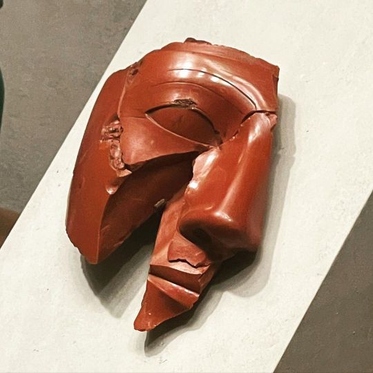
Fragmentary Head of a King (ca. 1400–1390 B.C.) | New Kingdom For many years 𓆳𓊪𓏏𓏤 “rnp.t” the Fragments of this face 𓁷𓏤 “ḥr” were dispersed in various collections, but in 1993 it became possible to reunite the pieces through an exchange with the Petrie Museum, London and a long-term loan from the Musée du Louvre, Paris. The original statue 𓂙𓏏𓏭𓀾 “ḫnty” was probably what we call a "composite statue" in which the face and other areas of exposed flesh 𓇋𓅱𓆑𓄻 “ı͗wf” (the hands and feet) were carved from red jasper 𓐍𓈖𓅓𓏏𓏸𓏬 “hnm.t” and rest of the statue was carved from Egyptian 𓂋𓐝𓎀𓀂𓀭𓏪 “rmṯ” alabaster 𓍱𓊃𓎱 “sh.s” limestone 𓇋𓈖𓂋𓊌𓈖𓂝𓈖𓁼𓊌 “inr-n-ˁı͗nw” or wood 𓇋𓅓𓏲𓆭𓏫 “ı͗mw”. Although such statues most frequently come from the the reign of Akhenaten (𓇋𓏏𓈖𓇳 𓅜𓐍𓈖) “3ḫ-n-ı͗tn” ‘The Spirit of Aten’ Amarna period (ca. 1353-1336 B.C.), this work has been dated to the reign of Akhenaten's grandfather, Thutmose IV, on stylistic grounds. Period: New Kingdom Dynasty: Dynasty 18 Reign: reign of Thutmose IV Date: ca. 1400–1390 B.C. Geography: From Egypt, Upper Egypt, Thebes Medium: Jasper Dimensions: fragments combined: H. 13.8 × W. 11.3 × D. 4.4 cm (5 7/16 × 4 7/16 × 1 3/4 in.) 𓋹𓎬𓋹𓎬𓋹𓎬𓋹𓎬𓋹𓎬𓋹𓎬𓋹𓎬𓋹𓎬𓋹𓎬𓋹𓎬𓋹𓎬𓋹𓎬𓋹𓎬𓋹𓎬𓋹𓎬𓋹𓎬 📸 @egyptologylessons © (pictures 1-3) 𓋹𓊽𓋴𓆖𓎛𓇳𓎛 (@metmuseum 📷 4-5 and Description) 𓊁𓊁𓊁𓊁𓊁𓊁𓊁𓊁𓊁𓊁𓊁𓊁𓊁𓊁𓊁𓊁𓊁 https://www.instagram.com/p/CahxxWxO2Z6/?utm_medium=tumblr
17 notes
·
View notes
Text
Synthesis, Characterization and Biological Evaluation of Copper (ii) Metal Complex with 1,10-Phenanthroline-JuniperPublishers

Abstract
Novel copper(II) complex, [Cu(phen)2(OH2)(NO3)2] (1) (where phen = 1,10-phenanthroline) have been synthesized and characterized by elemental analysis, FTIR, EPR, thermo gravimetric analysis, X-ray crystallography and biological activity. Crystal structure determination reveals that complexes (1) possess a neutral monomeric structure. In complex (1), five atoms (N and O) are coordinated with copper (II) ion to form distorted square pyramid structure. Amongst five coordinating atoms, four are nitrogen atoms from two 1,10-phenanthroline moities and one oxygen atom. The thermal decomposition of the complex (1) under an inert atmosphere has also been studied. Furthermore the complex (1) was screened for their anti-microbial activity, and antifungal cytotoxicity. Antibacterial and antifungal results indicate that the metal complexes are more active than the ligand.
Keywords: Copper(II) complex; X-ray diffraction; Crystal structure; Cytotoxicity; Anti-microbial activity
Introduction
Transition metal complexes have been extensively studied due to their potential applications in biological processes [1-3], catalysis [4], molecular reorganization, host-guest chemistry, photochemistry, analytical chemistry and magneto-chemistry [5-9]. Among transition metals, copper has its own unique identification due its coordinating ability with various ligands to from variety of geometrical structures such as square planar, square pyramidal, distorted square pyramidal, octahedral. The copper (II) complexes have also been investigated against a variety of bacterial, fungal as well as viral strains and showed profound activities against various diseases [10-12].
The copper complexes with N-donor ligands have proven to be active catalysts for the hydrolysis of phosphate ester [13], synthesis of crown ester [14], catenates and catenands [15] for formation of macromolecules [16]. Both 1,10-phenanthroline and 2,2-bipyridine are attractive building blocks that are incorporated with various transition metals to form complexes. Both nitrogen atoms in each ligand are ideally placed for cooperative binding with many metal cations. In addition, metal complexes of these two ligands are frequently employed for catalytic reactions. The metal ion complexes with 1,10-phenanthroline have been used as catalyst for various organic reactions such as the enantioselective hydrolysis of N-protected amino acid [17], for the oxidative cleavage of DNA [18], in palladium catalyzed allylic substitution [19] and enantioselective reduction of acetophenone [20]. Similarly, 2,2'-bipyridine have also been employed in the enantioselective alkylation of aldehydes [21], in the enantioselective hydrosilylation of ketone [22], in asymmetric alkylation of reaction [23] and as herbicides [24].
In continuation of our previous work on copper(ll) complex of various chelates such as 1,10-phenanthroline, 2,2'-bipyridine, suucccinate and malonate [1,25-27], we are here in reporting another copper(ll) complexes synthesized from 1,10-phenanthroline, [Cu(phen)2(H2O)2(NO3)2] (1). The synthesized compound was characterized by elemental analysis, IR spectra, EPR analysis, thermo gravimetric analysis and X-ray crystallography. The thermal decomposition of the complex (1) under an inert atmosphere has also been studied. Furthermore the complex (1) was initially screened for their anti-microbial activity, and antifungal cytotoxicity. As it showed good cytotoxicity, therefore it was e also screened for in vitro cytotoxic activity in human cell lines and the results show a moderate level of cytotoxicity against these human cancer cell lines, comparable to that of the less active standard chemotherapeutic drugs used for comparison.
Experimental
Materials and Methods
All the reagents and solvents used for the synthesis and analysis were of analytical grades and used without further purification. Elemental analyses were carried out with CHNS/O Analyzer (Thermo Scientific Flash 2000 Organic Elemental Analyzer). The copper analysis was carried out by Fisons Horizon Inductively Coupled Plasma Optical Emission Spectroscopy (ICP-OES). Melting points were determined using a Gallenkamp melting point apparatus. Infrared spectra were recorded on FTIR Bio-RAD Merlin (Varian) FTIR spectrometer in the range of 4000-200 cm-1. Perkin- Elmer and Seiko thermal analyzer was used to obtain thermo gravimetric analysis (TGA).
Synthesis of [Cu(phen)2(OH2)(NO3)2] (1)
An hot aqueous solution (25 ml) of dipotassium oxalate (0.24 g, 1.0 mmole) was added drop wise to the hot aqueous solution (25 ml) of copper(II) nitrate tri-hydrate (0.24 g, 1.0 mmole) followed by addition of 1,10-phenanthroline (0.54 g, 3.0 mmole) in ethanol (25 ml) to the resulting mixture. The mixture was stirred for 01 hour. The resulting blue solution was filtered and allowed to stand at room temperature for six months. The green crystals of compound (1) were obtained. The crystals were suitable for X-ray analysis. Yield = 76%; Elemental analysis: Calculated for C16 H24 Cu N6 O7: C, 51.1; H, 3.2; N, 15.0; Cu, 11.4%.Found: C, 50.9; H, 2.9; N, 14.8; Cu, 11.3 %. Main IR bands (ATAR): u(O-H), 3058(m); (C=C), 1428(vs), 1307(vs); (C-H), 655(vs), 616.47(s); (C=N), 735(vs); (C-N), 1380(m); (Cu-N), 445(vs).
Crystal structure analysis
Single-crystal X-ray diffraction data of the complexes (1) was collected through AXS SMART APEX (Bruker) diffractometer equipped with graphite crystal monochromator, using Mo-Kα radiation (λ = 0.71073 Å) at 100(2) K. Unit cell dimensions were obtained by least-squares refinement based on the setting angles of 935 reflections with theta (ϴ) angles ranging from 2.65° to 20.50°. The structures were solved by direct method and refined by full-matrix least-square on F2 (SHELX97) [28,29]. All non-hydrogen atoms were refined with anisotropic atomic displacement parameters. Hydrogen atoms were placed in calculated positions, assigned by isotropic atoms.Crystallographic details and selected interatomic distances and angles are given in Tables 1&2.
Results and Discussion
X-ray crystal structure of complex (1)
The monomeric molecular structure of complex (1) is shown in Figure 1. The crystallographic data and selected bond lengths and bond angles are given in Tables 1 & 2 respectively The crystals of compound (1) consist of a packing of monomeric molecule. The X-ray structure analysis shows that copper atom is five-coordinated in a distorted square pyramidal environment. Four of the coordinated atoms are nitrogen atoms (N1, N2, N3, N4) from two chelating 1,10-phenanthroline groups (Cu-N(1) =2.018(6)Â, Cu-N(2) =2.023(6)Â, Cu(1)-N(3)= 2.023(6)Â, Cu- N(4) =1.998(6)Â, and it is noteworthy that one oxygen atom is the same side of plane with copper(II) ion and bonded at distance of 2.174(6) Â. All the nitrogen atoms are almost at equal distances.
The molecular packing diagram (Figure 2) for compound (1) shows that the complex is of triclinic geometry. The units are connected in a packing diagram through a hydrogen bonding. The oxygen atoms of nitrate ion are bonded with hydrogen atoms of 1,10-phenanthroline molecule. Very weak interaction between the copper atom and oxygen atom of nitrate ion has been observed.
General Characterization
The Infrared spectrum of the complex (1) exhibits several characteristic strong bands. The strong bands at 1428, 1307, 919 and 735 cm-1 are due to C=C and C=N stretching vibrations of the coordinated 1,10-phenanthroline molecules. The spectrum shows a wide range of bands below 500 cm-1, which may be considered to 1,10-phenanthroline ligand, but it seems reasonable to assign the very sharp band at 445(s) cm-1 as a copper-nitrogen stretching mode. The spectrum of complex (1) shows a single strong band at 3057 cm-1 which may be due to the O-H vibration of water molecule (Table 3).
(b) = Broad; (m) = Medium; (s) = Strong; (vs) = Very Strong; as = Asymmetric and s = Symmetric
Thermal decomposition of several copper (II) complexes containing phen and bipy and a dicarboxylic acid or amino acids have been studied [30-31]. The thermal decomposition of complex (1) was carried up to 600°C at the rate of 5°C/minute. The decomposition of complex completed in two steps mass loss process before 450°C and the first step between 80-200°C corresponds to the endothermic elimination of water molecules. The loss of water molecule occurs at a significantly higher temperature. This suggests that the coordinated water molecule is held more tightly to the metal centre, consistent with the relative magnitude of the Cu-O bond distances of 2.174 Å. The experimental mass loss of 3.4% agrees well with the calculated mass loss of 3.2%. The degradation of phen ligand takes place in second step at 250-450°C range and copper obtained as the final product at 450°C (Weight loss: found 44.0%; calculated 44.0%). The residue product of complexes (1) was further confirmed by XRD.
The compounds were also analyzed by electron spinning resonance (ESR) and observed g value for the crystalline samples lie in the range 2.07-2.35. The x-band spectra of compound (1) shows a broad band isotropic signal that is clearly resolved in the Q-band towards an axial-type spectra with g-tensor values of gll = 2.27, g┴ = 2.05. The axial spectrum is consistent with elongated octahedral geometry of the copper (II) chromophore found in the structural work.
Biological Assays
In vitro antibacterial activity
Antibacterial screening of the synthesized compound (1) was carried out by using four bacterial strains two gram positive Staphylococcus aurous (ATCC 6538), Micrococcus luteus (ATCC 10240) and two Gram negative, Escherichia coli (ATCC 15224), Salmonella setubal (ATCC 19196). Cefixime and roxithromycine were used as standard antibiotics. Agar well diffusion method was applied as experiment was performed in triplicates readings of the zones of inhibition represent the mean value of the three readings with standard deviation [STDEV]. Activity was determined based on the zones of the inhibition (mm). Zone more than 12 mm is considered significant activity, 10-12 mm good activity, 7-9 mm low, and below 7 mm non significant activity. The results revealed that ligand showed good activitiy against M. leuteus, S. setubal, and S. aureus, while its Cu complex did not show any activity. MIC values were calculated ranging from (1-0.4mg/ml). while as-prepared compound does not showed activity against E.coli. Observations of the results showed that Phen ligands did not show antibacterial activity (Table 4).
Conc. of the standard drug “Imipenum” = 10 µg/disc. Concentration of samp!e =3 mg/mL (Stock so!ution) and 10 µg/disc
Antifungal Activity
As prepared compound (1) was subjected to antifungal assay by using Agar tube dilution method against four fungal strains i.e. Mucor species, Aspergillus niger, Aspergillu flavus, and Fusarium solani. In this assay terbinafine was used as standard drug (Table 5). Activity was investigated on the basis of percent growth inhibition; inhibition more than 70% is considered significant activity, 60-70% inhibition good, 50-60% inhibition, moderate, below 50% inhibition, non-significant. Results show that complex (1) showed good activities against Mucar, A. niger, and F. solani.
Standard drug = Terbinafine; Concentration of drug = 200 µg/mL. Concentration of tested compounds = 200 µg/mL.
Conclusion
In summary, this work describes the preparation, crystals structures, spectroscopic characterization, thermal decomposition studies of copper (II) complexes containing N-donor ligands. The structures of complexes (1) consist of a packing of monomeric molecules. In complex (1) copper (II) is five-coordinated in a distorted square pyramidal environment. Four of the coordinated atoms are nitrogen atoms from two 1,10-phenanthroline molecules and one oxygen atom from water molecule. The complex (1) was initially screened for their antimicrobial activity, and antifungal cytotoxicity. As it showed good cytotoxicity, therefore it was also screened for in vitro cytotoxic activity in human cell lines and the results show a moderate level of cytotoxicity against these human cancer cell lines, comparable to that of the less active standard chemotherapeutic drugs used for comparison.
Acknowledgement
Authors are grateful to Government of Pakistan for the financial assistance and School of Chemistry, The University of Manchester, UK, for providing us the research facilities.
To know more about Journal of chemistry,
Click here: https://juniperpublishers.com/omcij/index.php
To know more abour juniper Publishers,
click here: https://juniperpublishers.com/index.php
#juniper publishers#juniper publisher reviews#juniper publishers group#chemistry#Open access Journal of chemistry#chemistry journal#juniperpublishers#organic chemistry#inorganic chemistry
0 notes