#Euronoxx Medical Group
Text
Who needs breast screening?
The question of who needs breast screening has been a topic of debate and discussion for many years, with differing opinions based on individual risk factors.
The current standard in the UK is to offer routine mammography breast screening for women aged 50-70 every three years, a practice that has come under scrutiny.
Over 2 million women are screened annually, but the effectiveness of this approach has been called into question, particularly for certain age groups and risk profiles.
Elderly women, for example, may not always benefit from routine breast screening, especially if they are likely to die of other causes if diagnosed with breast cancer.
The discovery of cancer in women aged 66 to 79 is relatively rare through routine screening, and in many cases, the cancers detected are low-risk.
This has led to discussions about adjusting the age range for routine breast screening in the UK, with trials being conducted in some areas to assess the potential benefits of screening women from age 47 to 73.
As a result, invitations for screening may be sent to women outside the standard 50-70 age frame.
For women under the age of 50, routine breast screening is not normally available, except in specific circumstances.
These circumstances include a personal history of breast cancer, a family history of breast cancer at a young age, or a known genetic predisposition to breast cancer, such as the presence of genes like BRCA1, BRCA2, and TP53.
Radiation treatment to the chest, particularly at a young age, can also start earlier screening.
Furthermore, the decision to undergo breast cancer screening and the choice of a screening test depends on various factors, including individual risk profiles and genetic predispositions.
Women with specific risk factors for breast cancer or genetic syndromes may call for earlier and more frequent screening. The presence of certain genetic mutations, previous radiation treatment to the chest, and a family history of breast cancer can all contribute to the decision-making procedure.
Do screening tests cause harm?
Breast cancer screening has long been a subject of controversy, with growing recognition that not all cancers detected will ultimately pose a threat to a woman's health.
While these tests can potentially lead to early detection and treatment, it's important to acknowledge that they also carry inherent risks. Understanding these complications is crucial in making informed decisions about breast cancer screening.
One of the most significant concerns is the occurrence of false-positive test results:
False positives can bring about may lead to follow-up tests, including biopsies, which can cause undue anxiety and stress for women. The potential for further testing and its associated stress should be carefully considered before going for breast cancer screening.
An additional area of concern is the possibility of false-negative results, meaning that breast cancer may go undetected:
This can lead to delayed diagnosis and treatment, ultimately impacting a woman's health outcomes. Moreover, there is the concept of overdiagnosis, which refers to the detection of cancers that may never have caused harm. Treating these non-threatening cancers can expose individuals to unnecessary side effects without providing tangible health benefits.
Radiation exposure from mammography:
Another aspect to consider is the impact of radiation exposure from mammography. While the risk is minimal, it's still a factor that needs to be considered in the decision-making process, repeated mammography screening may lead to radiation-induced breast cancer, particularly for women with larger breasts or breast implants.
Pain and discomfort during mammograms also pose a challenge for some women:
Pain and discomfort during mammograms also present challenges for some women, adding to the complexity of the screening experience. It's important to acknowledge and address these potential physical and emotional discomforts when discussing screening options with patients.
Adding to the complexity of the screening experience. It's important to recognise and address these potential physical and emotional discomforts when discussing screening options with patients.
So what's the solution then?
Given these complexities and potential drawbacks, it's essential to explore alternative technologies that could potentially improve the screening process. The Koning Vera Breast CT technology is a promising example, offering the potential for enhanced visualisation and evaluation of breast tissue.
With its anticipated ability to optimise early disease detection, diagnosis, intervention, and treatment, this medical device holds the promise of improving outcomes and survival rates for countless patients. Moreover, the potential for the Koning breast CT FDA-approved medical device is a greater saviour, serving as a substitute for traditional mammogram imaging applications highlighting the ongoing evolution and precision of breast cancer screening methods.
So, the decision to undergo breast cancer screening is multifaceted, and individuals must be aware of both the benefits and potential harms. Discussing the issue with healthcare providers can empower women to make choices aligned with their individual women's values and preferences.
As Koning 3D breast CT screening technology continues the growing landscape of early breast cancer detection procedures, it is likely to evolve, offering new hope among women above 40 and improving the balance between breast cancer and breast health.
So, easily seen or understood here that breast screening is a complex issue, with no one-size-fits-all approach. The discussions about who should be screened for breast cancer and at what age reflect the evolving landscape of healthcare, as research and trials seek to refine and improve screening practices.
youtube
As our understanding of breast cancer risk factors and inherited genetic predisposition to breast cancer, the approach to breast screening is likely to become more personalised, ultimately leading to improved outcomes for women at risk.
#Koning breast CT FDA-approved medical device#Koning Vera Breast CT technology#breast implants#mammography#breast cancer#digital mammography#medical equipment#euronoxx medical group#koning breast ct#Youtube
0 notes
Text
Early Detection and Diagnosis of symptomatic patients is important for combating breast cancer so doesn't spread!
Breast cancer is a topic that often evokes fear and uncertainty, but the more we know about it, the better equipped we are to face it.
It is important to remember that around 5% of women with breast cancer have already reached an incurable stage by the time of diagnosis. Breast cancer is the most common type of cancer in the UK, and around 12,000 women die from it every year.
This underscores the significance of raising awareness about breast cancer and the crucial role of regular breast checks and screening.
Despite these statistics, survival rates from breast cancer have been improving over time. Nowadays, around 3 out of 4 women diagnosed with breast cancer are alive ten years later. One of the keys to achieving this is through early detection.
What is breast cancer?
Breast cancer is a disease that starts when cells in the breast begin to grow in an uncontrolled manner and build up to form a lump or tumour. As the cancer grows, cells can spread to other parts of the body, which can be life-threatening.
By detecting breast cancer early, you can increase your chances of surviving and minimise the need for more invasive treatments.
Breast screening is performed to detect breast cancer at an early stage, before the development of externally observable symptoms or objective signs, such as a lump.
This is why it is essential to undergo regular screening to detect any issues before they become more serious.
Breast screening is a procedure of digital mammography, which takes an X-ray picture of each breast. During a mammogram, each breast is compressed for a few seconds while an image is taken.
This compression can be slightly uncomfortable but only lasts for a few seconds, and the whole procedure is over quickly.
If something abnormal is detected, such as an unusual lump, you may need further tests, such as an ultrasound or biopsy.
As you get older, your risk of developing breast cancer rises!
Approximately 4 out of 5 cases of breast cancer occur in women aged 50 and above. It's important to note that the majority of women diagnosed with breast cancer do not have a family history of the disease.
If breast cancer is found early, you are more likely to be able to have breast-conserving surgery, which can help you avoid a more invasive mastectomy.
Early detection can also significantly increase your chances of surviving breast cancer in the long term.
It's important to note that early detection doesn't mean that you have to wait for your screening appointment.
By regularly checking your breasts at home, you can become familiar with how they look and feel, making it easier to spot any potential changes. If you notice anything unusual, such as a lump, swelling, or discharge, it is important to seek medical advice as soon as possible or go for breast screening.
So, we put some awareness programmes over this blog post - to get more people to regularly check their breasts and go for breast screening. With regular screening and self-checks, we can help catch breast cancer early and increase survival rates.
Rethinking Breast Cancer Screening: Are Mammograms the Best Option?
In the UK, routine breast screening tests, or mammograms, are offered to women aged 50-70 every three years. This widespread screening program aims to detect breast cancer early when it is most treatable.
With over 2 million women getting screened each year, the impact of these tests cannot be understated.
However, the age range for routine breast screening is being reconsidered in some parts of the UK.
A trial is underway to assess the benefits of extending screening from age 40 to 73.
This means that in these age groups, women may be invited for screening before turning 40 or even after reaching 70.
But what about those who fall outside of this age range?
For women under 50, routine breast screening is currently not available unless certain risk factors are present. These can include a personal history of breast cancer, a family history of the disease, or carrying specific genetic mutations that increase the likelihood of developing breast cancer.
If you are unsure whether you should be screened earlier, consulting your GP is crucial.
While mammograms have been a cornerstone of breast cancer detection, they do come with limitations.
The process of breast compression during a mammogram can be uncomfortable or painful and sometimes may lead to false recall of cancers.
Additionally, frequent exposure to radiation through mammograms can potentially contribute to an increased risk of cancer.
Furthermore, waiting time is required of at least 7 days to get the screening test results.
This highlights the importance of exploring alternative screening methods that may offer better outcomes.
Let's empower individuals to make informed decisions about their breast health!
Let's continue the conversation on breast cancer screening, raise awareness about the available options, and empower individuals to make informed decisions about their breast health.
Zero breast compression:
One such alternative gaining attention is the Koning Vera Breast 3D Imaging system. This technologically advanced medical device eliminates the need for breast compression, a factor that contributes to discomfort during mammograms.
Less scanning time:
The scanning time is significantly reduced, with each breast being scanned in less than 7 seconds compared to the longer duration of a mammogram. This not only improves the patient experience but also streamlines the screening process.
Better scanning angle irrespective of dense breast tissue:
Moreover, the Koning 3D isotopic breast imaging system caters to women of all breast sizes. It provides better coverage (360-degree scanning angle) of breast tissue, particularly in dense breasts where mammograms may struggle to detect abnormalities.
This is a crucial advancement, as dense breast tissue can make it challenging to obtain clear and accurate results from traditional mammograms.
Higher detection accuracy:
Breast cancer screening should constantly need improvements and innovations that enhance detection accuracy and patient comfort. While mammograms have been instrumental in saving lives, exploring more accurate and faster technologies like the Koning Breast CT FDA-approved system opens up new possibilities for more efficient and effective screening.
opens up new possibilities for more efficient and effective screening.
Easily find lesions:
Koning Breast CT is an FDA-approved medical device that can identify any cancerous lesions as early as stage 0 and stage 1. It can find lesions as small as 2mm and calcifications as small as 200 microns.
Easy Takeaways:
Increasing awareness is important to get more people to know the severe consequences and does regularly check their breast health. With regular screening and self-checks, we can help catch breast cancer early and increase survival rates.
As we guide the importance of breast cancer detection at an early stage, it is crucial to stay informed and open to exploring new screening options that may offer superior outcomes.
By bridging the gap between traditional mammograms and innovative technology like Koning 3D breast imaging, we can make better breast health for a future where early detection and improved screening methods play a pivotal role in combating breast cancer.
Euronoxx Medical now aim towards better breast health outcomes across the UK and dreams about a future where breast cancer is caught early and treated effectively.
#breast cancer#digital mammography#medical equipment#euronoxx medical group#Koning 3D breast imaging#Koning Breast CT
0 notes
Text
Unravelling the Link Between Breast Cancer and the Microbiome!
With the spilling beans of BC, the rate of incidence of breast cancer has reached alarming levels, impacting the lives of women across the globe. As the most commonly diagnosed cancer among women, the need for early detection and effective treatment has become more crucial than ever.
Traditionally, cancer diagnosis often involved invasive procedures, posing risks and challenges to patients. However, recent research has sparked a new wave of interest in non-invasive diagnostic approaches, using samples such as saliva, stool, and plasma, to explore the potential role of the microbiome in cancer detection.
The human microbiome, composed of diverse microorganisms residing within our bodies, has been found to influence various aspects of health and disease.
In the context of cancer, the microbiome's indirect influence on the onset and progression of breast cancer needs significant attention from the scientific community.
Can gut microbes impact induce cancer cells?
Studies have revealed that gut microbes can impact dormant cancer cells, and potentially induce chronic inflammation to disease recurrence in breast cancer. While much of the research has centred on the role of bacteria in cancer, there is a growing interest in exploring the potential involvement of fungi, viruses, and even archaea in cancer development.
Interestingly, metabolites from certain single-celled organisms (archaea) have been linked to various cancers, shedding light on the intricate relationship between microbial populations and the progression of the disease.
Despite being less studied due to their relative rarity, these findings underscore the complexity of the microbiome's impact on human health.
The burgeoning understanding of the proliferation microbiome's significance extends to its pivotal role in the gastrointestinal tract, where the largest concentration of microorganisms is found.
This complex ecosystem is influenced by both genetic and lifestyle factors, and its dynamic interplay with the host exerts both local and systemic effects.
As we highlight the backdraws of cancer proliferation, the exploration of the microbiome's influence on breast cancer is opening new avenues for innovative diagnostic methods and potential therapeutic strategies.
Unraveling the Microbiome and Lifestyle Factors in Breast Cancer: A Thought-Provoking Discussion!
The intricate trap of Microbiome influences breast cancer (BC), as new research sheds light on the potential impact of the microbiome and various lifestyle components.
Let's get into these compelling findings and the thought-provoking questions they inspire.
Microbial Players in Breast Cancer!
Recent studies have revealed a strong relation between the microbial composition of breast cancer patients and the abundance of specific bacteria, such as Bacillus, Enterobacteriaceae, and Staphylococcus.
The discovery that certain bacteria isolated from breast cancer patients, including Staphylococcus epidermidis and Escherichia coli, can induce DNA double-stranded breaks in cells raises exciting questions about the potential role of these microorganisms in cancer development.
Moreover, the presence of Fusobacterium nucleatum, a bacterium implicated in inducing tumour growth and metastasis, in breast tissue further underscores the complex relationship between the microbiome and cancer progression.
Other Lifestyle and Environmental Influences!
In addition to microbial factors, the influence of lifestyle and environmental components on breast cancer risk adds layers of complexity to our understanding of
the disease. While genetic factors like the loss of the BRCA1/2 gene command attention, lifestyle factors present compelling avenues for exploration.
The impact of diet, alcohol consumption, and radiation exposure on breast cancer incidence has been well-documented. Equally noteworthy are the associations between hormone exposure and breast cancer risk.
The interplay between physiological variations associated with puberty, pregnancy, menopause, and the use of hormonal contraceptives or hormone replacement therapy reveals the multifaceted nature of BC risk factors.
Framing Thought-Provoking Questions!
The emerging intersection of microbiome research, lifestyle factors, and breast cancer prompts thought-provoking questions that invite further exploration and discourse:
Microbiome and Cancer Development: How do specific microbial populations contribute to DNA damage in breast cancer cells, and what mechanisms underlie their potential role in tumour progression?
Fusobacterium nucleatum and Tumor Growth: In what ways does Fusobacterium nucleatum promote tumour growth and metastasis in breast tissue, and what importance does this hold for potential interventions targeting the microbiome?
Lifestyle and Hormonal Influences: How do variations in estrogen metabolism, influenced by environmental and lifestyle factors, impact breast cancer risk, especially among postmenopausal women? What implications does this have for personalised prevention strategies?
As we confront the complexities of breast cancer etiology, these key findings inspire a call for collaborative research and early diagnose.
By exploring the complex interplay between microbial influences, lifestyle components, and genetic predisposition, Euronoxx Medical Group strongly recommend an "Early Detection could minimize the risk of breast cancer mortalities". By getting this holistic approach to understand, prevent and early diagnose of breast cancer.
Here we shown some intricate relationships, we hope to illustrate the non-invasive, microbiome-based approaches that could revolutionise breast cancer early detection and awareness.
Meanwhile, the hidden interactions between the microbiota and breast cancer may hold the key to discussing new insights and transformative breakthroughs in our fight against cancer.
0 notes
Text
What Are The Possible Risks of Radiation-induced Breast Cancer & Mortality From Digital Mammography Screening Process?
Here, we express the thoughts and scientific discourse of cancer screening and diagnostic radiology. A crucial aspect that emerges at the forefront of discussion is the profound impact on women's health, particularly concerning breast cancer.
Breast cancer, a disease that affects millions of women worldwide, underscores the critical importance of early detection through screening programs such as mammography.
The advent of digital imaging technologies has shown us a new dimensional hope in breast cancer diagnostic radiology, offering enhanced precision and clarity in detecting abnormalities within breast tissue.
However, the discussion surrounding healthcare quality and services in the context of breast cancer screening remains a dynamic and evolving domain of research.
One of the key considerations in this discourse is the estimation of radiation-induced breast cancer risks associated with mammography screening.
Traditional mammography screening assessments often overlook the variability in dose exposure and the subsequent diagnostic work-up following abnormal screening results.
It is important to acknowledge that the potential risks of ionising radiation from repeated mammography examinations cannot be underestimated, especially when considering the cumulative effects of radiation exposure on breast cancer incidence and mortality.
Exploring the Impact of Breast Cancer Screening and Diagnostic Radiology on Women's Health!
The National Health Service Breast Screening Programme in England significantly studies that regular screening intervals and the ongoing randomised control trials exploring age extensions underscore the need for a comprehensive evaluation of screening strategies after careful consideration of exposure from both screening and diagnostic mammography, along with potential dose variations among women with radiation-induced cancers.
Here we need to understand the concern - radiation-induced breast cancer risks.
The relative risk of breast cancer induction!
As per Research conducted on August 1, 2002, such as the work by Preston et al., provides invaluable insights into quantifying the excess absolute risk and relative risk of breast cancer induction, offering a foundation for assessing risk factors across diverse populations and age groups.
By integrating such data into the broader landscape of research quality assessment, we can enhance our capacity to tailor screening programs effectively and mitigate potential risks associated with radiation exposure.
The conversation surrounding breast cancer screening, diagnostic radiology, and radiation-induced risks necessitates a multidisciplinary approach that prioritises the well-being of women and underscores the imperative of evidence-based decision-making in healthcare.
By promoting discussion, sharing insights, and high-grade technological advancements in breast cancer research such as the Koning 3D breast CT screening process, we can continue to advance the field of breast cancer management and contribute to improved outcomes for women worldwide.
Understanding the Complexities of Breast Cancer Screening and Radiation Exposure: Unravelling the Facts!
Breast cancer screening has long been a cornerstone of women's healthcare, offering a vital tool in the early detection and management of this prevalent disease.
However, recent discussions have shed light on the potential risks associated with radiation exposure during screening procedures, sparking a minute conversation about balancing the benefits and potential drawbacks.
A recent study explored the impact of radiation exposure from mammography screening regimens, revealing intriguing insights into the predicted incidence of radiation-induced breast cancer and associated mortality.
The findings, based on a cohort of 100,000 women undergoing screening from age 40 to 55 years and biennially thereafter to age 74 years, projected 86 induced cancers and 11 deaths due to radiation-induced breast cancer.
These results underscore the significance of age and age at exposure in influencing the risks, emphasising the complexity and multifactorial nature of radiation-induced breast cancer.
Showing deeper into the technical aspects, the excess absolute risk model was used to predict the number of radiation-induced breast cancers attributable to a typical digital mammography examination.
This modelling approach offers a valuable framework for understanding the potential impact of radiation exposure within the context of screening, enabling a more tailored and nuanced assessment of associated risks.
The average glandular ionising radiation dose for conventional mammography is from 2.2 to 15 mGy and cone-beam Koning 3D breast CT offers a lens through which to gauge the potential risks related to ionizing radiation dose from 4 to 12.8 mGy.
Furthermore, the collaboration with Koning Corporation - Euronoxx Medical Group now supplying Koning breast CT FDA-approved screening machines presents an example of the intersection between technological innovation and healthcare delivery, underscoring the dynamic nature of advancements in breast imaging.
Solution:
The balance between the risks and benefits of radiation exposure in the context of breast cancer screening remains a pivotal aspect of healthcare decision-making.
Despite the projected risks associated with radiation-induced breast cancer, the overarching consensus emphasises that the benefits of mammographic screening significantly overshadow these concerns.As we forge ahead in this journey, it is imperative to harness the power of research, technological innovation, and collaborative efforts to continue enhancing the landscape of breast cancer management and screening strategies, Koning breast CT FDA-approved screening machines facilitate the most advanced screening techniques with low risks of radiation exposure women with the best possible care and outcomes.
0 notes
Text
Sharing Life-Changing Tips to Empower Our Community in the Fight Against Breast Cancer in the UK!
Breast cancer is a subject that deeply affects many of us. With over 55,900 people diagnosed with breast cancer in the UK every year, we cannot ignore the need for action.
It's time for us to come together as a community and empower ourselves with knowledge and resources to combat this prevalent disease.
One key fact to remember is that early detection is key!
Over 90% of breast cancer cases have an early-stage inflammation at the time of diagnosis.
However, among them, around 25% will finally break down to distant metastasis. This alarming statistic highlights the urgency to take proactive steps to reduce the risk of recurrence and prevent the disease from progressing.
Welcome Diverse & Inclusive Change: Join the Movement to Prevent Breast Cancer!
The journey that begins with a breast cancer diagnosis is one filled with uncertainty, fear, and a profound desire for empowerment and control.
Many patients seek information from various sources on behaviours that may reduce the risk of cancer recurrence.
It's a path that's not just about physical health but also about regaining sensible knowledge gain and practice.
Let's Free the Grip From Breast Cancer!
In this hunt for knowledge and empowerment, the influence of Prevent Breast Cancer, a dedicated UK charity, looms large. This organisation has committed itself to raising awareness, focusing on prediction and prevention, and ultimately striving for a world free from the grip of breast cancer.
Prevent Breast Cancer in its unwavering focus on prevention rather than solely on finding a cure. They understand that early detection is the key, and that belief is something we wholeheartedly support at Euronoxx Medical.
We stand behind the initiatives that drive early diagnosis, screening, and promoting vital lifestyle changes. We firmly believe that we can prevent the problem before it even begins, and we are ready to champion behavioural interventions to make that vision a reality.
Now is the time for our community to come together and act!
Established in the limelight of the battle against breast cancer, as the leading Medical Group in the UK for spreading awareness of breast cancer prevention, Euronoxx Medical is fully committed to supporting Prevent Breast Cancer and their tireless efforts.
We implore you to stand with us in endorsing our advanced medical equipment and devices, such as the Koning Breast CT scanning machine to install in your charity hub.
This Koning breast CT FDA-approved scanning machine is a standard 3D isotropic breast imaging test often used to clarify breast tomosynthesis through screening procedure and has no compression of breasts, no pain and minimum radiation exposure for early detection and prevention. It can show a positive change in the battle against breast cancer.
Please don't hesitate to get in touch with us if you have any questions or concerns. Let's rally together to form a community that is informed, empowered, and united in our resolve to eradicate breast cancer. Join us today and take your place in the movement to shape a future free from the shadows of breast cancer. Together, we can effect change and make a tangible difference.
As we march forward, let's stand together as a flare of faith, knowledge, and strength. Let's affirm our commitment to preventing breast cancer and shaping a healthier future for all. Join us in this vital mission - together, we can create a world where breast cancer is a thing of the past.
Actual Change We Intended To Impose in The UK by 2025
From today and 2025, we're:
Doing intense attention to breast screening and encouraging women above 40 must conduct breast screening regularly.
Searching for innovation to find new methodologies for better patient outcomes.
Advocating for easy access to effective breast screening in the UK.
Encourage women to conduct regular exercise to mitigate the risk of breast cancer.
Spreading awareness on radiation-induced breast cancer prevalence and mortality from digital mammography screening!
Last but not least, Introducing Koning 3D Breast CT Imaging medical device for screening of breast health check.
Conduct awareness programs for " Early detection could save lives & improves intervention "
We are dedicated to creating a future where breast cancer is a chapter of history, not a part of our present.
Today is your chance to become part of this transformative activity.
#Koning 3d breast CT#Koning breast CT FDA#breast cancer#digital mammography#medical equipment#euronoxx medical group
0 notes
Text
Don't forget to decide whether or not to undergo breast screening for early diagnosis!
It's important to have all the information you need when making decisions about your health, and breast screening is no exception.
This leaflet aims to help you decide whether or not to have breast screening, because it's ultimately your choice.
So, why does the NHS offer breast screening?
The main goal is to save lives from breast cancer. By detecting breast cancers at an early stage, screening can find tumours that are too small to see or feel.
However, it's important to note that screening doesn't prevent breast cancer from occurring. Like any medical procedure, breast screening does come with some risks.
Some women who have screening may end up being diagnosed and treated for breast cancer that would never have been found or caused them harm otherwise.
Now, you might be wondering why you've been invited for breast screening. Well, if you're a woman aged 50 to 70, the NHS invites you for screening every three years.
However, there are also studies being conducted to assess the effectiveness of screening in different age groups, so some older and younger women may also receive invitations.
It's worth mentioning that even if you're over 70, you're still at risk of breast cancer.
Although you won't automatically get screening invitations after the age of 70, you can still have breast screening every three years. Just reach out to your local breast screening unit and ask for an appointment.
Let's show you some stories of famous British women's breast cancer journey!
Let's take a moment to remember some famous women who have had experiences with breast cancer.
Maggie Smith!
Maggie Smith is known for her fabulous act in some famous movies. A talented British actress we all know and love was diagnosed with breast cancer when she was 73.
She received chemotherapy while filming Harry Potter and the Half-Blood Prince. It's incredible to see how she tackled her treatment and continued to pursue her passion.
Carly Simon!
Carly Simon, the renowned singer-songwriter, had a successful surgery in 1997 to remove a stage I tumour.
Her doctor gave her the choice of whether or not to have chemotherapy, and she decided to go for it just to be safe. Simon later revealed that she was going through a deep depression during that time.
However, she has since written a New York Times best-selling memoir and continues to create music with her children. It's heartwarming to see how she used her experience to inspire others.
Teresa Heinz Kerry!
Teresa Heinz Kerry, a philanthropist and the wife of the former presidential nominee who became Secretary of State John Kerry, had a rare case of breast cancer in both breasts.
Not only that, but she also had different types of early-stage cancer in each breast. This meant that the treatment and chances of cancer-free survival depended on the more aggressive tumours. Kerry underwent lumpectomies and radiation therapy (as mastectomy) for her treatment.
Jaclyn Smith!
Lastly, another example is Jaclyn Smith, a well-known actor, entrepreneur, and mother of two. When her doctor wanted to take a biopsy of her left breast tissue, Smith was too busy and felt too healthy to worry.
However, when she received the news that it was cancerous, she was shocked. Today, she advises women in similar situations to have a family member or friend with them to help process the medical information.
Remember, it's your choice whether to have breast screening or not. Ultimately, what matters most is that you feel informed and empowered to make the decision that's right for you and your health.
Recovering the Spins in Life: Embodying Innovation in Breast Cancer Screening!
As women age, the importance of breast cancer screening becomes increasingly significant.
Despite the potential risk of radiation-induced breast cancer, the need for regular screening cannot be overstated.
Various factors, such as dosage variability, initiation age, and frequency of screening, can influence the incidence and mortality of radiation-induced breast cancer from digital mammography screening.
Potential ionising radiation exposure during mammography screenings!
Exposure to ionising radiation during mammography screenings raises concerns about the development of radiogenic breast cancer. The likelihood of repeated exposure to this radiation poses a potential risk that cannot be ignored.
In light of these concerns, finding a safer and more effective alternative is essential.
Introducing Koning 3D breast screening – a revolutionary medical device that offers minimal radiation exposure, eliminates breast compression, and ensures a pain-free experience.
With a scanning process that takes just 7 seconds, Koning 3D breast screening provides a comfortable and efficient way to prioritise your breast health.
Visit us at euronoxxmedical.com to order today.
Breast Cancer 2024 Live Conference!
To further promote awareness and education around breast cancer prevention, early detection, and management, a groundbreaking conference titled "Breast Cancer 2024" is set to take place.
Organised by the London International Medical School (LIMS) in collaboration with Euronoxx Medical and Koning Corporation (USA), this conference aims to bring together experts and professionals from the UK and the USA.
This innovative program welcomes GMC registered doctors, NMC registered nurses, and final-year medical students, providing a unique platform for learning and collaboration.
The conference will cover a range of topics, including primary prevention of breast cancer, secondary prevention strategies, and advancements in breast cancer treatment.
We Shed Light on early breast cancer detection awareness!
One of the conference's highlights is the discussion on early breast cancer detection using the Koning Breast 3D imaging procedure. This advanced medical equipment offers a more comprehensive and accurate method for detecting breast abnormalities, leading to better outcomes for patients.
Featuring a lineup of esteemed speakers, including Dr. Penny Kechagioglou, Dr. Qaiser Malik, Dr. Sarah Sadrudin, Ms. Jalini Varghese, and Professor Kefah Mokbel, the conference promises to deliver insights into the latest research, best practices, and patient experiences in breast cancer screening and treatment.
For those unable to attend in person, the conference will be live-streamed globally, allowing participants from all over the world to engage with the content in real-time.
This inclusive approach aims to ensure that individuals with a special interest in breast cancer, including those with a family history of the disease, can access valuable information and resources.
Join us on Saturday, September 7, 2024, at the Royal Society of Medicine in London for an enriching and informative conference on breast cancer prevention and management.
Be part of this transformative journey in reclaiming control over your breast health and well-being, followed by early detection of breast screening procedures.
Don't miss the opportunity to secure your spot and contribute to this vital initiative.
To learn more and register, visit euronoxxmedical.com today. Let's unite in the fight against breast cancer and pave the way for a healthier future together!
1 note
·
View note
Text
Early Signs & Symptoms of Breast Cancer and Best Koning Vera Breast CT Diagnostic Intervals
Breast cancer is a major concern for many women, and early detection is crucial for successful treatment. While a breast lump is the most commonly known early Signs and other various symptoms should not be ignored.
A research study titled "Symptoms of Breast Cancer and Their Associations with Diagnostic Intervals: Evidence from a National Audit of Cancer Diagnosis" sheds light on the importance of recognising these symptoms and the potential impact on diagnosis intervals.
The study highlights that although a breast lump is the most common symptom among women with breast cancer, around 1 in 6 women had symptoms other than a lump. This finding suggests that women need to be aware of a broader range of breast cancer symptoms. The study also indicates that, on average, women experienced longer intervals between experiencing such symptoms and seeking medical help than intervals between primary care consultations and diagnosis.
According to the study, of women with "non-lump" or "both lump and non-lump" symptoms 31.4% of respondents delay seeking medical help. This delay may be attributed to various factors, such as the misattribution of symptoms to non-malignant causes like hormonal changes, trauma, or breastfeeding. It is crucial to address these misconceptions and increase awareness of non-lump breast symptoms.
Some of the symptoms associated with breast cancer!
In addition to a new lump in the breast or armpit, include breast skin texture changes, such as a rash, redness, or dimpling.
Swelling in the armpit or near the collarbone may also be a sign that breast cancer has spread to lymph nodes.
Pain and tenderness, although lumps don't usually cause discomfort, and a flat or indented area on the breast may also indicate breast cancer.
Other symptoms to be aware of include changes in the breast's size, shape, or temperature.
Changes in the nipple, such as Inverted Nipples or pull hard, Breast dimpling, burning, itching, or the development of sores, should also be taken seriously.
Unusual nipple discharge can also be a symptom, whether clear, bloody, or colour changes.
Additionally, epidermoid cysts (or epidermal inclusion cysts) a marble-like area under your skin, which feel different from any other part of the breast, can be a sign of breast cancer.
It is important to note that breast cancer symptoms can vary from person to person, and some women may not notice any signs at all. Regular self-examinations and being aware of these symptoms can greatly help in the early detection of breast cancer.
Improving both the treatment outcomes and overall experience for breast cancer patients!
Cancer is a global health concern, and its incidence and mortality rates are rapidly increasing, especially in emerging economies.
Breast cancer, in particular, is a prevalent and deadly form of cancer among women worldwide. According to a report from 2018, breast cancer accounted for 25% of all female cancer cases globally, with 2.1 million cases diagnosed [1]. It is the most commonly diagnosed cancer among females in over 150 countries and the leading cause of mortality among all female cancers in 100 countries.
For surprising breast cancer incidence rates, the recent GLOBOCAN 2018 report reveals some interesting findings.
Age-specific breast cancer incidence rates per 100 thousand females were particularly high in Australia (94.2), Western Europe (92.6), and Northern Europe (90.1) [1].
On the other hand, South-Central Asia recorded the lowest incidence rate at 25.9. However, it is important to note that the mortality rate in South Asian countries is similar, if not higher, compared to other regions, highlighting the need for early detection of cancer and access to quality healthcare in developing nations.
Early Signs and Symptoms of Breast Cancer
Recognising the early signs and symptoms of breast cancer is crucial for early detection and improved treatment outcomes. However, many women may not exhibit any symptoms at first, underlining the importance of regular screenings such as mammograms, especially for women over the age of 40 or those with a family history of breast cancer.
Breast lumps
One of the most common early signs of breast cancer is a breast lump. It is important to note that not all breast lumps indicate cancer. Cysts, fibroadenomas, fibrocystic breasts, breast infections, clogged milk glands, and even injuries that form scar tissue can all cause lumps in the breast [2].
However, if a lump has irregular edges or is hard, it may be more likely to be cancerous. It is essential to consult with a healthcare professional to determine the underlying cause of any breast lumps.
Ductal Carcinoma Symptoms
Ductal carcinoma is the most common type of breast cancer, accounting for approximately 1 in 5 new breast cancer cases [2].
In the early stages of ductal carcinoma, known as ductal carcinoma in situ (DCIS), there are often no symptoms. Over 90% of DCIS cases are detected through imaging tests such as mammograms.
A breast lump
Galactorrhea or Milk discharge from your nipple
Itchy breasts
However, in some cases, individuals may experience symptoms such as a lump, nipple discharge, or itching in the breast area. It is important to note that ductal carcinoma can also progress to become invasive, spreading beyond the milk ducts and potentially causing more severe symptoms.
Revolutionising Breast Cancer Early Detection with Koning Vera Breast CT!
The Koning Vera Breast CT (KBCT) medical imaging, innovative technology continues to reshape the landscape of healthcare, improving both patient experience and diagnostic capabilities.
The Koning Vera Breast CT (KBCT) is a prime example of cutting-edge 3D imaging that is transforming the early detection and diagnosis of breast cancer.
This state-of-the-art system provides crystal clear 3D images of the breast without the discomfort of compression, low doses of radiation effects, and rotates around the breast, offering a more patient-friendly experience while significantly enhancing the accuracy of tumour detection, especially in women with dense breast tissue.
A New Era for Breast Imaging for Early Diagnosis!
Community Radiology NY, renowned for its commitment to leveraging advanced technology to provide the best care for its patients, has highly acknowledged Koning Vera Breast CT with open arms.
Charles Piccinnini, the Executive Administrative Director of Community Radiology, emphasises the impact of this Koning Breast CT technology on breast cancer early detection, stating, "The addition of the Koning Vera Breast CT to our diagnostic instrument allows us to provide our patients with the latest in medical imaging. This addition in healthcare driven new fold of breast cancer hope, we can detect and diagnose breast cancer more conveniently and devote treatment protocols for better breast cancer recovery."
The integration of this technology is aimed at empowering patients to access mammography without apprehension and marks a major leap in breast imaging capabilities.
Euronoxx Medical shares in this vision, expressing confidence that the Koning Vera Breast CT will revolutionise the visualisation and evaluation for mapping breast tissue types. With a focus on optimising early cancer lesion detection, diagnosis, intervention, and treatment.
The potential impact of this advanced imaging technology extends beyond borders. The hope is for improved survival rates and outcomes for millions of patients across the United Kingdom, the United States, UAE, Africa, Asia, and the Caribbean.
In addition, the adaptability and cost-effectiveness of the Koning Vera Breast CT diagnostic hospital Instrument is a viable alternative for traditional imaging applications.
Significance of Early Detection Of Cancer and Diagnosis!
The significance of early detection of cancer cannot be overstated, as it has a direct impact on the prognosis of breast cancer patients.

Traditional mammogram diagnostic instruments, have limitations, missing a substantial percentage of cancers, especially in women with dense breast tissue.
The introduction of the Koning Vera Breast CT presents a transformative solution, equipping physicians with value-based care with the ability to detect cancers as early as stage 0 and stage 1, identifying cancer lesions <2mm and calcifications as small as 200 microns.

These capabilities hold the potential to significantly improve five-year survival rates for individuals diagnosed with breast cancer in its early stages.
Joining Forces for Improved Patient Care!
The collaboration with Euronoxx Medical, and healthcare providers worldwide heralds a new chapter in breast cancer Signs and symptoms detection and diagnosis.
We are dedicated to connecting with UK hospitals, UK medical institutes, and USA healthcare systems to deliver this state-of-the-art Koning Vera Breast CT Patient Monitoring Diagnostic Instruments, set to promote the global standard breast cancer patient care.
This collective effort signifies a shared commitment to leveraging innovation in Patient Monitoring Diagnostic facilities to make proactive initiatives toward the Early detection of breast cancer and positively impact individuals facing the challenges of breast cancer.
The Koning Vera Breast CT represents a pivotal advancement in breast imaging, offering a compassionate and accurate approach to early detection and diagnosis of breast cancer.
With its potential to drive revolutions in healthcare standards, this Patient Monitoring Diagnostic Technology stands as a sign of the possibility of getting the outcomes from difficulty for improved patient experiences in the fight against breast cancer.
While a breast lump is commonly associated with breast cancer, it is crucial to recognize other symptoms that could indicate the presence of this disease. The research study highlights the need to raise awareness about non-lump breast symptoms and encourages women to seek medical help promptly if they experience any concerning changes in their breasts. Early detection is key to improving both the treatment outcomes and overall experience for breast cancer patients.
References:
1..GLOBOCAN 2018: Global Cancer Observatory, International Agency for Research on Cancer.
2..Breast Cancer: Symptoms and Signs
#Early detection of cancer#Signs and symptoms#Primary health care#Delayed diagnosis#Early diagnosis#breast cancer#digital mammography#medical equipment#euronoxx medical group
0 notes
Text
Most reliable 3D Breast Imaging produces with Koning CT Breast Imaging!
Breast cancer is a prevailing women's cancer and potentially life-threatening disease that affects millions of women worldwide. Early detection of cancer lesions lowers morbidity and improves patient outcomes, making accurate and reliable breast imaging technologies essential.
The Koning CT Breast Imaging presents a revolutionary diagnostic solution for Breast Imaging, an advanced diagnostic instrument that produces real 360-degree angle images of the breast, transforming the way clinicians visualise and evaluate breast tissue while enhancing the patient exam experience.
Unlike traditional mammography techniques, Koning CT Breast Imaging uses a 3-dimensional imaging approach. That rotation surrounds the woman's breast, providing a comprehensive view from every angle.
This advanced diagnostic instrument eliminates the possibility of cancers hiding within overlapping breast tissue, ensuring that no abnormality goes undetected.
By capturing detailed images with exceptional spatial resolution, Koning CT Breast Imaging enables earlier and more accurate diagnosis procedures, leading to better treatment options and improved patient outcomes.
Dr. Etta Pisano, a renowned expert in breast cancers, explains the significance of Koning CT Breast Imaging. She highlights the unique advantage of the device, stating, "The Koning CT Breast Imaging is surrounding the woman's breast, and so the cancers have nowhere to hide in the breast or may be less than 2mm. There's no way for overlapping tissue to be in the way from every angle."
Her comprehensive insights help breast cancer survivals get hope in medical history and Koning CT Breast imaging offers exceptional clarity and detail of deep breast tissues, enabling physicians to make more informed decisions and tailor treatment plans to individual patients.
Koning CT Breast Imaging prioritises the patient's comfort zone!
Traditional 2D mammograms can be uncomfortable for many women because of the compression of the breast. However, Koning CT Breast Imaging eliminates the need for breast compression, providing a more comfortable and less invasive examination process.
This upgraded version of breast imaging significantly improves patient satisfaction and encourages more women to undergo weekly/monthly screenings, promoting early detection and ultimately saving lives.
Koning CT Breast Imaging is backed by extensive clinical studies and research!
Extensive clinical studies and research backed by the technological advancements of Koning CT Breast Imaging have undergone rigorous testing to ensure its accuracy, reliability, and performance.
Additionally, the results from clinical studies showcase the superior diagnostic capabilities of Koning CT Breast Imaging in detecting breast cancer compared to traditional 2D mammography.
It represents a commitment to improving the lives and outcomes of women worldwide. By revolutionising breast imaging with Koning CT Breast Imaging, clinicians can provide more accurate diagnoses, personalised treatment plans, and better overall care.
This advanced hospital or lab diagnostic instrument helps patients take control of their health, and it offers hope for a future where breast cancer is detected early and effectively treated.
Solution:
Koning CT Breast Imaging sets a new standard in breast imaging technology. Euronoxx Medical is fully equipped with the best quality surgical equipment, diagnostic instruments, and hospital equipment supply mechanisms.
With 3D imaging, exceptional spatial resolution, and a patient-centred approach, it is revolutionising the field and improving the lives of women everywhere.
As the medical community continues to prioritise early detection and patient care, Koning CT Breast Imaging shines brilliantly, offering hope and advances in the fight against breast cancer.
#breast cancer#health care#treatment#digital mammography#medical equipment#euronoxx medical group#public health#doctors#health
0 notes
Text
How HOLMIUM LASER-BLAZE Are Helped In Treating Urological Conditions?
Over the past few decades, significant advancements in medical technology have preexisting procedures and have gained the popularity of lasers in the treatment of various urological conditions. Patients suffering from benign prostatic hyperplasia (BPH) or prostate enlargement, bladder stones, and kidney stones have found respite in laser-based treatments, particularly the Holmium laser.
The prevalence of health issues associated with the enlargement of the prostate gland, dating back to early human civilisation, cannot be overstated. Symptoms such as poor urinary stream (Urinary hesitancy), pain during urination (Dysuria - a burning sensation), and increased frequency of urination accompanied by involuntary leakage are often disregarded by patients because of embarrassment or fear of seeking medical assistance.
However, ignoring these symptoms can exacerbate the condition, leading to complications like recurrent urinary tract infections, Hematuria, urinary retention, and even renal failure.
While medical management can relieve symptoms in many cases, surgical intervention becomes imperative for those unresponsive to conservative measures. Unfortunately, the apprehension associated with surgery often deters individuals from seeking timely treatment, resulting in avoidable complications.
Advancements In urological interventions!
Advancements in science have initiated in the era of minimally invasive procedures through the miniaturisation of endoscopic equipment and improvements in energy sources.
While open surgery was once the foundation of urological interventions, it has now become a rarity.
The field of urology has undergone a remarkable transformation, especially with the emergence of laser technologies.
The traditional Transurethral Resection of the Prostate (TURP) was the gold standard for treating enlarged prostates or BPH in the past.
However, with the evolution of lasers in urology surgical instruments, surgeries for enlarged prostate conditions have significantly enhanced safety profiles, reduced bleeding, and improved efficacy, with the
Holmium laser urology surgical instrument emerging as the preferred choice in urological procedures.
The Holmium laser Blaze for urology surgical instruments has a diverse range of applications that continue to expand. It can be used for lithotripsy and urothelial tumour ablation throughout the urinary tract, prostate resection, incision of various urinary tract strictures, and even carbon dioxide laser vaporisation of cutaneous lesions in the external genitalia.
The Holmium laser's 2100-nm wavelength provides a distinctive combination of vaporisation and coagulation, enabling precise cutting actions when higher energy levels are applied.
With a shallow penetration depth of less than 0.5 mm in water and tissue, the Holmium laser allows for precise energy delivery, ensuring safety and minimising potential complications.
The Advantages of Holmium Laser in Urological Treatments!
As laser technology continues to evolve, the Holmium laser has emerged as a powerful surgical instrument or precision diagnostic instrument in urology medical procedures.
It has many advantages that make it an attractive option to treat a range of urological conditions.
Let's explore the key advantages of the Holmium laser in urological procedures:
1..Reduced Bleeding and Faster Recovery:
One of the significant advantages of the Holmium laser is its ability to cause minimal bleeding during and after surgery. This not only improves patient safety but also accelerates the recovery process. With reduced blood loss, patients require fewer blood transfusions and experience less post-operative discomfort, leading to a shorter hospital stay and faster overall recovery.
2..Improved Urinary Flow:
For prostate surgery, a Laser Prostate invasive procedure or HoLEP using the Holmium laser Blaze offers superior results in relieving urinary flow obstruction compared to traditional procedures like Transurethral Resection of the Prostate (TURP). HoLEP with the Holmium laser ensures better urinary flow, thereby improving the quality of life for patients suffering from enlarged prostates.
3..Long-Term Results:
The Holmium laser treatment offers long-lasting results with minimal chances of recurrence. This means patients can experience durable relief from symptoms, reducing the need for further interventions or treatments in the future. The Holmium laser's precision in targeting problem areas ensures a thorough treatment, resulting in a lower chance of the condition reoccurring.
4..Expanded Safety for High-Risk Patients:
Another remarkable advantage of the Holmium laser is its ability to safely treat patients who are considered unfit for traditional surgeries like TURP because of their critical medical conditions. Patients with significant comorbidities, such as major cardiac illnesses, renal diseases, advanced diabetes, bleeding disorders, anticoagulant dependency, respiratory problems, and significant anaemia can undergo Holmium laser treatment with safety and confidence. This expands the treatment options for patients who need it the most, allowing them to benefit from the latest advancements in urological care.
The Holmium laser has revolutionised urological medical procedures, from minimising bleeding and enabling faster recovery to providing superior relief in urinary flow obstruction.
As technology continues to advance, we can expect even more innovations in laser-based treatments, providing better outcomes and improved quality of life for patients with urological conditions.
Moreover, the Holmium laser's user-friendly urology surgical instrument is designed to facilitate its operation by urologists and allied health professionals. Its versatility in addressing a myriad of urologic conditions has made it an invaluable hospital instrument or surgical instrument in medium to large urological units, appealing to practitioners and patients alike for its efficiency and safety.
Holmium laser has revolutionised the treatment landscape of urological conditions by offering a safer, more effective, and minimally invasive urological treatment alternative to traditional surgical approaches.
By exploiting inclusive laser technology, urologists can provide patients with advanced urology treatments that deliver optimal outcomes and improve their quality of life, marking a significant milestone in the field of urology procedures.
To all allied healthcare personnel in the UK, USA, Asia, and Africa, all NHS, and private please visit our website at Euronoxx Medical. and order best quality Diagnostic Instruments, Patient Monitoring Instruments, Rehabilitation equipments, Laboratory Equipments, etc. We're ready to export in your medical hubs.
#laser technology#Holmium laser#surgical instrument#surgical instrument tracking system market trends#invasive procedure#Euronoxx Medical Group
0 notes
Text
Know the Reasons to upgrade to 3D Mammography advanced Medical Equipment to detect early breast cancer among women!
Breast cancer is the most common type of cancer among females in the UK, accounting for around 30%; which is approximately 55,000 cases diagnosed breast cancer each year, of all new female cancer cases.
However, it is not among the 20 most common cancers among males in the UK, representing less than 1% of all new male cancer cases. In the UK, 99% of breast cancer cases occur in females, while just 1% occurs in males.
Making Early diagnosis is a key component in successful treatment. EuroNoxx Medical Group is a leader in offering a comprehensive range of mammography solutions to the NHS and private healthcare sectors, tailored to meet the unique needs of healthcare facilities across the nation. Beyond just providing equipment, EuroNoxx Medical Group places the utmost importance on patient comfort and safety with an advanced solution of Medical Equipment and Healthcare Supplies. We understand the anxiety that can come with mammography, including discomfort and exposure to radiation. That's why our mammography equipment and accessories are expertly crafted to address these concerns, allowing for effective and safe breast cancer screening.
Full-field digital mammography improves its ability to detect and diagnose breast diseases in women. Digital mammography, computer-aided detection, and breast tomosynthesis have all contributed to greater accuracy, improved image quality, and enhanced diagnostic capabilities. These advancements in mammography play a vital role in the early detection of breast diseases, ultimately leading to better outcomes and more effective patient care.
What is Mammography?
Mammography is a specialised medical imaging technique that plays a crucial role in the early detection and diagnosis of medical conditions of breast diseases in women. That uses a low-dose weight-bearing X-ray system. Mammography allows doctors to identify abnormalities or signs of potential breast conditions early.
At the core of the mammography unit lies an X-ray tube that consists of a cathode and anode. This unit is essential in the process of capturing detailed images of the breast tissue.
Doctors can diagnose and treat various medical conditions through this procedure by exposing patients to a small dose of ionizing radiation. This radiation possesses enough energy to break an electron away from an atom, producing precise images of the body's inner structures. It is worth noting that X-rays are not only the oldest but also the most commonly utilised form of medical imaging.
Three different types of mammography are available in the market!
In recent years, mammography has witnessed notable advancements that have further improved its effectiveness. Three key developments have significantly influenced the field of mammography, namely digital mammography, computer-aided detection, and breast tomosynthesis.
Digital mammography also named full-field digital mammography (FFDM), has provided a significant upgrade compared to traditional film-screen mammography. Instead of using X-ray film, digital mammography uses specialised computer-aided detection (and breast tomosynthesis) to convert X-rays into advanced digital images to visualise breast tissue. These images are then displayed on a computer screen, allowing radiologists to enhance, manipulate, and analyze the images with greater precision. Compared to film-screen mammography, digital mammography offers better image quality and the ability to store and share images electronically. This technology improves the accuracy of breast cancer detection and reduces the need for retakes due to technical issues.
Computer-aided detection (CAD) is another noteworthy advancement in mammography. CAD utilises advanced computer algorithms to analyze mammographic images and detect subtle signs of potential abnormalities. The system functions as a second pair of eyes for radiologists, drawing their attention to potential areas of concern that may require further examination. This technology has been shown to increase the detection rates of breast cancer, ultimately aiding in more accurate diagnoses and reducing the chances of missed abnormalities.
One of the most groundbreaking advancements in mammography is Breast tomosynthesis (DBT), commonly known as 3D mammography. This technology goes beyond the traditional 2D mammography by capturing multiple images from different angles as the X-ray tube moves over the breast. This creates a three-dimensional image, enabling radiologists to view the breast tissue layer by layer. By eliminating overlapping structures that can sometimes hide abnormalities in 2D mammograms, breast tomosynthesis enhances the accuracy of breast cancer detection and reduces false positives. This technology has proven particularly beneficial for women with dense breast tissue.
Standard recommendations we must follow!
Avoid wearing deodorant, Carbonate and Perfumed Talc powder, or lotion under your arms or on your breasts before a mammogram test to prevent calcium spots from showing up on the images.
Inform the technologist about any breast symptoms or problems you have been experiencing.
Arrange to have your previous mammograms available for the radiologist to compare with the current exam, especially if they were performed at a different location. This information can often be obtained on a CD.
Examine the expected timeframe for receiving your mammogram results. Do not assume that everything is normal if you do not hear from your doctor or the mammography facility.
How does the Mammography procedure work?
Mammography is typically performed as an outpatient procedure. Here is an overview of how the procedure is carried out:
Preparation: You will be asked to undress from the waist up and put on a gown. It is advisable to wear comfortable clothing that can easily be removed.
Positioning: A specially trained radiologic technologist will guide you to the mammography unit. You will be positioned in front of the machine, and one breast at a time will be examined. The technologist will help you position your breasts on a special platform.
Compression: To get accurate and clear images, breast compression is necessary. The technologist will gently compress your breast with a clear plastic paddle. This compression may be uncomfortable or momentarily painful, but it should not be overly painful. The pressure is gradually increased until adequate compression is achieved.
Capturing Image Quality: Once the breast is properly positioned and compressed, the technologist will step behind a protective barrier and initiate the image capture process. The X-ray machine will generate low-dose X-rays that will pass through the breast tissue and be detected by a specialised detector. This detector will convert the X-rays into digital images.
Repositioning: After the first image is taken, the compression is released and the breast is repositioned to capture images from different angles. This process may be repeated for multiple views of each breast, ensuring a comprehensive examination.
Examination of the Other Breast: The same steps will be repeated for the other breast, starting with the positioning and compression process.
Completion: Once all the necessary images have been captured, you will be allowed to dress. This procedure would take around 20-30 minutes.
It is important to note that while breast compression may cause temporary discomfort; it is vital for obtaining accurate images and optimising the quality of the mammogram. The technologist will ensure that the compression is within a safe and tolerable range.
After the procedure, the images will be reviewed by a radiologist who specialises in interpreting mammograms. It is the radiologist who will evaluate the images and provide a report to your healthcare provider.
What Sets EuroNoxx Mammography Equipment Apart?
Advanced Detection Technology: Our state-of-the-art technology facilitates the identification of smaller, more subtle tumours, enabling earlier intervention and significantly improving patient outcomes.
Boost physician's ability: Early detection of breast cancer cells could aid doctors in conducting early treatment and minimise the chances of fatality.
Enhanced Patient Comfort: Our systems incorporate features like pressure-adjusting paddles and warmer plates to ensure a more comfortable mammogram experience.
Optimised Safety with Lower Radiation: EuroNoxx equipment employs innovative technology to deliver superior image quality while substantially reducing radiation exposure.
Optical insights followed by innovations:
We invite you to explore the detailed brochures available on our website, featuring visuals and infographics that highlight the unique capabilities of our mammography equipment. From ergonomic designs to cutting-edge imaging software, discover how our innovations are setting new standards in breast cancer screening.
Hearing from Our Users
"Introducing EuroNoxx mammography Medical Equipment and other healthcare supplies into our best practice has been a game-changer. The rates of early detection have improved significantly, and the feedback from patients has been overwhelmingly positive. The reduced discomfort and anxiety have made a noticeable difference," shared Dr. Elizabeth Harding, a renowned radiologist.
Comparative Advantage
Our medical equipment and Medical Devices stand out not only for their technological expertise but also for their proven effectiveness. Studies indicate that facilities utilising EuroNoxx's Patient Monitoring medical equipment and Diagnostic Instruments are highly classified and have international standards. It reports higher detection rates and lower recall rates, compared to conventional systems, ensuring a more accurate and less stressful screening process for patients.
Support and Accessibility
EuroNoxx is committed to providing unparalleled support, from installation and technical integration of Patient Monitoring and Diagnostic Instruments to comprehensive training programmes for your staff. Our customer service team is always on hand to assist with any queries or support needs, ensuring your facility can maximise the benefits of our mammography solutions.
Next Steps to follow:
Do you need medical equipment and other healthcare supplies used for the low-dose weight-bearing X-ray system (Mammography)? Are you ready to upgrade your mammography services for 3D-quality images? Contact us today to schedule a demonstration or to speak with one of our experts about how EuroNoxx Medical Group can meet your needs.
Visit our website to download our detailed brochures and learn more about our innovative solutions.
We understand you may have questions. Visit our FAQ section at www.euronoxxmedical.com for answers to common queries about our mammography equipment, from safety standards to the integration process, helping you make an informed decision.
#mammography equipment#medical equipment#healthcare supplies#mammography services for 3D-quality images#EuroNoxx Medical Group
0 notes
Text
How the MAM VENUS+ Series by EuroNoxx Medical Group is Revolutionising Breast Cancer Detection!
Breast cancer remains one of the most prevalent and deadly forms of cancer, making early detection and accurate screening essential in improving patient outcomes.
Traditional screening methods, such as mammography, have been medical instruments in detecting breast cancer at early stages.
However, challenges arise when screening patients with dense breast tissue, where the sensitivity of mammography is reduced because of the masking effect of overlying dense fibrous and glandular tissue. As a result, additional imaging, often through MRI, is frequently required for these patients.
A recent study published in Radiology:
Imaging Cancer, a journal of the Radiological Society of North America (RSNA), has brought to light a groundbreaking development in breast cancer screening. The study revealed an innovative breast imaging technique with high sensitivity for detecting cancer while also significantly reducing the likelihood of false-positive results.
According to the researchers, this MAM VENUS+ series has the potential to provide more reliable breast cancer screening for a broader range of patients, marking a significant advance in the fight against breast cancer.
In response to these challenges, an innovative MAM VENUS+ series has emerged as a potential game-changer in breast cancer screening. This novel approach, as highlighted by the study published in Radiology, has showcased high sensitivity for detecting cancer while simultaneously reducing the likelihood of false-positive results. The implications of this advancement are far-reaching, offering the potential to provide more reliable breast cancer screening for a broader range of patients, including those with dense breast tissue.
The significance of this breakthrough lies not only in its ability to enhance detection sensitivity, particularly in patients with dense fibroglandular tissue, but also in its potential to alleviate the anxiety and burden associated with false-positive results.
False-positive findings often lead to additional follow-up tests and unnecessary emotional distress for patients.
Therefore, by reducing the likelihood of false-positive results, this innovative imaging technique has the potential to significantly improve the overall screening experience for patients, providing them with greater peace of mind and confidence in the accuracy of their results.
The Challenge of Dense Fibroglandular Tissue:
MAM VENUS+ series 3D Tomosynthesis has proven to be an effective screening Diagnostic Instrument for breast cancer identification. However, its sensitivity can be compromised in patients with dense breast tissue because of the masking effect caused by overlapping dense fibroglandular tissue.
Approximately half of the screening population is affected by this issue, necessitating additional breast imaging after mammography, often through the use of MRI.
Introducing Low-dose Positron Emission Mammography (PEM):
The MAM VENUS+ series employs a low-dose positron emission mammography (PEM) technique, which acts as a molecular imaging tool. This novel approach offers superior diagnostic performance while maintaining a radiation dosage comparable to that of traditional mammography screenings. With its advanced capabilities, MAM VENUS+ addresses the limitations posed by dense breast tissue, enhancing detection sensitivity and reducing false-positive rates.
Full Field Digital Mammography with 3D Tomosynthesis:
The MAM VENUS+ series incorporates full-field digital mammography with 3D tomosynthesis, revolutionising breast cancer screening. This combination provides high diagnostic accuracy, ensuring faster detection without traditional distortion and shadowing. It offers improved image quality for patients with large and dense breast tissue, enhancing the reliability of the screening process.
Enhanced Patient Experience:
The MAM VENUS+ series goes beyond technological advancements to prioritise the comfort and convenience of patients. It enables both 2D and 3D imaging to be conducted in a single sitting, streamlining the diagnostic process. The compatibility of MAM VENUS+ with stereotactic biopsy further enhances the overall patient experience, reducing the need for subsequent procedures and minimising patient anxiety.
EuroNoxx Medical Group's MAM VENUS+: Illuminating Breast Health:
The MAM VENUS+ series offered by EuroNoxx Medical Group brings a new era of innovation and precision to mammography. With its striking clarity and unparalleled detail, it unveils the complexities of breast health during breast cancer screenings. By improving sensitivity and reducing false-positive results, MAM VENUS+ enhances the diagnostic accuracy of breast cancer screening, ultimately leading to earlier detection and improved patient outcomes.
By continually pushing the boundaries of existing technologies and exploring new avenues for Hospital Equipment and diagnostic Instruments field. Now healthcare professionals are working towards ensuring that all individuals, regardless of their unique physiological characteristics, have access to reliable and effective breast cancer screening.
Conclusion:
EuroNoxx Medical Group's MAM VENUS+ series represents a significant step forward in breast cancer screening in Hospital Diagnostic Equipment Supplies UK. The integration of low-dose positron emission mammography with full-field digital mammography and 3D tomosynthesis offers an innovative solution to the challenge of dense breast tissue.
With enhanced sensitivity, reduced false-positive rates, and improved patient comfort, MAM VENUS+ 3D tomosynthesis provides valuable Diagnostic Instruments to fight against breast cancer, ensuring that more women receive timely and effective detection, ultimately saving lives.
It's a reminder of how far we've come and a flare of hope for what's yet to be accomplished in the realm of medical science.
0 notes
Text
Top 7 Cutting Edge Surgical Devices Transforming Healthcare in 2024
1. Neurosurgical Marvels: Dive into the future of brain surgery with our state of the art Neurology devices. From precision guided neurostimulation to advanced imaging tools, unlock the potential of next gen neurosurgery.
2. Orthopaedic Wonders: Step into a realm of innovation with Orthogastor equipment. From robotic assisted joint replacements to bioengineered bone grafts, witness the evolution of orthopaedic surgery right before your eyes.
3. Revolutionary Radiology: Explore the forefront of medical imaging technology with our Radiology solutions. Featuring the KoninCT scanner, experience unparalleled clarity and diagnostic accuracy for improved patient outcomes.
4. Urological Breakthroughs: Discover groundbreaking advancements in urology with our cutting edge devices. From minimally invasive procedures to precision targeted therapies, revolutionise the treatment of urological conditions.
5. Cardiovascular Excellence: Unlock the secrets of the heart with our innovative Cardiology equipment. From high resolution imaging systems to advanced cardiac monitoring devices, redefine cardiovascular care with precision and efficiency.
6. Oxygen Plant Solutions: Ensure uninterrupted supply with our Oxygen Plant systems. With seamless automation and advanced monitoring capabilities, guarantee optimal oxygen delivery for critical care scenarios.
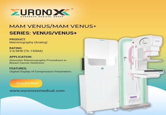
Embark on a journey of innovation and excellence with EuroNoxx Medical Group. Explore our cutting edge surgical devices today at www.euronoxxmedical.com revolutionise healthcare delivery for a brighter tomorrow.
0 notes
Text
Strengthening Healthcare Systems through Recruiting International Talent!
Exploring the hidden gems in the healthcare system - a crucial link between human resources for health and tangible health outcomes. The latest findings from the WHO dataset will restructure the way we feel the emergence of healthcare staffing.
This is not just a demystifying fact of vital connection and paving the way for a healthier future. World healthcare providers and advanced patient care through data-driven insights make a real difference in healthcare for all.
In the complex and dynamic landscape of healthcare, the challenge of addressing skill health professional shortages remains a persistent issue. International recruitment agencies came into force with a deeper strategy to coordinate with challenges by boosting healthcare systems with a diverse pool of talent.
EuroNoxx Medical Group stands at the forefront of this initiative, actively recruiting skilled locum doctors, dentists, nurses, and allied health professionals from abroad to work in the NHS and private hospitals.
This article delves into the benefits of international recruitment, illustrating how it not only mitigates staffing shortages but also enhances the quality of care provided to patients.
Delving deep into the data to uncover key insights that matter!
Our recent analysis sheds light on vital healthcare trends, exploring the intricate link between human resources for health and critical mortality rates.
From maternal to infant and under-five mortality rates, each data point tells a powerful story. And we're here to decode it all. Sharing our thoughts as we solve the importance of our findings. Let's transform numbers into actions that make a difference!
A...Addressing Critical Staff Shortages!
The demand for healthcare services continues to rise, driven by an ageing population, advances in medical treatment, and higher patient expectations.
This increased demand places significant pressure on the existing healthcare workforce, leading to critical staff shortages in various specialities and regions. International recruitment offers a practical solution to this issue, enabling healthcare systems to fill vacancies with highly qualified professionals from around the globe.
By doing so, EuroNoxx Medical Group plays a crucial role in ensuring that healthcare providers can continue to offer essential services without compromise.
B..Enriching the Workforce with Diversity!
One of the most significant advantages of international recruitment is the diversity it brings to the healthcare workforce. Healthcare professionals from different cultural and clinical backgrounds contribute varied perspectives, skills, and approaches to patient care.
This diversity promotes a more inclusive atmosphere, improves communication with patients from diverse backgrounds, and enhances the overall quality of care.
Moreover, the exchange of knowledge and best practices among internationally recruited staff and their UK counterparts leads to continual learning and improvement in clinical practices.
C..Meeting the Demand for Specialised Skills!
Certain areas of healthcare are in alarming need of professionals with specialised skills. Which can be challenging to source domestically.
International locum staff recruitment agency allows healthcare institutions to access a global talent pool of healthcare specialists by measuring their expertise to address specific healthcare challenges.
As the UK's best international locum staff recruitment company, we bridge this essential pool of healthcare talents by providing an advanced medical learning and training ecosystem.
Supporting Sustainable Workforce Development
While international recruitment addresses immediate staffing needs, it also supports the long-term development of the healthcare workforce. By incorporating international professionals into the UK healthcare system, EuroNoxx Medical Group contributes to a more robust and resilient workforce capable of meeting future healthcare demands.
Furthermore, for an international staff recruitment culture of mentorship and professional development, please visit our career page and fill out the form to get the next generation of UK healthcare and medical workforce.
Conclusion
International recruitment is more than just a strategy to fill staffing gaps; it is a means of enriching the UK healthcare system with global talent and expertise.
The efforts of EuroNoxx Medical Group in recruiting healthcare professionals from abroad are pivotal in enhancing the diversity, quality, and sustainability of healthcare services. By accommodating international locum talent on a permanent and temporary basis, our contribution remains a priority in the global as well as UK healthcare sectors to address critical staff shortages and foster an environment of Medical Equipment and healthcare supplies as well for supper speciality patient care.
As we move forward, the continued integration of international professionals will remain key to the strength and resilience of healthcare systems.
0 notes
Text
Key Developments And Emerging Technologies In Medical Devices - A Decade of Innovation!
In the past decades, the field of medical devices has seen amazing growth and innovation.
EuroNoxx Medical Group is a leading provider to the NHS and private hospitals. It is at the forefront of this revolution. It makes sure healthcare facilities have the newest technologies.
They use these to give top care to patients. Several key developments have changed the global landscape. They have transformed healthcare management and technology use. These include breakthrough technologies and revolutionary advancements.
Let EuroNoxx Medical dig deeper into top emerging technologies that have made a significant impact on medical devices in the last decade, revolutionising patient care and improving treatment outcomes.
mRNA Technology:
mRNA (messenger RNA) technology has changed vaccine development. It has also changed treatment for various diseases. This technology was showcased a lot during the COVID-19 pandemic.
It lets the body make specific proteins. These proteins trigger the immune response we want.
The success of mRNA vaccines against COVID-19 has opened doors. They lead to better treatments for cancer, heart disease, and genetic disorders.
Virtual Reality (VR):
Virtual Reality technology has entered the medical device field. It offers immersive experiences and has many uses. Surgeons use VR to simulate complex procedures.
This allows for detailed practice and honing of skills. It avoids risking patient safety. VR therapy is also a promising tool for pain management. It can provide distraction during procedures and aid in exercises for rehabilitation.
Neurotechnology:
Neurotechnology has seen big advances. This is especially true for brain-computer interfaces (BCIs) and neurofeedback. BCIs enable direct brain-to-device communication. They let paralyzed people control prosthetic limbs and communicate through thought commands.
Neurofeedback devices are for brain training. They help people enhance their brain patterns. This improves mental health and thinking.
Artificial Intelligence (AI):
Medical devices now integrate Artificial Intelligence. It offers capabilities like data analysis, pattern recognition, and predictive modelling. AI algorithms provide valuable insights for diagnosis, treatment planning, and personalised medicine.
Machine Learning algorithms help identify diseases in medical images. They improve accuracy and efficiency. AI-driven healthcare systems also facilitate real-time monitoring. They also enable early detection of potential health risks.
3D Printing:
3D printing has revolutionised the making of medical devices. They range from prosthetic limbs to custom implants. This technology allows for detailed, patient-specific designs. They ensure a perfect fit and better function.
Also, 3D-printed surgical models help with pre-surgical planning and education. They let surgeons understand complex procedures better and improve patient outcomes.
Precision Medicine:
The concept of precision medicine has gained considerable traction in recent years. It involves tailoring medical treatment to an individual's specific traits.
These traits include genetic makeup, lifestyle, and the environment. Genomics, diagnostics, and data analytics have advanced. They allow for personalised treatment and targeted therapies. This improves patient outcomes and minimises adverse effects.
CRISPR:
CRISPR is a gene-editing technology. It has sped up research on genetic disorders and potential treatments. CRISPR modifies or deletes specific genes. It can treat genetic diseases. It can improve current treatments. It can enhance disease prevention.
Health Wearables:
Health wearables are very popular these days. They include smartwatches, fitness trackers, and biosensors. They monitor health and promote wellness.
These devices provide data in real-time. They show heart rate, sleep patterns, and activity. They also show more. Wearables now work with smartphone apps and cloud platforms. This lets users and healthcare providers track and manage health better.
Technology in Mental Health:
Advancements in technology have also extended to the field of mental health. Digital mental health platforms offer innovative solutions for therapy, counselling, and self-care.
These platforms use AI-powered chatbots, virtual therapists, and mobile apps. They provide mental health support, symptom tracking, and personalised interventions. They fill gaps in traditional mental healthcare systems.
The Impact of Technology on Diagnostics:
The first step in effective treatment is accurate diagnosis. Advanced diagnostic instruments, like MRI, CT, and X-ray machines, have improved doctors' ability. They can now diagnose conditions with precision and speed.
These patient monitoring technologies produce detailed images of the body's internal structures.
They let us detect diseases and conditions early stages.
These would be invisible to the naked eye. High-resolution images let medical professionals make better decisions about patient care. This leads to better outcomes and happier patients.
Enhancing Treatment with Surgical Instruments:
The development of tech-driven surgical tools has revolutionised the way surgeries are performed. Hospital equipment such as laparoscopes, robotic surgical systems, and laser tools, has made minimally invasive surgeries accessible.
They reduce the need for open incisions. This minimises the risk of infection and complications. It also substantially reduces recovery times. This allows patients to return to their daily lives more rapidly.
EuroNoxx Medical Group is committed to supplying these new hospital equipment and surgical instruments. This commitment ensures that hospitals can offer the best treatment options. It improves the quality of healthcare.
Patient Recovery and Rehabilitation
Medical equipment's role goes beyond diagnosis and treatment. It is also crucial in patient recovery and rehab. For example, modern physiotherapy equipment helps patients. It helps them regain mobility and strength after surgery or illness.
Similarly, advanced patient monitoring systems allow continuous patient vital illness observation. They ensure that any problems are found promptly. This tech-driven approach to patient care improves patient recovery. It also significantly adds to patient safety and comfort during their hospital stay.
The Future of Healthcare Technology
As technology rapidly advances. This creates an enormous opportunity for more medical and hospital equipment to be incorporated into real-world healthcare systems.
AI and ML improve diagnosis accuracy. Telemedicine expands access to care. EuroNoxx Medical Group is dedicated to adopting these innovations.
We ensure that the NHS and private hospitals are always at the forefront of medical tech.
Conclusion:
The last decade has seen avant-garde in medical devices. They entrust healthcare professionals and help patients. New technologies, such as mRNA and virtual reality, are transforming diagnostic instruments.
They are also changing how we diagnose ecosystems, manage healthcare facilities, and control diseases. This includes intertwined Neurotechnology and AI, 3D printing, precision medicine, CRISPR, telemedicine, health wearables, and mental health tech.
State-of-the-art medical equipment and surgical devices are very important in modern healthcare. Their importance cannot be emphasized enough. They greatly improve diagnosis, treatment, and patient recovery. They enhance healthcare services a lot.
EuroNoxx Medical Group is key in supplying these technologies to the NHS and private hospitals. They ensure top-notch patient care. Advancements in medical technology will improve healthcare accessibility, efficiency, and effectiveness.
#Enhancing Treatment with Surgical Instruments#Patient Recovery and Rehabilitation#Future of Healthcare Technology
1 note
·
View note
Last Seen Blogs

aceforge12-blog
Ace Forge Pvt Ltd

harri-podd-blog
Arty stuff

dolorem-ipsum-au
Still Not Ready For the Responsibility

professorshape
I wanna talk about my favorite gay people

montliyets
@girlbosselrond :]