#Handheld Ultrasound Machine
Explore tagged Tumblr posts
Visit Tumblr Blog
Explore Tumblr blogs with no restrictions, modern design and the best experience.
Last Seen Tumblr Blogs


Fun Fact
Kazakhstan’s Minister of Communications and Informatics has blocked the Tumblr site because it contained 60 sites of terrorism, extremism, and pornography in 2015.
Text
How to Choose the Right Ultrasound Machine for Your Needs

In today's fast-paced medical environment, the need for efficient, portable diagnostic tools is paramount. Handheld ultrasound machine have revolutionized the field by offering portability without compromising image quality. Whether you're a medical professional in a busy hospital or a practitioner in a remote clinic, selecting the right handheld ultrasound machine can significantly impact your diagnostic capabilities. Here’s a guide to help you make an informed decision.
Understanding Handheld Ultrasound Machines
Handheld ultrasound machines are compact, portable devices designed for diagnostic imaging. Unlike traditional ultrasound machines, they can easily fit in a pocket or bag, making them ideal for on-the-go use. These devices typically connect to smartphones or tablets via WiFi or Bluetooth, providing high-resolution images and advanced diagnostic capabilities.
Key Features to Consider

1. Image Quality
The primary purpose of an ultrasound machine is to provide clear and accurate images. When choosing a handheld ultrasound machine, look for devices that offer high-resolution imaging. Check the specifications for details on image depth, resolution, and frame rate. A device like the Sonosif Portable WiFi Ultrasound Scanner offers excellent imaging capabilities, ensuring you get the clarity needed for accurate diagnoses.
2. Portability and Ease of Use
One of the main advantages of handheld ultrasound machines is their portability. Ensure the device you choose is lightweight, compact, and easy to carry. Additionally, the user interface should be intuitive, allowing for quick and easy operation. Devices compatible with both iOS and Android platforms can offer more flexibility and ease of use.
3. Battery Life
Since handheld ultrasound machines are designed for portability, battery life is a crucial factor. Look for devices with long battery life to ensure they can last through multiple examinations without needing a recharge. Some models come with rechargeable batteries that can be easily swapped, further extending their usability.
4. Connectivity
Handheld ultrasound machines often rely on connectivity with smartphones or tablets for image display and analysis. Ensure the device you choose has reliable WiFi or Bluetooth connectivity and is compatible with the operating systems of your devices (iOS and Android). This feature allows for easy sharing of images and integration with other medical software.
5. Cost and Value for Money
While handheld ultrasound machines are generally more affordable than traditional units, prices can still vary widely. Consider your budget and compare features across different models to find a device that offers the best value for your money. The Sonosif handheld mobile ultrasound probes provide high-quality imaging at an affordable price, making them a great option for many healthcare providers.
6. Durability and Warranty
Medical devices are an investment, so it's important to choose a handheld ultrasound machine that is built to last. Check the durability of the device and the materials used in its construction. Additionally, consider the warranty and customer support provided by the manufacturer. A good warranty can provide peace of mind and protect your investment.
7. Application-Specific Features
Depending on your specific field of practice, you may need a handheld ultrasound machine with specialized features. For example, certain models are better suited for cardiovascular imaging, while others may excel in obstetrics or musculoskeletal applications. Assess your needs and choose a device that meets those requirements.
Why Choose Sonosif Handheld Ultrasound Machines?

Sonosif offers a range of high-quality handheld ultrasound machines designed to meet the diverse needs of medical professionals. Their devices are known for:
High-Quality Imaging: Providing clear and detailed images to support accurate diagnoses.
Portability: Lightweight and easy to carry, making them ideal for use in various settings.
Compatibility: Works seamlessly with both iOS and Android devices, offering flexibility and ease of use.
Affordability: Competitive pricing without compromising on quality.
Durability: Built to last with robust construction and backed by excellent customer support.
Conclusion
Choosing the right handheld ultrasound machine involves considering several factors, including image quality, portability, battery life, connectivity, cost, durability, and application-specific features. By carefully evaluating your needs and comparing different models, you can find a device that enhances your diagnostic capabilities and fits your budget. The Sonosif range of handheld ultrasound machines offers an excellent balance of quality, affordability, and ease of use, making them a top choice for healthcare professionals.
For more information and to explore the range of handheld ultrasound machines, visit Sonosif.
0 notes
Note
Gutten Morgen, Ich bin back once again👹 So, I just wanted to ask if you can write about the triplets. Of course, if you don't want to you don't have to.
With love, Deutsch Nonnie❤️👹

Summary: Noah x Reader finding out having triplets
Pairing: Noah x Reader
TW: none, maybe fainting? But just a cute little blurb.
N/A: for my Deutsch Nonnie, sorry it took me so long to post this, been through chaos, but I hope you like it! almost I'm sorry if this isn't accurate, I don't know anything about pregnancy or babies to be quite honest with you, but I tried my best <3
Divider by @bernardsbendystraws

Ever since Noah and I found out I was pregnant a few weeks ago everything has been like a dream. It feels so surreal to me that I’m starting a little family with the love of my life. He's been very attentive to my needs.
Today we had our first check up just to make sure everything was going to plan and hopefully we would be able to hear the heartbeat of our little.
“Are you nervous?” I asked Noah as he pulled out of the driveway of our home.
“I'm more excited than anything.. i can't believe we're having a baby” he replied back, sounding ecstatic. “I know where probably not doing this the conventional way, i mean were not married yet and were having a baby now but i wouldn't want to do it with anyone else”
A small sigh escaped my lips and I spoke once more “I wouldn't want to do it with anything else either. Plus we don't need to be married or we don't need to do anything how everyone else thinks it should be. i'm happy just the way we are”
He grabbed my hand over the middle console with one of his while his other hand remained on the steering wheel. “Yeah me too” he said in almost a whisper.
We had the radio as background for the rest of the drive. It didn't take long to arrive at the clinic.
“Hi im here for my 2 p.m. appointment” i spoke with the receptionist outside of the office of my doctor,
“Yes, Mrs. Y/L/N, follow me” she signalled with her hand.
I intertwined my finger with Noahs and started to follow the receptionist.
She knocked on the door and poke her head in “Dr. Quinn your next appointment is here”
“Yes thank you Martha, i'll take it from here” i hear the Dr, voices right as we walked in
After a couple of minutes of introduction we started a regular check up and made my vitals where they needed to be.
“Now it's a bit early to determine the sex of the baby but we can listen to its heartbeat, if that's okay with you?” Dr Quinn asked with a smile on her face.
Both Noah and I nodded in unison, too excited to speak.
“Okay i'm gonna have to lay down on the bed here and please pull your top up just a little bit for me please. I'll start getting the machine ready” she commanded.
I did as she said and laid there with my baby bump exposed to the cold air, it was starting to show more and more everyday and I was starting to feel the baby move around.
She inch closer with the machine in hand and started to also put on gel on my belly.
“Okay here we go”
At first there wasn't any sound coming from the little speaker by the machine and made me very anxious that something might be wrong but after a minute of moving the handheld device around we could hear your baby's heartbeat.
Thump, thump, thump..
It was so magical, my eyes started to water from the emotion, I reached out to grab Noah's hand once more and squeeze it and I could feel the same way as his eyes started to crystallize with adoration.
Thump, thump, thump…
“Heartbeat is where it should be everything seems to be normal but i do want to do an ultrasound to see if we can't get a look at how big the baby is” Dr Quinn said already grabbing whatever she needed to complete the ultrasound
“I'm so happy you have no idea” Noah admitted with a sigh
“So am i can't believe we got to hear your baby's heartbeat” i said
Noah gave me a forehead just as Dr Quinn walked right back in, she placed more gel on my belly and continued “Ok let's look here” she pointed at the monitor that hung from the wall.
“ok here's the head and those are its arms and legs…”
“I don't know how you can define a baby form, it all just looks like a blob to me,” Noah said probably without thinking.
“Noah!” I said, thankfully Dr Quinn thought it was funny since she let out a small laugh.
She started to move around my belly once more with the machine. “oh well you look at that..there's…”
“Where's Dr? Is everything okay?” I was a bit worried.
“Everything is okay, there one, two, and three heads, looks like you're having triplets! the heart beats we hear a beat earlier where from different little babies”
Did I hear correctly? there's 3 babies growing inside of me?
“TRIPLETS?” Noah shouted before he took a stumble down, he fainted with the impression of the news
“Noah!” I screamed once more but this time to try to get to him. “Dr please you gotta do something”
Dr Quinn got as fast as she could to grab a cotton ball and rubbing alcohol to put it under Noah's nose so he would come back to his senses.
“What..happened.. ?” he said with a disoriented tone.
Dr Quinn looked at me as if to say I should let him know what went down.
“You fainted..because..we just found out we were having triplets..” i said with an almost guilty smile,
He shook his head as if he wasn't understanding what just said to him “Triplets?” he asked again with a bit of a calmer voice.
Dr Quinn nodded her head before saying “Yes, but i'm gonna leave you two alone for a couple of minutes so you cant talk” with that she walked out of the room.
“I feel like I'm in a fever dream,” Noah spat out.
“ A good one or a bad one?” i asked
“I don't know yet honestly..” he sighed.
“I know you're scared…and to be quite honest with you so am i” i took a pause “but i know we will get through this together. remember i wouldn't want anyone else to do this with. i love you”
“you're right, we're in this together, I can't wait to have 3 little goblins running around our home” he squeezed my hand this time. “I love you too.. now where have to start tripling up on everything” he said with a small laugh.
I know that this pregnancy would probably be beating the shit of me, once i got the last trimester but I couldn't wait to have 3 babies in our home causing the best kind of chaos. Life would be a bit haptic, but I knew with Noah by my side everything will be alright.
#bad omens#bad omens cult#noah sebastian#noah fan fic#dad noah#noahsebastian#dee's works#deutsch nonnie
17 notes
·
View notes
Text

Physicians work to help prevent vision loss associated with space travel
Physicians at the Medical College of Georgia at Augusta University are working with Polaris Dawn, the first of the Polaris Program's three human spaceflight missions, to better understand the eye changes many astronauts experience during spaceflight that can leave them with a wide range of symptoms once they return to Earth—from a new need for glasses to significant loss of vision.
The Polaris Program is a first-of-its-kind effort to rapidly advance human spaceflight capabilities while continuing to raise funds and awareness for important causes on Earth.
More than 70% of astronauts experience a phenomenon known as Spaceflight Associated Neuro-Ocular Syndrome, or SANS, according to NASA. The syndrome can have "a constellation of symptoms, including these changes in vision," said Matt Lyon, MD, director of the MCG Center for Telehealth.
Astronauts can experience other maladies as well when body fluids, such as cerebrospinal fluid, shift, which can lead to structural changes in the brain.
"The changes start happening on day one," said Lyon, who is also the J. Harold Harrison M.D. Distinguished Chair in Telehealth.
"We are not entirely sure what causes these issues with vision, but we suspect it has to do with a shift in cerebrospinal fluid in the optic nerve sheath. On Earth, gravity pushes that fluid down and it drains out, but in space, it floats up and presses against the optic nerve and retina."
With the help of portable, handheld ultrasound machines, Lyon and his team hope to not only figure out the mechanism behind the changes, but also be able to predict which astronauts will be most likely to experience them.
MCG patented the concept of using portable ultrasound to rapidly visualize damage from pressure and fluid changes in the optic nerve sheath, the layers of protective membranes around the nerve. Lyon is investigating how this part of the brain is affected by elevated cranial pressure and mild traumatic brain injuries. When the brain is injured, like many other organs, it swells and fills with fluid.
Researchers worked with URSUS Medical Designs LLC, a Pennsylvania-based biotech company with expertise in ultrasound, to build a 3D ultrasound machine that could help. A one-year grant enabled the project to build a device that added another dimension to 2D transducers.
Lyon and the MCG research team are now using those ultrasound machines to screen astronauts and determine who among them may already have these incompetent or damaged optic nerve sheaths. They suspect that those are the ones who will be more susceptible to the vision changes associated with SANS.
"We discovered that when the cerebral spinal pressure goes up with mild traumatic brain injuries (TBIs), there is resulting damage to the sheath that is likely lifelong," he explained.
"We think that when astronauts who have experienced concussions or mild TBIs go into space and experience the low-gravity fluid shifts, the sheath dilates from the increase in volume. It is like a tire—a normal tire remains its normal shape as it is filled with air, and the shape doesn't change.
"When it's damaged, like bulges on the side of a tire, the fluid fills the bulges up and the sheath expands. This can cause pressure on the nerve and retina. A damaged sheath is less of a problem on Earth, but in space, the excess fluid has nowhere to go."
The researchers don't yet know if the vision changes are caused by the sheer volume of fluid, by the associated pressure, or both. They are training Polaris Dawn crewmembers to use these ultrasound machines to measure both fluid and pressure in real time during spaceflight.
"If it's just volume, we suspect the cerebrospinal fluid goes up, fills this floppy bag and gets stuck. It's almost like not flushing your toilets. You're creating this toxic environment, because the cerebral spinal fluid (CSF) is what carries toxins away from your eyes and nerves, and instead the toxins sit against the optic nerve, killing it," Lyon said.
"But it could be that combined with the increased pressure that comes with increased CSF, which would be like getting intermittent hypertension in your eye."
Next steps could include working to develop and fine tune countermeasures that would decrease fluid volumes in the head during spaceflight. For example, the U.S. space program and their international space partners use a vacuum-pant device, called lower-body negative pressure, that pulls blood and other fluids down in the body.
This experiment will be part of an extensive suite of science and research performed throughout the mission. Polaris Dawn will collaborate with 23 institutions, including MCG, on the selected experiments.
Other participating institutions include the Translational Research Institute for Space Health (TRISH); University of Texas, Houston; University of Colorado, Boulder; Baylor College of Medicine; Pacific Northwest National Laboratory; and the U.S. Air Force Academy.
4 notes
·
View notes
Text
Christine feels her heart warming at the sight of the two - especially of Stephen being so happy, delighted, finally having reached a point in life where he does not need to lie about it. It causes her to just look at her patient(s) for a moment longer before her attention flicks back to the screen...
And Khan hums out a low sound as he looks back up at his husband, the smile returning to his lips, even though he is still very much shocked about the news.
"I... don't think it is common for my kind to have twins.", is what he says then, thoughtful. "But... I'm not sure."
"It isn't exactly super common for humans either.", Christine replies as she moves the handheld part of the ultrasound across Khan's belly once more. "It happens, yes, and there's a lot of twins out there, but most parents expect one child at a time. ---Fun fact: If a family is blessed with twins, the children are more likely to also have twins later... and so on."
Genetics, she thinks, and Khan blinks as he looks back at the screen, at their two babies, with the surprise one still moving the little arms and legs...
Christine chuckles at the sight.
"Baby number two is definitely not happy about the ultrasound. We don't hear the noise it makes, but... it's been said that fetuses can hear the machine working, and to them it can feel loud at times."
Khan thinks he can hear it, a very faint, low buzzing noise... thanks to his advanced hearing. His children must perceive it even louder than it already is, and while one twin doesn't seem to care much, the other is not really having it. ... That's actually a little funny, and Khan feels amusement tugging on the edges of his mouth.
"By the way, I think I should be able to find out the gender of the two - they're sitting in a promising position. Do you want to know?" Christine looks back at the two, waiting. Some parents want to know, others don't, after all.
Three more months at the most.
They needed to pick out names, organize the nursery and do so many other things to make sure everything was ready for the child - children. Stephen was trying to figure out if they wanted to come to the hospital for the birth or do it just with the two of them. He wanted to do a sort of comforting birth....but where?
Stephen glanced down at his husband, smiling down at him as he spoke.
"I was shocked....startled, actually. I felt the magic of the first baby pulsing, but then I thought I felt another, different but still similar....and then it seemed to come out of hiding and I felt the second magical presence within." He explained, squeezing Khan's hand gently.
"I'm glad we had an appointment today, because I wanted to be sure that there are two inside." He said.
2K notes
·
View notes
Text
All You Need to Know About Fetal Echocardiography in Pune
When a baby is growing inside the mother's womb, their heart starts forming in the early stages of pregnancy. Sometimes, doctors may want to take a closer look at the baby's heart to check if everything is developing properly. This is where a special test called Fetal Echocardiography comes in.
In this article, we will explain everything about Fetal Echocardiography in Pune in a simple and easy-to-understand way—perfect for school-level students or first-time parents looking for clarity. We'll also introduce you to one of the most trusted radiologists in Pune, Dr. Sagar Sujanyal, who provides expert fetal echocardiography services at Pearl Diagnostics.
🧠 What is Fetal Echocardiography?
Fetal Echocardiography is a special type of ultrasound that focuses only on the unborn baby’s heart. It gives detailed pictures and allows doctors to see how the baby's heart looks and functions while still in the womb.
Just like a regular sonography, this test uses sound waves to create images. It is safe, non-invasive, and does not harm the baby or the mother.
🍼 Why is Fetal Echocardiography Important?
Sometimes, babies may have heart problems even before birth. These are called congenital heart defects (CHDs). Detecting such conditions early helps doctors and parents prepare for treatment and care after birth.
Here are some common reasons why a doctor might recommend Fetal Echocardiography in Pune:
The mother has diabetes or certain infections
There is a family history of heart disease
Previous baby had a heart problem
Doctor noticed something unusual during a routine ultrasound
Multiple pregnancies (like twins or triplets)
Mother is on certain medications during pregnancy
🧑⚕️ Meet Dr. Sagar Sujanyal – Specialist in Fetal Echocardiography
Dr. Sagar Sujanyal, founder of Pearl Diagnostics, is one of the most experienced and trusted radiologists in Pimpri Chinchwad and Pune. With years of experience in ultrasound diagnostics, he specialises in Fetal Echocardiography in Pune, along with a wide range of other advanced imaging tests.
At Pearl Diagnostics, every fetal echo scan is performed with utmost care, accuracy, and sensitivity. Dr. Sujanyal explains the results in simple language and helps the expecting parents understand every detail about their baby’s heart health.
🏥 About Pearl Diagnostics – Best Diagnostics Centre in PCMC, Pune
If you're searching for a reliable place for Fetal Echocardiography in Pune, look no further than Pearl Diagnostics. Here's why we are trusted by hundreds of families:
Modern ultrasound machines for clear and accurate imaging
Comfortable and hygienic facility with friendly staff
All sonography services under one roof
Managed by Dr. Sagar Sujanyal, a highly qualified and compassionate radiologist
Authorised partner of Golwilkar Metropolis for all types of blood and urine tests
📍 Address: Jhamtani Building, near GANESH HOTEL, Shastri Nagar, Pimpri Colony, Pune, Maharashtra 411017 📞 Phone: +91-91569 59267 📧 Email: [email protected]
🔍 What Happens During a Fetal Echocardiography?
The test is very similar to a regular pregnancy ultrasound. Here’s what happens:
You’ll lie down comfortably on a bed.
A special gel is applied to your belly.
A handheld device (called a probe) is moved over the belly to capture images of the baby’s heart.
The screen shows how the heart is beating, how blood flows, and if all parts of the heart are growing properly.
The test usually takes 30 to 60 minutes, and you can go home right after.
🗓 Best Time to Do the Test
Doctors usually recommend Fetal Echocardiography between the 18th and 24th weeks of pregnancy. But in some special cases, it can be done earlier or later as well.
✅ Benefits of Fetal Echocardiography
Doing this test at the right time helps:
Detect heart defects early
Plan safe delivery at a hospital with NICU
Get early opinions from child heart specialists
Prepare emotionally and medically
Many parents in Pimpri and Pune have found peace of mind after getting Fetal Echocardiography in Pune done at Pearl Diagnostics.
💡 Real-Life Example
A couple from Pimpri visited Pearl Diagnostics for a second opinion after their regular scan showed some doubts about the baby's heart. Dr. Sagar Sujanyal performed a detailed fetal echo scan and explained everything clearly. Thankfully, the baby’s heart was perfectly normal, and the parents felt reassured and stress-free.
🧪 Other Services at Pearl Diagnostics
Apart from Fetal Echocardiography in Pune, Pearl Diagnostics also offers:
Pregnancy Scans (Level 1 & 2)
NT Scan, Anomaly Scan
Follicular Study & Ovulation Monitoring
2D Echo, ECG, and Stress Echo
3D/4D Sonography
Color Doppler Ultrasound
Pathology Tests – Blood, Urine, Thyroid, Diabetes, Vitamin D, and more
We focus on Quality, Accuracy, and Compassionate Care.
🔁 When Follow-up Is Needed
If the first fetal echo shows any issue or if the baby’s position is not ideal for viewing, your doctor might ask for a follow-up scan. It is important not to panic—sometimes, small adjustments and time help in getting clearer images.
🌟 Why Parents Prefer Dr. Sagar Sujanyal for Fetal Echo
He explains reports in simple and clear words
Focuses on detailed evaluation
Gives parents the emotional support they need during pregnancy
Works with leading paediatric cardiologists if any advanced care is needed
💬 Final Words
Bringing a baby into the world is a beautiful journey, and knowing that your baby's heart is growing healthy brings peace of mind. If your doctor recommends it, don’t delay the Fetal Echocardiography in Pune.
With Dr. Sagar Sujanyal at Pearl Diagnostics, you’re not just getting an ultrasound—you’re getting expert guidance, modern technology, and personal attention.
📞 Book Your Appointment Today
Whether you’re in Pimpri, Chinchwad, or anywhere in Pune, make sure your baby’s heart is in safe hands.
📍 Visit us: Jhamtani Building, near GANESH HOTEL, Shastri Nagar, Pimpri Colony, Pune, Maharashtra 411017 📞 Call Now: +91-91569 59267 📧 Email: [email protected]
Get your Fetal Echocardiography in Pune done with care and confidence at Pearl Diagnostics.
0 notes
Text

Chinese rescue efforts on Thursday provide medical service in #Mandalay City, the most devastaded area by last week's M7.9 earthquake. Medical staff from 14 departments were on site, equipped with bedside ECG machines, handheld ultrasound devices, portabal X-ray machines. Over 250 people recieved treatment and consultations.
0 notes
Text
Echocardiography Devices Market is driven by Technological Advancements

The echocardiography devices market encompasses a diverse range of ultrasound-based imaging systems designed for non-invasive evaluation of cardiac anatomy and function. Key products include 2D and 3D transthoracic and transesophageal echocardiography systems, Doppler imaging devices, portable and handheld scanners, as well as advanced software suites for strain imaging and AI-enhanced analysis. These devices offer significant advantages such as real-time visualization, high spatial resolution, safety (no ionizing radiation), cost-effectiveness, and user-friendly interfaces that streamline workflow in hospitals, clinics, and point-of-care settings.
The growing need for early detection and management of cardiovascular diseases, coupled with rising healthcare expenditure and favorable reimbursement policies, drives adoption. Innovations in miniaturized probes and wireless connectivity support telemedicine and remote monitoring, expanding market scope. Furthermore, integration of machine learning algorithms enables automated measurements and improved diagnostic accuracy, addressing market challenges related to operator dependency. Robust R&D investments and strategic collaborations among market players fuel continuous product enhancements and launch of next-generation platforms. As healthcare providers seek efficient, scalable imaging solutions, the demand for versatile Echocardiography Devices Market continues to rise.
The echocardiography devices market is estimated to be valued at USD 10.82 Bn in 2025 and is expected to reach USD 16.27 Bn billion by 2032, growing at a compound annual growth rate (CAGR) of 6.00% from 2025 to 2032. Key Takeaways
Key players operating in the Echocardiography Devices Market are:
-B. Braun Melsungen AG
-Medtronic
-BD (Becton, Dickinson and Company)
-Fresenius Kabi AG
-Baxter According to recent market analysis, these market companies command a significant market share through comprehensive portfolios featuring portable handheld scanners, advanced 3D/4D imaging platforms, and AI-driven diagnostic software. B. Braun leverages its expertise in catheter-based imaging technologies, while Medtronic’s strategic collaborations with software developers enhance its solution suite. BD’s targeted acquisitions have broadened its product offerings and global footprint, and Fresenius Kabi exploits its robust distribution network to penetrate emerging markets. Baxter’s focus on probe innovation and ergonomic design bolsters its competitive positioning. These key players continuously invest in R&D, product launches, and strategic alliances, underscoring intense market dynamics. Their concerted efforts to optimize performance, reduce costs, and improve user experience are central to shaping industry trends and sustaining business growth. Growing demand for echocardiography devices is driven by a surge in cardiovascular disease prevalence worldwide and a shift toward non-invasive diagnostic modalities. Market research highlights increasing adoption of point-of-care ultrasound in emergency departments, outpatient clinics, and remote care settings. The rising geriatric population, along with higher rates of obesity and diabetes, fuels the necessity for routine cardiac monitoring and early disease detection. Telemedicine initiatives and mobile health programs further stimulate demand for compact, wireless-enabled echocardiography systems. Government healthcare reforms and favorable reimbursement frameworks in developed regions bolster market opportunities. Moreover, the integration of AI-based analytics enhances diagnostic accuracy and workflow efficiency, aligning with healthcare providers’ goals of cost containment and improved patient outcomes. These factors collectively propel market growth and underscore the technology’s critical role in modern cardiology.
‣ Get More Insights On: Echocardiography Devices Market
‣ Get this Report in Japanese Language: 心エコー��査装置市場
‣ Get this Report in Korean Language: 심장초음파장비시장
0 notes
Text
Medical Power Supply Devices Market Introduction: Powering the Future of Healthcare Technology
The Medical Power Supply Devices Market is a critical segment within the broader medical technology industry, driving innovation and reliability in healthcare equipment. As medical devices become increasingly sophisticated, the demand for specialized power supply units designed to meet stringent safety, efficiency, and performance standards has surged. This article explores the current landscape, emerging trends, challenges, and future outlook of the medical power supply devices market, shedding light on how these unseen components are vital in transforming patient care.

The Vital Role of Medical Power Supply Devices
Medical power supply devices serve as the backbone for an array of critical medical equipment—ranging from diagnostic machines like MRI scanners and CT devices to therapeutic tools such as ventilators and infusion pumps. Unlike conventional power supplies, medical-grade units must adhere to rigorous regulatory standards to ensure patient safety, minimize electromagnetic interference, and maintain continuous, uninterrupted operation in life-supporting environments.
The market’s growth is fueled by the ongoing digitization of healthcare, increasing adoption of portable and wearable medical devices, and the rising demand for home healthcare solutions. These devices require power supply systems that are compact, energy-efficient, and capable of supporting battery operation alongside mains power, creating a niche for highly specialized power technologies.
Market Dynamics and Key Drivers
Growing Prevalence of Chronic Diseases The surge in chronic illnesses such as diabetes, cardiovascular diseases, and respiratory disorders has accelerated the adoption of advanced medical devices for continuous monitoring and treatment. This rise has directly increased the need for reliable power supplies that can support long-term device functionality, especially in wearable and portable formats.
Advancement in Healthcare Infrastructure Developments in healthcare infrastructure, particularly in emerging economies, have led to an increase in diagnostic and treatment facilities equipped with state-of-the-art machines. As hospitals and clinics modernize their equipment, demand for sophisticated medical power supply devices that ensure safety and efficiency is rising.
Technological Innovation and Miniaturization Continuous innovation in power electronics has resulted in power supplies that are smaller, lighter, and more efficient. Technologies such as digital power controllers and gallium nitride (GaN) semiconductors enable better thermal performance and higher power density, allowing the integration of power units into compact medical devices without sacrificing reliability.
Stringent Regulatory Landscape Medical power supply devices are subject to strict standards and certifications—such as IEC 60601 for medical electrical equipment—to ensure they meet safety and electromagnetic compatibility requirements. Manufacturers investing in compliance are gaining market trust and access to global markets, pushing the industry towards higher quality and safer products.
Emerging Trends in the Market
Integration of Smart Power Supplies Power supplies embedded with IoT connectivity and smart diagnostics are gaining traction. These smart units can communicate device status, predict failures, and optimize power consumption, enhancing maintenance and operational efficiency in healthcare setups.
Battery-Operated and Portable Medical Devices The rise of portable medical devices, such as handheld ultrasound machines and wearable biosensors, demands power supplies optimized for battery efficiency and long operational life. This trend is encouraging innovation in rechargeable power modules and energy harvesting techniques.
Sustainability and Energy Efficiency With global emphasis on reducing carbon footprints, medical power supply manufacturers are focusing on eco-friendly designs, utilizing materials that reduce waste and energy consumption. High-efficiency power conversion and low standby power are becoming critical features.
Customization for Specialized Applications The diverse range of medical devices—from surgical robots to dental equipment—requires customized power solutions tailored for specific voltage, current, and form factor needs. Companies offering adaptable and modular power supply designs are gaining competitive advantage.
Challenges Facing the Market
Despite promising growth, the medical power supply devices market faces several hurdles:
High R&D Costs Developing power supplies that meet medical standards demands significant investment in research, testing, and certification. Smaller manufacturers may struggle to keep pace with these requirements.
Complex Regulatory Approvals Navigating the labyrinth of medical regulations can delay product launches and increase costs, particularly for companies aiming for global distribution.
Competition from Low-Cost Suppliers Price-sensitive buyers in emerging markets often prefer low-cost alternatives that may not meet stringent quality standards, affecting overall market quality perception.
Supply Chain Disruptions The reliance on specialized components and semiconductor chips makes the market vulnerable to global supply chain interruptions, which can delay production and increase costs.
Regional Insights
North America and Europe remain dominant markets due to advanced healthcare infrastructure and high adoption of innovative medical devices. Stringent regulatory frameworks in these regions also drive demand for high-quality medical power supplies.
Asia-Pacific is the fastest-growing region, driven by expanding healthcare services, rising government healthcare expenditure, and increasing awareness about medical technologies in countries like China, India, and Japan.
Latin America and Middle East & Africa markets are emerging, supported by investments in healthcare infrastructure and rising incidence of chronic diseases.
Future Outlook
Looking ahead, the medical power supply devices market is poised for sustained growth fueled by continuous technological advancements and expanding healthcare needs worldwide. Key future developments likely to shape the market include:
Enhanced Integration with AI and IoT The fusion of AI and IoT technologies with power supply units will create smarter medical devices capable of self-monitoring and predictive maintenance, reducing downtime and improving patient outcomes.
Increased Focus on Portable and Home Healthcare Devices As healthcare shifts towards personalized and home-based care, demand for compact, lightweight, and efficient power supplies tailored for portable devices will surge.
Growth in Telemedicine and Remote Monitoring Remote diagnostics and monitoring require devices powered reliably in various environments, highlighting the need for robust and adaptable power supply solutions.
Sustainability-Driven Innovation Manufacturers will continue to innovate in energy-efficient designs and recyclable materials to meet regulatory demands and corporate social responsibility goals.
Conclusion
The Medical Power Supply Devices Market stands as an essential pillar supporting the evolution of modern healthcare technology. From enabling life-saving equipment in hospitals to powering wearable health monitors, these specialized power systems ensure safety, reliability, and efficiency in critical medical applications. With growing healthcare demands and rapid technological advancements, the market promises dynamic growth and exciting innovation opportunities in the years ahead, fundamentally powering the future of healthcare delivery.
0 notes
Text
What to Expect at a Trusted General Ultrasound Clinic

When visiting a General Ultrasound Clinic in Noida, knowing what to expect can ease your mind and help you prepare better for your appointment. Ultrasound imaging, also known as sonography, is a painless and non-invasive diagnostic tool used to view internal organs, soft tissues, and blood flow. Whether your doctor recommends it for a routine check-up or a specific health concern, a reliable clinic ensures accuracy, comfort, and professional service.
What Is a General Ultrasound?
Ultrasound technology uses high-frequency sound waves to create images of internal body structures. It is commonly used to examine organs such as the liver, kidneys, pancreas, bladder, and thyroid. The procedure is safe and does not involve radiation, making it a preferred choice for various diagnostic needs.
At a General Ultrasound Centre in Noida, trained sonographers and radiologists conduct the tests with advanced machines to ensure precise and reliable results. These clinics are equipped to handle a wide range of ultrasound procedures for both men and women.
Preparing for Your Ultrasound
Before your appointment, the clinic will usually provide you with preparation instructions depending on the type of scan. For abdominal scans, fasting for 6-8 hours may be required, while for pelvic scans, you may be asked to drink water to ensure a full bladder. Proper preparation is key to obtaining clear images and accurate diagnosis.
Patients undergoing a General Ultrasound Scan in Noida should also bring any previous medical records or prescriptions. This helps the radiologist compare findings and give a more comprehensive report.
What Happens During the Procedure?
The ultrasound procedure typically lasts between 15 to 30 minutes. You’ll be asked to lie down on an examination table, and a water-based gel will be applied to the area being examined. This gel helps transmit sound waves effectively.
A handheld device called a transducer is moved over your skin, sending sound waves into the body and receiving echoes back. These echoes form real-time images displayed on a monitor. Throughout the scan, the sonographer may ask you to change positions or hold your breath briefly for clearer imaging.
The experience is painless, and most patients report only mild discomfort from the pressure of the transducer.
Benefits of Visiting a Trusted Clinic
Choosing a trusted General Ultrasound Centre in Noida ensures several key advantages:
Accurate Diagnosis: Expert radiologists and technicians interpret your scan with precision.
Advanced Equipment: High-resolution imaging machines ensure detailed visuals for better analysis.
Patient Comfort: Clinics prioritize patient well-being with minimal wait times and friendly staff.
Comprehensive Care: Many clinics provide follow-up consultations or can directly coordinate with your physician.
Reliable clinics maintain hygiene standards, follow up with patients, and often provide digital reports for easy sharing with your doctor.
Types of General Ultrasounds Offered
Some common types of general ultrasounds include:
Abdominal Ultrasound: Evaluates organs like the liver, gallbladder, and spleen.
Pelvic Ultrasound: Often used to examine reproductive organs or the urinary bladder.
Thyroid Ultrasound: Checks for abnormalities in the thyroid gland.
Kidney Ultrasound: Identifies kidney stones, infections, or cysts.
Depending on your symptoms, your doctor may refer you to a General Ultrasound Scan in Noida to investigate specific concerns.
After the Ultrasound
Once the procedure is complete, the radiologist will analyze the images and prepare a detailed report. This report is either handed to you or sent directly to your referring doctor. You can usually resume your daily activities immediately unless instructed otherwise.
If you were required to fast before the test, don’t forget to hydrate and eat something light after the procedure. In some cases, your doctor may recommend further testing based on the ultrasound findings.
0 notes
Text
Strategic Developments Reshaping the Competitive Dynamics of the Veterinary Ultrasound Scanner Market
The veterinary industry has experienced a notable transformation over the past decade, driven by the convergence of advanced technology, growing awareness of animal health, and the increasing role of pets as family members. Among the many innovations reshaping veterinary care, ultrasound imaging has become one of the most essential diagnostic tools. As a result, the veterinary ultrasound scanner market has seen steady growth, evolving rapidly to meet the changing demands of veterinarians and pet owners alike.

The Role of Ultrasound in Veterinary Care
Ultrasound scanners are used to visualize soft tissues, monitor pregnancies, diagnose internal abnormalities, and guide surgical procedures in animals. The technology is prized for being non-invasive, real-time, and relatively affordable when compared to other imaging techniques such as MRI or CT scans. Whether in small animal clinics, large animal practices, or wildlife conservation projects, ultrasound plays a key role in modern veterinary diagnostics.
Ultrasound scanners used in veterinary medicine fall into two primary categories: portable/handheld units and cart-based systems. Handheld devices are growing in popularity due to their convenience, mobility, and increasing accuracy. These are particularly favored by mobile vets, equine practitioners, and those working in rural areas where on-site diagnostics are necessary.
Market Drivers
Several factors are fueling the growth of the veterinary ultrasound scanner market. First and foremost is the rising pet population. With pet ownership booming globally, especially in emerging markets, there is a growing demand for better and more frequent veterinary care.
Another key driver is increased spending on pet healthcare. Pet owners today are more willing to invest in the health and wellness of their animals, leading to a surge in diagnostic services. This trend is particularly evident in developed countries where pets are treated with the same level of care as human family members.
In addition, livestock health monitoring has gained traction. The health of food-producing animals like cows, pigs, and sheep is crucial to both economic productivity and food safety. Ultrasound scanners are used widely in reproduction management, making them valuable tools for farm operators and veterinary professionals alike.
Technological advancements are also playing a significant role. Innovations like wireless probes, 3D/4D imaging, and AI-assisted diagnostics are making ultrasound machines more powerful, accurate, and user-friendly. These improvements are attracting not just veterinarians but also institutions and training centers, further expanding the market.
Regional Trends
North America and Europe currently dominate the market, largely due to high pet ownership rates, well-established veterinary infrastructures, and strong R&D presence. In the U.S., for example, the number of veterinary clinics equipped with ultrasound devices has increased significantly over the past few years.
However, Asia-Pacific is emerging as a fast-growing market. Countries like China, India, and South Korea are witnessing rising disposable incomes, urbanization, and awareness around pet healthcare. Moreover, government initiatives aimed at livestock welfare are also contributing to the adoption of veterinary ultrasound devices in this region.
Latin America and parts of Africa represent smaller but promising markets, where expanding agricultural sectors and increasing efforts in animal disease control could drive future demand.
Competitive Landscape
The veterinary ultrasound scanner market is moderately competitive, with a mix of established medical imaging companies and niche veterinary equipment manufacturers. Companies like Esaote, Mindray, FUJIFILM SonoSite, and IMV Imaging are well-recognized players, offering a range of ultrasound solutions tailored for veterinary applications.
Meanwhile, new entrants and startups are focusing on compact, affordable, and AI-powered scanners to gain a foothold in the market. These companies are pushing innovation, offering products that cater to mobile vets, smaller clinics, and even educational institutions.
Challenges and Opportunities
Despite its growth trajectory, the market is not without challenges. High initial costs, particularly for advanced ultrasound systems, can be a barrier for small clinics and solo practitioners. Training and skill development are also critical, as accurate ultrasound interpretation requires expertise that may not be readily available in all regions.
However, these challenges bring opportunities. There's a growing demand for cost-effective, easy-to-use ultrasound systems that require minimal training. Companies that can strike the right balance between performance, portability, and price will find ample room to grow.
Final Thoughts
The veterinary ultrasound scanner market is in the midst of a significant evolution. Driven by rising demand, technological innovation, and global awareness of animal health, the market is poised for steady growth in the years ahead. For stakeholders across the board from manufacturers to veterinarians this is a space worth watching closely.
As technology becomes more accessible and animal healthcare more prioritized, ultrasound is set to become even more indispensable in veterinary diagnostics, bridging the gap between advanced care and everyday practice.
0 notes
Text
HIFU in Gurgaon
Discover the Power of HIFU in Gurgaon at Skin Aura Brain and Spine Neuro Center
In today’s fast-paced world, achieving youthful, glowing skin without undergoing surgery is a dream come true for many. One revolutionary treatment that’s making waves in the aesthetic world is HIFU (High-Intensity Focused Ultrasound). At Skin Aura Brain and Spine Neuro Center in Gurgaon, HIFU has emerged as one of the most sought-after non-invasive procedures for skin tightening and facial rejuvenation. Let’s explore how HIFU is changing the face of cosmetic dermatology and why Skin Aura is your go-to destination for this advanced treatment.

What is HIFU?
HIFU stands for High-Intensity Focused Ultrasound. It’s a non-invasive cosmetic procedure designed to tighten and lift the skin using focused ultrasound energy. Unlike lasers, which target the surface of the skin, HIFU penetrates deep into the dermis and SMAS (Superficial Musculoaponeurotic System) layer to stimulate collagen production. This results in firmer, younger-looking skin without the need for surgical intervention.
The treatment is FDA-approved and is especially popular among individuals who wish to delay or avoid going under the knife. Whether it’s sagging skin on the face, drooping jowls, or fine lines around the eyes, HIFU in Gurgaon at Skin Aura Brain and Spine Neuro Center can help restore your youthful charm.
Why Choose HIFU in Gurgaon?
Gurgaon has rapidly grown into a hub for advanced medical and cosmetic treatments. With the increasing demand for non-invasive facial rejuvenation, HIFU stands out as a top choice among locals and visitors alike. Here’s why:
Non-Surgical and Painless: HIFU is an outpatient procedure that doesn’t involve cuts, needles, or anesthesia. It’s ideal for people who prefer natural results without the downtime of surgery.
Minimal Downtime: You can return to your daily activities immediately after the session. It’s often called the “lunchtime facelift” because of its convenience.
Natural-Looking Results: Unlike fillers or Botox, HIFU works by stimulating your body’s natural collagen production. This means results appear gradually, offering a more natural and subtle transformation.
Targeted and Effective: HIFU can target specific areas, including the forehead, jawline, neck, cheeks, and even décolletage.
Skin Aura Brain and Spine Neuro Center – The Leader in HIFU in Gurgaon
When it comes to advanced cosmetic treatments in Gurgaon, Skin Aura Brain and Spine Neuro Center stands head and shoulders above the rest. With state-of-the-art equipment and a highly experienced medical team, the center offers safe and effective HIFU in Gurgaon.
Here’s what sets Skin Aura apart:
1. Expertise and Experience
Led by a team of experienced dermatologists and neurospecialists, the center combines medical expertise with aesthetic precision. Each HIFU session is personalized based on the patient’s skin type, age, and aesthetic goals.
2. Advanced Technology
Skin Aura is equipped with cutting-edge HIFU machines that deliver precise energy to different skin layers. This ensures optimal results with minimal discomfort.
3. Comprehensive Consultation
Before initiating the treatment, clients undergo a detailed consultation where skin type, concerns, and expectations are thoroughly evaluated. This ensures a customized approach and better outcomes.
4. Hygienic and Comfortable Environment
Patient safety and comfort are top priorities at Skin Aura. The clinic maintains the highest standards of hygiene and provides a welcoming environment for a pleasant experience.
What to Expect During a HIFU Session
Wondering what happens during a HIFU treatment in Gurgaon at Skin Aura Brain and Spine Neuro Center? Here’s a brief overview:
Consultation: You’ll start with a consultation to determine if HIFU is right for you.
Cleansing: Your skin will be thoroughly cleansed, and a numbing cream may be applied to minimize discomfort.
Ultrasound Application: A special handheld device is used to deliver focused ultrasound waves to targeted areas beneath the skin.
Session Duration: The procedure usually takes 30 to 90 minutes, depending on the area being treated.
Post-Treatment: There may be slight redness or swelling, but it typically subsides within a few hours.
Benefits of HIFU Treatment
If you’re considering HIFU in Gurgaon, here are the key benefits that make it a smart choice:
Non-invasive facelift without surgery or needles
Long-lasting results that improve over time
No recovery time needed
Improves skin texture, elasticity, and tone
Reduces fine lines and wrinkles
Contours the jawline and tightens sagging skin
Is HIFU Right for You?
HIFU is suitable for people in their 30s and above who are beginning to notice signs of aging like mild to moderate skin laxity. It’s ideal for those looking for a lift without going under the knife.
However, individuals with severe skin sagging or very heavy jowls may require more intensive procedures. At Skin Aura, specialists will guide you on whether HIFU in Gurgaon is the right treatment or if other options should be considered.
Aftercare and Results
Post-treatment care is minimal. You might experience slight tingling or tenderness for a few days, but this is completely normal. There’s no need for special skincare, although sunscreen and good hydration are always recommended.
Results typically begin to show after 2–3 weeks and continue to improve over 2–3 months as collagen production ramps up. The effects can last up to 18 months, depending on your skin type and lifestyle.
Book Your HIFU Appointment Today
If you’ve been searching for an effective, safe, and non-invasive way to rejuvenate your appearance, it’s time to experience HIFU in Gurgaon at Skin Aura Brain and Spine Neuro Center. With world-class technology, experienced professionals, and personalized care, you’re in expert hands.
Say goodbye to sagging skin and hello to a younger, more radiant you—without surgery, pain, or downtime.
Contact Skin Aura Brain and Spine Neuro Center today and take your first step towards ageless beauty.
0 notes
Text
Find the Best 2D Echo Test in Dombivli – Mahajan Heart Clinic
The heart is one of the most vital organs in the human body, and keeping it healthy is crucial for overall well-being. One of the most effective, non-invasive ways to assess heart function is through a 2D Echo Test. If you’re looking for a 2D Echo Test in Dombivli, Mahajan Heart Clinic offers advanced diagnostic facilities with expert care, ensuring accurate results and personalized treatment recommendations.
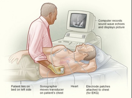
What is a 2D Echo Test?
A 2D Echo Test, also known as a 2D Echocardiography, is a painless, non-invasive ultrasound-based test that provides real-time images of the heart. It helps doctors visualize the heart chambers, valves, and blood flow to evaluate its overall function and detect any abnormalities.
This test plays a crucial role in diagnosing a variety of cardiac conditions, including:
Valve disorders
Congenital heart disease
Cardiomyopathy
Heart failure
Blood clots within the heart
Pericardial diseases
At Mahajan Heart Clinic in Dombivli, we use high-resolution echocardiography machines to deliver precise and detailed reports that form the foundation of a correct diagnosis.
Why Do You Need a 2D Echo Test?
Doctors recommend a 2D Echo Test for several reasons, including:
Unexplained chest pain or discomfort
Shortness of breath or difficulty exercising
Suspected heart murmurs
Irregular heartbeat (arrhythmia)
High blood pressure or history of heart disease
Monitoring after a heart attack or surgery
If you experience any of these symptoms, it is essential to consult a cardiologist promptly. At Mahajan Heart Clinic, you can schedule a 2D Echo Test in Dombivli and get a thorough evaluation by an experienced heart specialist.
What to Expect During the Test?
A 2D Echo Test is safe and typically takes about 20-30 minutes. Here's what happens during the test:
Preparation: You may be asked to remove your upper clothing and wear a gown.
Electrode Placement: Electrodes may be placed on your chest to monitor your heart's electrical activity.
Gel Application: A special gel is applied to your chest to help conduct sound waves.
Image Capture: A transducer (a handheld device) is moved over the chest area. It sends sound waves that bounce off the heart structures and return as echoes, which are then converted into live images on a monitor.
Observation and Recording: The cardiologist observes the heart's movements, blood flow, valve functions, and measures heart dimensions.
Since the procedure is non-invasive and radiation-free, it can be repeated if necessary to monitor progress or changes over time.
Why Choose Mahajan Heart Clinic for 2D Echo Test in Dombivli?
When it comes to your heart, precision and experience matter. Mahajan Heart Clinic is a trusted name in Dombivli, known for comprehensive heart care services. Here's why patients prefer us for their cardiac diagnostics:
1. Experienced Cardiologist
Our clinic is led by Dr. Vivek Mahajan, an experienced and compassionate cardiologist with a deep understanding of cardiovascular health. His accurate diagnostic skills and patient-centric approach ensure that every test leads to the right course of treatment.
2. State-of-the-art Technology
We use advanced echocardiography machines that provide high-resolution, real-time imaging. This enables us to detect even minor heart abnormalities with precision.
3. Prompt and Accurate Reports
Our clinic ensures timely and accurate reporting, so you don’t have to wait for long periods to understand your heart condition. Same-day reporting is often available for routine tests.
4. Comfortable and Hygienic Environment
Patient comfort and hygiene are our top priorities. Our clinic follows stringent sanitization protocols and ensures a safe environment for all diagnostic procedures.
5. Affordable Pricing
We believe in providing quality healthcare at affordable rates. Our 2D Echo Test in Dombivli is reasonably priced without compromising on the quality of care.
Book Your 2D Echo Test in Dombivli Today
Regular heart check-ups are crucial, especially if you have a family history of cardiac issues or existing health conditions such as diabetes, hypertension, or high cholesterol. A 2D Echo Test can provide early detection of potential heart problems, allowing for timely intervention and lifestyle changes.
To know more or to book your test, visit our dedicated 2D Echo service page here: 👉 www.mahajanheartclinic.in
Our team will guide you through the appointment process and answer any questions you may have.
Testimonials from Patients
Mr. Suresh P."I had mild chest discomfort, and my physician referred me to Mahajan Heart Clinic. The 2D Echo Test was smooth, quick, and the staff were very courteous. Dr. Mahajan explained the results clearly and guided me well."
Mrs. Neeta D."Very professional service. The clinic was clean, and the test was done without any waiting. Highly recommend for anyone looking for a 2D Echo Test in Dombivli."
Contact Us
Mahajan Heart Clinic – Dombivli📍 Address: Shop No 2, Siddhivinayak Plaza, M G Road, Dombivli West, Thane - Mumbai (Maharashtra) - 421202. 📞 Phone: +91-9870270751, 9773329131📧 Email: [email protected]🌐 Website: www.mahajanheartclinic.in
Final Thoughts
Your heart deserves the best care, and regular diagnostic testing is a key part of preventive health. If you are experiencing symptoms or have risk factors for heart disease, don’t delay. Schedule your 2D Echo Test in Dombivli at Mahajan Heart Clinic today and take the first step towards a healthier heart.
0 notes
Text
How next-gen ultrasound improves cardiac care | Bilmed Group of Institutions

Cardiovascular disease remains the leading cause of death globally, making accurate and timely diagnosis a critical part of saving lives. In this evolving landscape of medical diagnostics, next-generation ultrasound technology is transforming the way healthcare providers assess and treat heart conditions. At Bilmed Group of Institution, we are committed to integrating cutting-edge innovations into both our academic curriculum and clinical training—ensuring our students and healthcare professionals are equipped with the tools that shape the future of cardiac care.
What Is Next-Gen Ultrasound?
Next-generation ultrasound refers to advanced echocardiographic systems that go beyond traditional 2D imaging. These systems incorporate:
High-resolution 3D/4D imaging
Automated AI-powered diagnostics
Strain and speckle tracking imaging
Portable, handheld devices for point-of-care ultrasound (POCUS)
These enhancements enable a more precise, complete, and efficient evaluation of cardiac structures and functions.
Transforming Cardiac Diagnosis and Treatment
1. Unparalleled Image Quality
Next-gen ultrasound offers high-definition imaging that allows clinicians to visualize the heart in exceptional detail. With 3D and 4D capabilities, cardiologists can assess structural heart diseases—such as valve abnormalities and congenital defects—with greater clarity and confidence. These improvements lead to earlier detection and more effective treatment plans.
2. Advanced Functional Assessment
Strain imaging and tissue Doppler techniques provide insights into myocardial performance that traditional imaging often misses. This allows for:
Detection of subclinical heart failure
Precise measurement of ejection fraction
Real-time evaluation of diastolic function
These capabilities are especially crucial in monitoring patients with cardiomyopathies or those undergoing chemotherapy.
3. Artificial Intelligence Integration
Modern ultrasound machines use AI to assist clinicians in:
Automatically measuring heart chambers and valve functions
Identifying abnormalities with real-time suggestions
Reducing variability between operators
This not only speeds up the diagnostic process but also ensures consistency—making it an invaluable tool for both experienced cardiologists and medical trainees.
4. Portability and Point-of-Care Use
The advent of portable ultrasound devices has revolutionized emergency and critical care. These compact machines provide real-time cardiac imaging at the patient’s bedside, in ambulances, or even in remote rural settings. At Bilmed, we utilize these devices in our clinical simulation labs and outreach programs, demonstrating their effectiveness in real-world scenarios.
A Teaching Tool for the Future
As a premier medical education institution, Bilmed Group of Institution integrates next-gen ultrasound into its academic and clinical training programs. Students benefit from:
Hands-on experience with the latest technology
Simulation-based learning for better understanding of cardiac anatomy and pathology
Exposure to AI tools that are reshaping the future of diagnostic medicine
Our approach ensures that graduates are not only competent in using modern diagnostic equipment but are also critical thinkers prepared for real-world challenges.
Better Care, Better Outcomes
Ultimately, the goal of implementing advanced ultrasound technology is to improve patient outcomes. With faster diagnostics, fewer invasive procedures, and more accurate monitoring, next-gen ultrasound supports:
Early intervention
Personalized treatment plans
Improved patient satisfaction and safety
Conclusion
The integration of next-gen ultrasound into cardiac care represents a major step forward in non-invasive diagnostics. At Bilmed Group of Institution, we proudly stand at the forefront of this transformation—educating tomorrow’s healthcare leaders and empowering them with the tools that will define the future of cardiac medicine.
As the demand for faster, smarter, and more precise cardiac diagnostics grows, so does our commitment to innovation and excellence in education and clinical practice.
For more information and to download the brochure, visit our official website or contact our admissions team at: 📞 +91-9311964890 🌐 www.bilmedgroup.com
0 notes
Text
A New Era in Non-Invasive Fat Reduction and Body Contouring

The pursuit of a toned, sculpted body has never been more accessible thanks to modern aesthetic technology. One of the most effective innovations in this space is the Body Slimming Machine, a device designed to reshape the body without the need for surgery or long recovery periods.
Understanding the Technology Behind It
This advanced device typically combines multiple non-invasive technologies such as radiofrequency, infrared light, vacuum therapy, and ultrasound. These components work together to target stubborn fat, improve skin texture, and enhance body contours. The main goal is to break down fat cells while also tightening the skin and improving circulation.
Unlike traditional methods like liposuction, this treatment doesn't require incisions, anesthesia, or downtime. It's a solution that appeals to people looking for safe, efficient, and noticeable body improvements with minimal disruption to their daily lives.
Target Areas and Treatment Goals
This approach is particularly effective for areas that are resistant to diet and exercise. Common treatment zones include the abdomen, thighs, upper arms, buttocks, and flanks. Some devices can even treat smaller, delicate areas such as the chin and neck.
Beyond fat reduction, the treatment also aims to lift and tone the body. Many users experience not only a reduction in circumference but also an improvement in skin firmness and elasticity.
How Sessions Are Performed
Each session typically begins with a brief consultation to identify the client’s goals and determine the most suitable settings for the treatment. After the targeted area is cleaned and prepared, a technician uses the device to deliver energy through the skin using a handheld applicator.
The sensation during the session is often described as warm and soothing. There is usually no pain involved, although some people may feel mild suction or vibration, depending on the technology being used. A session usually lasts between 30 and 60 minutes.
Who Benefits the Most?
This type of treatment is ideal for individuals who maintain a healthy lifestyle but struggle with isolated fat deposits. It's best suited for people who are near their target weight and are looking to fine-tune their shape rather than achieve significant weight loss.
Those who hydrate well, follow a balanced diet, and stay active tend to see better and faster results. It’s also recommended for those who want gradual, natural-looking changes rather than dramatic alterations.
Skin Tightening and Collagen Stimulation
In addition to breaking down fat, the energy delivered by the device often stimulates collagen and elastin production. This is especially beneficial for individuals concerned about loose or sagging skin. As collagen levels increase, the skin becomes firmer and more elastic, adding to the overall body contouring effect.
Many users appreciate this dual action—fat reduction and skin tightening—in a single, convenient treatment plan.
Recovery and Aftercare Tips
One of the key benefits of this approach is the lack of downtime. Clients can usually return to their regular activities immediately after a session. To support results, practitioners often recommend drinking plenty of water, engaging in light exercise, and avoiding alcohol or heavy meals for a day or two post-treatment.
Depending on the treatment area and goals, multiple sessions may be needed, often spaced one to two weeks apart. Maintenance treatments can also help sustain the results over the long term.
Safety and Side Effects
The process is widely regarded as safe when performed by trained professionals. Most side effects are mild and temporary, such as slight redness, swelling, or a warm sensation in the treated area. These usually subside within a few hours. For more info Click Here.
Because it is non-invasive, the risk of serious complications is minimal. Clients often appreciate the balance between effectiveness and comfort that this method offers.
Evolving Aesthetic Preferences
The growing interest in natural-looking results and minimal recovery has fueled the popularity of non-surgical body shaping. This method aligns perfectly with these preferences, offering a flexible and effective way to enhance physical appearance without going to extremes.
0 notes
Text
Everything You Should Know About Ultrasound Anomaly Scans in Pimpri Pune
When you’re expecting a baby, there are many things you do to make sure everything goes well. One of the most important steps in pregnancy is getting an Ultrasound Anomaly Scan, also known as a Level 2 scan.
In this article, we will talk about the importance of Ultrasound Anomaly Scans in Pimpri Pune, what happens during the test, when it is done, and why Dr. Sagar Sujanyal at Pearl Diagnostics is one of the best doctors for this test in the Pune area.
This article is simple enough for a 9th or 10th class student to understand and explain to others too.
👶 What is an Ultrasound Anomaly Scan?
An Ultrasound Anomaly Scan is a special type of ultrasound test done during the second trimester of pregnancy, usually between 18 to 22 weeks.
This scan checks the baby’s growth and development and helps find any birth defects or abnormalities early. That’s why it’s also called a “mid-pregnancy scan” or “detailed fetal scan.”
📍 Purpose of the Anomaly Scan
The main reason for doing Ultrasound Anomaly Scans in Pimpri Pune is to look closely at the baby’s:
Head and brain
Heart (chambers and valves)
Spine
Kidneys and bladder
Stomach and intestines
Limbs (hands, legs, feet)
Face (to detect cleft lip or other issues)
It also checks:
The position of the placenta
The amount of amniotic fluid
The umbilical cord
The cervix and uterus for any signs of early labor
This test can help doctors take early action if any problem is found.
🩺 Who Performs the Test?
At Pearl Diagnostics, all Ultrasound Anomaly Scans in Pimpri Pune are performed by Dr. Sagar Sujanyal, a well-known and experienced radiologist and ultrasound expert.
About Dr. Sagar Sujanyal:
MBBS, DMRD (Radiology)
Over 10+ years of experience
Expert in prenatal scans and fetal health imaging
Explains reports clearly to the expecting parents
Known for accurate results and a caring approach
Dr. Sujanyal personally supervises each scan and ensures no important detail is missed.
🏥 Pearl Diagnostics – Trusted Centre for Ultrasound in Pimpri Pune
If you're searching for the best place to get Ultrasound Anomaly Scans in Pimpri Pune, Pearl Diagnostics is a leading and reliable option.
Why Choose Pearl Diagnostics?
Advanced machines for detailed fetal imaging
Clean, safe, and peaceful environment
Friendly and skilled staff
Fast and trusted reporting
Easy appointment booking
Affordable charges for all ultrasound tests
📍 Address: Jhamtani Building, near GANESH HOTEL, Shastri Nagar, Pimpri Colony, Pune, Maharashtra 411017 📞 Phone: +91-91569 59267 📧 Email: [email protected]
🧪 What Happens During an Anomaly Scan?
Many first-time mothers are nervous before the test. But don’t worry—this is a completely safe and painless scan.
Here’s what will happen:
You will be asked to lie down.
A cool gel is applied to your tummy.
A handheld probe (transducer) is gently moved across your belly.
The screen will show your baby’s organs and body parts.
The doctor will carefully check the baby's measurements and development.
🕒 The scan takes around 30 to 45 minutes.
🧠 Why This Scan Is Very Important
Some parents wonder, “Is this scan really needed?”
Here’s why Ultrasound Anomaly Scans in Pimpri Pune are important:
It helps detect birth defects like heart problems, spinal defects (spina bifida), cleft lip, and kidney issues.
It shows if the baby's growth is normal.
It helps in planning early treatment or specialist referral, if needed.
Gives peace of mind to the parents when everything is normal.
💡 Remember: This scan does not tell the baby’s gender. It is only for health and safety of the baby and mother.
💬 Real Life Example
Mrs. Priya, a 30-year-old woman from Pimpri, came for her Ultrasound Anomaly Scan in Pimpri Pune at Pearl Diagnostics. During the scan, Dr. Sagar Sujanyal found a minor heart issue. Thanks to this early detection, Priya’s baby got timely treatment after birth and is now healthy and doing well.
🧑⚕️ Services Offered at Pearl Diagnostics
Along with Ultrasound Anomaly Scans in Pimpri Pune, Pearl Diagnostics also provides:
Pregnancy Ultrasounds (Level 1, NT Scan, Growth Scan)
Fetal Echocardiography
Color Doppler Ultrasound
Follicular Study for fertility monitoring
2D Echo, ECG, Stress Echo
3D/4D Ultrasound Scans
All Blood & Urine Tests through Golwilkar Metropolis partnership
Pearl Diagnostics is your one-stop solution for pregnancy scans and routine diagnostics.
❓ Frequently Asked Questions (FAQs)
Q1. Is the anomaly scan painful? No, not at all. It is safe and painless.
Q2. Can I eat before the scan? Yes, there is no need to fast. You can eat normally.
Q3. Can my husband or family member come with me? Yes, at Pearl Diagnostics, we allow one person to join you for emotional support.
Q4. What if something abnormal is found? Don’t panic. Dr. Sagar Sujanyal will guide you on the next steps and refer you to specialists if needed.
Q5. How do I book an appointment? Call +91-91569 59267 or email: [email protected]
🌟 Final Thoughts
Every mother wants the best for her baby. That’s why doing the Ultrasound Anomaly Scan at the right time is so important. It helps to catch problems early, gives you peace of mind, and ensures your baby gets the best care from the beginning.
If you are pregnant and staying in Pune or Pimpri-Chinchwad, don’t wait. Visit Pearl Diagnostics, and let Dr. Sagar Sujanyal help you with a safe, clear, and trusted Ultrasound Anomaly Scan in Pimpri Pune.
🩷 Take care of your baby’s health from the very start. 📞 Book your scan today: +91-91569 59267 📧 Email: [email protected] 📍 Address: Jhamtani Building, near Ganesh Hotel, Shastri Nagar, Pimpri Colony, Pune
0 notes
Text

Chinese rescue efforts on Thursday provide medical service in #Mandalay City, the most devastaded area by last week's M7.9 earthquake. Medical staff from 14 departments were on site, equipped with bedside ECG machines, handheld ultrasound devices, portabal X-ray machines. Over 250 people recieved treatment and consultations.
0 notes