#Intraoperative Mode
Explore tagged Tumblr posts
Visit Tumblr Blog
Explore Tumblr blogs with no restrictions, modern design and the best experience.
Last Seen Tumblr Blogs


Fun Fact
Average visit duration of Tumblr.com is 10 mins and 25 secs.
Text
Anesthesia Information Management Systems Market to Grow on Enhanced Efficiency

The Global Anesthesia Information Management Systems Market is estimated to be valued at USD 340.64 Bn in 2025 and is expected to exhibit a CAGR of 6.0% over the forecast period 2025 to 2032. Anesthesia Information Management Systems (AIMS) streamline perioperative data collection, electronic anesthesia records, and clinical decision support for anesthesiologists and surgery teams. This advanced software populates real-time vitals, medication dosing, and procedural notes into a unified platform, enhancing patient safety and operational efficiency. The benefits include reduced paperwork, standardized documentation, error minimization, and seamless integration with hospital information systems. AIMS solutions support advanced analytics for market insights and audit trails, aiding regulatory compliance and quality improvement initiatives. Anesthesia Information Management Systems Market Insights as healthcare providers seek to optimize resources, AIMS adoption helps control costs and elevates care standards across surgical departments. Growing focus on precision medicine and data-driven protocols fuels demand for interoperable platforms capable of integrating with electronic health records and anesthesia machines. Furthermore, rising cases of complex surgeries and the need for robust risk management drive market growth and business growth strategies in the global healthcare sector. Get more insights on,Anesthesia Information Management Systems Market
#Coherent Market Insights#Anesthesia Information Management Systems#Anesthesia Information Management Systems Market#Anesthesia Information Management Systems Market Insights#Intraoperative Mode
0 notes
Text
Upgrade Your Surgical Skills with Short Term Phaco Training in India
In the continuously changing world of ophthalmology, phacoemulsification is now the gold standard in cataract surgery. Becoming a master of this method is a prerequisite for any aspiring ophthalmologist who wants to provide world-class care. But not all have the time and resources to spare for long-term fellowships or one-year programs. That's where short term phaco training in India saves the day—providing high-quality, intensive learning experiences for ophthalmologists worldwide.
India, the excellence of which is well-established when it comes to medical education, has been recognized as one of the major locations for training in cataract surgery. More specifically, short term phaco training in India has also come into fashion as an intense, hands-on, and productive option. Either a practicing ophthalmologist or an immediate postgraduate, such training can enhance your expertise and gain your confidence in a short time frame.
What is Short Term Phaco Training?
Short term phaco training in India usually means courses of one week to three months' duration. The courses are structured to give intensive hands-on exposure to phacoemulsification with emphasis on technique development, complication management, and live surgical exposure. The concept is straightforward: optimize learning in a short time without compromising quality.
Who Should Opt for Short Term Phaco Training in India?
This training mode is best suited for:
Ophthalmologists with initial surgical exposure wishing to upgrade to phacoemulsification
Surgeons wishing to master the technique or acquire new methods
Practitioners requiring a refresh before establishing their own surgical practice
International candidates who wish to have a quality, affordable option instead of Western training
The short term phaco training in India is customized to accommodate different levels of learning and objectives and hence is highly flexible and inclusive.
Why India for Phaco Training?
The reason why most people prefer short term phaco training in India is because of the country's combination of surgical volume, expert teachers, and affordability. India does millions of cataract surgeries every year, providing trainees with plenty of opportunities to see and do varied cases.
Additionally, many institutions offering short term phaco training in India are run by experienced surgeons who have trained hundreds, if not thousands, of ophthalmologists. Their mentorship ensures you’re not just learning technique but also gaining insights into patient selection, case planning, and intraoperative decision-making.
What to Expect in a Short Term Phaco Program
A typical short term phaco training in India includes:
Didactic sessions on phaco physics, instrumentation, and fluidics
Wet lab and simulator practice to develop preliminary hand-eye coordination
Observation of live surgeries and step-by-step explanations
Guided hands-on surgeries with constructive feedback from seasoned mentors
Postoperative assessment and complication management workshops
Everything is squeezed into a tight timetable, so that you get maximum value out of every hour in training.
State-of-the-Art Infrastructure
Another reason to opt for short term phaco training in India is the availability of the latest equipment. Training facilities have advanced phaco machines, high-quality microscopes, and video recording equipment for case analysis in detail. You practice in the same type of setting you'll be performing in after graduation—making you job-ready.
Certification and Career Boost
Completing a well-recognized short term phaco training in India not only improves your clinical skills but also brings considerable value to your professional resume. Several institutions offer certificates that are highly respected among the medical fraternity, making you a cut above the rest in job applications and collaborations.
Flexibility for Working Professionals
For most practicing eye surgeons, time is a limited commodity. One of the strengths of short term phaco training in India is that it permits you to advance your skills without having to step away from your duties for long. You can go back to your hospital or clinic not just with enhanced skills but also with increased confidence in your surgical abilities.
Cost-Effective Excellence
Short term phaco training in India is highly inexpensive compared to other programs available in the U.S. or Europe. Being inexpensive, it makes it easily affordable by both Indian and foreign ophthalmologists. You receive top-class training at a small fraction of the price, without any compromise on quality.
Alumni Network and Global Recognition
Hundreds of practicing ophthalmologists globally have gained short term phaco training in India. Several of them attribute their success to the solid foundation and learning that they obtained under the program. The alumni of these programs also provide a useful professional networking opportunity, providing ongoing support and collaborative opportunities.
Post-Training Support
Most of the centers that provide short term phaco training in India continue to aid their trainees even after completion of the course. This can range from online webinars, surgical videos, and advanced workshops. This continuous mentorship keeps you updated and confident even after going back to your normal practice.
Conclusion
Whether you’re looking to begin your journey with phacoemulsification or refine your surgical approach, short term phaco training in India offers a fast, focused, and highly effective path to mastery. With expert guidance, high surgical volume, and cutting-edge infrastructure, these programs are your gateway to becoming a more skilled and confident surgeon.
So, if you’re serious about taking your surgical career to the next level, don’t wait. Enroll in a short term phaco training in India and step into the future of cataract surgery with skill and confidence.
0 notes
Text
The Era of Precision Medicine in Minimally Invasive Maxillofacial Surgery
The Collision of Minimally Invasive Techniques with Maxillofacial Surgery
The so-called minimally invasive surgery refers to the advanced surgical mode in which modern photoelectric imaging system, micro-surgical device and energy generation equipment, replace traditional open operation with endoscopic technology, and precision controlled surgical instruments with traditional scalpel to complete the diagnosis and treatment of lesions through tiny wounds.
Maxillofacial surgery is an important branch of oral medicine for the surgical treatment of cranial and maxillofacial facial bones, soft tissues and temporomandibular joints.
According to the surgical symptoms and treatment purpose can be divided into the following categories:
1. Trauma Repair Surgery
Treatment range: facial fracture
Typical surgery: open reduction and internal fixation of the fracture
2. Orthognathic Surgery
Treatment range: jaw development deformity
Typical surgical method: type Le Fort I osteotomy, lost split osteotomy
3. Tumor Resection and Reconstructive Surgery
Treatment range: jaw bone cystic lesions, benign and malignant tumors
Typical surgical method: lesion enlargement resection combined with vascularized free tissue flap grafting
4. Temporomandibular Joint Surgery
Treatment scope: joint ankylosis, structural disorder
Typical surgical methods: joint disc reduction, joint molding
Important structures such as trigeminal nerve branches, facial nerve and external carotid artery are centrally distributed in the maxillofacial area. Traditional surgery is easy to cause nerve damage and vascular bleeding. At the same time, the incision scar formed after the traditional surgery is more obvious, while the minimally invasive technology can realize the incision concealment and non-trace healing. Therefore, the application of minimally invasive techniques is important for maxillofacial surgery.

Application of Micro Power Tools in Minimally Invasive Maxillofacial Surgery
1. Bone Cutting and Plastic Surgery:
Zygomatic and Mandibular Plastic Surgery: Micro bone drills and bone saws can be used to precisely cut bones and reshape bone contours through oral or small skin incisions, avoiding scars and nerve damage caused by traditional large incisions
Fracture Reduction and Fixation: In complex fractures such as zygomatic arch and mandible, micro power tools can finely polish the bone surface, assist in reduction, and cooperate with micro titanium plate fixation to reduce intraoperative bleeding
2. Neurodecompression Technique: For facial nerve compression (such as Bell's palsy), micro drills can be used with endoscopic assistance to remove bone (such as temporal bone) that compresses the nerve, avoiding damage to surrounding soft tissues
3. Dental Implantation and Bone Augmentation:
Preparation of Implant Cavity: Micro implant drill bit (diameter can be less than 2mm) reduces bone thermal damage and improves initial stability of the implant by accurately controlling the speed and torque
Bone Augmentation Surgery: used in bone splitting or bone compression surgery to increase bone mass while preserving autogenous bone activity, promoting bone regeneration
4. Endoscopic Assisted Surgery:
Temporomandibular Joint (TMJ) Surgery: Through endoscopic channels, micro power tools can clean joint cavity adhesions, repair joint discs, or grind bone spurs to improve joint function
Salivary Gland Stone Extraction: Micro drills are used to crush stones in the submandibular gland or parotid duct, avoiding gland resection
5. Tumor Resection and Biopsy:
Removal of Jaw Cysts and Small Tumors: Through minimally invasive approaches, precise removal of lesions and preservation of surrounding healthy bone tissue are achieved, reducing the risk of postoperative deformities

Advantage of Minimally Invasive Techniques
1. Clinical Advantage of Functional Recovery
2. Multi-dimensional Improvement in Patient Benefit
Physiological Level: the blood loss was <50ml (traditional surgery> 200ml), and the hospital stay was reduced to 1-3 days (traditional 5-7 days)
Psychological Level: the wound is invisible, and the recovery time of social activities is 2-3 times earlier
Economic Benefits: reduced cost of comprehensive treatment
3. Innovation at the Operational Level
Improved visualization ability
Operation accuracy breakthrough
Contrast Dimensions
Traditional Surgery
Minimally Invasive Surgery
Bone Healing Time
Six to eight weeks after surgery
4-5 weeks after surgery (Piezosurgery promotes osteocyte activity)
The Occlusal Function Restore
Six weeks after surgery
2-3 weeks after surgery (Precise reduction reduces error)
Opening Training Starts
Four weeks after surgery
1 week after surgery (Arthroscopic techniquereduces adhesions)
Complex surgery simplified
With the continuous innovation of technology, minimally invasive maxillofacial surgery will continue to develop in the direction of precision, personalization and intelligence, to provide safer and more effective treatment plans for patients.
0 notes
Text
Introduction Gallbladder disease, especially cholelithiasis (gallstones) affects over 20 million Americans every year. The condition often goes undiagnosed because cholelithiasis rarely presents symptoms. Abdominal discomfort, nausea, jaundice and biliary colic are some symptoms of the condition. Imaging techniques are the most accurate diagnosis tools for gallbladder diseases. However, laboratory values such as CBC, serum amylase, liver-function testing and lipase can help differentiate the type of gallbladder disease/or identify related issues. Surgery is the most effective treatment for gallbladder disease patients. Exercise, diet, and nutrition affect gallbladder disease. It is important for patients to integrate the healthy habits into their lifestyle to lower the risk of developing gallbladder disorders (Jugenheimer, et al., 2008). Cholelithiasis (gallstones) is the most common type of gallbladder disease. It affects over 20 million Americans every year, translating to over $6.3 billion in direct costs. Generally, gallstones are asymptomatic. The stones are usually identified during autopsy or a surgical procedure of an unrelated condition. The condition is the most common inpatient diagnosis among liver and gastrointestinal diseases in the United States. Although the disease is asymptomatic, patients can progress into symptomatic condition of the disease. Cholecystitis (gallbladder inflammation) is the main clinical manifestation and effect of cholelithiasis. Severe cases of the disease may develop gallbladder perforation, gallstone pancreatitis or any other gallbladder disease (In Cox et al., 2018). Cholecystectomy is a surgical procedure aimed at gallbladder removal. The organ lies below the liver on the top right side of the abdomen. It is responsible for bile collection and storage. Bile is a digestive fluid secreted in the liver. The surgery has a small risk of complications, with the possibility of same-day discharge after the surgery. A tiny video camera and other special surgical tools are inserted into the abdomen via four small incisions for gallbladder observation and removal. The process is known as laparoscopic cholecystectomy. On the other hand, open cholecystectomy involves the use of one large incision during surgery. The surgery minimizes trauma that may be experienced during the interventional process while facilitating satisfactory therapeutic outcomes (Jugenheimer, et al., 2008). The surgery promotes faster recovery and hastens return to normal life, shortens hospital stay, and reduces postoperative pain and pulmonary complications, explaining its preference as the mode of treatment for cholecystitis. It also reduced stress response, postoperative wound infection rate, respiratory function impairment, intraoperative bleeding and cosmetic appearance. Although it shortens hospital stay, it has no general effect on postoperative mortality. Clinical findings, patient characteristics, and the experience of a surgeon determine the patient’s risk factors for perioperative complications. The benefits of the procedure must outweigh the effects of carbon dioxide used during surgery (In Cox et al., 2018). The patient’s name is Marie Peter, born on 19/09/38. The female patient’s URL is 012345. She was rushed to St. Thomas hospital emergency department at 1730. The patient was admitted after being diagnosed with post-cholecystectomy- TF ongoing abdominal pain. She was accompanied to the hospital by her husband and daughter. She requires ongoing care forward: D/C still drain Insitu. The paper looks into her case from the pathophysiology of cholecystectomy and pharmacokinetics of her medication, including GORD and T2DM (Jugenheimer, et al., 2008). Pathophysiology of Cholecystectomy Cholecystectomy has respiratory and cardiovascular effects, including other body systems. Gallstones are hard, stone-like masses that block the cystic duct. The presence of biliary sludge, calcium deposits, a viscous mixture of glycoproteins, and cholesterol crystals in biliary ducts or the gallbladder lead to the development of gallstones (Borzellino & Cordiano, 2008). Gallstones among patients in the U.S mainly comprise of bile with high saturation of cholesterol. The super saturation (cholesterol is higher in concentration than its solubility percentage) results due to hyper secretion of cholesterol resulting from hepatic cholesterol metabolism alteration. A change in balance between antinucleating (crystallization-inhibiting) and pronucleating (crystallization-promoting) proteins in the bile can speed up cholesterol crystallization in the bile. Biliary epithelial cells secrete mucin, a glycoprotein mixture and a pronucleating protein. Decreased mucin degradation by lysosomal enzymes facilitates the development of cholesterol crystals (Borzellino & Cordiano, 2008). Gallstone development also results from excessive sphincteric contraction and gallbladder muscular-wall motility loss. The hypomotility results in prolonged bile stasis (delayed emptying on the gallbladder), and reduction function of the reservoir (Jugenheimer, et al., 2008). Increased predisposition for stone development and bile accumulation results from failure of bile to flow. Increased hepatic bile proportion being diverted to the small bile duct from the gallbladder and ineffective filling can result due to hypomotility. Sometimes, gallstones comprise of a chemical produced from RBCs standard breakdown known as bilirubin. Bilirubin stone development results from increased enterohepatic bilirubin cycling and biliary tract infection. Bilirubin stones, also known as pigment stones, manifest in patients with biliary tract infections or chronic hemolytic diseases (or damaged RBCs). Pigment stones are more prevalent in Africa and Asia (Jugenheimer, et al., 2008). Cholecystitis pathogenesis includes Hartmann’s pouch, effect of gallstones in the neck of the bladder, or the cystic duct; however, cholecystitis does not always present gallstones. Enlargement of the organ, increase in gallbladder pressure, decrease in blood supply, thickening walls, and formation of an exudate can also occur. Cholecystitis is either chronic or acute, with a cycle of acute inflammation. The inflammation may lead to the condition becoming chronic. Various microorganism such as the gas-forming types can infect the gallbladder. Gangrene and necrosis can occur in an inflamed gallbladder, progressing into symptomatic sepsis if not treated. Lack of proper cholecystitis treatment may lead to gallbladder perforation, a phenomenon that is rare, but life-threatening. If stones dislodge in the gallbladder down to the Oddi Sphincter, gallstone pancreatitis develops if clearance of the stone traces does not take place. The result is the pancreatic duct getting blocked (Borzellino & Cordiano, 2008). Pharmacokinetics Related to Her Medication Variations in patient positioning and intra-abdominal CO2 insufflation’s physiological effects can heavily impact the cardiorespiratory function. Moreover, anesthesia’s resulting effects produce a distinct hemodynamic response. Proper understanding of the physiological changes is key to maximum anesthetic care. Inhalation agents, intravenous drugs and muscle relaxants are used for anesthetic care. Drugs with short actions such as atracurirm, propofol, sevoflurane, vecuronium or desflurane are used as maintenance drugs. Assessments and procedures before the surgery, proper monitoring and a high suspicion index can help with diagnosis and treatment of complications in their early stages (Vizi et al., 2014). The patient was given a prescription of Paracetamol and Panadol, among other pain management medications. She was also given antibiotics and other medications for pain management. The drugs are readily absorbed from the gastrointestinal (GI) tract. After 30 minutes to an hour of taking an oral dose, plasma concentrations hit peak levels. Ranging from 70-90%, the drugs’ systemic bioavailability is dependent on the dosage given. The drugs are distributed across various body tissues and the liver is responsible for metabolizing them. With half-life varying from 1 to 6 hours, the metabolites of the drugs are excreted in urine. However, they are inactivated if therapeutic doses are administered. Therefore, the medications are not just effective, but also safe (Vizi et al., 2014). Pathophysiology of GORD After eating, many people experience reflux of the food in the stomach into the oesophagus, making it a normal physiological occurrence. Grastro-oesophageal reflux disease (GORD) is when gastric reflux results in symptoms and/or complications. It is a range of disorders such as erosive oesophagitis, non-erosive reflux disease, oesophageal adenocarcinoma, and Barrett’s oesophagus. At least every couple of days in a week, about 15% to 20% of adults experience heartburn, a typical symptom of GORD. Proton pump inhibitors (PPIs) are used for short-term GORD diagnosis, and to support erosive lesions healing, including long-term control of symptoms on a daily basis or “as needed.” Further investigation is recommended for symptoms that do not respond to treatment or uncertain diagnosis of patients (Bullock et al., 2012). Gastric reflux results from periodic lower oesophageal sphincter relaxation. It exposes the oesophagus’ squamous mucosa, prone to damage, to bile salts, proteolyticenzymes (such as trypsin and pepsin) and acid. Consistent gastric reflux exposure can result in oesophagitis, visible on endoscopy in some patients. However, two-thirds of patients diagnosed with GORD do not present visible signs. GORD symptoms, for many patients, results from abnormal spaces being present in the mucosa epithelium. It causes excessive stimulation of peripheral sensitization and nerve endings. Heartburn also occurs in the form of gas reflux, without any gastric fluid reflux. GORD symptoms that do not respond to PPI treatment could point to gas reflux causing mechanoceptors distension in the oesophageal wall (Talley, 2011). An empty stomach produces the highest amount of acid. However, patients experience GORD after taking a meal, when the production of the acid is lowest. After eating, the stomach’s proximal region forms an unbuffered acid volume known as the acid pocket. Hiatus hernia can worsen or cause GORD; the condition develops due to displacement of the oesophageal junction (Talley, 2011). Severe GORD is often related to hiatus hernia, a condition that endoscopy can easily diagnose. It is also related to impaired gastric clearance or oesophageal, reducing the speed at which food moves down the digestive tract. Central obesity, also associated with GORD, increases the pressure gradient between the thorax and the abdomen, hence increasing reflux episodes and the risk of hiatus hernia. About 60% of patients with GORD attribute their symptoms to stress. Lifestyle and diet such as spicy foods, high-fat foods, alcohol, caffeine and smoking can worsen GORD symptoms (Bullock et al., 2012). Pharmacokinetics Related to the Medication She is taking Esomeprazole tab (EC) or omeprazole 20mg daily is used to treat GORD and/or manage its symptoms. The absorption of the drug occurs in the small intestine within 3 to 6 hours. The systemic bioavailability of the drugs after being taken repeatedly is almost 60%. Its distribution volume is 0.4 L/kg. The drugs have a high plasma protein binding of 95%. PPIs such as omeprazole and esomeprazole, are effective on active K /H -ATPase pumps only. The presence of food stimulates the pumps to facilitate digestion. Therefore, the drug should be taken with a glass of water on an empty stomach (Anderson et al., 2010). Moreover, patients should not eat for a minimum of half an hour after taking the drug. However, sodium bicarbonate drugs such as Zegerid and immediate-release omeprazole requires up to an hour before eating. The cytochrome P450 system totally metabolizes the drug, in the liver. Sulfide and sulfone metabolites, and hydroxyl-esomeprazole exert no major effect on secretion of acid in the stomach. Almost 77% of the oral drugs are excreted as metabolites in urine, and the remaining 23% in feces, mainly from bile secretion. The drug has a half-life of 0.5 to 1 hour. Esomeprazole’s pharmacological effects last longer because it is bonded to proton pump covalently on parietal cells to induce effects (Anderson et al., 2010). Pathophysiology of T2DM Type 2 diabetes mellitus is a heterogeneous disorder. Its prevalence varies among various ethnicities. Hispanic Americans, Native Americans mostly in the desert Southwest, and Asian-Americans are the populations in the U.S most vulnerable to T2DM. Peripheral insulin resistance, declining ?-cell function (may lead to ?-cell failure), and impaired hepatic glucose production regulation make up the pathophysiology of T2DM. In most patients, the primary pathophysiology event is an initial deficit in secretion of insulin. The deficit is relative to peripheral insulin resistance (Nahikian-Nelms & Sucher, 2015). Pharmacokinetics Related to the Medication She is taking Administration of insulin through SQ injection results in its direct absorption into the bloodstream. The lymphatic system plays a small role in the transportation of insulin. After SQ absorption, insulin absorption into the bloodstream is insulin activity’s limiting step. The variation coefficients of T50% (the time it takes for 50% of the insulin dose to be absorbed) varies, making absorption inconsistent. The rate is 25% in individual patients and 50% between different patients. The difference in blood flow at various injection sites (gluteus, deltoid, thigh and abdomen) is attributed to the variation in insulin absorption. Unlike other subcutaneous injection sites, the absorption of regular insulin from the abdomen in twice faster. Patients should avoid random use of different parts of the body for insulin injections (Krentz, 2012). For instance, if the thigh is used for noontime injections, it should be maintained. However, the abdomen is less susceptible to factors impacting absorption of insulin, making it the preferred injection site. Insulin glulisine, aspart and lispro vary less in rates of absorption on a day-to-day basis, and based on different injection sites on the body. For insulin glargine, the pharmacokinetic profile after deltoid, abdominal or thigh SQ injections is similar. Similarly, insulin degludec’s effect on lowering glucose is consistent when injection is done on the upper arm, abdomen or thigh SQ sites (Krentz, 2012). Alteration of local blood flow in SQ sites is responsible for altering insulin absorption. Factors that increase blood flow at SQ sites raise the rate of absorption. Exercise of injected areas, temperature, local massage, lipohypertrophy, injection site, jet injectors, insulin dose, insulin mixtures and soluble vs. suspensions insulin (physical status) influence the absorption rate of insulin into the bloodstream. Insulin degradation occurs in the liver and kidneys. Whereas the kidneys degrade 35% to 45% of insulin the pancreases releases into the portal vein, 50% to 60% is degraded in the liver. Exogenous insulin injection alters its degradation profile because it is not delivered directly into the vein. The liver degrades 30-40% of SQ insulin while the kidneys partake 60%, making it a major player in insulin degradation. Renal dysfunction reduces insulin clearance and prolongs its effect because kidneys degrade a large portion of insulin injected in the body. This is true for both exogenous insulin injections and endogenous insulin production (oral-agent stimulated or normal production). However, this happens only after kidney’s renal function is greatly diminished. With renal function deterioration, exogenous insulin requirements declines progressively, and the risk for hypoglycemia increases (Krentz, 2012). Investigation The patient has a background of PMHx: T2DM, GORD, osteoarthritis, HTN, Depression, Asthma, Cholecystitis, IVAD, Right total hip replacement, and NSTEMI. Gallbladder disease diagnosis is currently less invasive, enabling patients to recover faster than with traditional diagnostic procedures. Despite the high cholelithiasis prevalence in the United States, most patients do not present symptoms. This does not just complicate diagnosis, but prolongs it. Liver-function testing, CBC, serum amylase and lipase are laboratory tests included in the diagnosis process to screen for the various gallbladder disease types and/or identify related complications. Pancreatic gallstones (abnormal liver function test; high amylase and lipase), biliary colic (no major changes in lab data; acute pain with high liver enzymes and bilirubin may present), acute cholecystitis (mild increase in alkaline phosphate and/or bilirubin; leukocytosis), choledocholithiasis (high liver enzymes and bilirubin) and chronic cholecystitis (normal lab values) are some common gallbladder diseases (AL-alem et al., 2017). Various imaging technologies can verify the various gallbladder diseases. Ultrasonography and cholescintigraphy are the most common imaging techniques for cholecystitis and cholelithiasis diagnosis. Positive findings from the ultrasonography imaging include gallbladder wall thickening, stones, Murphy’s sign (pain) upon contact with the probe in use, and pericholecystic fluid. When performed in the fasting state, correct diagnosis is done in over 90% of patient cases. However, 50% of patient cases miss bile-duct stones. Also known as hepatobiliary iminodiacetic acid (HIDA) scan, cholescintigraphy screens gallbladder function to diagnose cholecystitis. However, the scans are not effective for chronic cholecystitis or cholelithiasis diagnoses (In Wang & In Portincasa, 2017). Cholescintigraphy offers accurate diagnosis in over 95% of ambulatory patients. However, it may produce incorrect findings in 30% - 40% of hospitalized patients, especially those under parenteral nutrition. Therefore, ultrasonography is the preferred diagnostic tool for such patients. If the dye or radioactive tracer does not visualize the gallbladder, cholescintigraphy results are categorized as abnormal. The same applies to cases where the gallbladder is detected outside the biliary system or the dye moves slowly across the bile ducts. Endoscopic retrograde cholangiopancreatography (ERCP) is handy if choledocholithiasis is a likely diagnosis. ERCP identifies common bile-duct stones for removal. ERCP is related to complications such as pancreatitis. Endoscopic ultrasonography and other noninvasive techniques detect cholelithiasis, but does not support stone removal (AL-alem et al., 2017). Although CT can also be used for diagnosis, it is less accurate than other imaging techniques, and detects about 75% of gallstones, only. On the other hand, magnetic resonance, cholangiopancreatography (MRCP) detects biliary tract abnormalities such as choledocholithiasis. Its sensitivity is about 98%. Generally, lab Investigation include a general report XR Chest, blood test, CT drainage, DF drainage biliary tube check, Troponin T level, CT brain check, CT Abdo pelvis, PTH drain, and AN cholangiogram. On the other hand, clinical assessment include MRSA screen. Action after surgical review includes IVAB’s, MSSU, ACAT for respite TCP, Observation 4/24, Drain Insitu, PT/OT, BGL-QID, Oral analgesia, multiple allergies, Triflow (In Wang & In Portincasa, 2017). Assessment Cholecystectomy puts the patient at risk of various complications. They include bleeding, bile leak, infection, injury to nearby organs such as liver, bile duct and small intestine, pneumonia and blood clots from anesthesia use, tear of abdominal wall, pneumothorax, pneumopericardium, urinary tract injuries, gastrointestinal tract injuries, etc. Postcholecystectomy syndrome (PCS) is a temporary diagnosis (Lemone, 2013). A functional or organic diagnosis is carried out. Read the full article
0 notes
Text
Electrosurgical Instruments: Revolutionizing Modern Surgical Procedures
Electrosurgical instruments play a crucial role in modern surgical procedures, enabling precision, efficiency, and enhanced control. These tools utilize high-frequency electrical currents to cut, coagulate, or ablate tissue, minimizing the need for traditional surgical methods like scalpels. This not only reduces blood loss but also accelerates the healing process, making them an indispensable component in operating rooms around the world.
How Electrosurgery Works
Electrosurgery involves the application of electrical energy to biological tissue to achieve a desired surgical effect. The energy is typically delivered through an electrode or specialized instrument, and the type of effect (cutting, coagulating, or both) depends on the waveform of the electrical current and the settings used. There are two main modes of electrosurgery: monopolar and bipolar.
Monopolar Electrosurgery: In monopolar procedures, the electrical current passes from an active electrode through the patient’s body to a grounding plate, completing the circuit. This method is widely used for cutting and coagulation in a variety of surgical specialties, including general surgery, gynecology, and urology.
Bipolar Electrosurgery: In bipolar procedures, the current flows only between two tips of a forceps-like instrument, offering more precise control. Bipolar electrosurgery is often used in delicate surgeries like neurosurgery or procedures involving small, confined areas, as it minimizes the risk of damaging surrounding tissues.
Key Types of Electrosurgical Instruments
Electrosurgery Forceps: These instruments allow for precise tissue manipulation while delivering electrical energy to seal vessels or tissues. They are often used in minimally invasive surgeries where precision is critical, such as laparoscopic procedures.
Sealers and Scissors: Combining the functions of cutting and coagulation, these instruments are particularly useful in procedures where both actions are required simultaneously. They are highly effective in reducing intraoperative bleeding, making surgeries faster and safer.
Electrosurgery Cables: These cables connect the power source to the active electrode or instrument, ensuring reliable energy transfer during the procedure. High-quality cables are essential for the smooth functioning of electrosurgical instruments, as they help prevent interruptions or malfunctions during critical operations.
Gynecology Instruments: In gynecology, electrosurgical instruments are commonly used for procedures like hysterectomies or the removal of fibroids. These tools offer surgeons enhanced control, reducing the risks of complications such as excessive bleeding or infection.
Diathermy Pencils: These versatile instruments are used for cutting or coagulating tissues, particularly in open surgeries. Diathermy pencils come with interchangeable tips to adapt to the surgeon's needs and ensure precision in various types of tissue.
Diathermy Electrodes: These specialized tips come in a variety of shapes and sizes to meet the needs of different surgical procedures. They are attached to diathermy pencils and are used for precise cutting or coagulation. The variety of electrode designs allows for flexibility, making them suitable for a wide range of applications.
Diathermy Patient Plates: Also known as grounding pads, these are essential for monopolar electrosurgery. They safely return the electrical current from the patient back to the electrosurgical generator, completing the circuit and preventing electrical burns.
Advantages of Electrosurgical Instruments
The use of electrosurgical instruments offers numerous advantages over traditional surgical tools. Firstly, these instruments significantly reduce blood loss, as the electrical current cauterizes blood vessels as it cuts. This minimizes the need for blood transfusions and reduces the risk of infection. Additionally, electrosurgical instruments allow for greater precision, enabling surgeons to operate on delicate tissues without damaging surrounding areas.
Another key benefit is the speed of the procedure. Electrosurgical instruments often make surgeries faster, reducing the time patients spend under anesthesia, which contributes to faster recovery times. Moreover, they support minimally invasive techniques, which lead to smaller incisions, less postoperative pain, and shorter hospital stays.
Conclusion
Electrosurgical instruments have transformed modern surgery, offering greater precision, safety, and efficiency. Their ability to perform both cutting and coagulation simultaneously makes them versatile tools across various medical disciplines, from general surgery to specialized fields like gynecology and neurosurgery. As technology continues to advance, the use and functionality of electrosurgical instruments are likely to expand, further enhancing their role in patient care.
0 notes
Text
Xrumon Arigah’s Surgical Extraction Procedure
AS WRITTEN AND DICTATED BY ULLANE WISTIM, M.D, IN THE CROWN CLINIC
PREP BEFORE OPERATION:
All surgeons will be fed, watered, well rested, and having used the restroom. They will have woken up shortly before the operation starts due to its duration. All surgeons will be wearing proper attire - head covers, masks, and scrub suits. Sterile scrub suits, shoes, gloves, and goggles will be provided by the clinic, donned upon entry into the operating room to provide the lowest possible bacterial contamination during the operation.
The operating theater will be prepared with sufficient room to remove, lift, and rotate sections of the suit to provide access to all the systems requiring detachment. These sections will be held and maneuvered with hover-tech above the operating surgeons for maximum ease of access to the patient and a clear field for operations.
The patient will have been scanned by intraoperative CT prior to the operation, and 3D imagery of these scans will be displayed on wall screens to give surgeons access to all information about his internal organs. These images can be changed at will with simple voice commands.
Lard’s note: The person in the suit would be kept standing while being worked on - picture a sort of medical stand, keeping the patient upright even while unconscious, which allows mechanics/doctors access to everything except the literal soles of the feet. Which they shouldn't need access to, and even if they did, they could just move the stand onto a lift of some kind.
Surgeons will be sustained via nutritionally sufficient meal drinks provided. Short rest breaks will be allowed in shifts. Surgeons requiring restroom breaks will need to be sterilized before returning to the operating room.
Robots will be present with biological waste containers to ferry waste away for autoclaving. Robots will also be present to bring and take away tools and equipment.
The suit will be put into maintenance mode so that the subject can be safely operated on, and general anesthetic will be performed. A catheter will be inserted and IVs will be connected to supply sources on movable, levitating robots. The subject will have already undergone fasting, being shaved, and using clinic-issue painkillers for two weeks instead of his standard variety.
LIST OF SURGICAL TOOLS:
Harmonic Scalpel - A surgical instrument used to simultaneously cut and cauterize tissue. Ultrasonic energy is converted to mechanical energy at the active blade to apply pressure and then seal with a denatured protein coagulum. It has almost no thermal spread and smoke production, making it the safest model possible.
Protective Goggles - In appearance they are ordinary surgical goggles, and provide protection from spurts of blood and other bodily fluids the same way. They can be adjusted with the tap of a button or voice command to provide advanced perception of oxygenated versus un-oxygenated blood, and generate vein road maps.
LIST OF CLINIC INVENTIONS:
Internal Laryngeal Rebreather - A liquid that goes down the patient’s throat and expands into the airway, becoming an internal, independent structure instead of an external mask that would impede other surgeries. The oxygen intake is reduced, so it is inadvisable to wear for long periods - this is why it will only be utilized in later stages of the surgery.
Regenerative Serum - A substance derived from Thrixe Varzim’s tissue that allows for controlled regeneration of a troll’s body. It speeds healing and can allow for minor regrowth of lost biological matter. It must be used in small doses to not accidentally cause cancerous activity or undo surgical work, as it only regrows tissue in its natural form.
Bio-Sponge Buds - A modified version of the synthetic troll flesh already utilized by the clinic, these have been custom engineered by Ullane Wistim out of Xrumon Arigah’s DNA, monofilament fibers, and a bioabsorbable scaffold for optimal integration and structural integrity in his stomach wall.
Nanotechnology - A specific brand created by Friday Lovely, these tiny repair vehicles made of her own radiation-resistant DNA can be programmed to fulfill a variety of tasks during surgery.
SURGERY PROCESSES, IN ORDER:
VISUAL SENSOR SUITE DISCONNECTION STEPS:
Due to it hampering the eventual necessary removal of the helmet, the visual sensor suite will be disconnected first.
The head will be held in an altered 3-pin skull fixation device (to accommodate the patient’s standing position) to keep it absolutely still during the surgeries.
3D image-guidance will be used to display the patient’s internal condition to the 3D computer model created from the CT scan.
The helmet sections will be adjusted and slide aside so that the suite may be accessed. The skin will be prepped with an antiseptic. A cranial drill will be used to drill through the skull, exposing the dura, which will be peeled back to allow access to the brain.
The optic nerve will be held and isolated via forceps as it is packed with cottonoids for protection. The cells grown into the conduits will be dissociated by injections of TrypLE, so that the conduits may be safely removed without damaging the vitreous humor.
His eyes will be monitored for their light response (and integrity of the optic nerve) at all times. Regenerative serum will be immediately applied via infused cottonoids if damage is sustained.
The skull bone will be sealed back in place with laser soldering.
It will be likely the patient has sustained minor vision loss due to scar buildup around the conduits, but this will be addressed post-surgery.
PSIIONIC DAMPENER REMOVAL STEPS:
Dioscuri’s Area is the part of the troll brain where psiionic energy is generated; it is tied to voluntary motor functions, as while psiionic abilities are not muscularly based, even passively present powers require focus from the user to manipulate at will.
Fortunately for Xrumon Arigah, he has no psiionic powers, so removal is safe in that regard.
The dampener will be removed second, after the visual sensor suite. After its removal, psiionic influence on the surgery will be possible via Ullane Wistim and Friday Lovely’s psiionics.
Once the helmet sections are slid back to expose the head, the skull will be cut into via cranial drill and the dampener will be located. Once it is located the wires attaching it to the Dioscuri region will be carefully detached via a stent-retriever inserted by way of a micro catheter.
Once all wires have been detached and removed from the body, the dampener itself will be extracted.
As with all neurological operations during this surgery, laser soldering will once more be used to seal the brain tissue after incision and prevent damage and blood loss.
VITAL SIGNS MONITOR REMOVAL STEPS:
The monitor must first be removed from where it is hooked into the patient’s brain stem by PEDOT clusters. Sterile saline solution will be used to sanitize and avoid excess heating while drilling through the skull to reach the clusters, microspheres meshed with the brainstem via hydrogels.
A lighted scope will be used to view the site clearly, and it will be clipped to prevent full circulation so the patient does not die of blood loss. The clusters will be detached via a syringe inserted into the capillary of the correct cerebellar blood vessel to extract all the PEDOTS clusters, earlier located during the CT scan.
A small window will be drilled into the bone above the spinal cord to observe the monitor site and to make it possible to extract after the wires are detached, all the way down to the base of the vertebrae at the conus medullaris.
A few key incisions along the spine will be made to access the scar tissue around the wires, and nanotech delivering extracorporeal shockwave treatment will be used to loosen and draw them out by gentle tissue dissolution around the sites.
The neck muscles will be spread apart to allow extraction via retractor, then a bony well will be drilled to access the monitor, using a silicone replica of the monitor as a guide to ensure the exact necessary depth and no further.
The monitor will then be set aside, along with the wires.
ADVANCED WARFARE CENTER REMOVAL STEPS:
Located prior via CT scan, the microchips will be reached by syringes piercing the brain matter exactly where the chips in their capsules are located.
Care must be taken to ensure the brain tissue itself is not damaged in the process. As is standard, regenerative serum injections will be prepped in case damage is sustained.
The microchips will be set aside and later prepped for hazardous waste disposal.
BREATH OF LIFE SYSTEM REMOVAL STEPS:
Infrared fluorescent imaging will be used to provide a real-time model of the patient’s lungs and the structures within during this operation.
A bite block followed by an endoscope will be placed down the patient’s throat. This will deliver nanotech to dissolve the anti-clotting coating on the cannulas so that hemostatic nanoparticles may temporarily clot his blood to prevent the patient choking to death.
The oxygenator membrane and pump will be disabled by nanotech to safely cut off its power source, then the lung pumps’ tubes loosened and detached gradually. The tubes - cannulas- will be disassembled into their glass and wire components, and removed back up the endoscope via nanotech, and set aside. This step will need to be done incrementally over several minutes to ensure safe disconnect.
The stomach tube will also need to be disconnected and disassembled. All tubes require time to do so properly, as the cannulas are structured to avoid bubbling in the blood vessels and support a smooth transition to prevent improper drainage and maintain regular flow levels.
As the drainage is redirected, more hemostatic particles will both absorb the excess and other nanoparticles will temporarily graft and re-direct blood to other vessels so the patient does not die of blood loss.
In the likely case of damage caused during the surgery, nanotech carrying regenerative serum will also be accompanying the disassembly units. Regardless of additional harm, the vessels the tubes were attached to will need to be repaired by this method before the nanotech is removed once more.
Finally, a ventilator will need to be attached afterward to help his lungs transition back to their usual functionality.
BLOODFLOW REGULATOR REMOVAL STEPS:
The chest will be cut open below the collarbone, and pacing leads will be attached to the cardiac veins. Plastic tubing will be inserted over leads as sheaths to break up the scar tissue that has formed around the vein sites. The leads will be anchored via suture after fixation to prevent dislodging.
The regulator will first be disconnected from the devilfish reflex kit via surgical scissor and the wound sealed via harmonic scalpel, then disconnected from the heart itself.
A smaller, less powerful biventricular pacemaker will be installed to ease the loss of the old one, as his heart has come to depend on it. It will be implanted inside the heart itself, with a lead attached to a vein under the collarbone on one side and the pulse generator on the other.
In time, he will ideally be able to survive without one entirely.
DEVILFISH REFLEX KIT DISCONNECTION STEPS:
All conduits must be disconnected from the relevant nerve clusters without paralyzing the patient from damage to his nervous system or incurring fatal amounts of blood loss. This stage of the surgery will be the longest; surgeons will be swapped out every three hours to ensure focus and quality of work is retained.
The conduits will be cut via robotically operated surgical scissors, and harmonic scalpels will be used to seal openings.
In case of any damage, nerves can later be repaired via a few different methods: suture, grafting, or transfer. This will be a future operation, as any damage will only be able to be patched and not fully repaired in the present moment due to time constraints.
Intraoperative cell salvage will be used to prevent the patient from dying of blood loss. Blood will be collected, combined with anticoagulant, centrifuged, washed in saline, and reinfused periodically by robots.
One to two surgeons will operate a robotic set of instruments to detach the conduits - allowing for tremor-free and magnified views of the vital nerve clusters - as others perform the cell salvage and set the conduits aside, ready to pause in case of emergencies.
ANTI-TOXIN FILTER DISCONNECTION STEPS:
The dialysis units in the suit must be safely disconnected from the subject’s kidneys and liver.
The hazards primarily involve not tearing the fistula connection site, as the vein and artery connection will bleed heavily if torn during removal of the tubing. Excessive clotting should also be avoided.
The units must be prepared for removal. First, they will be switched off, then the units’ lines must be clamped to prevent blood loss. Saline will be kept on hand for emergency infusions, and to flush the tubes in case of any clotting.
Nanotech will be used to disconnect the tubing by loosening it safely so the needles may be removed nigh-instantly via scalpel and needle pliers. To best prevent damage and complications, they will be removed at the same angle they were inserted, and with moderate compression via nanotech to both prevent excess clotting and circulatory issues.
The kidney units themselves must be removed via disassembly via laparoscopic tube (similarly to the breath of life regulator) and the wound sealed shut using subcuticular stitches (placed below the epidermal layer).
The liver unit will follow the same process, using the same supracostal incision.
An external dialysis machine will then be connected following the removal of all three units, to ensure his body does not fail from the dependence it will have likely developed during its time connected to the units.
IRON JAW REMOVAL STEPS:
The front portion of the helmet will be opened, and the mouth propped open with gags.
The patient will be switched from breathing tubes to the laryngeal airway mask substitute, allowing direct access to the tongue.
This substitute will also contain the solvent to dissolve the hardening compound. When the compound is fully dissolved, it and the solvent can be safely swallowed.
WAVEFORM ALIGNMENT RETICULATOR DISCONNECTION STEPS:
First, the stomach will be inflated via tube-delivered carbon dioxide gas, to have a clear view of its lining.
The reticulator itself will first be secured via medical cinch. The clips holding the tube in place will be removed and the primed buds containing bio-sponge will be placed in the clips’ former locations via syringe.
The tube will be pulled through the stomach wall as the buds generate and weave a mesh plug over the hole in under a second. This allows for the minimum possible leakage.
The tube removal site will then be covered with a sterile dressing to further contain any later leakage and later changed periodically.
Afterward, the patient will receive medication to reduce his stomach acid to facilitate the healing and successful graft of the stomach hole.
CLEAN CHAIN REACTOR REMOVAL STEPS:
The final operation. Before it is undertaken, the vitals of the suit must be checked to ensure all other disconnects are stable.
Xrumon must still be under anesthesia, and he must be stabilized with fresh IV fluids to prepare him for full life-support disconnect.
Much like the reticulator, the reactor will follow similar securement and removal steps, and the stomach hole will need to be plugged using the same method as above. Once this hole is plugged, Xrumon will be fully detached from the suit.
Once this is done, the patient laid down to rest and stabilize with the aforementioned post-surgery procedures and the suit itself must be set aside.
---
ULLANE’S BACKUP PLAN, SHOULD XRUMON SEEM IRRECOVERABLY ON THE BRINK OF DEATH, AS A PROJECTED MESSAGE THAT WILL SHOW IF SHE IS COMPELLED TO USE UP ALL HER PSIIONIC ENERGY IN ONE BURST:
To my staff, should this be necessary -
I have had ports installed under my skin like those of helms. So that if I must overclock my psiionics to save Xrumon’s life, I will be immediately stabilized, preventing brain damage if not other damage from burnout, though the ports are also designed to release regenerative serum if such occurs. I have fallen unconscious, and can be safely revived after an hour’s rest.
I know you’ll question me; you’re right to. But even if this is the wrong decision, it’s the one I’m making. Ideally, it won’t be necessary.
I would still rather do it to ensure his survival. He must live.
No matter what.
- Ullane Wistim
5 notes
·
View notes
Text
Lupine Publishers | Radiofrequency Ablation for Snoring and Sleep Apnoea

Lupine Publishers | Journal of Otolaryngology
Abstract
Radiofrequency proves to be a useful tool for snoring/ sleep Apnoea cases. Its advantage includes relative precision in incision making, relative bloodless fields if used appropriately, decrease postoperative pain and excellent healing with fibrosis which aids in stiffening tissues. Radiofrequency is high frequency alternating current used to ablate (cut/coagulate) tissues. It can be applied to nasal turbinate’s, soft palate, tongue base, tonsils etc. and it can be used to perform various procedures in the cutting mode to improve obstructive sleep disordered breathing. The objective/aim was to assess efficacy of radiofrequency as a tool for procedures/surgeries for snoring/ sleep apnoea. The parameters assessed were post-op pain, post- op blood loss, reduction in subjective snoring sounds by patients and partner, reduction in AHI post operatively.
Methods
The procedures were carried out over a period of three years. All cases that came to us had complaints of snoring, difficulty in breathing and sleep disturbances at the hospital departments were included in the study [1]. A total of 25 cases were studied. A thorough history, clinical examination in all and flexible endoscopy /sleep study were carried out according to the case. The radiofrequency SUTTER BM 7180 machine was used to treat patients. The power settings used were from 2 - 6 in the cutting and coagulation mode. The procedures were carried out under; local or general anaesthesia with oral intubation and a throat pack.
RF Tonsillectomy
Exposing the tonsil on either side, the To-bite radiofrequency forceps or the RF needle was used to incise /open the plane for tonsillar dissection. Dissection was carried out with the same achieving haemostasis at the same time. If properly done bleeding was minimal and pain scores were low post operatively. Fossa deepened and stiffened post operatively. RF setting of 2-3 in cutting mode and 5-6 in coagulation mode was used.
RF Adenoidectomy
Can be performed after retracting lower edge of the palate with tongue depressors or tourniquets and coagulating the adenoid with bipolar forceps, the lower edge of the adenoid can be dissected using RF needle or ball point. Bleeding is negligible and wound heals well. There was no case of postoperative haemorrhage. Ideal for recurrent adenoids. RF setting of 5-6 in the coagulation mode.
RF Palate
It is temperature controlled RF volumetric reduction of the palate in order to stiffen or scar the soft palate. The Sutter RF bipolar probe is used to deliver energy to the soft palate at various points. Blanching has to be avoided. The subsequent stiffening occurs over 6 weeks. It was done under local anaesthesia as an outpatient procedure with no bleeding and low pain scores. Subjective decrease in snoring was achieved even in one sitting.
RF Tongue Base
It is temperature controlled volumetric tongue base reduction by giving RF energy to multiple sites of post tongue base with Sutter RF bipolar forceps [2]. Three sittings of reduction gave a significant reduction in tongue base tissue. There was no incidence of tongue base oedema or infection. The procedure could be done under local or general anaesthesia.
RF UP3
It is achieved by uvular and periuvular lateral cuts and trimming of lower edge soft palate with rf in cutting mode and subsequent suturing and tonsillectomy with pillar suturing. The postoperative widening contracture /stiffening helps in achieving a good result.
RAUP
For snoring is done by uvular and lateral cuts and redefining the post pillars. Tonsillectomy may be combined. It achieves its result due to removal of the redundant mucosa and subsequent healing with fibrosis. Subjective decrease in snoring is achieved by most patients. RF is used in the cutting mode.
Discussion
Of the 27 patients who underwent treatment with radiofrequency, of the 5 palate cases 2 patients got a pain score of 4 and 3 patients 0-1. RAUP patients had a varied score of 1 to 4. RF adenoidectomy was relatively pain free and tonsillectomy was between 4-5. Rf tongue base had very low pain scores. There was no postoperative bleeding in any of the cases. Intra operative bleeding was encountered in tonsillectomy when rf was used in the cutting mode. RF Palate in one sitting can give a reduction in snoring by 50-70%. Rf in cutting mode if used inappropriately can give rise to bleeding issue otherwise not.
Conclusion
Rf appears to be an efficient tool for snoring/sleep apnoea procedures because of:
a) Ability to cut fast and maintain a relatively bloodless field.
b) Ability to cut and coagulate at various settings.
c) Decrease intraoperative blood loss.
d) Induces fibrosis and stiffening of tissues.
e) Decrease postoperative pain.
Other Advantages
a) The instrument /unit appears dynamic with a good unique feel.
b) Procedures can be performed under local / gen. Anaesthesia.
c) Instruments are autoclavable/recurring cost is lower.
d) Machine is ambulatory.
e) Minimally invasive.
For more Lupine Publishers Open Access Journals Please visit our website: h http://lupinepublishers.us/ For more Journal of Otolaryngology-ENT Research articles Please Click Here: https://lupinepublishers.com/otolaryngology-journal/
To Know More About Open Access Publishers Please Click on Lupine Publishers
Follow on Linkedin : https://www.linkedin.com/company/lupinepublishers Follow on Twitter : https://twitter.com/lupine_online
#Lupine Publishers#Lupine Publishers Group#Open Access Publishers#Otolaryngology Journal#ENT Research Journal
2 notes
·
View notes
Text
Global Intraoperative Radiation Therapy Market Reach $ 66 Million : Technological Advancements In The Field Of Electron IORT
The rising incidence of cancer, technological advancements, and advantages offered by IORT over conventional radiotherapy are the major factors driving the growth of the global intraoperative radiation therapy market. Additionally, growing clinical trials exploring the use of IORT for various cancer applications is expected to offer lucrative growth opportunities to market players.
[159 Pages Report] The global intraoperative radiation therapy market is projected to reach USD 66 million by 2025 from USD 48 million in 2020, at a CAGR of 6.4% during the forecast period.
This study involved the extensive use of both primary and secondary sources. The research process involved the study of various factors affecting the industry to identify the segmentation types, industry trends, key players, competitive landscape, fundamental market dynamics, and key player strategies.
Market Dynamics
1. Growing global prevalence of cancer 2. Improving reimbursement scenario 3. Procedural advantages over EBRT 4. Technological advancements
COVID-19 Impact on the global Intraoperative radiation therapy market
The intraoperative radiation therapy market was significantly impacted by the COVID-19 pandemic. Various screening, diagnostic, and surgical procedures were restricted or postponed at hospitals and cancer treatment centers, which resulted in disruptions in the cancer diagnosis as well as treatment market. The pandemic also impacted the availability of healthcare resources. Major regulatory authorities across the globe, such as the CDC, WHO, MHRA, TGA, and EMA, have identified cancer patients at a greater risk of contracting coronavirus disease than healthy adults. In order to reduce the burden on the patient and staff while reducing the risk of exposure, few alternate dose-fractionation regimens were suggested by regulatory authorities.
Download PDF Brochure @ https://www.marketsandmarkets.com/pdfdownloadNew.asp?id=245000083
The IORT procedure is associated with various advantages such as targeted radiation, limited treatment time, high precision and radiation control, and minimal radiation exposure of normal tissue. This is especially true for breast cancer, as this mode offers women a choice of breast-sparing treatment (lumpectomy). The increasing prevalence of breast cancer is, therefore, a key factor driving the market growth. The growing prevalence of cancer will increase the adoption of IORT procedures.
The geographical regions mapped in the report are: 1. North America 2. Europe 3. Asia-Pacific 4. Latin America 5. Middle East & Africa
North America, comprising the US and Canada, accounted major share of the intraoperative radiation therapy market in 2019. The major factors driving market growth in this region are the easy availability and high adoption of IORT for cancer treatment, the rising incidence of cancer, and significant per capita annual healthcare expenditure in the US and Canada. Furthermore, associations such as the American Society for Radiation Oncology (ASTRO) have advocated the use of low-energy IORT, which has contributed to its increased adoption in North America..
Request for sample pages @ https://www.marketsandmarkets.com/requestsampleNew.asp?id=245000083
0 notes
Text
C-reactive Protein Market Key Drivers, Constraints and Challenges – Future Market Insights
Market Analysis and Insights : C-reactive Protein Market
Global C-reactive Protein Market By Assay Type (Immunoturbidimetric Assay, ELISA, Chemiluminescence Immunoassay, Others), Detection Range (hs-CRP, Conventional CRP, cCRP), Disease (Cardiovascular Diseases, Cancer, Rheumatoid Arthritis, Inflammatory Bowel Disease, Endometriosis, Lupus, Syphilis, Inflammatory Diseases, Diabetes, Others), Analysis Mode (Serum, Plasma, Blood), End- User (Clinics, Hospitals, Laboratories, Assisted Living Healthcare Facilities, Home, Others), Product Type (Kits, Reagents), Geography (North America, South America, Europe, Asia-Pacific, Middle East and Africa) – Industry Trends & Forecast to 2026.
Global C - reactive protein market is expected to rise to an estimated value of USD 1.99 billion by 2026, registering a steady CAGR in the forecast period of 2019-2026. Increasing cardiovascular diseases among population and increasing ageing population is driving the growth of this market.
Get absolutely free Sample Copy of report Click on the link @ https://www.databridgemarketresearch.com/request-a-sample/?dbmr=global-c-reactive-protein-market
Market Definition: Global C-reactive Protein Market
C - reactive protein is a protein which is found in the liver whose concentration usually increases due to rise in the inflammation. To detect the inflammation in infection, cancer, autoimmune disorders and other diseases, C- reactive protein test are used. These tests are also used to detect the heart attack C-reactive protein test cannot identify the direct cause of inflammation, but indicates inflammation level triggered by other causes. Increasing cases of diabetes worldwide is the major factor fueling the market growth.
Market Drivers
Increasing R&D in C- reactive protein is driving the growth of this market
Rising inflammatory disease cases among population is another factor driving the market growth
Rising geriatric population worldwide is driving the market growth
Market Restraints
Availability of alternatives in the market is restraining the market growth
Lack of skilled and trained professional is another factor restraining the market growth
Segmentation: Global C-reactive Protein Market
By Assay Type
Immunoturbidimetric Assay
ELISA
Chemiluminescence Immunoassay
Others
By Detection Range
hs-CRP
Conventional CRP
cCRP
By Disease
Cardiovascular Diseases
Cancer
Rheumatoid Arthritis
Inflammatory Bowel Disease
Endometriosis
Lupus
Syphilis
Inflammatory Diseases
Diabetes
Others
By Analysis Mode
Serum
Plasma
Blood
By End- User
Clinics
Hospitals
Laboratories
Assisted Living Healthcare Facilities
Home
Others
By Product Type
Kits
Reagents
By Geography
U.S.
Canada
Mexico
Brazil
Rest of South America
Germany
France
United Kingdom
Italy
Spain
Russia
Turkey
Belgium
Netherlands
Switzerland
Rest of Europe
Japan
China
South Korea
India
Australia
Singapore
Thailand
Malaysia
Indonesia
Philippines
Rest of Asia-Pacific
South Africa
Rest of Middle East & Africa
Get Full Table of Contents with Charts, Figures & Tables @ https://www.databridgemarketresearch.com/toc/?dbmr=global-c-reactive-protein-market
Competitive Analysis:
Global C–reactive protein market is highly fragmented and the major players have used various strategies such as new product launches, expansions, agreements, joint ventures, partnerships, acquisitions, and others to increase their footprints in this market. The report includes market shares of C–reactive protein market for global, Europe, North America, Asia-Pacific, South America and Middle East & Africa.
Key Market Competitors:
Few of the major competitors currently working in the global C–reactive protein market are Randox Laboratories Ltd., Abaxis, Abbott., Beckman Coulter, Inc., F. Hoffmann-La Roche Ltd, Laboratory Corporation of America Holdings, Merck, Quest Diagnostics, Randox Laboratories Ltd., Siemens Healthcare Private Limited, Thermo Fisher Scientific, GeTein BioMedical Inc., Goldsite Diagnostics Inc., Biomerica Inc, GESAN PRODUCTION, Arlington Scientific, Inc., SD Biosensor Healthcare Pvt. Ltd., Teco Diagnostics and others
Research Methodology: Global C-reactive Protein Market
Data collection and base year analysis is done using data collection modules with large sample sizes. The market data is analysed and forecasted using market statistical and coherent models. Also market share analysis and key trend analysis are the major success factors in the market report. To know more please request an analyst call or can drop down your enquiry.
Access Full Report @ https://www.databridgemarketresearch.com/reports/global-c-reactive-protein-market
The key research methodology used by DBMR research team is data triangulation which involves data mining, analysis of the impact of data variables on the market, and primary (industry expert) validation. Apart from this, other data models include Vendor Positioning Grid, Market Time Line Analysis, Market Overview and Guide, Company Positioning Grid, Company Market Share Analysis, Standards of Measurement, Top to Bottom Analysis and Vendor Share Analysis. To know more about the research methodology, drop in an inquiry to speak to our industry experts.
Primary Respondents
Demand Side: Doctors, Surgeons, Medical Consultants, Nurses, Hospital Buyers, Group Purchasing Organizations, Associations, Insurers, Medical Payers, Healthcare Authorities, Universities, Technological Writers, Scientists, Promoters, and Investors among others.
Supply Side: Product Managers, Marketing Managers, C-Level Executives, Distributors, Market Intelligence, and Regulatory Affairs Managers among others.
Reasons to Purchase this Report
Current and future of global C–reactive protein market outlook in the developed and emerging markets
The segment that is expected to dominate the market as well as the segment which holds highest CAGR in the forecast period
Regions/Countries that are expected to witness the fastest growth rates during the forecast period
The latest developments, market shares, and strategies that are employed by the major market players
Customization of the Report:
All segmentation provided above in this report is represented at country level
All products covered in the market, product volume and average selling prices will be included as customizable options which may incur no or minimal additional cost (depends on customization)
Read other Report
Asia-Pacific Flexible Digital Video Cystoscopes Market
Asia-Pacific Intraoperative Imaging Market
Asia-Pacific Skin Tightening Market
Dental Diagnostic & Surgical Equipment Market
Europe Cardiac Pacemakers Market
About Data Bridge Market Research:
Data Bridge Market Research set forth itself as an unconventional and neoteric Market research and consulting firm with unparalleled level of resilience and integrated approaches. We are determined to unearth the best market opportunities and foster efficient information for your business to thrive in the market
Contact: Data Bridge Market Research
Tel: +1-888-387-2818
Email: [email protected]
0 notes
Text
Introduction Gallbladder disease, especially cholelithiasis (gallstones) affects over 20 million Americans every year. The condition often goes undiagnosed because cholelithiasis rarely presents symptoms. Abdominal discomfort, nausea, jaundice and biliary colic are some symptoms of the condition. Imaging techniques are the most accurate diagnosis tools for gallbladder diseases. However, laboratory values such as CBC, serum amylase, liver-function testing and lipase can help differentiate the type of gallbladder disease/or identify related issues. Surgery is the most effective treatment for gallbladder disease patients. Exercise, diet, and nutrition affect gallbladder disease. It is important for patients to integrate the healthy habits into their lifestyle to lower the risk of developing gallbladder disorders (Jugenheimer, et al., 2008). Cholelithiasis (gallstones) is the most common type of gallbladder disease. It affects over 20 million Americans every year, translating to over $6.3 billion in direct costs. Generally, gallstones are asymptomatic. The stones are usually identified during autopsy or a surgical procedure of an unrelated condition. The condition is the most common inpatient diagnosis among liver and gastrointestinal diseases in the United States. Although the disease is asymptomatic, patients can progress into symptomatic condition of the disease. Cholecystitis (gallbladder inflammation) is the main clinical manifestation and effect of cholelithiasis. Severe cases of the disease may develop gallbladder perforation, gallstone pancreatitis or any other gallbladder disease (In Cox et al., 2018). Cholecystectomy is a surgical procedure aimed at gallbladder removal. The organ lies below the liver on the top right side of the abdomen. It is responsible for bile collection and storage. Bile is a digestive fluid secreted in the liver. The surgery has a small risk of complications, with the possibility of same-day discharge after the surgery. A tiny video camera and other special surgical tools are inserted into the abdomen via four small incisions for gallbladder observation and removal. The process is known as laparoscopic cholecystectomy. On the other hand, open cholecystectomy involves the use of one large incision during surgery. The surgery minimizes trauma that may be experienced during the interventional process while facilitating satisfactory therapeutic outcomes (Jugenheimer, et al., 2008). The surgery promotes faster recovery and hastens return to normal life, shortens hospital stay, and reduces postoperative pain and pulmonary complications, explaining its preference as the mode of treatment for cholecystitis. It also reduced stress response, postoperative wound infection rate, respiratory function impairment, intraoperative bleeding and cosmetic appearance. Although it shortens hospital stay, it has no general effect on postoperative mortality. Clinical findings, patient characteristics, and the experience of a surgeon determine the patient’s risk factors for perioperative complications. The benefits of the procedure must outweigh the effects of carbon dioxide used during surgery (In Cox et al., 2018). The patient’s name is Marie Peter, born on 19/09/38. The female patient’s URL is 012345. She was rushed to St. Thomas hospital emergency department at 1730. The patient was admitted after being diagnosed with post-cholecystectomy- TF ongoing abdominal pain. She was accompanied to the hospital by her husband and daughter. She requires ongoing care forward: D/C still drain Insitu. The paper looks into her case from the pathophysiology of cholecystectomy and pharmacokinetics of her medication, including GORD and T2DM (Jugenheimer, et al., 2008). Pathophysiology of Cholecystectomy Cholecystectomy has respiratory and cardiovascular effects, including other body systems. Gallstones are hard, stone-like masses that block the cystic duct. The presence of biliary sludge, calcium deposits, a viscous mixture of glycoproteins, and cholesterol crystals in biliary ducts or the gallbladder lead to the development of gallstones (Borzellino & Cordiano, 2008). Gallstones among patients in the U.S mainly comprise of bile with high saturation of cholesterol. The super saturation (cholesterol is higher in concentration than its solubility percentage) results due to hyper secretion of cholesterol resulting from hepatic cholesterol metabolism alteration. A change in balance between antinucleating (crystallization-inhibiting) and pronucleating (crystallization-promoting) proteins in the bile can speed up cholesterol crystallization in the bile. Biliary epithelial cells secrete mucin, a glycoprotein mixture and a pronucleating protein. Decreased mucin degradation by lysosomal enzymes facilitates the development of cholesterol crystals (Borzellino & Cordiano, 2008). Gallstone development also results from excessive sphincteric contraction and gallbladder muscular-wall motility loss. The hypomotility results in prolonged bile stasis (delayed emptying on the gallbladder), and reduction function of the reservoir (Jugenheimer, et al., 2008). Increased predisposition for stone development and bile accumulation results from failure of bile to flow. Increased hepatic bile proportion being diverted to the small bile duct from the gallbladder and ineffective filling can result due to hypomotility. Sometimes, gallstones comprise of a chemical produced from RBCs standard breakdown known as bilirubin. Bilirubin stone development results from increased enterohepatic bilirubin cycling and biliary tract infection. Bilirubin stones, also known as pigment stones, manifest in patients with biliary tract infections or chronic hemolytic diseases (or damaged RBCs). Pigment stones are more prevalent in Africa and Asia (Jugenheimer, et al., 2008). Cholecystitis pathogenesis includes Hartmann’s pouch, effect of gallstones in the neck of the bladder, or the cystic duct; however, cholecystitis does not always present gallstones. Enlargement of the organ, increase in gallbladder pressure, decrease in blood supply, thickening walls, and formation of an exudate can also occur. Cholecystitis is either chronic or acute, with a cycle of acute inflammation. The inflammation may lead to the condition becoming chronic. Various microorganism such as the gas-forming types can infect the gallbladder. Gangrene and necrosis can occur in an inflamed gallbladder, progressing into symptomatic sepsis if not treated. Lack of proper cholecystitis treatment may lead to gallbladder perforation, a phenomenon that is rare, but life-threatening. If stones dislodge in the gallbladder down to the Oddi Sphincter, gallstone pancreatitis develops if clearance of the stone traces does not take place. The result is the pancreatic duct getting blocked (Borzellino & Cordiano, 2008). Pharmacokinetics Related to Her Medication Variations in patient positioning and intra-abdominal CO2 insufflation’s physiological effects can heavily impact the cardiorespiratory function. Moreover, anesthesia’s resulting effects produce a distinct hemodynamic response. Proper understanding of the physiological changes is key to maximum anesthetic care. Inhalation agents, intravenous drugs and muscle relaxants are used for anesthetic care. Drugs with short actions such as atracurirm, propofol, sevoflurane, vecuronium or desflurane are used as maintenance drugs. Assessments and procedures before the surgery, proper monitoring and a high suspicion index can help with diagnosis and treatment of complications in their early stages (Vizi et al., 2014). The patient was given a prescription of Paracetamol and Panadol, among other pain management medications. She was also given antibiotics and other medications for pain management. The drugs are readily absorbed from the gastrointestinal (GI) tract. After 30 minutes to an hour of taking an oral dose, plasma concentrations hit peak levels. Ranging from 70-90%, the drugs’ systemic bioavailability is dependent on the dosage given. The drugs are distributed across various body tissues and the liver is responsible for metabolizing them. With half-life varying from 1 to 6 hours, the metabolites of the drugs are excreted in urine. However, they are inactivated if therapeutic doses are administered. Therefore, the medications are not just effective, but also safe (Vizi et al., 2014). Pathophysiology of GORD After eating, many people experience reflux of the food in the stomach into the oesophagus, making it a normal physiological occurrence. Grastro-oesophageal reflux disease (GORD) is when gastric reflux results in symptoms and/or complications. It is a range of disorders such as erosive oesophagitis, non-erosive reflux disease, oesophageal adenocarcinoma, and Barrett’s oesophagus. At least every couple of days in a week, about 15% to 20% of adults experience heartburn, a typical symptom of GORD. Proton pump inhibitors (PPIs) are used for short-term GORD diagnosis, and to support erosive lesions healing, including long-term control of symptoms on a daily basis or “as needed.” Further investigation is recommended for symptoms that do not respond to treatment or uncertain diagnosis of patients (Bullock et al., 2012). Gastric reflux results from periodic lower oesophageal sphincter relaxation. It exposes the oesophagus’ squamous mucosa, prone to damage, to bile salts, proteolyticenzymes (such as trypsin and pepsin) and acid. Consistent gastric reflux exposure can result in oesophagitis, visible on endoscopy in some patients. However, two-thirds of patients diagnosed with GORD do not present visible signs. GORD symptoms, for many patients, results from abnormal spaces being present in the mucosa epithelium. It causes excessive stimulation of peripheral sensitization and nerve endings. Heartburn also occurs in the form of gas reflux, without any gastric fluid reflux. GORD symptoms that do not respond to PPI treatment could point to gas reflux causing mechanoceptors distension in the oesophageal wall (Talley, 2011). An empty stomach produces the highest amount of acid. However, patients experience GORD after taking a meal, when the production of the acid is lowest. After eating, the stomach’s proximal region forms an unbuffered acid volume known as the acid pocket. Hiatus hernia can worsen or cause GORD; the condition develops due to displacement of the oesophageal junction (Talley, 2011). Severe GORD is often related to hiatus hernia, a condition that endoscopy can easily diagnose. It is also related to impaired gastric clearance or oesophageal, reducing the speed at which food moves down the digestive tract. Central obesity, also associated with GORD, increases the pressure gradient between the thorax and the abdomen, hence increasing reflux episodes and the risk of hiatus hernia. About 60% of patients with GORD attribute their symptoms to stress. Lifestyle and diet such as spicy foods, high-fat foods, alcohol, caffeine and smoking can worsen GORD symptoms (Bullock et al., 2012). Pharmacokinetics Related to the Medication She is taking Esomeprazole tab (EC) or omeprazole 20mg daily is used to treat GORD and/or manage its symptoms. The absorption of the drug occurs in the small intestine within 3 to 6 hours. The systemic bioavailability of the drugs after being taken repeatedly is almost 60%. Its distribution volume is 0.4 L/kg. The drugs have a high plasma protein binding of 95%. PPIs such as omeprazole and esomeprazole, are effective on active K /H -ATPase pumps only. The presence of food stimulates the pumps to facilitate digestion. Therefore, the drug should be taken with a glass of water on an empty stomach (Anderson et al., 2010). Moreover, patients should not eat for a minimum of half an hour after taking the drug. However, sodium bicarbonate drugs such as Zegerid and immediate-release omeprazole requires up to an hour before eating. The cytochrome P450 system totally metabolizes the drug, in the liver. Sulfide and sulfone metabolites, and hydroxyl-esomeprazole exert no major effect on secretion of acid in the stomach. Almost 77% of the oral drugs are excreted as metabolites in urine, and the remaining 23% in feces, mainly from bile secretion. The drug has a half-life of 0.5 to 1 hour. Esomeprazole’s pharmacological effects last longer because it is bonded to proton pump covalently on parietal cells to induce effects (Anderson et al., 2010). Pathophysiology of T2DM Type 2 diabetes mellitus is a heterogeneous disorder. Its prevalence varies among various ethnicities. Hispanic Americans, Native Americans mostly in the desert Southwest, and Asian-Americans are the populations in the U.S most vulnerable to T2DM. Peripheral insulin resistance, declining ?-cell function (may lead to ?-cell failure), and impaired hepatic glucose production regulation make up the pathophysiology of T2DM. In most patients, the primary pathophysiology event is an initial deficit in secretion of insulin. The deficit is relative to peripheral insulin resistance (Nahikian-Nelms & Sucher, 2015). Pharmacokinetics Related to the Medication She is taking Administration of insulin through SQ injection results in its direct absorption into the bloodstream. The lymphatic system plays a small role in the transportation of insulin. After SQ absorption, insulin absorption into the bloodstream is insulin activity’s limiting step. The variation coefficients of T50% (the time it takes for 50% of the insulin dose to be absorbed) varies, making absorption inconsistent. The rate is 25% in individual patients and 50% between different patients. The difference in blood flow at various injection sites (gluteus, deltoid, thigh and abdomen) is attributed to the variation in insulin absorption. Unlike other subcutaneous injection sites, the absorption of regular insulin from the abdomen in twice faster. Patients should avoid random use of different parts of the body for insulin injections (Krentz, 2012). For instance, if the thigh is used for noontime injections, it should be maintained. However, the abdomen is less susceptible to factors impacting absorption of insulin, making it the preferred injection site. Insulin glulisine, aspart and lispro vary less in rates of absorption on a day-to-day basis, and based on different injection sites on the body. For insulin glargine, the pharmacokinetic profile after deltoid, abdominal or thigh SQ injections is similar. Similarly, insulin degludec’s effect on lowering glucose is consistent when injection is done on the upper arm, abdomen or thigh SQ sites (Krentz, 2012). Alteration of local blood flow in SQ sites is responsible for altering insulin absorption. Factors that increase blood flow at SQ sites raise the rate of absorption. Exercise of injected areas, temperature, local massage, lipohypertrophy, injection site, jet injectors, insulin dose, insulin mixtures and soluble vs. suspensions insulin (physical status) influence the absorption rate of insulin into the bloodstream. Insulin degradation occurs in the liver and kidneys. Whereas the kidneys degrade 35% to 45% of insulin the pancreases releases into the portal vein, 50% to 60% is degraded in the liver. Exogenous insulin injection alters its degradation profile because it is not delivered directly into the vein. The liver degrades 30-40% of SQ insulin while the kidneys partake 60%, making it a major player in insulin degradation. Renal dysfunction reduces insulin clearance and prolongs its effect because kidneys degrade a large portion of insulin injected in the body. This is true for both exogenous insulin injections and endogenous insulin production (oral-agent stimulated or normal production). However, this happens only after kidney’s renal function is greatly diminished. With renal function deterioration, exogenous insulin requirements declines progressively, and the risk for hypoglycemia increases (Krentz, 2012). Investigation The patient has a background of PMHx: T2DM, GORD, osteoarthritis, HTN, Depression, Asthma, Cholecystitis, IVAD, Right total hip replacement, and NSTEMI. Gallbladder disease diagnosis is currently less invasive, enabling patients to recover faster than with traditional diagnostic procedures. Despite the high cholelithiasis prevalence in the United States, most patients do not present symptoms. This does not just complicate diagnosis, but prolongs it. Liver-function testing, CBC, serum amylase and lipase are laboratory tests included in the diagnosis process to screen for the various gallbladder disease types and/or identify related complications. Pancreatic gallstones (abnormal liver function test; high amylase and lipase), biliary colic (no major changes in lab data; acute pain with high liver enzymes and bilirubin may present), acute cholecystitis (mild increase in alkaline phosphate and/or bilirubin; leukocytosis), choledocholithiasis (high liver enzymes and bilirubin) and chronic cholecystitis (normal lab values) are some common gallbladder diseases (AL-alem et al., 2017). Various imaging technologies can verify the various gallbladder diseases. Ultrasonography and cholescintigraphy are the most common imaging techniques for cholecystitis and cholelithiasis diagnosis. Positive findings from the ultrasonography imaging include gallbladder wall thickening, stones, Murphy’s sign (pain) upon contact with the probe in use, and pericholecystic fluid. When performed in the fasting state, correct diagnosis is done in over 90% of patient cases. However, 50% of patient cases miss bile-duct stones. Also known as hepatobiliary iminodiacetic acid (HIDA) scan, cholescintigraphy screens gallbladder function to diagnose cholecystitis. However, the scans are not effective for chronic cholecystitis or cholelithiasis diagnoses (In Wang & In Portincasa, 2017). Cholescintigraphy offers accurate diagnosis in over 95% of ambulatory patients. However, it may produce incorrect findings in 30% - 40% of hospitalized patients, especially those under parenteral nutrition. Therefore, ultrasonography is the preferred diagnostic tool for such patients. If the dye or radioactive tracer does not visualize the gallbladder, cholescintigraphy results are categorized as abnormal. The same applies to cases where the gallbladder is detected outside the biliary system or the dye moves slowly across the bile ducts. Endoscopic retrograde cholangiopancreatography (ERCP) is handy if choledocholithiasis is a likely diagnosis. ERCP identifies common bile-duct stones for removal. ERCP is related to complications such as pancreatitis. Endoscopic ultrasonography and other noninvasive techniques detect cholelithiasis, but does not support stone removal (AL-alem et al., 2017). Although CT can also be used for diagnosis, it is less accurate than other imaging techniques, and detects about 75% of gallstones, only. On the other hand, magnetic resonance, cholangiopancreatography (MRCP) detects biliary tract abnormalities such as choledocholithiasis. Its sensitivity is about 98%. Generally, lab Investigation include a general report XR Chest, blood test, CT drainage, DF drainage biliary tube check, Troponin T level, CT brain check, CT Abdo pelvis, PTH drain, and AN cholangiogram. On the other hand, clinical assessment include MRSA screen. Action after surgical review includes IVAB’s, MSSU, ACAT for respite TCP, Observation 4/24, Drain Insitu, PT/OT, BGL-QID, Oral analgesia, multiple allergies, Triflow (In Wang & In Portincasa, 2017). Assessment Cholecystectomy puts the patient at risk of various complications. They include bleeding, bile leak, infection, injury to nearby organs such as liver, bile duct and small intestine, pneumonia and blood clots from anesthesia use, tear of abdominal wall, pneumothorax, pneumopericardium, urinary tract injuries, gastrointestinal tract injuries, etc. Postcholecystectomy syndrome (PCS) is a temporary diagnosis (Lemone, 2013). A functional or organic diagnosis is carried out. Read the full article
0 notes
Text
Optical Imaging Market by Technique (OCT, NIRS, HSI, PAT) Product (Imaging System, Camera, Lens, Software) Therapeutic Area (Ophthalmology, Oncology, Dermatology), Application (Pathological, Intra-operative), End user (Research) - Global Forecast to 2026 published on
https://www.sandlerresearch.org/optical-imaging-market-by-technique-oct-nirs-hsi-pat-product-imaging-system-camera-lens-software-therapeutic-area-ophthalmology-oncology-dermatology-application-pathological-intra-ope.html
Optical Imaging Market by Technique (OCT, NIRS, HSI, PAT) Product (Imaging System, Camera, Lens, Software) Therapeutic Area (Ophthalmology, Oncology, Dermatology), Application (Pathological, Intra-operative), End user (Research) - Global Forecast to 2026
The global optical imaging market is projected to reach USD 3.3 billion by 2026 from USD 1.6 billion in 2021, at a CAGR of 14.8%.during the forecast period. Factors such as the rising aging population, increasing research & development activities in the life sciences sector, and increasing applications of optical imaging techniques in drug discovery processes and preclinical research are fueling the growth of this market.
“OCT segment to have the largest share in 2020”
Based on technique, the optical imaging market is broadly categorized into optical coherence tomography (OCT), hyperspectral imaging (HIS), near-infrared spectroscopy (NIRS), and photoacoustic tomography (PAT). The OCT technique commanded the largest share of the global optical imaging market; this segment will continue to dominate the global optical imaging market by 2026. The large share of this market can be attributed to its wide acceptance for research and clinical diagnostics in ophthalmology, oncology, cardiology, dermatology, neurology, and other clinical areas.
“The imaging system segment accounted for the largest market share in 2020”
Based on products, the optical imaging market is divided into imaging systems, cameras, software, lenses, illumination systems, and others. The imaging systems segment dominated the optical imaging market in 2020. The large share of this segment is mainly attributed to its wide applications in ophthalmology and rising adoption in medical diagnostics & clinical research.
“Pathological Imaging was the largest application for the Optical imaging market in 2020”
Based on application, the optical imaging market is divided into pathological imaging and intraoperative imaging. The pathological imaging segment dominated the optical imaging market in 2020. The large share of this segment is mainly attributed to the high-resolution imaging capabilities of optical imaging technologies, its non-invasive nature, and the increasing use of optical imaging in clinical applications.
“Ophthalmology was the largest therapeutic area for the Optical imaging market in 2020”
Based on therapeutic area, the optical imaging market is divided into ophthalmology, oncology, cardiology, neurology, dermatology, and others. The ophthalmology segment dominated the optical imaging market in 2020. The large share of this segment is mainly attributed to the large application areas of optical imaging in ophthalmology.
“North America dominated the Optical imaging market in 2019”
North America dominated the optical imaging market in 2019, followed by Europe and the Asia Pacific. North America’s large share in the global market is attributed to factors such as increasing government support for improving healthcare infrastructure, the need for reducing healthcare costs, and well-established government payers
The break-down of primary participants is as mentioned below:
By Company Type – Tier 1: 42%, Tier 2: 31%, and Tier 3: 27%
By Designation – C-level: 30%, Director-level: 45%, and Others: 25%
By Region – North America: 42%, Europe: 28%, APAC: 19%, LATAM:9%, MEA:2%
Research Coverage:
The report analyses the Optical imaging market and aims at estimating the market size and future growth potential of various market segments based on product, application, therapeutic area, imaging technique, and end-user. The report also provides a competitive analysis of the key players operating in this market, along with their company profiles, product offerings, recent developments, and key market strategies.
Reasons to Buy the Report
This report will enrich established firms as well as new entrants/smaller firms to gauge the pulse of the market, which, in turn, would help them garner a greater share of the market. Firms purchasing the report could use one or a combination of the below-mentioned strategies to strengthen their positions in the market.
This report provides insights into:
Market Penetration: Comprehensive information on the product portfolios of the top players in the Optical imaging market. The report analyzes this market by product type, delivery mode, and end-user
Market Development: Comprehensive information on the lucrative emerging markets, by-products, delivery mode, and end-user
Market Diversification: Exhaustive information about the product portfolios, growing geographies, recent developments, and investments in the Optical imaging market
Competitive Assessment: In-depth assessment of market shares, growth strategies, product offerings, and capabilities of the leading players in the Optical imaging market
0 notes
Text
Lupine Publishers | Management of Irritational Fibroma of the Tongue with Diode Laser: A Case Report

Lupine Publishers | Journal of Otolaryngology
Abstract
Irritational fibroma is a common presentation on the lateral surfaces of the tongue due to irregular and sharp surfaces of the dentition. It is a reactive lesion and is benign in nature. Irritational fibroma is well managed with conventional scalpel excision techniques. Of all the techniques mentioned for excision of the lesion, laser is found to produce the best results in terms of intraoperative bleeding, recurrence, scar formation and patient comfort. This case report presents the management of irritational fibroma in a middle aged female patient with diode laser.
Introduction
Fibroma is a common benign lesion of the oral cavity. It is often seen on the tongue, lips and some parts of the keratinized mucosa. They are painless and often do not cause any discomfort. However they can interfere with functions such as chewing and talking and hence some patients prefer to get them removed. Histologically, Irritational fibroma demonstrates a reactive focal fibrous hyperplasia due to local irritation [1,2]. The common irritational factors are plaque, calculus, overhanging margins and restorations [3,4]. This case report describes a case of irritational fibroma treated with diode laser in a middle aged female patient.
Case Report
The patient visited our outpatient department for routine dental examination. On clinical examination, a solitary nodular lesion, firm to soft in consistency, with no erythema or surface irregularities was noted on the left lateral surface of the tongue (Figure 1). Patient did not complain of any pain or discomfort on palpation. On extra oral examination, no lymphadenopathy or signs of infection were noted. Hard tissue examination revealed sharp lingual cusps of mandibular posterior teeth. Based on the findings, a provisional diagnosis of irritational fibroma was made. Patient was advised for complete blood investigations and an appointment was scheduled for the excision. Patient opted for laser excision while all the treatment options were explained to her. On the day of excision, the systemic health of the patient was reassured. An airotor with a straight fissure bur was used to regularize the sharp cusps to avoid further irritation to the soft tissues during mastication (Figure 2). After odontoplasty, local anesthetic gel (Lidocaine Hydrochloride 2 %) was applied over the area including the area surrounding the lesion. Local anesthetic infiltration was given surrounding the lesion. A FOX A.R.CTM, Diode laser was used for excision. The laser settings were continuous wave (CW) mode, 2. 5 mW (Power) and a 400 micron cutting fiber for excision. The tongue was immobilized by holding the tip with a dry gauze. The lesion was stabilized with a micro hemostat (Figure 3) and the laser tip was activated to excise the lesion from the posterior to anterior aspect of the tongue (Figure 4). The lesion was excised and sent for histological analysis. Throughout the procedure which took around fifteen minutes, the patient did not complain of any pain or discomfort. She was discharged after the procedure and analgesics were prescribed to be taken if necessary. On three months follow up, the area had healed completely with no scar (Figure 5).
Figure 1: Fibroma on the left lateral side of lip.
Figure 2: Odontoplasty to remove irregularities.
Figure 3: The lesion held with hemostat.
Figure 4: Excising the lesion with diode laser.
Figure 5: The surgical area after excision.
Figure 6: Histological view (20 X) of the excised lesion showing hyper keratinization and nodular mass composed of collagenized fibrous connective tissue.
Figure 7: Three months postoperative view showing healed wound with no scar.
Discussion
This case report describes a case of irritational fibroma of the tongue which was treated with a diode laser. The histological analysis showed nodular mass composed of collagenase fibrous connective tissue which confirmed the diagnosis of an irritational fibroma (Figure 6). Irritational fibroma, otherwise known by different names such as focal fibrous hyperplasia or fibromatosis fibroma has a female predilection. Although uncommon, it often poses functional problems to few patients and hence requires treatment. Irritational fibroma is commonly seen in the maxilla than the mandible [5]. In this case, lesion was present in relation to the mandibular molar region. They usually measure less than 1.5 cm in diameter. The surface of lesion may be ulcerated in 66% of cases and intact in 34% of cases. In the present case diameter of the lesion was 0.8*0.7 cm in diameter. The lesion usually represents multiple stages of fibroma with ossification [6]. However, we did not note any ossifications in this case. The most common way of treating fibromas are by scalpel incisions, but in this case, laser was used to reduce the bleeding, reduce the post-operative pain and improve the healing with less scar formation [7]. The same was achieved in our case. Laser continues to be optimal in treating several oral lesions with increased patient acceptance and enhanced clinicians comfort. The patient did not complain of any post treatment pain and analgesics were not necessary. She was recalled after three months and the area had healed completely without any scar formation.
Conclusion
Irritational fibroma, although benign, can be quite disturbing to the patient and requires surgical excision. Diode laser excision is an effective method of excising fibroma form the patient as well as the clinician’s perspective.
For more Otolaryngology Journals please click on below link https://lupinepublishers.com/otolaryngology-journal/
For more Lupine Publishers Please click on below link https://lupinepublishers.com/
#Lupine Publishers#Lupine Publishers Group#Journal of Otolaryngology#Scholarly Journal of Otolaryngology#SJO
0 notes
Text
Juniper Publishers| Left-Sided Gastroschisis: Case Report and Some Anatomic and Technical Considerations
Journal of Surgery
-
JuniperPublishers
Abstract
Conclusion: Gastroschisis, a congenital periumbilical abdominal wall defect/malformation, allows intrauterine herniation of intestines at the right side of the umbilical cord. Its pathogenesis is still blurred as no tenable explanations or specific causes are found that can elucidate its increasing incidence presently in place among teenager and very young pregnant women. Left-gastroschisis is a rare variant of the commoner right-sided gastroschisis. It is sporadic in its prevalence and has as well an etiology unknown. When left-sided gastroschisis is present, one should be aware that an increased number of associated extra-intestinal abnormalities are likely. Our case has HIV exposure as a potential factor that could have had a role in the ontogenesis of the defect.
Keywords: Left-sided gastroschesis; HIV exposure; Fascia alba split technique
Go to
Introduction
Gastroschisis is a well known clinical entity of a congenital periumbilical abdominal wall defect/malformation through which intestinal herniation occurs during the foetal life [1-3]. Its incidence is increasing worldwide [4-6]. Intrauterine exposure of the eviscerated intestines to amniotic fluid makes them oedematous, chronically inflamed and covered by yellow-greenish peels as end-result of amniotic deposition over them of fluid pigments and other body waste. This is the obvious clinical picture of the infants at birth. Other hidden characteristics are the obligatory association of intestinal malrotation and the universally reported female predominance [7,8].
Left-sided gastroschisis, though uncommon, is suitably well described and reported in literature [9-15]. However the exact incidence of such a peculiar form of gastroschisis is unknown in South Africa. Its rarity when coming to South African reports may risk to confine this type of rare congenital anomaly into an anatomical curiosity more than an autonomous abdominal wall defect/malformation within the broader subject of gastroschisis. The HIV exposure of our patient seems purely serendipitous while exposure to other health conditions is well established risk for gastroschisis [16-18].
Go to
Case Report
A normal vaginal delivery of a young pregnant woman attending the antenatal surveillance programme at a peripheral clinic of our referral territory was marred by the discovery that her newly delivered baby-girl had a large gastroschisis. The transfer to neonatal intensive care unit (NICU) of Grey’s Hospital had been speedily organized. After the haemodynamic stabilization the baby was taken to theatre for correction of the defect and repositioning of the oedematous bowel into the abdominal cavity.
The intraabdominal reduction of the eviscerated and oedematous intestines was facilitated by our technique of generously splitting, subcutaneously, the fascia alba toward the xiphisternum and the pubic bone. Intestines were accommodated without difficulty. The patient tolerated well the procedure and was extubated at the end of the operation. She remained on full parenteral nutrition (PN) for a few days.After the opening of the intestines she was gradually introduced to breast milk and was able to be on full breast feeding after a week from the operation day. Shortly thereafter she was discharged home. The follow-up was uncomplicated and the patient transferred to the Paediatric outpatient Clinic for the routine control of growth and vaccination programme.We were unable to find extra intestinal malformations and any causal link between HIV exposure and gastroschisis (Figure 1).
Go to
Discussion
Gastroschisis with its increasing trend of occurrence in very young pregnant women represents a challenge for clinicians as well as for researchers. Both groups have not yet come out with definitive answers about aetiology and pathogenesis of this congenital defect/malformation. Many are the embryologic hypotheses put forward but none has reached a solid universal consensus [19-21]. No certain risk factors have been identified which make weak the numerous published epidemiological surveys [22]. Sex ratio in all studies have always confirmed female predominance that is even more pronounced for the patients affected by the left-sided gastroschisis [7,8].
Though the abdominal wall defect is usually small, still the entire intestine and stomach can herniate through. Despite such a massive intestinal herniation there is no a preferred mode of delivery infants with gastroschisis. None has a prognosticator value and as a consequence caesarian section should not necessarily be seen as the only way of delivering infants with prenatal diagnosis of gastroschisis [23-25]. Gut oedema present in the herniated intestines after birth is the combined result of their chronic oedema from the continuous amniotic bath during the intrauterine time and of a potentially impaired portal venous return that after birth may be worsened. Their deterioration may become very rapidly sinister in the postnatal period if none cares for securing an appropriate venous drainage to the herniated bowel.
The venous axis must be kept straight to avoid venous kinking and the onset of an acute venous obstruction that would irreversibly worsen the state of intestines by aggravating their oedema and eventually by compromising their viability. A silo may represent a temporary solution to this pressing problem before taking the patient to theatre for bowel reduction into the abdominal cavity and for the final repair of the abdominal wall defect. However, grossly dilated gut containing inspissated meconium represents more a quandary than an ideal condition to work with. Extensive bowel manipulation may indeed worsen its oedema and make serosal tears possible. Intraabdominal hypertension is the risky consequence of an oedematous gut that is brought back into an abdominal cavity in which its wall has never been stretched during the intrauterine life by the presence of growing intestines, its inside anatomical space is limited and is “unprepared” to accommodate organs with a volume well over its capacity.
The familiar intraoperative abdominal stretching is most of the times unable to increase significantly the compliance of the abdominal wall to make the abdomen more receptive. The fascial layers are inelastic and their expanse limited, unless stretched to a point of their laceration. The habitual operative maneuvering of abdominal wall stretching then cannot always be successful [26,27]. We have introduced in our approach to gastroschisis the breach of the hernial ring and a generous split of the fascia alba toward xiphisternum and pubic bone before the intraabdominal intestinal reduction. The skin above the fascial layer is much more malleable and the intestine has no great difficulty to be housed back into the abdominal cavity. This is what we have observed since the introduction of our technical approach.
This patient was not an exception, her intraabdominal pressure remained low as it was demonstrated by a low airway pressure during the operative time and her extubation at the end of the operation. Though the acquired knowledge of left-sided gastroschisis is considerable, its aetiology remains obscure and pathogenesis controversial as there are no answers for its incidence [28-32]. The economic burden of gastroschisis on countries with high natality rates is conspicuous and the measures to prevent it concealed [33-43].
Go to
Conclusion
Left-sided gastroschesis is a rare neonatal condition and the role of maternal HIV infection is not well known. Many techniques are used to manage this condition. At Grey’s Hospital, we use a generous split of the fascia Alba toward the xiphisternum and the pubic bone to breach the hernia ring.
To read more articles in
Journal of Surgery
Please Click on:
https://juniperpublishers.com/oajs/index.php
For More
Open Access Journals
in
Juniper Publishers
Click on:
https://juniperpublishers.com/journals.php
juniper publishers Linkedin
0 notes
Text
Treatment of Gingival Melanin Hyperpigmentation using Dental Diode Laser
A beautiful smile surely enhances the individual’s self-confidence. The esthetics of smile though majorly influenced by the color, shape, and position of the teeth, is also influenced by the color of the gingival tissues. The color of gingiva is influenced by the vascular supply, thickness and degree of keratinization of the epithelium, and presence of various pigments. Among these, the major factor contributing to the color of gingiva is melanin. Gingival hyperpigmentation does not pose any medical problem, but many patients may consider this an esthetic problem, particularly patients who have excessive gingival display or gummy smile. The most common cause is physiologic pigmentation i.e. melanin production, although some adverse habits such as smoking can further stimulate melanin pigmentation, and so intensity of pigmentation is also related to the duration of smoking and number of cigarettes consumed.
Treatment Modalities Gingival depigmentation is a treatment to remove melanin hyperpigmentation of the gingiva and various methods have been used for this procedure with different degrees of success including gingivectomy, gingivectomy with free gingival autografting, acellular dermal matrix allograft, electrosurgery, cryosurgery and abrasion with diamond bur. The drawbacks of these techniques are intraoperative and post-operative bleeding, need for anaesthesia, need of periodontal pack/dressing, risk of post-operative infection, edema etc.
Laser with different wavelengths have been used easily with well-tolerated and good esthetic outcomes. In depigmentation process the epithelial tissue exhibiting excessive melanin pigments is ablated using diode lasers. PIOON Laser offers various wavelengths that can either be used in contact or non-contact manner to perform this procedure. In the present case only topical anesthesia was applied, wavelength of 980nm was used in contact mode in scrapping motion at ablative settings for removal of the pigmented epithelium and a part of connective tissue, leaving behind the rest of connective tissue intact. After the procedure, laser bandage was done, postoperative instructions were given and patient was kept on follow up.
Hanaa M. El Shenawy et al in 2015 used 980nm wavelength and concluded that the use of diode laser is a safe and effective treatment modality that provides optimal aesthetics with minimal discomfort in patients with gingival hyperpigmentation. Eser Elemek in 2018 also suggested that the use of 810nm diode laser for gingival depigmentation resulted in complete healing with maximum esthetic outcome and minimum patient discomfort.
Rationale behind Use of Lasers Laser is absorbed by pigments (melanin and haemoglobin) in the soft tissue, thus making it an excellent modality for depigmentation procedure with minimal amount of bleeding. It also offers easy handling of tissues, less chairside time, potential decontamination and sterilization. It also forms protein coagulum on the wound surface which act as a biological wound dressing, sealing the ends of sensory nerve endings hence obsoleting the need of periodontal packs/dressings, thereby making the aesthetic laser treatment more acceptable for the patients.
Conclusion The use of a diode laser is a safe, effective, quick, and minimally invasive. They provide optimal aesthetics with reduced discomfort to the patients during and after procedure.
References - Hanaa M. El Shenawy et al. Treatment of Gingival Hyperpigmentation by Diode Laser for Esthetical Purposes. Open Access Maced J Med Sci. 2015 Sep 15; 3(3): 447–454 and Eser Elemek. Gingival melanin depigmentation by 810 nm diode laser. Eur J Dent 2018; 12:149-52.

Preoperative View (Courtesy- Dr. Sana Farista)

Postoperative View (Courtesy- Dr. Sana Farista)
#wavelength of 980nm#soft tissue#post-operative infection#minimally invasive#laser bandage#gummy smile#gingivectomy#Gingival Hyperpigmentation#Gingival Depigmentation#diode lasers
0 notes
Text
Intraoperative Radiation Therapy Market Reach USD 66 Million : Technological Advancements In The Field Of Electron IORT
The rising incidence of cancer, technological advancements, and advantages offered by IORT over conventional radiotherapy are the major factors driving the growth of the global intraoperative radiation therapy market. Additionally, growing clinical trials exploring the use of IORT for various cancer applications is expected to offer lucrative growth opportunities to market players.
[159 Pages Report] The global intraoperative radiation therapy market is projected to reach USD 66 million by 2025 from USD 48 million in 2020, at a CAGR of 6.4% during the forecast period.
This study involved the extensive use of both primary and secondary sources. The research process involved the study of various factors affecting the industry to identify the segmentation types, industry trends, key players, competitive landscape, fundamental market dynamics, and key player strategies.
Market Dynamics
1. Growing global prevalence of cancer 2. Improving reimbursement scenario 3. Procedural advantages over EBRT 4. Technological advancements
COVID-19 Impact on the global Intraoperative radiation therapy market
The intraoperative radiation therapy market was significantly impacted by the COVID-19 pandemic. Various screening, diagnostic, and surgical procedures were restricted or postponed at hospitals and cancer treatment centers, which resulted in disruptions in the cancer diagnosis as well as treatment market. The pandemic also impacted the availability of healthcare resources. Major regulatory authorities across the globe, such as the CDC, WHO, MHRA, TGA, and EMA, have identified cancer patients at a greater risk of contracting coronavirus disease than healthy adults. In order to reduce the burden on the patient and staff while reducing the risk of exposure, few alternate dose-fractionation regimens were suggested by regulatory authorities.
Download PDF Brochure @ https://www.marketsandmarkets.com/pdfdownloadNew.asp?id=245000083
The IORT procedure is associated with various advantages such as targeted radiation, limited treatment time, high precision and radiation control, and minimal radiation exposure of normal tissue. This is especially true for breast cancer, as this mode offers women a choice of breast-sparing treatment (lumpectomy). The increasing prevalence of breast cancer is, therefore, a key factor driving the market growth. The growing prevalence of cancer will increase the adoption of IORT procedures.
The geographical regions mapped in the report are:1. North America2. Europe3. Asia-Pacific4. Latin America5. Middle East & Africa
North America, comprising the US and Canada, accounted major share of the intraoperative radiation therapy market in 2019. The major factors driving market growth in this region are the easy availability and high adoption of IORT for cancer treatment, the rising incidence of cancer, and significant per capita annual healthcare expenditure in the US and Canada. Furthermore, associations such as the American Society for Radiation Oncology (ASTRO) have advocated the use of low-energy IORT, which has contributed to its increased adoption in North America..
Request for sample pages @ https://www.marketsandmarkets.com/requestsampleNew.asp?id=245000083
0 notes
Link
The robotic-assisted technology ensures precision and accuracy like never before. Here, the robotic-assistance is integrated with handheld instrumentation aimed to guide the surgeon at every step. It focuses on customized patient-planning considering the shape, size and motion of the knee joint. Correct placement and alignment of knee joint is extremely crucial as it determines the life of your implant, and so the procedure is designed to guarantee precision. The Robotic-assisted surgical system with its advanced procedures also eliminated the CT/MRI Scans contrary to its counterparts. It has reported success and efficiency in both, partial and total knee replacement surgeries.
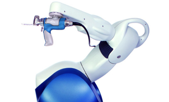
Advance Robotic Surgery in Ahmedabad
How it works: The Unicondylar and Patellofemoral (UNI and PFJ) guide the surgeon in creating implant plans that localize components and balance soft-tissue. Then, the robotic-assisted technology translates this information into a virtual plan, thereby eliminating the need of standard mechanical procedures, cuts and jigs. The robotic-assisted execution provides the surgeon with a 3D colour map of the knee replacement plan to be executed through handheld instrumentation. This offers multiple control modes, enabling the surgeon to accurately prepare the bone for knee replacement.
tkr Surgery Doctor in Ahmedabad
Salient Features
Hand-held Robotics- Ensures precision and enables producing repeatable results.
CT-Scan Free- The intraoperative technology enables mapping o f anatomy, creating a virtual 3D model for visualizing during the surgery.
Patient Planning- The structure, shape, size and motion of knee is unique to every patient. So a customized surgical plan enables the surgeon to perform the procedure with higher accuracy and precision.
Small Footprint- OR-friendly design and streamlined pre-operative set up integrates easily into existing ORs.
Multi-implant platform- It allows free use of any of our unicondylar implants.
This majorly involves surgical procedures pertaining to brain and spinal cord. It may become inevitable in cases of birth defects, traumatic injury, cancer etc.
Excellence of Robotic-assistance
Handheld instrumentation enables more control
Smaller Incisions
Less Pain
No CT/MRI Scan
Higher Precision and Accuracy
Comfort of natural Knee joint
Faster Recovery
Quicker Rehabilitationy
#Best Surgeon For Joint Replacement Surat#Robotic knee surgery in Ahmedabad#Robotic knee surgery in Gujarat#Knee robotic surgery in Ahmedabad#Advanced Knee Replacement In Mehsana#Advanced Knee Replacement In Udaipur#Best Doctor For Knee Replacement In Gujarat#Best Doctor For Knee Replacement In Nadiad#Best Doctor For Knee Replacement In Kota#Best Doctor For Knee Replacement In Kadi#Best Knee Surgeon In junagadh
0 notes