#Zeiss Surgery Operating Microscope
Text
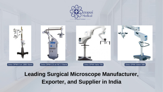
Octopus Medical's Role as a Leading Surgical Microscope Manufacturer, Exporter, and Supplier in India
In the realm of modern medicine, precision is paramount. Surgical procedures, especially those involving delicate areas like the brain, eyes, and ears, demand unparalleled accuracy and clarity. This is where surgical microscopes emerge as indispensable tools, enabling surgeons to perform intricate procedures with enhanced visibility and precision. Among the pioneering names in this field stands Octopus Medical, a renowned manufacturer, exporter, and supplier of surgical microscopes in India.
Octopus Medical prides itself on delivering cutting-edge solutions tailored to the diverse needs of medical professionals across specialties. With a commitment to quality, innovation, and affordability, the company has established itself as a trusted partner in the healthcare industry. Let's delve into the world of Octopus Medical and explore their impressive range of surgical microscopes.
Unveiling Excellence: Octopus Medical's Product Line
At the heart of Octopus Medical's offerings lies a spectrum of surgical microscopes designed to meet the evolving demands of modern healthcare. Among their flagship products are the renowned Zeiss OPMI series, renowned for their exceptional optical clarity and ergonomic design.
Zeiss OPMI 6 on 1880 Stand: Combining advanced optics with ergonomic engineering, the Zeiss OPMI 6 on 1880 Stand redefines precision in surgical microscopy. With its versatile features and superior image quality, this microscope is a preferred choice for neurosurgical procedures, offering unparalleled clarity and depth perception.
Zeiss OPMI CS on NC-2 Stand: Engineered to deliver optimal performance in ENT surgeries, the Zeiss OPMI CS on NC-2 Stand sets a new standard for precision and versatility. Equipped with intuitive controls and customizable settings, this microscope empowers surgeons to navigate intricate anatomical structures with ease and confidence.
Zeiss OPMI Vario 700: Designed for versatility and adaptability, the Zeiss OPMI Vario 700 offers unparalleled flexibility in surgical microscopy. Whether in ophthalmic, neuro, or ENT procedures, this microscope delivers superior visualization and ergonomic comfort, enhancing surgical outcomes across specialties.
Zeiss OPMI Vario/S88: The Zeiss OPMI Vario/S88 stands as a testament to innovation and excellence in surgical microscopy. With its state-of-the-art optics and seamless functionality, this microscope caters to the diverse needs of modern surgical practice, ensuring precision and reliability in every procedure.
Empowering Healthcare: Octopus Medical's Commitment
As a leading manufacturer, exporter, and supplier of surgical microscopes in India, Octopus Medical is dedicated to empowering healthcare professionals with advanced tools and technologies. Their collaboration with renowned brands like Zeiss underscores a commitment to quality and innovation, ensuring that surgeons have access to the finest equipment for optimal patient care.
Navigating the Competitive Landscape: Octopus Medical's Differentiators
In a market flooded with options, Octopus Medical stands out for several reasons:
Quality Assurance: Octopus Medical adheres to stringent quality standards, ensuring that every microscope meets the highest benchmarks of performance and reliability.
Affordability: Despite offering cutting-edge technology, Octopus Medical strives to keep its products accessible, making precision surgery attainable for healthcare facilities of all sizes.
Customer-centric Approach: Octopus Medical places a premium on customer satisfaction, offering comprehensive support and assistance to healthcare providers at every stage, from procurement to after-sales service.
Conclusion: Elevating Surgical Precision with Octopus Medical
In the fast-paced world of modern medicine, precision is non-negotiable. Surgical microscopes serve as invaluable tools, enabling surgeons to navigate intricate procedures with enhanced clarity and accuracy. Octopus Medical emerges as a beacon of excellence in this domain, offering a diverse range of surgical microscopes tailored to the unique needs of healthcare professionals across specialties.
With a steadfast commitment to quality, innovation, and affordability, Octopus Medical continues to redefine the standards of surgical microscopy in India and beyond. As the healthcare landscape evolves, Octopus Medical remains poised to empower surgeons with the tools they need to achieve optimal patient outcomes, one precise incision at a time.
#Refurbished Medical Equipment Supplier and Exporter in India#Surgical Microscope Manufacturer#Exporter#and Supplier in India#Uncategorized#TagsENT Microscope Manufacturer In India#For Hospital Use#leading Surgical Operating Microscope manufacturers#Neuro Surgical Microscopes Manufacturer Supplier India#Operating Microscope at Best Price#Operating Microscope Suppliers In India#Surgical Microscope in India#Surgical Microscope manufacturer in India#Surgical Operating Microscope suppliers and exporters from India#Surgical Ophthalmic Microscope manufacturers#Surgical Ophthalmic Microscope suppliers and exporters from India#Top Operating Microscope Products#ZEISS ENT surgical microscopes#Zeiss OPMI 6 on 1880 Stand#Zeiss OPMI CS on NC-2 Stand#Zeiss OPMI Vario 700#Zeiss OPMI Vario/ S88#Zeiss Surgery Operating Microscope
0 notes
Text
Ophthalmic Operating Room OR Microscopes Market Size, Type, segmentation, growth and forecast 2023-2030

Ophthalmic Operating Room OR Microscopes Market
The Ophthalmic Operating Room OR Microscopes Market is expected to grow from USD 2.10 Billion in 2022 to USD 2.90 Billion by 2030, at a CAGR of 4.60% during the forecast period.
Get the Sample Report: https://www.reportprime.com/enquiry/sample-report/10941
Ophthalmic Operating Room OR Microscopes Market Size
Ophthalmic Operating Room OR Microscopes market research report covers the market segment based on type which includes Basic Microscope, Standard Microscope, and Advanced Microscope. Its applications include Ambulatory Surgery Center (ASC), Hospital Outpatient Department (HOPD), and Others. The report analyses the market players such as Carl Zeiss, Leica, Haag-Streit Group, Alcon, Zhenjiang Zhongtian Optical Instrument, 66 VISION TECH, Xintian Medical Devices, Zhenjiang Yihua Operation Instrument, Seiler Medical, Karl Kaps, Shanghai Eder Medical Technology, and Topcon Corporation. The region-wise segmentation focuses on North America, Asia Pacific, Middle East, Africa, Australia, and Europe. Regulatory and legal factors specific to market conditions are also discussed in the report. Ophthalmic Operating Room OR Microscopes market research report provides a comprehensive analysis of the market.
Ophthalmic Operating Room OR Microscopes Market Key Player
Carl Zeiss
Leica
Haag-Streit Group
Alcon
Zhenjiang Zhongtian Optical Instrument
Buy Now & Get Exclusive Discount on this https://www.reportprime.com/enquiry/request-discount/10941
Ophthalmic Operating Room OR Microscopes Market Segment Analysis
The Ophthalmic Operating Room OR Microscopes market comprises medical equipment used by ophthalmologists to perform surgeries and procedures related to the eyes. The market is expected to grow at a steady rate due to the increasing prevalence of eye-related diseases and disorders, rising geriatric population, and advancements in technology that are improving surgical outcomes.
One of the major factors driving revenue growth in the Ophthalmic Operating Room OR Microscopes market is the growing demand for minimally invasive surgeries. These procedures are less invasive and require smaller incisions, resulting in reduced recovery times and higher patient satisfaction. As a result, there is a shift towards incorporating microscopes with advanced imaging capabilities and improved illumination in ophthalmic surgeries.
Another significant driver of the market is the rising elderly population. With age, comes an increased risk for eye-related disorders such as cataracts, age-related macular degeneration, and glaucoma, among others. As a result, ophthalmic surgeries are becoming more common, and the demand for advanced operating room equipment is growing.
Moreover, technological advancements are also expected to propel the market forward. The incorporation of digital imaging systems, augmented reality, and robotic-assisted surgeries are some of the latest trends in the Ophthalmic Operating Room OR Microscopes market, offering more precision in surgeries and improved patient outcomes.
However, the market also faces some significant challenges. One of the major challenges is the high cost associated with premium equipment and procedures. Reimbursement policies can be complex, thus limiting the adoption of advanced equipment by healthcare facilities. Moreover, the COVID-19 pandemic has further slowed down the market as elective surgeries are postponed due to a focus on pandemic-related care.
The report's main findings highlighted the growing demand for advanced equipment in ophthalmic surgeries driven by increasing geriatric population, advancements in technology, and a trend towards minimally invasive procedures. It also pointed out the significant challenges faced by the market such as complex reimbursement policies and the impact of the pandemic on elective surgeries.
Recommendations include exploring opportunities in developing countries, targeting niche procedures, and focusing on the development of cost-effective equipment without compromising quality. Additionally, healthcare providers should collaborate with insurance providers and governments to address reimbursement policies and improve patient access to advanced equipment and procedures.
This report covers impact on COVID-19 and Russia-Ukraine wars in detail.
Purchase This Report: https://www.reportprime.com/checkout?id=10941&price=3590
Market Segmentation (by Application):
Ambulatory Surgery Center (ASC)
Hospital Outpatient Department (HOPD)
Others
Information is sourced from www.reportprime.com
0 notes
Text
Are vasectomies reversible? | John C. McHugh M.D.
Vasectomy Reversal Experience, Success, and the Best all-inclusive price in the Southeast.
Are vasectomies reversible? Yes! Vasectomy Reversal: Dr. McHugh is a board certified urologist who has performed hundreds of microscopic vasectomy reversals in our practice owned and accredited urological surgery center using a Zeiss operating microscope. General anesthesia is provided by a board certified…

View On WordPress
0 notes
Text
Life Science Microscopes Industry Foresees Skyrocketing Growth in the Coming Years
Life Science Microscopes Industry | Forecast 2030
Life Science Microscopes Industry Data Book Covers Surgical, In-vitro Fertilization, Super Resolution And Scanning Electron Microscopes Markets.
The global life science microscopes industry generated over USD 3.35 billion in 2021 and is expected to grow at a CAGR of 8.9% over the forecast period.
Access the Global Life Science Microscopes Industry Data Book, 2022 to 2030, compiled with details like market sizing information & forecasts, trade data, pricing intelligence, competitive benchmarking, macro-environmental analyses, and regulatory & technological framework studies
Surgical Microscopes Market Growth & Trends
The global surgical microscopes market size is expected to reach USD 2.9 billion by 2030, growing at a CAGR of 11.37% from 2022 to 2030, based on a new report by Grand View Research, Inc. With the advent of surgical microscopes, it became easier for surgeons to perform such procedures more accurately in less time. In addition, the introduction of technologically advanced products is driving the demand for surgical microscopes as they are more precise, offer better illumination sources, and provide options for customization and technology integration based on the complexity of the procedures.
Emerging technologies such as wide-angle illumination, Red Reflex illumination, automation and augmented reality microscopy are expected to boost the market growth. International players like Carl Zeiss Meditec AG and Leica Microsystems are contributing to the market by providing highly advanced, automated, and robotic surgical microscopes for more precision.
In response to the COVID-19 pandemic, hospitals decided to suspend all the elective and non-urgent surgeries, which has negatively impacted the market. However, with the ease of restrictions, treatments are resuming in many countries, including developing nations. Also, many companies, such as Alcon and Carl Zeiss, have resumed their business operations with the given government guidelines to deliver their orders.
In-vitro Fertilization Microscopes Market Growth & Trends
The global in-vitro fertilization microscopes market size is expected to reach USD 193.2 million by 2030, according to a new report by Grand View Research. The market is expected to expand at a CAGR of 8.4% from 2022 to 2030. The market has witnessed advancements in microscopes and microscope-related equipment for use in in-vitro fertilization. Artificial Intelligence (AI), embryo assessment, and sperm selection are some of the applications targeted for innovation.
Manufacturers are providing upright microscopes, digital microscope cameras, and software to view images together as a sperm analysis system to provide the complete solution for semen analysis. Moreover, as sperm analysis can be better studied by maintaining a certain temperature of the sample, manufacturers are coupling the use of such devices along with an upright microscope. For instance, PROiSER provides ISAS HEAT, a slide warmer, compatible with its UB200i upright microscope used along with Computer Assisted Semen Analysis (CASA) systems.
The demand for In-vitro Fertilization (IVF) procedures is now increasing due to the relaxation of travel restrictions and supporting government guidelines. For instance, the American Society of Reproductive Medicine (ASRM) announced that there is a need to ensure reproductive care with maximal safety as we will have to continue to live with a COVID-19.
Super-resolution Microscopes Market Growth & Trends
The global super-resolution microscopes market size is expected to reach USD 6.6 billion by 2030, according to a new report by Grand View Research, Inc. The market is expected to expand at a CAGR of 9.04% from 2023 to 2030. Increasing application in cell biology and biomedical imaging is a key factor expected to drive the market over the forecast period.
Governments all over the world are supporting R&D for innovative technology, majorly nanotechnology research to help build improved nanoimaging technology-based super-resolution microscopes. The National Institutes of Health in the U.S. established a trans-NH bioengineering nanotechnology program to solicit grant applications for biomedical nanotechnologies. The U.K. government spent EUR 81 million (USD 105 million) on the development of advanced 3D imaging technology in February 2020.
The COVID-19 pandemic accelerated technological advancement, culminating in the creation of diverse uses for super-resolution microscopes since 2020. Top players have recently introduced new super-resolution microscopes having a variety of applications, including operations, cancer research, COVID-19 investigations, and academic research. For instance, Hitachi High-Tech Corporation introduced the simple-to-use AFM 100 and AFM100 plus Atomic Force Microscopes in July 2021, which provides improved dependability and ease for quality control applications or high-throughput R&D.
Order your copy of the Free Sample of “Life Science Microscopes Industry Data Book – Surgical, In-vitro Fertilization, Super-Resolution and Scanning Electron Microscopes Market Size, Share, Trends Analysis And Segment Forecasts, 2022 - 2030” Data Book, published by Grand View Research
Scanning Electron Microscopes Market Growth & Trends
The global scanning electron microscopes market size is expected to reach USD 6.5 billion by 2028, registering a CAGR of 8.52% over the forecast period, according to a new report by Grand View Research, Inc. Rising demand for nanotechnology-based research and growing R&D innovation in application areas are anticipated to serve as key growth drivers. Rapid growth witnessed in application areas, such as semiconductors, automobiles, pharmaceuticals, and nanotechnology, globally is among the key factors responsible for the significant growth of the SEM market.
Technological advancements in SEM improves the quality control procedures of research laboratories in a wide range of industries, such as semiconductors, automobiles, and pharmaceutical manufacturing. Scanning electron microscopy plays a critical role in the imaging and elemental analysis of products. However, the advanced SEMs offer advantages, such as rapid analysis, compact size, and efficient results with higher resolution and 3D imaging. Furthermore, the COVID-19 pandemic is expected to increase the sale of SEMs.
Due to the growing prevalence of communicable diseases, the market is expected to observe substantial growth over the coming years. Market participants are entering into partnerships and collaborations to prove their technical capabilities. For instance, in November 2020, Thermo Fisher Scientific Inc. partnered with Nanoimaging Services (NIS)-a provider of Transmission Electron Microscopy (TEM) services. The partnership helped Thermo Fisher Scientific Inc. obtain better accessibility to NIS’s cryoelectron microscopy (cryoEM) technology for pharmaceutical applications and biotechnology.
Competitive Landscape
Key players operating in the life science microscopes industry are –
Carl Zeiss Meditec AG,
Leica Microsystems,
Olympus Corporation,
Nikon Corporation,
Applied Precision (GE Healthcare),
Haag-Streit Surgical GmbH,
Synaptive Medical,
Alcon, Inc. (Novartis),
Topcon Corporation,
ARI Medical Technology Co., Ltd.,
Takagi Seiko Co., Ltd.,
Seiler Instrument Inc.,
Chammed Co. Ltd, etc.
Grand View Research’s life science microscopes Industry data book is a collection of market sizing information & forecasts, regulatory data, reimbursement structure, competitive benchmarking analyses, macro-environmental analyses, and regulatory & technological framework studies. Within the purview of the database, all such information is systematically analyzed and provided in the form of presentations and detailed outlook reports on individual areas of research.
Check out more Industry Data Books, published by Grand View Research
About Grand View Research
Grand View Research, U.S.-based market research and consulting company, provides syndicated as well as customized research reports and consulting services. Registered in California and headquartered in San Francisco, the company comprises over 425 analysts and consultants, adding more than 1200 market research reports to its vast database each year. These reports offer in-depth analysis on 46 industries across 25 major countries worldwide. With the help of an interactive market intelligence platform, Grand View Research helps Fortune 500 companies and renowned academic institutes understand the global and regional business environment and gauge the opportunities that lie ahead.
Contact:
Sherry James
Corporate Sales Specialist, USA
Grand View Research, Inc.
Phone: 1-415-349-0058
Toll Free: 1-888-202-9519
Email: [email protected]
Web: https://www.grandviewresearch.com/sector-reports-list
Follow Us: LinkedIn | Twitter
1 note
·
View note
Text
ENT Hospital in Indore | Ear, Nose, Throat

https://choithramhospital.com/departments/ear-nose-and-throat/
The Department of Ear, Nose and Throat provides comprehensive care for all types of ear ailments along with audiology and speech therapy, cochlear implant surgery and post operative rehabilitation. The detailed list of our services is provided in the services section.
Services
Ear, Nose and Throat offers the following services:
Laryngostroboscopy-with Storz Pulsar II
Diagnostic nasal endoscopy with Storz camera
Examination of ear under Zeiss microscope
Diagnostic video laryngoscopy with Storz camera
Audiometry – Sound proof chamber for Audiometry and Middle Ear Analysis
Otology
Cochlear implant
Rhinology
Endoscopic Sinonasal Procedures
Revision Endoscopic Surgery
0 notes
Text
Are you searching for the best hospital near me for Neurosurgery?
Datta Meghe is having chains of neurosurgery hospitals. Acharya Vinoba Bhave Hospital at Wardha, Maharashtra offers neurosurgery and neuro-imaging services with a team of experienced neurosurgeons, neuro-radiologists, neuro-physiotherapists, neuro-psychologists, neuro-neurologists, neuro-nurses, neuro-anesthetists.
Neurosurgery is the super specialty branch of medicine concerned with the surgical management of diseases of the nervous system composed of the brain, spinal cord, and spinal column, as well as the nerves that travel through all parts of the body.
The Department of Neurosurgery at AVBRH is fully equipped to perform all types of surgeries for a wide range of neurological illnesses.
These include:
1. Pediatric Neurosurgery: Congenital diseases of the brain and spine and other illnesses affecting children
2. Neuro-Oncology: Tumours of the brain, spine, and spinal cord
3. Cerebrovascular Surgery: Stroke, hemorrhage in the brain and spinal cord, vascular diseases such as aneurysms and vascular malformations
4. Spine Surgery: Instrumentation of the spine and the craniovertebral junction, degenerative disc, and other spinal diseases
5. Skull Base Surgery: Diseases of the pituitary gland, skull base tumors, etc
6. Minimally invasive Neuro navigation guided surgery, Intra op Neuro monitoring
7. Surgery for epilepsy and movement disorders
8. Peripheral nerve surgery
9. Brain and spine trauma management
The department is supported by state-of-the-art dedicated neurosurgical operation theatres, equipped with the world’s latest Carl Zeiss Kinevo 900 Operating Microscope, Medtronic’s Stealth S8 Neuronavigation system, Medtronic’s intra op neuro monitoring system, a Karl Storz Neuroendoscope, a Midas Rex drill system, NSK drill system, a Misonix Ultrasonic Surgical aspirator, C—arm with imported micro neurosurgical instruments.
A dedicated Neurosurgical Intensive Care Unit provides comprehensive care for postoperative and acutely ill patients.
Department of neurosurgery has successfully performed more than 1000 brain tumour surgeries, 500 paediatric neurosurgery, 300 cerebrovascular surgeries, 2000 spine surgeries, 2000 head injuries, etc in last 15 years
#Best hospital near me#Private hospital near me#Private hospital near nagpur#Rural hospital near nagpur#Best multispeciality hospital near me
0 notes
Text
Surgical Microscopes Market Driving Factors And Highlights of The Market 2022-2030
The global surgical microscopes market size is expected to reach USD 2.9 billion by 2030, according to a new report by Grand View Research, Inc. The market is expected to register a CAGR of 11.37% from 2022 to 2030. With the advent of surgical microscopes, it became easier for surgeons to perform such procedures more accurately in less time. In addition, the introduction of technologically advanced products is driving the demand for surgical microscopes as they are more precise, offer better illumination sources, and provide options for customization and technology integration based on the complexity of the procedures.
Emerging technologies such as wide-angle illumination, Red Reflex illumination, automation and augmented reality microscopy are expected to boost the market growth. International players like Carl Zeiss Meditec AG and Leica Microsystems are contributing to the market by providing highly advanced, automated, and robotic surgical microscopes for more precision.
In response to the COVID-19 pandemic, hospitals decided to suspend all the elective and non-urgent surgeries, which has negatively impacted the market. However, with the ease of restrictions, treatments are resuming in many countries, including developing nations. Also, many companies, such as Alcon and Carl Zeiss, have resumed their business operations with the given government guidelines to deliver their orders.
Surgical Microscopes Market Report Highlights
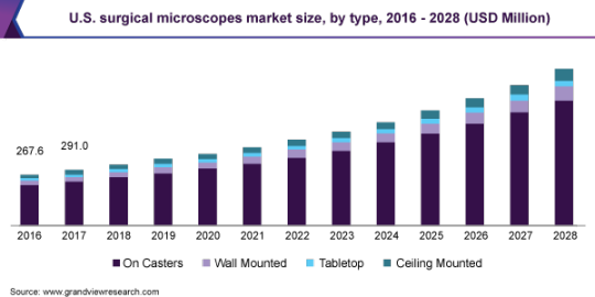
• On casters was the largest type segment in 2021 due to the high portability & flexibility and low maintenance requirements of these tools. Moreover, the segment is expected to register the fastest growth over the forecast period due to the high use of casters microscope in various surgical fields
• The ophthalmology application segment accounted for the maximum revenue share in 2021. However, the ENT surgery segment is expected to register the fastest CAGR from 2022 to 2030. According to data published by The Children’s Hospital of Philadelphia, tympanostomy tube insertion surgery, also known as ear tube surgery, is the most commonly performed surgery in children in the U.S. More than 4,000 ear tube surgeries are performed each year at the hospital. Thus, an increasing number of ENT procedures is likely to favor segment growth
• Carl Zeiss Meditec AG and Leica Microsystems (Danaher Corporation) are the major players in the surgical microscopes market. Launch of new products, acquisitions, and geographic expansion are some of the strategies being adopted by key players in the market. In February 2019, Danaher Corporation acquired the Biopharma business of GE Healthcare, which also includes microscopy products the company
• Manufacturers such as Alcon, Nikon, and Carl Zeiss reported the decreases in the revenue in the second quarter of the year 2020 due to the impact of the COVID-19 pandemic which affected the supply chain of most of the companies. Currently, most of the companies have resumed their business and are delivering orders within the given guidelines pertaining to the COVID-19 pandemic
Life Science Microscopes Industry Size was valued at USD 3.36 Billion in 2021 and is projected to expand at a CAGR of 8.9% during 2022 to 2030.
Surgical Microscopes Market Segmentation
Grand View Research has segmented the global surgical microscopes market report based on type, application, end use, and region:
Surgical Microscopes Market – Type Outlook (Revenue, USD Million, 2017 – 2030)
• On Casters
• Wall Mounted
• Tabletop
• Ceiling Mounted
Surgical Microscopes Market – Application Outlook (Revenue, USD Million, 2017 – 2030)
• Neurosurgery and Spine Surgery
• ENT Surgery
• Dentistry
• Gynecology
• Urology
• Ophthalmology
• Plastic & Reconstructive Surgeries
• Other Surgeries
Surgical Microscopes Market – End Use Outlook (Revenue, USD Million, 2017 – 2030)
• Hospital
• Physician Clinics and Other Settings
Surgical Microscopes Market – Regional Outlook (Revenue, USD Million, 2017 – 2030)
• North America
• Europe
• Asia Pacific
• Latin America
• Middle East & Africa (MEA)
List of Key Players in Surgical Microscopes Market
• Carl Zeiss Meditec AG
• Leica Microsystems
• Olympus Corp.
• Hagg-Streit Surgical GmbH
• Synaptive Medical
• Alcon, Inc.
• Topcon Corp.
• Takagi Seiko Co., Ltd.
• ARI Medical Technology Co., Ltd.
• Chammed Co., Ltd.
• Seiler Instrument, Inc.
Request free sample copy of Life Science Microscopes Industry Data Book @ https://www.grandviewresearch.com/sector-report/life-science-microscopes-industry-data-book/request/rs1
0 notes
Text
Best Spine Surgeon in Jalandhar — Capitol Hospital

Department of Neuro Surgery
Through world-class, patient-focused care; innovative clinical and laboratory research; and specialized training, Department of Neuro Surgery at Capitol provides complete neuro-surgical expertise to patients; both clinical and surgical with cranial & spinal diseases and disorders. Our specialists offer an array of services including cerebro vascular, spinal, functional, traumatic, tumor, and pediatric neurosurgery.
The Department of Neuro Surgery at Capitol is fully equipped to perform all types of surgeries for a wide range of illnesses.
These include:
Congenital diseases of the brain and spine and other illnesses affecting children
Tumours of the brain, spine and spinal cord
Vascular diseases such as aneurysms and vascular malformations
Degenerative disc and other spinal diseases
Instrumentation of the spine and the cranio-vertebral junction
Diseases of the pituitary gland
Stroke and hemorrhage in the brain and spinal cord
Features:-
Microneurosurgery:
Brain tumours (Supra Tentorial and Infra Tentorial)
Skull base tumours
Spine tumours
Anterior Cervical Micro Discectomy
Lumbar Micro Discectomy
Head injuries
Spine injuries
Paediatric Neurosurgery:
VP shunt surgery
Congenital anomaly correction
Brain & spine tumours excision
Spinal dysraphism anomaly correction
Spine Surgery
Stabilization of fracture spine
Spinal tumors
Stroke surgery
Medical management of Neurological disorders:
Stroke
Seizure disorders
Movement disorders
Parkinson’s disease
Headache
Backache
Disorders Treated
Brain tumors:
The management of brain tumors includes treating benign and malignant tumors.
Vascular disorders of brain:
Capitol is one of the few hospitals in the region treating abnormalities of the blood vessels of the brain and spine. This includes aneurysms and vascular malformation of complex types.
Traumatic and other spinal disorders:
Age related and other degenerative conditions of the spine like disc prolapse, spondylolisthesis, complex craniovertebral junction anomalies and other conditions of the vertebral column like traumatic spine fractures are treated here regularly.
In treatment options, we offer range from routine microdiscectomies to artificial disc replacement surgeries of the lumbar and cervical spine.
Spine tumors:
Various spinal tumors including meningiomas, schwannomas, ependymomas, gliomas etc are treated using most advanced intra-operative monitoring modalities to give an optimal outcome and maximize functional recovery of the patient.
Congenital disorders:
Disorders like congenital hydrocephalus, spina bifida and tethered cord are treated . Congenital craniofacial anomalies are managed in conjunction with the specialists of maxillofacial surgery and plastic surgery.
Brain and spine trauma:
The neuro surgical trauma care attend to wide range of brain and complex spine trauma which usually need emergent surgical treatment.
Services Offered
Neurooncology
Brain Tumour surgery
Brain Aneurysm Surgery
Head Injury Treatment
Disc Replacement
Cerebrovascular surgery
Minimally invasive surgery- Stereotactic, Endoscopic and endoscope-assisted surgery
Spine surgery
Skull base surgery
Facilities
The Department of Neuro Surgery at Capitol is supported by top of the line dedicated neurosurgical operation theaters equipped with:
Carl Zeiss Operative Microscopes
Kapalin Drill System
Neuro Endoscopy
Siemens C-arm machine for DSA
EEG
EMG
NCV
Digital recording and documentation facilities
Read more: https://www.capitolhospital.com/department-of-neurological-sciences/
#spine surgeon in jalandhar#brain and spine#Spine tumors#Neuro Surgery#Best Neurologists in Jalandhar Punjab#Capitol Hospital Jalandhar
0 notes
Link
Process of doing Micro Ear Surgery is done using a surgical microscope. The structure of ear very small this surgical microscope was developed to treat the ears. Visibility within the ear is poor and the nature of surgery being delicate we have latest Carl Zeiss operating microscope for ear surgeries at our institute.
#Dr. Manoj Gupta#dr manoj gupta varanasi#ear doctor in varanasi#best ent doctor in varanasi#cochlear implant surgery in varanasi#Digital Hearing Aid Varanasi#Hearing Aid in Varanasi#Best ent specialist in varanasi#best ENT Hospital in Varanasi#best ent clinic in varanasi#ear specialist in varanasi#Sinus Doctor in Varanasi#sugar specialist doctor in varanasi#ENT Surgeon in Varanasi#Micro Ear Surgery in varanasi
1 note
·
View note
Text
Microsurgery Market Growth, Size, Opportunities, Key Growth Factors, Revenue Analysis Till 2027
Microsurgery, a procedure that combines magnification and specialized precision tools & techniques is helpful in healing wounds caused in an accident (Trauma) and in restoring the function after the surgical procedures conducted in the treatment of chronic diseases. However, these surgeries are also predominantly used for the treatment of congenital deficiencies and cancer.
The microsurgery market is growing at the rapid pace on the global platform, owing to the increasing prevalence of cancer, ophthalmic diseases, and other chronic diseases. Rising uptake of these surgeries due to their efficacy is the key driving force escalating the microsurgery market growth, garnering huge prominence for the market.
According to a recent study report published by Market Research Future (MRFR), the global microsurgery market is booming and is projected to grow at a CAGR of 10.2 % during 2017-2023.
Additional factors substantiating the market growth include the increasing number of organ transplantation, adoption of the microscopes in surgeries and advances in the surgical procedures. Furthermore, increasing healthcare expenditure backed by the improving economic conditions that allow access to the quality healthcare and quality of life especially in the developing countries is fuelling the market growth.
On the other hand, factors such as the high cost of the treatment and the poor reimbursement policies for the medical devices are impeding the market growth. Nevertheless, growing demand for the quality treatment of the diseases is expected to increase the market size over the forecast period.
Key Players:
Prominent players operating in the market include Tisurg Medical Instruments Co., Ltd (China), AROSurgical Instruments (US), Carl Zeiss Meditec AG (Germany), Synovis Micro Companies Alliance, Inc. (US), MicroSurgical Technology (US), Peter LAZIC GmbH (Germany), Microsurgery Instruments, Inc. (US) and BIONIKO (US). Profiling them in its analysis, MRFR explores strategies helping them to stay at the forefront of competition.
Global Microsurgery Market - Segments
The MRFR analysis is segmented into three key dynamics for an easy grasp and enhanced understanding.
By Application : General Surgery, Ophthalmology, Plastic Surgery, Gynaecological Surgery, Orthopaedic Surgery, Oncology, Neurosurgery, and Oral Surgery among others.
By Procedure : Free Tissue Transfer, Replantation (fingers & thumbs, ear, scalp, nose, and others.), Transplantation, and Treatment of Infertility (tubal obstructions, vas deferens obstructions and varicocele.) among others.
By End-User : Hospitals & Clinics, and Research Organizations.
By Region : North America, Europe, APAC and the Rest-of-the-World.
Global Microsurgery Market - Regional analysis
Globally, North America leads the global microsurgery market with the largest market share. The market is estimated to accrue exponentially by 2023, registering a phenomenal CAGR from 2017 to 2023. Factors supporting the market growth include favorable reimbursement scenarios and substantial healthcare expenditure. Also, wide uptake of new technologies in the US is also a key driver for the regional market growth.
Additionally, the increasing cancer population, along with the increasing prevalence of neurological diseases and deficiency diseases, drives the market growth in the region. Also, the rising government support for R&D is proving impetus to the market growth.
The Europe market is expected to be the second-largest market for microsurgery is expected to grow at a considerable CAGR. Undoubtedly, the resurging economy in the region is the major driving force acting as a tailwind to the market growth in the region.
Availability of funds for R&D activities, well-proliferated healthcare sector and the increasing awareness about the availability of these surgeries drives the market of microsurgery in the region.
The Asia Pacific market for the microsurgery is expected to emerge as the fastest growing market during the estimated period (2017 to 2023). The market growth is expected to be led by China and India owing to the huge population and the availability of low-cost procedures.
Moreover, the fastest growing healthcare sector coupled with the large unmet needs over the forecast period will foster the market growth. Vietnam, Thailand, and Malaysia among other South East Asian countries are projected to contribute significantly to the regional market growth. Besides, the growing penetration of healthcare insurance is expected to drive the microsurgery market in the Asia Pacific region.
Global Microsurgery Market - Competitive Analysis
The global market for Microsurgery appears to be fiercely competitive with the several well-established and small players operating in the market. These players incorporate collaboration, acquisition, expansion, strategic partnership, & technology launch. Substantial investments are transpired in clinical trials and development of effective surgeries.
These key players compete based upon pricing, technology, and services. The Microsurgery market demonstrates a high growth potential which is likely to attract many entrants to the market resulting in intensified competition further with an increase in product/service extensions, technological innovations. Well established players are emphasizing upon expansion of their network and product distribution mainly into the developing economies.
Access Report @ https://www.marketresearchfuture.com/reports/microsurgery-market-4214
About Market Research Future:
At Market Research Future (MRFR), we enable our customers to unravel the complexity of various industries through our Cooked Research Report (CRR), Half-Cooked Research Reports (HCRR), & Consulting Services. MRFR team have supreme objective to provide the optimum quality market research and intelligence services to our clients.
Contact us:
Market Research Future (part of Wantstats Research and Media Private Limited),
99 Hudson Street, 5Th Floor,
New York, New York 10013
United States of America
+1 628 258 0071
Email: [email protected]
0 notes
Text
Surgical Microscopes Market Worth $2.3 Billion By 2028 | CAGR: 11.1% | Global and Regional Forecast | Grand View Research, Inc.
The global surgical microscopes market size is expected to reach USD 2.3 billion by 2028, based on a new report by Grand View Research, Inc. The market is expected to expand at a CAGR of 11.1% from 2021 to 2028. The rising need for ophthalmic procedures, rapid adoption of surgical microscopes in cosmetic surgeries, and new product launches are factors driving the growth of the market. For instance, in October 2018, Med X Change, Inc. launched 4Klear, a 4K Camera & Medical Video Recorder for surgical microscopes. Ophthalmic, neurological, cosmetic, and other minimally invasive surgeries require utmost precision and accuracy.
This can only be achieved through superior visualization tools. Surgical microscopes offer improved visibility, stability, recording capabilities, illumination, and high magnification of minute veins. This has led to increased demand for the product among physicians. These tools have revolutionized the field of minimally invasive surgeries as they allow surgeons to perform surgeries with maximum precision on actual pathology by magnifying the surrounding anatomical structures, resulting in improved patient outcomes with a shorter duration of procedures and rapid recovery.
In addition, limitations of open surgeries have been significantly eliminated by integrating the workflow in operating rooms. Thus, the majority of surgeons are adopting minimally invasive surgeries in various medical fields worldwide. Furthermore, people in emerging economies are willing to pay for premium-quality services due to increasing disposable income, which is anticipated to drive market growth during the forecast period. North America dominated the market in 2020 and will grow at a steady CAGR from 2021 to 2028. The growth is credited to the rising adoption of microsurgeries, favorable health reimbursement programs, and the presence of major manufacturers in the region.
Asia Pacific is expected to witness the fastest CAGR from 2021 to 2028 owing to rising cases of ophthalmic and neurological disorders that require microsurgical treatment and the growing demand for advanced surgical microscopes in hospitals for complex procedures. In response to the COVID-19 pandemic, hospitals decided to suspend all the elective and non-urgent surgeries, which have negatively impacted the market. However, with the ease of restrictions, treatments are resuming in many countries, including developing nations. Also, many companies, such as Alcon and Carl Zeiss, have resumed their business operations with the given government guidelines to deliver their orders.
Request Free Sample Report: https://www.grandviewresearch.com/industry-analysis/surgical-microscopes-market
Contact Us:
Grand View Research, Inc.
201 Spear Street 1100,
San Francisco, CA 94105
United States
Phone:
1-415-349-0058
Toll Free:
1-888-202-9519
0 notes
Text
ENT Surgery Microscopes Market : Global Market Revenue and Share by Manufacturers
ENT Surgery Microscopes Market
Careful magnifying lens assists a specialist with accomplishing ideal careful results through top notch optical pictures. ENT stands for the body parts namely; the ear, the nose, and the throat, and experts use ENT microscopes to complete routine surgeries. ENT surgery microscopes assume a significant part in assisting specialists with doing a surgery by survey the careful site accurately. During ENT surgery, exploring the life systems through complex designs and profound channels gets troublesome without appropriate enlightenment. Along these lines, brightening and amplification assume a huge part in ENT surgery and are significant to do the method by finding the careful site appropriately and without harming the nearby organs or tissue. Key components considered while utilizing ENT microscopes are enlightenment, amplification, goal, free-drifting suspension, center, and conveyability.
Read Report Overview: https://www.transparencymarketresearch.com/ent-surgery-microscopes-market.html
The increasing awareness about the upsides of ENT microscopes appeal for insignificantly intrusive ENT medical procedures, and expansion in the quantity of ENT surgical techniques in medical care centers are the main considerations projected to help the development of the worldwide ENT surgery microscopes market. Moreover, ascend in accessibility of ENT microscopes is additionally expected to help the development of the market. Furthermore, the increasing adoption of various therapeutics such as septoplasty, laryngeal surgery, endoscopic skull base surgery, endoscopic sinus surgery, are likely to augment the demand for ENT surgeries in the coming years, thereby boosting the growth of this market.
ENT Surgery Microscopes Market - Introduction
Surgical microscope helps a surgeon to achieve optimal surgical outcome through high quality optical images. Ear, nose, and throat (ENT) specialists use ENT microscopes to carry out routine surgical procedures. ENT surgery microscopes play an important role in helping surgeons to perform a surgery by viewing the surgical site precisely. During ENT surgery, navigating the anatomy through complex structures and deep channels becomes difficult without proper illumination. Therefore, illumination and magnification play a significant role in ENT surgery and are absolutely essential to carry out the procedure by locating the surgical site properly and without damaging the nearby organs or tissue. Key factors considered while using ENT microscopes are illumination, magnification, resolution, free-floating suspension, focus, and portability.
Request a PDF Brochure - https://www.transparencymarketresearch.com/sample/sample.php?flag=B&rep_id=68057
ENT Surgery Microscopes Market – Competitive Landscape
Prominent players operating in the global ENT surgery microscopes market are Olympus Corporation, Carl Zeiss Meditec AG, Haag-Streit Group, Labo America, Inc., ATMOS MedizinTechnik GmbH & Co. KG, Leica Microsystems, Seiler Instrument, Inc., Karl Kaps GmbH & Co. KG, Takagi Ophthalmic Instruments Europe Ltd., and Optofine Instruments, among others.
Olympus Corporation
Launch of new products is a key strategy adopted by the company to increase market share. For instance, in September 2017, Olympus Corporation launched ORBEYE Surgical Microscope, based on latest advances in 4K 3D video technology, in Japan and America. The product was developed by Sony Olympus Medical Solutions, Inc., a joint venture between Sony Imaging Products & Solutions, Inc. and Olympus Corporation. The company focuses on developing technological advanced microscopes that produce high-resolution stereoscopic images.
Leica Microsystems
Leica Microsystems manufactures microscopes and scientific instruments for analysis of various microscopic structures having application in life science research, medical microscopy and other fields. In October 2018, Leica Microsystems launched PROVIDO multidisciplinary surgical microscope at the American Academy of Otolaryngology, thereby expanding its product portfolio and offering technological advanced microscopic systems to ENT and spine surgeons.
Request for Analysis of COVID19 Impact on ENT Surgery Microscopes Market- https://www.transparencymarketresearch.com/sample/sample.php?flag=covid19&rep_id=68057
Carl Zeiss Meditec AG
Carl Zeiss Meditec AG offers pioneering technologies for various surgical applications. Key applications of products offered by the company are ophthalmology & optometry, neurosurgery, dentistry, ENT, gynecology and others. ZEISS EXTARO 300, ENT surgery microscope is one the major products offered by the company in the market. The microscope can be sued for tympanoplasty, transoral laser surgery, myringotomy, mastoidectomy and stapedectomy.
ENT Surgery Microscopes Market – Dynamics
Increase in awareness about the advantages of ENT microscopes high demand for minimally invasive ENT surgeries, and increase in the number of ENT surgical procedures in health care centers are the major factors projected to boost the growth of the global ENT surgery microscopes market. Additionally, growing popularity of minimally invasive ENT surgeries such as transoral robotic surgery (TORS), endoscopic sinus surgery, tonsillectomy, septoplasty, endoscopic skull base surgery, and laryngeal surgery has led to increase in demand for ENT microscopes, especially in developed countries in North America. Furthermore, rise in availability of ENT microscopes is also expected to support the growth of the market.
Request for Custom Research - https://www.transparencymarketresearch.com/sample/sample.php?flag=CR&rep_id=68057
Increase in awareness about the advantages of ENT surgery microscopes to drive the growth of the market
Increase in awareness about the advantages of ENT surgery microscopes on casters such as portability and better cleaning maintenance capability is a major factor expected to drive the growth of the ENT surgery microscopy market during the forecast period. Furthermore, increase in familiarity of ENT microscopes among surgeons and rise in the number of ENT surgeries, and surge in patient population are anticipated to boost the growth of market. Also, healthcare professionals are aware about the importance of functional properties of these microscopes and rise in the number of application in ENT surgery. A number of healthcare institutes and research centers offer training to medical staff regarding the use of ENT surgery microscope and its advantages. Therefore, knowledge about use and application of ENT surgery microscope is increasing among the healthcare providers.
Pre-book ENT Surgery Microscopes Market Report - https://www.transparencymarketresearch.com/checkout.php?rep_id=68057<ype=S
North America dominated the ENT Surgery Microscopes Market
North America is projected to dominate the global ENT surgery microscopes market owing to rise in the number of market players offering microscopes having application in ENT surgery, technological advancements in the medical field, and positive impact of medical technologies in the delivery of health care in the U.S. and Canada. Also, the market is anticipated to be driven by increase in the number of initiatives to create awareness about technological advancements in ENT surgery and rise in demand for new visualization technologies by ENT surgeons.
Asia Pacific expected to register significant growth
The ENT surgery microscopes market in Asia Pacific is driven by increase in patient population, availability of skilled & qualified personnel in public as well as private health care facilities, and rapid adoption of technologically advanced medical devices. Moreover, increase in the number of health care institutes and medical centers offering training to health care professional in ENT surgery is projected to boost the growth of the market in the region.
More Trending Reports by Transparency Market Research:
https://www.prnewswire.com/news-releases/mobile-hospitals-market-to-expand-at-cagr-of-10-1-key-role-played-during-covid-19-pandemic-strengthens-growth-prospects-finds-tmr-301388801.html
https://www.prnewswire.com/news-releases/surgical-glue-market-size-to-surpass-us-2-9-billion-by-2027-end-says-tmr-301395333.html
https://www.prnewswire.com/news-releases/veterinary-supplements-market-to-be-worth-us-13-76-billion-by-2031--increasing-demand-for-natural-supplements-and-products-with-prebiotic-content-to-bolster-market-growth-says-tmr-301398612.html
About Us Section:
Transparency Market Research is a global market intelligence company, providing global business information reports and services. Our exclusive blend of quantitative forecasting and trends analysis provides forward-looking insight for thousands of decision makers. Our experienced team of Analysts, Researchers, and Consultants, use proprietary data sources and various tools and techniques to gather, and analyse information. Now avail flexible Research Subscriptions, and access Research multi-format through downloadable databooks, infographics, charts, interactive playbook for data visualization and full reports through MarketNgage, the unified market intelligence engine. Sign Up for a 7 day free trial!
Contact
Transparency Market Research,
90 State Street, Suite 700,
Albany, NY 12207
Tel: +1-518-618-1030
USA - Canada Toll Free: 866-552-3453
Website: https://www.transparencymarketresearch.com/
0 notes
Text
Atlanta, GA Vasectomy Reversal Doctor | John McHugh M.D.
Georgia's leader in no scalpel vasotomies and microscopic vasectomy reversal.
Vasectomy Reversal Experience, Success, and the Best all-inclusive price in the Southeast.
Atlanta, Ga Vasectomy Reversal: Dr. McHugh is a board certified urologist who has performed hundreds of microscopic vasectomy reversals in our practice owned and accredited urological surgery center using a Zeiss operating microscope. General anesthesia is provided by a board certified…

View On WordPress
0 notes
Text
Use of Operating Microscope in Spine Surgery
🔰Bombay Spine Society with ZEISS in collaboration with OrthoTV presents:
“Use of Operating Microscope in Spine Surgery”
🔺Saturday, 11th December, 8pm – 9pm IST
🔺Click to watch: https://bit.ly/OrthoTV-Zeiss_for_Spine
🔅Speakers:
Dr. Uday Pawar
Dr. Amit Sharma
Dr. Satyen Mehta
Dr. Sameer Dalvie
Dr. Sandeep Sonone
🔅OrthoTV Team: Dr. Ashok Shyam, Dr. Neeraj Bijlani
Streaming Live on OrthoTV

0 notes
Text
Dr. Aphale’s Eye Hospital,Thane- Cataract Specialty Hospital.
Dr. AAPHALE EYE HOSPITAL is a place where elegance and technology, skill and knowledge, care and sincerity, art and science live together. A hard working, ethical, sincere, firm but caring, disciplined CEO Dr Kshipra Aphale, has transferred the same qualities in her honest and dedicated staff. Here every patient is examined personally by Dr Kshipra Aphale after preliminary examination by her assistant. All patients are treated by Dr Kshipra Aphale. The privacy of the patient is fully maintained through out the eye check up. We are empaneled with many insurance companies for Cashless Services.
About Us:Dr Kshipra R. Aphale M. S. (Ophthalmology)
The Director of Aaphale Eye Hospital, is a passionate, perfectionist and perseverant person when it comes to treating eye problems.
In addition to knowledge and skills, treating the patient needs compassion.
For Dr. Kshipra, CARE comes FROM THE BOTTOM OF THE HEART. Hence, even cases of difficult eye surgery or long standing eye problems are managed well with effective results.
Dr Kshipra Aphale passed her MBBS with special rank in ophthalmology. She received training for her MS ophthalmology from Govt Medical College from best of the teachers.
After passing MS ophthalm, she underwent extensive training in PHACO cataract surgery, LASIK surgery, GLAUCOMA management and RETINAL lasers.
Services:
Routine Eye check up
Refraction for adults
Paediatric (child) eye check up and spectacle number
Diet counseling for children regarding eye health
Management of progressive myopia of children
Intra-vitreal injection
Contact lens fitting
Dry eye management
Diabetic eye care and
Green Laser photocoagulation
Amblyopia management
Management of watering of eye
Management of uveitis
Management of ocular allergy
Computer eye care
Management of aging eye problem
Trauma and emergency eye problems
Corneal infection treatment
YAG capsulotomy
Equipment, Facilities:
OPD
Slitlamp examination
Autorefractometry
Tonometry
Goldman Applanation Tonometer
Non Contact Tonometer
Schultz Tonometer
A scan biometry
Optical
Immersion
Perimetry - essential test for glaucoma
Field examination
Ophthalmoscopy
Direct
Indirect
Gonioscopy
OCT
YAG laser
Green Laser
Operation Theater
Alcon phacoemulcification machine
Zeiss Lumera I microscope
Class B autoclave
Vertical Autoclave
Boyle’s apparatus
Suction machine
RF cautery
Standby microscope
Inverter back up.
Hospital is extensively protected for fire safety. Staff is well trained in fire safety
Cashless and Insurances:
Dr Aaphale Eye Hospital is registered with Gipsa and many insurance companies and TPAs (Third Party Administrators) like-
National Insurance Co Ltd
United India Insurance Co Ltd
The New India Assurance Ltd
The Oriental Insurance Co Ltd
Bajaj Allianz General Insurance
ICICI Lombard General Insurance Co Ltd
Star Health and Allied Insurance Ltd
Paramount TPA
Mediassist TPA
MDIndia TPA
Raksha TPA
Good Health TPA
Our Specialty - Cataract
Although we do many types of operations successfully at Dr. Aaphale’s Eye Hospital, Dr. Aphale is specialised in CATARACT SURGERY.
What is cataract?
There is a crystal clear lens in our eye which focuses the light rays on retina, where image of the object falls. Because of various reasons like aging, injury, diabetes, prolonged steroid use, this lens gets opacified, or it is no longer transparent. Hence it cannot focus light rays on retina. In fact it obstructs the passage of light rays in the eye. So the person starts seeing everything blurred.
Though operation, without cut etc. It is not true. Actually, laser energy is used to perform some of the steps in cataract surgery e.g., cornea is cut with the help of laser instead of blade. The removal of cataract is still not possible with laser and phaco is still only method to crush and remove the cataract. Besides, insertion of lens in the eye needs cutting of the eye.
Cataract Surgery
The good news that with latest technology, it is not only possible to regain good vision after cataract surgery but it is also possible to get even better and spectacle free vision after cataract surgery, than the pre-cataract stage.
In simple language, if a person has −3 or +4 spectacle number before he developed cataract, it is possible that he can see everything without spectacles after cataract surgery. It is the experience of most of the operated patients at Dr. Aaphales’ Eye Hospital that the quality of vision which they have after cataract operation is much better than the vision they used to have in their forties. This means they almost always get the vision of their twenties after cataract operation. This is the magic of good quality intra-ocular lenses.
Cataract Operation at Dr Aaphale Eye Hospital
Dr. Aphale has been specially trained for cataract surgery in USA and in India. It is Dr. Aphale’s mission and passion to give best vision to her patient of cataract without spectacles by the painless phacoemulsification procedure. This is made possible in part, by her latest technology phaco machine. It enables her to remove the cataract through smallest cut. It helps to remove cataract without disturbing any other structure of the eye. There is no need of injection, no need of closing the eye with a patch. There is no bleeding during operation. Operation takes 5 to 20 mins.
The skillfully taken cut heals on its own and doesn’t need stitches. So no pain, but all gain! No redness or pricking sensation is left after the operation. It is difficult to recognize which eye has been operated on next day! Of course no stay in the hospital or admission is required and it’s a matter of 2 to 3 hours and one can go home for lunch, if started in the morning.
The visual recovery is very fast and within a day or two people can resume to work, while those who want, can start cooking from the next day.
That’s why most of the corporate managers, busy executives, business persons and lonely ladies prefer to get operated at Dr. Aaphale’s Eye Hospital.
It is not only important to remove the cataract through smallest cut with world class machine by latest technology but it is also important to use the best quality intra-ocular lenses.
Just like a good quality camera lens gives good picture, a good quality intra-ocular lens gives good vision. Just seeing an object is not a good vision. Good vision includes sharp image, minute details, excellent colour contrast, ability to identify different colour hues, and ability to see objects at different distances. We offer excellent quality intra-ocular lenses to be implanted in the eye after cataract surgery, which give even youth-like vision. We offer hydrophobic acrylic lenses, unifocal toric and multifocal. In case of unifocals, person needs near spectacle number, though he can see distant objects clearly without spectacles.
Toric lenses are specially designed for the people who have corneal astigmatism. They get better vision without glasses after inserting this lens.
The multifocal lens offers spectacle-free good vision for near and distance.
Contact Us
Dr. Aphale’s Eye Clinic Thane
Teejadeep Mall, Castle Mill, LBS Road, Near Vikas Complex, Thane West. Dr. Aphale’s Eye Hospital 201 Teejadeep Mall, Castle Mill, LBS Road, Near Vikas Complex, Thane West, Thane Maharashtra 400601
Phone No.+919833699623, +022 25474995
Email: [email protected]
draphale.com
Designed by YCCINDIA.COM and Promoted by PICKMYURL.COM.
0 notes
Text
The Use of Operating Microscope for Removal of Broken Instruments from the Canal in Endodontic Treatment by Hakobyan G

Abstract
Purpose: To evaluate the success of using an operating microscope to remove broken instruments from different levels in curved and straight canals.
Patients and Methods: Removal of the broken instrument from the curved canals was performed on 61 teeth (2016 to 2020) using ultrasound under the imaging of an operating microscope (Carl Zeiss, Germany). The success of the tool removal methods used were evaluated, the success was determined by the complete removal of the broken fragment of the instrument.
Results: Postoperative clinical and radiological monitoring was regularly conducted, and criteria for the success were evaluated. In the present study, the and using an operating microscope successful at removing fractured rotary nickel titranium segments from narrow and curved root canals in clinical cases.
Conclusion: Removing broken instruments outside of the curvatures when direct vision is not possible can be very difficult. The clinical procedure of endodontic retreatment under the operating microscope allows to deal with highly complex cases and improve the scope of treatment and its prognosis.
Keywords: Endodontic Treatment; Operating Microscope; Removal of Broken Instruments.
Introduction
Endodontic treatment is a fairly predictable procedure, with success rates up to 86–98% [1]. Careful cleaning of the canals from any contaminated pulp tissue so that the canal space can be shaped and prepared for filling with inert material is. However, when endodontic treatment does not follow standard clinical guidelines, failure occurs [2]. If the canal filling protocol is not followed, rotating instruments tend to collapse in the canals; as a result of a fracture, access to the apical part of the root canal is reduced, and this can have a detrimental effect on canal disinfection, and then on obturation. When an instrument breaks in the canal, disinfection and obturation of the part of the canal distal to the broken instrument becomes difficult, which can lead to persistent infection in this area [3]. A clinical study of the relationship between broken rotating instruments and the prognosis of an endodontic case confirmed that in the absence of preoperative infection and periradicular changes, the instrument would not affect the prognosis [4]. For endodontic complications, 3 treatment methods are proposed: non-surgical treatment, surgical treatment or removal. Among all these treatment alternatives nonsurgical retreatment should be considered as the first choice of treatment [5].
The use of CBCT endodontic treatment technologies to diagnose dental clinical practice allows for procedures that are more predictable. Ultrasound is becoming a very useful tool in most stages of endodontic therapy, especially non-surgical and surgical treatments. Operating microscopes have been used for decades many other medical disciplines: ophthalmology, neurosurgery, reconstructive surgery, otorhinolaryngology, and vascular surgery. Its implementation in dentistry in the last fifteen years, especially in endodontics, has revolutionized the way endodontics is practiced worldwide. Precision is important for successful endodontics dental procedures, because operations are performed in small fields under poor lighting. The introduction of the operating microscope revolutionized the practice of endodontics. Incorporating microscopic approach in surgical endodontics, conceptualized by Prof. Kim in the 1990s with Use it is possible to carefully filling of the root canal system and all its branches along the longitudinal axis of the root [6]. Operating surgical microscope greatly increase the image of the structure of the object from 0.2 mm to 0.006 mm or 6 microns, improving the visible vision. The operating microscope is an instrument of great importance for solving various clinical difficulties and situations that arise during endodontic treatment.
Adequate coronal imaging is essential to prevent coronary leakage and to ensure the success of treatment methods, that is, the health of the periradicular periodontium, but according to the literature, it is not uncommon for filling materials to escape from the root of the tooth [7, 9]. The use of nickel-titanium rotary instruments in endodontic practice has gained popularity over the years, however sometimes they break despite their favorable qualities [10-16]. In case of instrument fractures during root canal treatment, the physician is prevented from optimal preparation for obturation of the entire root canal system and negatively affects the long-term prognosis of root canal treatment.
If the instrument is broken during root canal preparation procedures, the approaches chosen are to remove the damaged segment, bypass and seal the fragment in the root canal space, or a true blockage. Many factors must be considered before attempting to remove broken tools [17]. The odds of success must be balanced against potential complications. These factors may include anatomy of the root canal system nature of the material, tools and devices for displacement and removal of individual tools location, size, position and diameter of the destroyed part the experience and ability of the specialist clinician [18].
There is no standardized procedure for successfully removing broken instruments. Previous methods and devices have shown limited success. There is currently no standardized procedure for safe and consistently successful removal. Removing broken fragments with traditional methods is time consuming, risky, and has limited success. Currently, removal of broken instruments is performed using ultrasound, operating microscopes, or micro tube delivery methods. Ultrasonic vibration of broken instrumental segments in combination with irrigation solution is performed under direct visualization and illumination of an operating microscope In the Ultrasonic method, first a direct access created by Gates-Glidden drills, then the ultrasonic tips mounted on the ultrasonic hand piece were used under the operating microscope. Dry ultrasonic tips with a diamond coating were used around the fragment, and then ultrasonic vibrations with ultrasonic tips made of nickel titanium (types 6-8) were used to remove the fragment. In terms of determining success, 74 out of 90 broken instruments were removed or successfully bypassed. This resulted in an 82.2% success rate. The failure rate was 17.7%. The overall success rate was found to be 93.3% ultrasonic hand pieces ultrasonic techniques were found to be more effective in removing instruments [19-22].
A versatile method is the use of fine ultrasonic magnification tips, preferably an operating microscope. If the instrument goes beyond the curvature of the canal or is not visible, the possibilities for removal are reduced, increasing the risk of complications. The first thing to be achieved is direct access to the instrument, which must be removed in order for the instrument to be exposed to 1 mm to 3 mm in its most coronal region using ultrasonic vibrations at that location. This situation can also lead to perforation in the absence of good vision and accurate movement. Several factors will determine whether or not to remove the broken piece. Firstly, its position in the root canal is significant, given that the more apical the fragment, the more difficult it is to remove. In addition, if the instrument goes beyond the curvature of the canal or is not visible, the possibilities are reduced from a few to zero, increasing the risk. If the file breaks during root canal treatment, there are several treatment options available. These solutions may include leaving the fragment where the fracture occurred and including the fragment to form part of the final obturation or removal from the root canal.
Removal of a surgical fracture of an instrument from root canals depends on the anatomy of the canal, the location of the fragment in the canal, the length of the separated fragment, the diameter and curvature of the canal itself, as well as the ingress of the instrument fragment into the canal [23]. If individual instruments lie partially around the curvature of the canal and direct access is prepared for the crown of the fractured instrument segments, they can be removed, and with broken instrument segments that are apically located to the curvature of the canal, it is usually impossible. To remove broken instruments, the use of dental microscopes is essential for improved vision. With magnification and microscope illumination, it allows clinicians to observe the most coronal aspects of broken instruments and remove them without any perforation [24]. One of the most difficult situations to address in endodontics is the removal of broken instruments from the canal. In the middle of the canal, 16 out of 21 (76.19%) instruments in straight canals and 9 out of 10 (90%) in curved canals were successfully removed independently from. Fragments located in the crown of one third of the root canal with curved and straight roots were completely removed, however, in the apical third of the canal, 13 of 21 (61.90%) instruments in straight canals and 5 of 10 (50%) in curved canals were removed [25].
Numerous methods have been described for removing broken instruments from a canal, from using hand files to capture and remove fragments to countless devices built for this purpose [26]. If a decision is made to remove the broken instrument, it must be borne in mind that the procedure can be one of the most difficult to treat.Experiencing an instrument fracture in clinical practice is not uncommon for complicating endodontics, and may include creating inadequate access to the root canal system, anatomical problems and extreme root curvatures, multiple treatments with the same instrument, and the skill set and experience of the treating physician [27]. The use of the dental operating microscope in endodontics, advocated by many professionals, has provided a breakthrough in endodontic treatment [28]. The final decision on the choice of the method should be based on a thorough knowledge of the success rates of each treatment option, balanced'[29]. Analysis of the literature has shown if treat well and there are no signs of apical disease, then the presence of a broken instrument should not reduce the prognosis [30].
Based on this, the long-term study of the results of endodontic therapy is very relevant, which justifies the need for this work.
Purpose
To evaluate the success of using an operating microscope to remove broken instruments from different levels in curved and straight canals.
Patients and Methods
Removal of the broken instrument from the curved canals was performed on 61 teeth using ultrasound under the imaging of an operating microscope or conventional methods. The success of the tool removal methods used were evaluated. The success was determined by the complete removal of the broken fragment of the instrument. All patients underwent a thorough clinical examination according to a generally accepted scheme. After the diagnostic workup was completed, a treatment plan was developed by using a cone beam computed.
The fractured instrument was then bypassed using 6, 8, 10, no K file (Mani inc Japan), and 17% EDTA gel and liquid (Prime Dental).In the present study, the ultrasound technique was successfully applied to the removed broken nickel and titanium segments from narrow and curved root canals under the Dental operating microscope (Carl Zeiss, Germany). The exact location of the damaged file was confirmed under the Dental operating microscope (Carl Zeiss, Germany). To remove broken instruments using the GG drill no. creating stage platform 4 (Mani Inc., Japan). Irrigation for RCTs used 3.5% sodium hypochlorite (Prime dental) and Final rinse 17% EDTA (Prime Dental) followed by 2% CHX (Neelkanth).
An ultrasonic tip # 3 and 4 (pro ultra) was used at power 4. It was placed between the open part of the file and the canal wall and activated in a counterclockwise direction to remove dentin around the separated file.
After loosening the broken instrument from the curved canals, it was caught and removed using special pliers with fine toothed branches. After the broken was removed, we proceeded to the next stage - removal of the residual cement and paste in the coronal part of the root canal with an ultrasonic tip. The sealant was removed from the mid- and apical root canal using ProTaper D1-D3 endo-denture rotary instruments. After ensuring the patency of the root canals (the second canal was detected palatally), the following procedure followed - mechanical and chemical cleaning and the formation of the root canal. Used Crown Down technology and K3 rotary nickel-titanium files. 3% H2O2 and 2.5% sodium hypochlorite were used for short term treatments. A 17% EDTA solution was used to remove the smear applied to the root canal for 1 min. After removal of the instrument, the tooth canal was filled and the tooth was filled.
Results
Postoperative clinical and radiological monitoring was regularly conducted, and criteria for the success were evaluated. In the present study, the ultrasonic technique successful at removing fractured rotary nickel titranium segments from narrow and curved root canals in clinical cases.
Discussion
When removing a broken instrument from a tooth canal The prognosis of endodontic treatment of a tooth depends on several factors, from the equipment to the separation of the instrument, the state of the pulpal or periradicular tissue before treatment, and whether it is possible to remove or bypass the damaged file [30].
A number of factors affect extractions, these factors can be grouped as (1) location, length and type of broken instrument, (2) tooth/canal and (3) doctor's qualifications and available weaponry [31]. A wide range of techniques and devices have been developed to remove the damaged segment of the instrument to make the process easier. These devices can be broadly classified as ultrasonic, microtube devices and pliers/forceps [32]. All methods have similar problems of excessive dentin removal, weakening of the root structure, predisposition to bulge, root perforation or fracture, and possible fragment extrusion. Removing broken instruments outside of the curvatures when direct vision is not possible can be very difficult.
Removal of NiTi instruments is more difficult than removal of stainless steel instruments due to the fact that NiTi instruments are usually split at a shorter fragment length, more apically, in curvature of narrow root canals, with a rota Due to damage from ultrasonic vibration when trying to remove a fragment, NiTi instruments can be further detached or shortened [35]. Many different methods have been used to remove fractured instruments, these methods usually require the use of an operating microscope [36].
Some authors suggest that it is more careful to bypass the broken instrument, especially in those cases when access to the fragment is limited (apical third channel or beyond the curvature of the channel) and its removal can lead to excessive dentin removal with corresponding consequences [37]. A number of factors affect removal. These factors can be broadly grouped as (1) the location, length and type of the broken instrument, (2) the tooth / canal, and (3) the qualifications of the doctor and the weaponry available [38]. Removal of the root canal posts provides access to the endodontic space for thorough cleaning and disinfection. Removal of pillars carries the risk of complications associated with the formation of protrusions, perforations and fractures of the roots of the teeth. The safe removal of metal posts requires knowledge of the appropriate weaponry and technology. The mechanism of ultrasonic vibration impact on the post is associated with the effect of adhesion of the cementitious agent with the subsequent loosening of the post [39,40]. There are three approaches to conservative treatment
Bypass of the separated instrument,
Removal of the fractured file,
Instrumentation and obturation of canal coronally to the fragment
When attempting to remove a fragment due to damage from ultrasonic vibration, NiTi instruments may additionally detach or shorten [41]. As removal of a fractured file is associated with considerable risk,bypassing the instrument should be considered. The removal of files can be expensive in terms of time and equipment and therefore a cost- benefit analysis of the treatment should be considered before selecting a definitive treatment for the patient. Patients should be informed if an instrument fractures during treatment or if a fractured file is discovered during a routine radiographic examination. It is essential legally that the treatment details and the information given to the patient are recorded accurately in the patient’s notes.
Conclusion
Removing broken instruments outside of the curvatures when direct vision is not possible can be very difficult. The clinical procedure of endodontic retreatment under the operating microscope allows to deal with highly complex cases and improve the scope of treatment and its prognosis.
For more information about Journal : https://ijclinmedcasereports.com/
https://ijclinmedcasereports.com/ijcmcr-rw-id-00072/
https://ijclinmedcasereports.com/pdf/IJCMCR-RW-00072.pdf
0 notes
Last Seen Blogs

individual-civil-democratic
Untitled

darkcyder
The Apple Tree

itsmecilica
Cilica

wiseclodzipperperson
Vitor Mendonça

ne0nic0
Byte Me