#difference between X-ray and Ct scan and MRI
Text
What are the difference between X-ray and Ct scan and MRI?
If you aren’t satisfied with your health then you should book an appointment with the concerned doctor or experts. You must go for X-ray, CT scan and MRI to know about the internal factor. First you must know these points:
What is X-ray?
What is CT scan?
What is MRI?
So, let’s introduced about these radiology’s:
What is X-ray?
X-rays are the imaging test, if you want body scan then you must go for the X-ray at nearest Modern Diagnostic & Research Centre. X-ray machines cannot diagnose issues like damage to the muscles, soft tissues, or internal organ damage.
Types of X-rays:
Soft X-ray
Hard X-ray
What is CT scan?
CT scan creates the quality images of bones, blood vessels, soft tissue and organs and may be used to help the doctor diagnose medical conditions such as-
Appendicitis
Cancer
Trauma
Heart disease
Musculoskeletal disorders
Infectious diseases
What is MRI?
MRI stands for magnetic resonance imaging and combines a strong magnet with radio waves. It will use extremely powerful magnets as well as radio waves in order to create the inside images in place of ionizing radiation.
Distinguish between X-ray, CT scan and MRI:
X-Ray
Creates 2D images
Used primarily to see bones and to detect cancers and pneumonia
Most common and widely available
Use radiation to produce images
CT scan
Creates 3D images
Used primarily diagnose condition in organ and soft tissues
More powerful than X-Ray
Take 360-degree image
MRI
Creates 3D images
Used primarily to scan the spine, brain, breasts, muscles, abdomen and men
Use a powerful magnet and radio waves
Creates cross section images
0 notes
Text
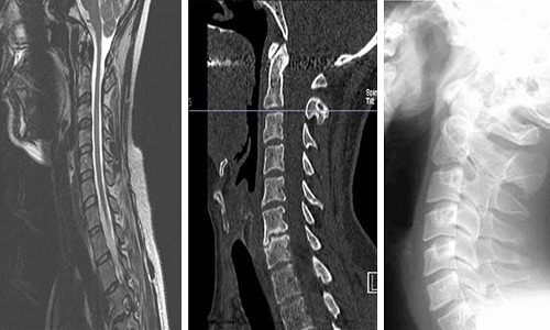
Listened to the FDC podcast episode with Dr Danika Bannasch on chondrodystrophy (CDDY). It was not dachshund-specific; since she breeds tollers she mostly focused on them. Here are my notes:
IVDD (intervertebral disc disease) is not the same thing as IVDH (intervertebral disc herniation). Dr Bannasch considers CDDY and IVDD to be interchangeable.
CDDY variant: Abnormal intervertebral discs. Can have bouts of back pain. Can have disc herniation. All discs are "diseased".
Not all dogs with disc herniation show clinical signs.
All CDDY dogs (in her study on tollers) have signs of diseased discs, but not all had clinical signs that the owners noticed. So not all CDDY dogs are brought in for medical care.
She emphasized that CDDY causes short legs, not long backs. So CDDY shows up in a lot of breeds that you wouldn't expect-- tollers, Portuguese water dogs, beagles, Chesapeake bay retrievers, etc. because the phenotype is shorter legs, not longer backs.
She feels that IVDH is the most painful disease in veterinary medicine.
The CDDY mutation is dominant. It is a "gain of function" mutation. There is no "normal" gene; it is an insertion of an entire gene on a chromosome where it does not belong.
There is a difference in calcification risk between dogs with one versus two copies of the CDDY mutation, but there is no difference in herniation risk with one or two copies.
All beagles are homozygous for CDDY. You cannot "fix" this unless you do an outcross. In breeds where the allele frequency is lower, you can select against it. But be careful-- if the frequency of the allele is high enough, there can be consequences if you remove all CDDY dogs from the gene pool.
Usual recommendations-- keep dogs lean and fit, avoid landing hard if possible, stairs seem to help. Pay attention to subtle signs of pain, eg. refusing to do things they like.
CDDY prematurely degenerates the discs. So a younger dog has "older" discs. All discs degenerate as dogs age, but a dog with CDDY degenerates faster.
What does CDDY do? It makes the legs a little shorter and the skull a little wider. Dr Bannasch believes it rounds the ear tips. This is a desirable phenotype (it seems to win in the show ring), so breeders selected for it before we knew it was bad.
Chondrodysplasia (CDPA) is not associated with IVDD but also produces short legs. Some breeds only have CDDY, some only have CDPA, and some have both. [Note: Dachshunds have both, but CDDY is more common.]
In a breed where there are a lot of homozygotes, it will take a long time to remove CDDY. First you will have to produce heterozygotes, then you will have to produce homozygotes of CDPA.
Chondrodysplasia (CDPA) may be associated with valgus (deformed/bowed legs) and elbow issues. So its not totally innocuous.
As a breeder, she worries about producing a dog that would experience back pain, or have a herniation event, or sell a dog to someone who cannot afford the $12,000 surgery.
X-rays are not great at detecting calcification. CT scans are a bit better, but much more expensive. They've been screening in Europe for a while, and have not seen much improvement. She has not seen much evidence that the amount of calcification correlates with the possibility of a disc herniation event. MRIs look at hydration status, and still aren't great at predicting herniation events, and are super fucking expensive. There are no good screening tests for predicting disc herniation.
Dr Bannasch has dealt with a lot of anger and pushback from breeders for her work on CDDY. Whenever she feels down about it, she goes into the neuro ward to look at the surgery patients for disc herniation events. Then she is rejuvinated to continue her research.
#ivdd#intervertebral disc disease#long post#canine health#there is some FDC nonsense that I'm like. what. at#but most of the episode is very informative
16 notes
·
View notes
Text
Dushyant Verma Shillong - Spinal Stenosis – Causes, Symptoms, Treatment

Dushyant Verma Maharani Bagh - Spinal stenosis is a condition that affects the spine and can cause pain, numbness, and difficulty in movement. It is important to be aware of its symptoms and causes as well as its different treatment options. In this Blog, Dushyant Verma Southern Avenue, explains what is spinal stenosis - what causes and symptoms are spinal stenosis, and which treatments are available.
What is Spinal Stenosis?
Dushyant Verma Shillong says, Spinal stenosis is a condition that occurs when the spaces in the spine narrow, putting pressure on the spinal cord and nerve roots.This can result in leg and arm pain, numbness, tingling, and weakness. Spinal stenosis is most common in the elderly.
There are two types of spinal stenosis: central and foraminal. Central stenosis occurs when the central canal, which contains the spinal cord, narrows. Foraminal stenosis happens when the foramina, which are openings where nerves exit the spine, become narrower. Both types of stenosis can occur in any part of the spine.
Causes of Spinal Stenosis
Dushyant Verma Maharani Bagh says, The most common cause of spinal stenosis is aging. As we get older, the spinal column can narrow and the discs between the vertebrae can deteriorate. This can result in the growth of bony spurs, which can further narrow the spinal canal. Other causes of spinal stenosis include:
Herniated discs
Arthritis
Degenerative disc disease
Spinal injuries
Bone diseases
Tumors
Aging and wear and tear on the spine
Symptoms of Spinal Stenosis
The symptoms of spinal stenosis vary depending on the location of the stenosis and the severity of the condition. According to Dushyant Verma Southern Avenue, Some common symptoms include:
Back pain
Leg pain or cramping
Numbness or tingling sensations in the legs or arms
Weakness in the legs or arms
Difficulty standing or walking for extended periods of time
Weakness in the limbs
Bowel or bladder incontinence (in severe cases).
You should see a doctor if you have any of these symptoms.
How is spinal stenosis diagnosed?
Spinal stenosis is typically diagnosed through a combination of medical history, physical examination, imaging tests (such as X-rays, MRI, or CT scan), and possibly nerve function tests (such as electromyography (EMG) or nerve conduction studies). The doctor will review the symptoms, evaluate the range of motion, and feel for any tenderness in the affected area. Imaging tests can help to confirm the diagnosis and determine the extent and location of the stenosis.
Treatments for Spinal Stenosis
There are a number of treatments that can be effective in managing the symptoms of spinal stenosis. Depending on the severity of your condition, your age, and other factors, your doctor may recommend one or more of the following:
Physical therapy to benefit improve flexibility and strength
Anti-inflammatory medications to help reduce swelling
Corticosteroid injections to help reduce inflammation
Surgery to remove the bone or tissue causing the stenosis
Alternative Treatments for Spinal Stenosis:
There are many alternative treatments for spinal stenosis. Some of these include:
Chiropractic care: This is a form of manual therapy that can help to relieve pain and improve mobility in people with spinal stenosis.
Acupuncture: Acupuncture is an ancient Chinese practice that involves inserting thin needles into specific points on the skin. It is said to help with pain relief and circulation.
Massage: Massage can help to relax muscles and relieve tension in the back and neck. It can also improve blood flow and reduce inflammation.
Exercise: Regular exercise is important for overall health, but it can also be helpful in managing symptoms of spinal stenosis. Low-impact activities such as walking, swimming, and yoga can all be beneficial.
Dushyant Verma Maharani Bagh says, Spinal stenosis is a common condition that can cause significant impairment and pain if left untreated. Fortunately, this condition can be managed with the help of medications, physical therapy and/or surgery. It is important to be aware of the causes and symptoms of spinal stenosis so that you can take steps to prevent it or seek treatment as soon as possible. With proper diagnosis and treatment, most people are able to find relief from their symptoms and return to their normal lives.
6 notes
·
View notes
Text
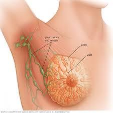
How is breast cancer diagnosed?
Diagnosing breast cancer typically involves a combination of medical history review, physical examination, imaging tests, and biopsy. Here's an overview of the diagnostic process for breast cancer:
Medical History and Physical Examination: A healthcare provider will start by taking a detailed medical history, including any symptoms you may be experiencing and any risk factors for breast cancer. They will then perform a physical examination of your breasts, looking for any lumps, changes in breast size or shape, nipple abnormalities, or skin changes.
Imaging Tests:
Mammogram: A mammogram is an X-ray of the breast tissue. It is commonly used for breast cancer screening and may also be used for diagnostic purposes if a lump or other abnormality is detected.
Breast Ultrasound: Ultrasound uses sound waves to produce images of the breast tissue. It can help differentiate between solid masses (which may be cancerous) and fluid-filled cysts (which are usually benign).
Breast Magnetic Resonance Imaging (MRI): MRI uses magnetic fields and radio waves to create detailed images of the breast tissue. It may be used in certain cases, such as for high-risk individuals or to further evaluate abnormalities detected on mammography or ultrasound.
Biopsy: If imaging tests reveal a suspicious area in the breast, a biopsy is typically performed to obtain a sample of tissue for further examination. There are different types of breast biopsies, including:
Fine Needle Aspiration (FNA): A thin needle is used to withdraw cells from the suspicious area.
Core Needle Biopsy: A larger needle is used to remove a small cylinder of tissue from the breast.
Surgical Biopsy: A surgical procedure is performed to remove a larger sample of tissue for examination.
Pathology Examination: The tissue samples obtained during the biopsy are sent to a pathology laboratory, where they are examined under a microscope by a pathologist. This allows for the determination of whether the tissue is cancerous (malignant) or non-cancerous (benign), as well as the specific type of breast cancer if present.
Once a diagnosis of breast cancer is confirmed, further tests may be performed to determine the extent (stage) of the cancer and to help guide treatment decisions. These additional tests may include imaging studies such as chest X-rays, bone scans, CT scans, or PET scans.
It's important to work closely with a healthcare team specialized in breast cancer diagnosis and treatment to ensure that all necessary tests are performed and that an accurate diagnosis is obtained. Early detection and diagnosis are key to improving outcomes and increasing the chances of successful treatment for breast cancer.
0 notes
Text
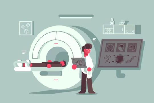
AI's Impact on Healthcare: Current Trends

Artificial intelligence (AI) has revolutionized the healthcare industry, bringing about profound shifts in diagnosis, treatment, research, and patient care. This thorough analysis examines the current trends in AI's influence on healthcare and explores how cutting-edge technology is revolutionizing this vital profession.
AI's Growing Importance in Healthcare
Artificial intelligence (AI) is becoming increasingly prevalent in the healthcare industry thanks to advancements in machine learning, deep learning, and natural language processing. Artificial Intelligence has the potential to significantly improve patient outcomes, lower costs, and improve medical decision-making.
Artificial Intelligence in Healthcare
Medical imaging is one of the areas where artificial intelligence (AI) in healthcare shows the most significant promise. AI algorithms can analyze medical images with great precision, which helps radiologists diagnose conditions including cancer, fractures, and abnormalities. AI systems' speed and accuracy are revolutionary advancements in this field, enabling faster diagnosis and better treatment planning.
Deep Learning's Place in Radiology
Convolutional neural networks (CNNs), in particular, are deep learning approaches that have demonstrated impressive performance in picture segmentation and classification applications. These neural networks significantly reduce human error and speed up diagnosis by distinguishing between healthy and diseased tissues in X-rays, MRIs, and CT scans.
The Role of AI in Pathology
Artificial Intelligence (AI) is progressing significantly in the realm of pathology. It can quickly examine histopathology slides, identifying abnormalities or malignant cells in a fraction of the time that a human pathologist would need. This improves accuracy and speeds up diagnosis, essential for treating cancer and other illnesses.
Predictive Analytics in the Prevention of Diseases
The ability of AI to anticipate outcomes is crucial in identifying those who may be at risk of getting different kinds of illnesses. By analyzing large datasets, AI can evaluate genetic predispositions, lifestyle characteristics, and past medical records. This enables predictions about the chance of developing conditions like diabetes, heart disease, or Alzheimer's.
Customized Therapy Schedules
Using AI-powered predictive analytics enables medical professionals to create unique treatment regimens for every patient. This precision medicine method maximizes treatment effectiveness and reduces adverse effects by tailoring treatments to each patient's genetic composition and medical background.
Healthcare Applications of Natural Language Processing (NLP) AI-driven NLP has created new opportunities for analyzing and extracting insightful data from unstructured research articles, patient records, and clinical notes.
Improving the Quality of Clinical Records
AI-powered natural language processing (NLP) can turn handwritten or spoken clinical notes into structured data, saving healthcare staff time and improving the quality and comprehensiveness of patient records. This makes better patient care and more informed decision-making possible.
Facilitation of Research and Drug Discovery
In the field of research, AI-driven natural language processing (NLP) can comb through enormous amounts of scientific literature to find pertinent studies and glean meaningful insights. Thanks to the acceleration of medication discovery processes, researchers can access current knowledge and possible scientific advances.
The Use of Virtual Health Assistants in Telemedicine
The COVID-19 pandemic has led to an increase in the use of telemedicine, and artificial intelligence is a significant factor in this development. AI-powered virtual health assistants can effectively assess patients, answer frequently asked questions about health, and offer advice on managing chronic illnesses.
Remote Patient Surveillance
Vital sign monitoring can be done continuously remotely with AI capabilities by wearable sensors. This feature enables medical professionals to identify health problems immediately and take appropriate action. As a result, hospital readmissions and medical expenses decrease, and patient outcomes improve.
Also Read: AI Developer
Despite its enormous promise, AI in healthcare presents ethical and data security challenges. One of the industry's most critical issues is ensuring that AI-generated insights are used responsibly and protecting patient privacy.
Data Privacy Protection Measures
Healthcare institutions need strong data encryption, access controls, and compliance mechanisms to protect patient data appropriately. It is crucial to balance patient privacy protection and data availability for research.
Taking Care of Bias and Maintaining Fairness
AI systems may unintentionally reinforce prejudices present in past medical data. Diverse datasets, rigorous evaluation, and a persistent commitment to transparency are necessary to guarantee AI systems' fairness and combat bias.
In summary
AI has a significant impact on healthcare, as seen by recent developments that show how it can improve patient care, diagnosis, and prediction. As the industry continues integrating AI certification, overcoming the associated challenges and maximizing benefits for patients, healthcare providers, and researchers is critical. The future of healthcare seems promising, with better outcomes and a more effective healthcare system possible with continued innovation and appropriate AI implementation.
0 notes
Text
The Cost of Ortho Cancer Treatment: What You Need to Know
Orthopedic cancer, often referred to as ortho cancer, involves the malignancies of bones and soft tissues, presenting significant treatment challenges. Understanding the cost of ortho cancer treatment is crucial for patients and their families, as the financial implications can be substantial. This article delves into the various components that influence the cost, options for managing these expenses, and the importance of choosing a specialized treatment center like Action Cancer Hospital.
Understanding Orthopedic Cancer
What is Ortho Cancer?
Ortho cancer encompasses a range of cancers that affect bones and soft tissues, including muscles, fat, blood vessels, nerves, and tendons. The most common types include osteosarcoma, chondrosarcoma, and Ewing's sarcoma. These cancers can be aggressive and require comprehensive treatment plans.
Symptoms and Diagnosis
Common symptoms of ortho cancer include persistent pain, swelling, fractures, and limited mobility. Early diagnosis is critical for effective treatment and often involves imaging tests such as X-rays, MRI, CT scans, and biopsy procedures to determine the cancer type and stage.
Factors Influencing the Cost of Ortho Cancer Treatment
Type and Stage of Cancer
The type and stage of ortho cancer significantly impact treatment costs. Early-stage cancers may require less aggressive treatment, while advanced stages often involve complex and multi-modal therapies, escalating expenses.
Treatment Modalities
Surgery: Often the primary treatment for ortho cancer, surgery can be expensive, especially if it involves limb-sparing techniques or complex reconstructive procedures.
Radiation Therapy: This treatment uses high-energy radiation to kill cancer cells. Costs vary depending on the number of sessions and technology used.
Chemotherapy: Involves the use of drugs to destroy cancer cells. The cost depends on the type and duration of the chemotherapy regimen.
Targeted Therapy: Uses drugs or other substances to precisely identify and attack cancer cells. This innovative approach can be costly but is often effective.
Immunotherapy: A newer treatment that helps the immune system fight cancer. It can be expensive due to the advanced technology involved.
Hospital and Healthcare Provider
The choice of hospital and healthcare provider plays a crucial role in the overall cost. Specialized cancer treatment centers like Action Cancer Hospital offer state-of-the-art facilities and expert care, which can influence costs.
Geographical Location
The location of the treatment center can affect costs due to variations in healthcare pricing across different regions and countries.
Duration of Treatment
The length of treatment required, including post-treatment care and rehabilitation, adds to the total cost. Longer treatment durations generally result in higher expenses.
Breakdown of Treatment Costs
Initial Consultation and Diagnostic Tests
The journey begins with initial consultations, which typically range from $200 to $500. Diagnostic tests, including imaging and biopsy, can cost anywhere from $1,000 to $5,000.
Surgery Costs
Surgical costs vary widely, with simple procedures costing around $10,000 and complex surgeries, especially those involving reconstruction, exceeding $50,000.
Radiation Therapy Costs
Radiation therapy sessions can cost between $2,000 and $10,000 per session, with a typical course involving multiple sessions.
Chemotherapy Costs
Chemotherapy costs depend on the drugs used and the treatment duration, ranging from $1,000 to $12,000 per month.
Targeted Therapy and Immunotherapy Costs
Targeted therapies and immunotherapies are among the most expensive, often exceeding $10,000 per month, depending on the specific treatment plan.
Rehabilitation and Follow-Up Care
Post-treatment rehabilitation and follow-up care are essential for recovery, adding another $5,000 to $20,000 to the total cost.
Managing the Financial Burden
Health Insurance
Having comprehensive health insurance is vital. Insurance can cover a significant portion of the treatment costs, but patients should thoroughly understand their policy details, including deductibles, co-pays, and coverage limits.
Financial Assistance Programs
Many hospitals and cancer treatment centers offer financial assistance programs to help patients manage costs. These programs can include grants, subsidies, and flexible payment plans.
Government and Non-Profit Organizations
Government programs and non-profit organizations often provide financial aid, support services, and resources to help cancer patients cover treatment costs.
Fundraising and Crowdfunding
Personal fundraising and crowdfunding have become viable options for many patients. Platforms like GoFundMe allow patients to share their stories and raise funds for their treatment from friends, family, and the public.
Choosing the Right Treatment Center
Why Choose Action Cancer Hospital?
Action Cancer Hospital is renowned for its expertise in ortho cancer treatment, offering a multidisciplinary approach and state-of-the-art facilities. Choosing a specialized center ensures access to the latest treatments and a team of experienced professionals dedicated to cancer care.
Factors to Consider
When selecting a treatment center, consider factors such as:
Reputation and Expertise: Research the hospital’s reputation and the expertise of its medical staff.
Available Treatments: Ensure the hospital offers a comprehensive range of treatments tailored to ortho cancer.
Support Services: Look for centers that provide holistic support, including psychological counseling, nutritional advice, and physical therapy.
The Importance of Early Detection and Treatment
Benefits of Early Detection
Early detection of ortho cancer significantly improves treatment outcomes and can reduce overall costs. Regular check-ups and prompt attention to symptoms are crucial for early diagnosis.
Preventive Measures
Adopting a healthy lifestyle, staying informed about cancer risks, and undergoing regular screenings can help in the early detection and prevention of ortho cancer.
Conclusion
The cost of ortho cancer treatment can be overwhelming, but understanding the factors involved and exploring financial assistance options can help manage the burden. Choosing a specialized treatment center like Action Cancer Hospital ensures access to the best care possible, improving the chances of successful treatment and recovery. Early detection remains a key factor in reducing costs and enhancing outcomes, highlighting the importance of regular health check-ups and prompt medical attention.
Navigating the financial landscape of cancer treatment is challenging, but with the right information and support, patients can focus on their health and recovery, knowing they are receiving the best possible care.
0 notes
Text
Understanding CT Scans: A Comprehensive Guide
CT scans, or Computed Tomography scans, are a pivotal diagnostic tool in modern medicine, offering detailed images of the body's internal structures. They have revolutionized the way doctors diagnose and treat various medical conditions. This article explores what a CT scan is, how it works, its applications, and what patients can expect during the procedure.
What is a CT Scan?
A CT scan combines X-ray technology with computer processing to create cross-sectional images of the body. Unlike regular X-rays, which produce a single flat image, CT scans generate multiple images or slices of the area being examined. These slices can be viewed individually or combined to create a three-dimensional image, providing much more detailed information.
How Does a CT Scan Work?
During a CT scan, the patient lies on a motorized table that slides into a doughnut-shaped machine called a gantry. Inside the gantry, an X-ray tube rotates around the patient, sending out multiple beams of X-rays from different angles. Detectors on the opposite side of the tube capture the X-rays that pass through the body. A computer then processes these signals to create detailed images of the internal structures.
Applications of CT Scans
CT scans are versatile and can be used to diagnose a wide variety of conditions. Some common applications include:
Head and Brain: Detecting tumors, bleeding, stroke, and skull fractures.
Chest: Evaluating lung conditions, heart problems, and blood clots.
Abdomen and Pelvis: Assessing organs such as the liver, kidneys, pancreas, and intestines for issues like tumors, infections, or inflammation.
Bones and Joints: Diagnosing fractures, infections, and degenerative diseases.
Cardiovascular System: Identifying blockages in blood vessels, aneurysms, and other vascular conditions.
Preparing for a CT Scan
Preparation for a CT scan depends on the type of scan and the area being examined. Here are some general guidelines:
Diet: You may be asked to avoid eating or drinking for a few hours before the scan, especially if a contrast material is used.
Clothing: Wear comfortable, loose-fitting clothes and remove any metal objects, such as jewelry, as they can interfere with the imaging.
Contrast Material: Sometimes, a contrast dye is used to enhance the images. This dye may be ingested orally, injected into a vein, or administered rectally. Inform your doctor if you have any allergies or kidney problems, as these could affect your ability to receive the contrast material.
What to Expect During the Procedure
A CT scan is typically quick and painless. Here’s what you can expect:
Positioning: You will lie on the scanning table, which will move slowly through the gantry. You may be asked to hold your breath for short periods to avoid blurring the images.
Duration: The entire process usually takes between 10 and 30 minutes, depending on the complexity of the scan.
Comfort: While the procedure is generally comfortable, the table might feel a bit hard, and the room may be cool. Inform the technician if you feel any discomfort.
Risks and Benefits
CT scans are generally safe, but they do involve exposure to a small amount of radiation. The benefits, however, usually outweigh the risks, as the detailed images provide crucial information for accurate diagnosis and treatment planning. If contrast material is used, there is a slight risk of allergic reaction, but this is rare.
Conclusion
CT scans are an invaluable tool in modern diagnostic medicine, offering detailed and accurate images that aid in diagnosing and treating a wide range of conditions. Understanding the process, preparation, and what to expect can help patients feel more comfortable and informed when undergoing a CT scan. Always consult with your healthcare provider if you have any concerns or questions about the procedure.
More Read:
https://www.tumblr.com/srivastavmri01/751170805936865280/srivastava-mri-and-imaging-centre-the-premier-ct?source=share
https://www.tumblr.com/srivastavmri01/751170629390188544/srivastava-mri-and-imaging-centre-the-premier?source=share
https://www.tumblr.com/srivastavmri01/751170530259943424/srivastava-mri-and-imaging-centre-the-premier?source=share
https://www.tumblr.com/srivastavmri01/751170283629690880/srivastava-mri-and-imaging-centre-the-best?source=share
https://www.tumblr.com/srivastavmri01/751355009366622208/srivastava-mri-and-imaging-centre-the-leading-mri?source=share
https://www.tumblr.com/srivastavmri01/751354876990291968/srivastava-mri-and-imaging-centre-the-premier-ct?source=share
https://www.tumblr.com/srivastavmri01/751354543616475136/srivastava-mri-and-imaging-centre-the-leading?source=share
https://www.tumblr.com/srivastavmri01/751354339601317888/srivastava-mri-and-imaging-centre-premier-x-ray?source=share
0 notes
Text
Best Knee Replacement Surgeons in India
Knee is the largest joint of the human body, it is a hinge joint which allows flexion and extension and also slight internal and external rotation. It connects our thigh to our lower leg. Similar to all other joints, the knees are part of our skeletal system. The knee helps to support our weight. It is synovial joint and has freedom to move. Any movement that involves our legs relies on our knees. It is also considered as compound joint, hence there is a chance of it to get injured easily.
The knee is made up of bones, cartilage, muscles, ligaments, nerves and tendon and three bones, femur, tibia and the patella. The ends of the femur and tibia and the back of the patella are covered with the articular cartilage. This cartilage is slippery substance and it helps the knees to glide smoothly when the leg is stretched or bend. There are two wedge-shaped pieces of meniscal cartilage which act as shock absorbers between the femur and tibia. Ligament in the knee act like a rope which holds the bones together, and keep our knee stable. Muscles are connected with bones by the tendons.
Among the most common injuries, the ligamentous and meniscal tears are most common. Anything that damages the bones or tissues affects the knees, they can be- arthritis, osteoarthritis, osteoporosis, ACL tears, MCL tears, meniscus tears, sprains, bone fractures, dislocation. Some of the common tests advised to diagnose the knees are X-rays, ultrasound, CT scan, MRI.
Knee transplant or knee replacement surgery is done to replace the inured parts or the worn-out knee joints. This can help ease pain and makes the knee to work better. In the surgery, the damaged bone and cartilages are replaced with metal parts. These surgeries are done in by best surgeons for knee replacement surgery in India in best knee replacement surgery hospitals in India. To be confirmed whether a knee replacement is required or not, the knee’s range of motion, stability and strength is checked by the surgeons.
Most common reason for knee transplant surgery is to relieve arthritis pain. Surgeons recommend either a total knee transplant or partial transplant. Surgeons provide the required diagnosis to get ready for a surgery. This includes- a whole body physical examination, blood tests, ECG to check heart health, dental exam to reduce the risk of having infection post-surgery, imaging tests. These tests may add up to a huge knee replacement surgery cost in India.
Knee Replacement Surgery cost in India
The minimum knee replacement surgery cost in India INR 1,50,000, while the maximum knee replacement surgery price in India is INR 6,00,000. However, the price varies depending upon the location of hospitals in different cities. The price of knee replacement surgery in India is based on method of surgery, cost of implant, doctor’s fees, type of replacement, hospital stay.
Best Knee Replacement Surgery Hospitals in India
Best hospitals for knee replacement surgery in India are- Fortis Hospital, Nanavati Hospital, and Artemis Hospital.
Best Knee replacement Surgeons in India
Some of the best knee replacement surgerons in India are- Dr. Ashok Rajgopal, Dr. Harshavardan Hegde, and Dr. Subhash Jangid.
My Med Trip is a leading medical tourism company. We are offering complete medical and healthcare services with medical consulting in India for foreign patients. We help patients in finding the best hospitals, top doctors, and good accommodations at affordable costs in India. We offer surgeries, treatment and transplant. Some of them are kidney transplant cost, heart transplant, bone marrow transplant cost, cancer treatment, liver transplant in India, prostate cancer ovarian cancer, hip replacement surgery cost, knee replacement, orthopedic surgery cost in India, shoulder surgeons,best hospital for prostate treatment in India, orthopedic surgeons, heart valve replacement knee surgeons, and so on.
Source: https://mymedtrips.blogspot.com/2023/10/best-knee-replacement-surgeons-in-india.html
0 notes
Text
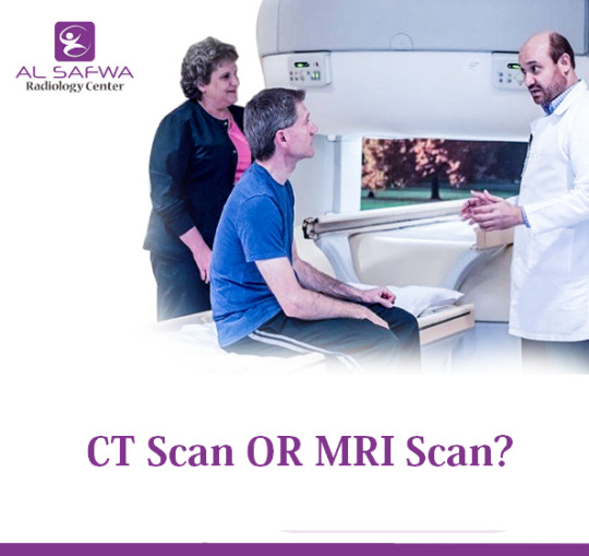
CT or MRI? Choosing the Right Imaging Test for You
When faced with the need for advanced imaging to diagnose a medical condition, patients often wonder which test is right for them: a CT scan or an MRI. Both of these imaging techniques are powerful tools used in modern medicine, but they serve different purposes and have distinct advantages and limitations. At Alsafwa Radiology Center in Sharjah, we provide both CT and MRI scans, and our expert radiologists help guide patients to the most appropriate choice based on their individual medical needs.
Understanding CT Scans
What is a CT Scan?
A Computed Tomography (CT) scan uses X-rays and computer technology to create detailed images of the inside of the body. CT scans are particularly useful for viewing bone injuries, diagnosing lung and chest problems, and detecting cancers. The procedure is relatively quick, usually taking only a few minutes.
When is a CT Scan Recommended?
CT scans are recommended in several scenarios:
Trauma: CT scans are excellent for detecting bone fractures and internal injuries in trauma patients.
Cancer Detection: They help in identifying tumors and assessing the extent of cancer.
Chest and Lung Issues: CT scans can detect lung infections, pulmonary embolisms, and other chest conditions.
Abdominal Concerns: They are used to diagnose appendicitis, kidney stones, and other abdominal problems.
Advantages and Limitations of CT Scans
Advantages:
Speed: CT scans are quick, making them ideal for emergency situations.
Detail: They provide detailed images of bone structures.
Availability: CT scanners are widely available and less expensive than MRI machines.
Limitations:
Radiation Exposure: CT scans involve exposure to ionizing radiation, which may be a concern, especially for repeated use.
Soft Tissue Imaging: CT scans are less effective than MRIs at imaging soft tissues like the brain and muscles.
Understanding MRI Scans
What is an MRI?
Magnetic Resonance Imaging (MRI) uses a powerful magnet, radio waves, and a computer to produce detailed images of the body’s internal structures. Unlike CT scans, MRI Scans do not use ionizing radiation. They are particularly useful for imaging soft tissues, including the brain, muscles, and ligaments.
When is an MRI Recommended?
MRIs are recommended for various conditions, including:
Neurological Issues: MRIs are the gold standard for imaging the brain and spinal cord, useful in diagnosing strokes, tumors, and multiple sclerosis.
Musculoskeletal Problems: They are ideal for imaging joints, muscles, and ligaments, helping diagnose tears, sprains, and other injuries.
Cardiac Conditions: MRIs can provide detailed images of the heart and blood vessels.
Soft Tissue Evaluation: MRIs are superior in evaluating soft tissues in the body, including organs like the liver and kidneys.
Advantages and Limitations of MRIs
Advantages:
No Radiation: MRIs do not use ionizing radiation, making them safer for repeated use.
Soft Tissue Imaging: They provide unparalleled detail for soft tissue structures.
Versatility: MRIs can be used to image almost any part of the body.
Limitations:
Duration: MRI scans take longer than CT scans, often up to an hour.
Cost: They are generally more expensive than CT scans.
Accessibility: MRI machines are not as widely available as CT scanners.
Claustrophobia: Some patients may feel uncomfortable or claustrophobic inside the MRI machine.
Factors to Consider When Choosing Between a CT Scan and an MRI
The Nature of Your Condition
The type of medical condition being investigated is a primary factor in deciding between a CT scan and an MRI. For instance, bone injuries and acute trauma are best evaluated with CT scans, while neurological conditions and soft tissue injuries are better suited for MRI.
Urgency of the Situation
In emergency settings where speed is crucial, CT scans are typically preferred due to their quick imaging times. MRI scans, while more detailed, take longer to perform and are less commonly used in emergency situations.
Radiation Concerns
For patients who require multiple imaging tests over time, or for those who are particularly sensitive to radiation (such as pregnant women and children), MRI may be a better choice because it does not involve ionizing radiation.
Cost and Availability
The availability of imaging equipment and the cost of the procedure can also influence the decision. CT scans are generally more accessible and less expensive than MRI scans. However, the choice should be made based on clinical necessity rather than cost alone.
Patient Comfort
Patient comfort is another consideration. Some individuals may feel claustrophobic in an MRI machine, which is a more enclosed space compared to a CT scanner. Additionally, patients with metal implants or devices cannot undergo MRI scans due to the magnetic field.
Why Choose Alsafwa Radiology Center?
At Alsafwa Radiology Center in Sharjah, we are committed to providing the highest quality imaging services with state-of-the-art technology and a patient-centered approach. Our team of experienced radiologists and technologists ensures that each patient receives personalized care and precise diagnostic information.
Expert Consultation
Our radiologists consult with referring physicians and patients to determine the most appropriate imaging test. We take into account the specific medical condition, patient history, and any other relevant factors to make an informed recommendation.
Advanced Technology
We utilize the latest imaging technology to ensure high-quality images and accurate diagnoses. Our CT and MRI machines are regularly updated and maintained to provide the best possible results.
Patient-Centered Care
We understand that undergoing an imaging test can be stressful. Our staff is dedicated to making the experience as comfortable and efficient as possible. We provide clear instructions and support throughout the process, ensuring patients are well-informed and at ease.
Conclusion
Choosing between a CT scan and an MRI depends on various factors, including the nature of the medical condition, urgency, radiation concerns, cost, availability, and patient comfort. At Alsafwa Radiology Center in Sharjah, we offer both imaging options and guide patients to the most suitable choice based on their individual needs. Our commitment to advanced technology and patient-centered care ensures that you receive the highest quality diagnostic services. If you have any questions or need to schedule an imaging test, please contact us today.
0 notes
Text
Signs Of Throat Cancer
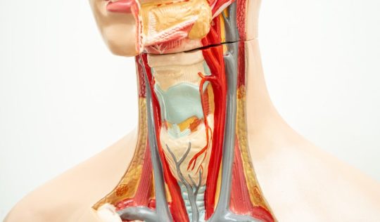
Usually individuals who suffer from throat cancer develop cancer in their voice box called larynx and face problems like sore throat, swelling of the neck, and difficulty swallowing. Possibly, the sufferer may note these indications of throat cancer in the beginning, but they might get to know it when the cancer gets more advanced and they face issues with breathing or speaking. However, throat cancer symptoms may differ based on their location in your body, and if the cancerous tumors grow within the voice box, they can make you ineligible to speak. These conditions require an immediate diagnosis, and you must visit a medical expert quickly!
Common Causes
However, the exact cause of throat cancer is not known, but due to the reasons mentioned, an individual might develop this illness:
Throat Cancer Signs/Symptoms
Here’s a list of some common signs that a person may face when developing throat cancer:
Voice Changes: If your voice is becominghoarse or husky or you are facing difficulty pronouncing certain sounds or words, then there might be a possibility that you are developing cancer in your larynx.
Difficulty or Pain in Swallowing: The most initial sign that you might come across would be difficulty swallowing, as you might experience burning or pain when chewing and swallowing food.
Chronic Sore Throat: Known as an early warning sign, where cancer can occur in the pharynx, causing pain in your throat, which doesn’t disappear when you swallow.
Weight loss: An unusual weight loss may occur as a common cancer symptom which can be exacerbated by swallowing problems.
Swollen Lymph Nodes Around the Neck: Swollen nodes or enlarged lymph glands around the neck can be a sign your body is developing a cancer illness, especially if it grows slowly.
Other Signs
Stages of Throat Cancer
Throat cancer staging can form an essential part done by your doctor to identify the stage of cancer and to analyze how far the cancer has grown and spread. Usually, these are the stages that your doctor will diagnose:
Stage 0: Denoted by the term carcinoma in situ where the throat will develop cancerous cells.
Stage I: Referred to as an early stage of cancer where the cancer is only two centimeters, limited to the throat.
Stage II: This stage forms where thetumor is usually between two-four centimeters, but yet not spread to the lymph nodes.
Stage III: Here, your throat cancer comes larger than four centimeters, or even spread to nearby lymph nodes.
Stage IV: During this, the cancer is no longer confined to the original site as it might spread to other body areas including your neck, trachea or jaw.
Throat Cancer Diagnosis
Usually the specialists follow the below mentioned methods to diagnose the throat cancer:
Physical Examination: Most initially when you see a doctor, they will usually examine your mouth, throat and neck to find any throat cancer symptom.
Larynx Endoscopy: To look for abnormalities of the throat, the doctors use an endoscope and insert it through the nose to analyze the illness.
Biopsy: The doctors do this to determine if there are cancerous cells present by taking a small sample of cells or tissue to examine using a magnifying glass.
Ultrasound: Ultrasound of the neck is utilized to detect any abnormalities present, which can subsequently be verified using CT or MRI scans.
X-rays: The doctors may opt for chest x-ray to determine the sufferer’s general health and to analyze the spread of lung cancer.
CT Scan: A radiological procedure is performed to confirm and pinpoint the presence of disease or cancer in the neck region, while also assessing any potential lymph nodes. This involves creating detailed cross-sectional images of the targeted structure from various angles.
MRI: Using radio waves and magnets, the test produces detailed images about what’s going on inside your body.
PET scan: The doctors usually inject radioactive material into the body to detect cancer cells. Treatment Options
Mentioned below are the treatment options that the experts suggest:
The order of treatment for throat cancer is especially if it’s involving voice box or adjacent structures is chemotherapy plus radiotherapy . This approach has evolved from the surgery first approach which was used few decades ago but with the principle of organ preservation and to preserve the voice of the patient .
Surgery: Your medical expert will suggest surgery depending upon the location of the cancer and the structures involved ,if erosion of the cartilages seen in the scan then surgery is the choice of treatment The surgery involves laryngectomy plus partial phryngectomy and also reconstruction of the pharynx which connects to the food pipe . However, surgery involves excision of the voice box and loss of voice but with recent development of voice prosthesis ,voice can be generated.
Laser Surgery: Another treatment option, usually opted by the medical experts in the very initial stages if the disease is limited only to the voice box that too partially .
Radiation Therapy: You may get advised by the doctor to undergo radiation therapy which is the primary treatment in some cases as these doses target the cancer cells to eliminate them.
Chemotherapy: To treat certain cancer conditions, chemotherapy may be needed, especially if the tumor is large, or if the cancer has spread into the lymph nodes as this surgery can shrink tumors possibly.
Targeted Therapy: Since this type of treatment reduces the risk of side effects, these drugs target specific cancer cells or proteins that affect the growth of cancer.
Immunotherapy: This new approach boosts the ability of the immune system to protect the body from cancer.
Prevention
Final Words
Since we tried our best to cover every relatable aspect about Signs Of Throat Cancer for our readers, it is essential to note that visiting a trusted medical hospital for your treatment will help you recover better from the illness. To receive personalized advice and a discussion regarding Throat Cancer, consult with Dr. Sanjog Singh at Samsara Cancer Care Nagpur one of the best for Throat Cancer treatment in Nagpur, a highly respected medical expert today. With years of expertise working in this field, doctors can assist you understand the process of throat cancer better and help you through the course of your illness
0 notes
Text
Revolutionizing Radiology: The Rise of Specialized Staffing in Houston
In the bustling healthcare landscape of Houston, Texas, the demand for specialized medical imaging and radiology services has seen an unprecedented rise. As healthcare facilities strive to offer top-notch patient care, the need for skilled professionals in medical imaging and radiology departments has never been more critical. This article delves into the significance of Medical Imaging Staffing Houston and Radiology Staffing Solutions Houston, highlighting how specialized staffing services are transforming patient care in the region.
The Crucial Role of Medical Imaging and Radiology Staffing
Medical imaging and radiology are at the heart of modern diagnostics, playing a pivotal role in the early detection, diagnosis, and treatment of various diseases. From X-rays and MRIs to CT scans and ultrasound, the array of imaging techniques available today is vast and sophisticated. However, the effectiveness of these technologies is heavily dependent on the expertise of the professionals who operate them. This is where specialized Medical Imaging Staffing Houston services come into play, ensuring that healthcare facilities have access to the highly skilled personnel needed to deliver accurate and timely diagnostics.
Similarly, Radiology Staffing Solutions Houston is integral to maintaining the efficiency and reliability of radiology departments. With the advent of digital imaging and teleradiology, the scope of radiology services has expanded, necessitating a workforce that is not only technically proficient but also adaptable to the evolving landscape of medical diagnostics.
The Impact of Specialized Staffing on Healthcare
Specialized staffing services in medical imaging and radiology have a profound impact on healthcare delivery. By providing access to a pool of qualified professionals, these services enable healthcare facilities to maintain high standards of care, even in the face of staffing shortages or sudden increases in demand. This flexibility is particularly crucial in large metropolitan areas like Houston, where the population density and healthcare needs can vary significantly across different communities.
Moreover, specialized staffing solutions contribute to the overall efficiency of healthcare services. They allow for the seamless operation of imaging and radiology departments, minimizing delays in diagnostics and treatment. This not only improves patient outcomes but also enhances the patient experience, fostering trust and satisfaction with the healthcare system.
The Future of Healthcare Staffing in Houston
As technology continues to advance, the demand for specialized medical imaging and radiology staffing is expected to grow. Houston, with its robust healthcare infrastructure and diverse population, is at the forefront of this trend. Healthcare facilities in the region are increasingly recognizing the value of partnering with specialized staffing agencies to navigate the complexities of medical imaging and radiology services.
Conclusion
The evolution of medical imaging and radiology services in Houston underscores the importance of specialized staffing solutions. As healthcare facilities strive to meet the growing demands of patient care, services like riscstaffing.com emerge as vital partners in this endeavor. In the dynamic field of healthcare, the partnership between specialized staffing agencies and medical facilities is not just beneficial but essential for the future of patient care
0 notes
Text
What is the difference between CT scan and MRI?
CT (computed tomography) scans and MRI (magnetic resonance imaging) are both imaging techniques used in medicine to produce detailed images of the body's internal structures, but they operate on different principles and have distinct advantages and applications:
Principle of Imaging:
CT Scan: CT scans use X-rays to create cross-sectional images of the body. The X-ray machine rotates around the patient, taking multiple images from different angles, which are then processed by a computer to create detailed cross-sectional images.
MRI: MRI uses a strong magnetic field and radio waves to create detailed images of the body's internal structures. It does not use ionizing radiation like X-rays do.
Contrast Resolution:
CT Scan: CT scans are better at detecting bone and calcium deposits and are often used to visualize bone fractures, tumors, and abnormalities in soft tissues.
MRI: MRI provides superior contrast resolution in soft tissues, making it especially useful for imaging the brain, spinal cord, muscles, ligaments, tendons, and internal organs like the liver, pancreas, and prostate.
Radiation Exposure:
CT Scan: CT scans involve exposure to ionizing radiation, although the amount is typically low and considered safe for diagnostic purposes. However, repeated CT scans over time can increase cumulative radiation exposure.
MRI: MRI does not involve exposure to ionizing radiation, making it a safer option for imaging, especially for pregnant women and children.
Metallic Implants and Artifacts:
CT Scan: Metallic implants and objects can cause artifacts on CT scans, which may degrade image quality.
MRI: MRI is more sensitive to metallic implants and objects, which can cause significant artifacts and distortions in the images.
Contrast Agents:
CT Scan: CT scans can be performed with or without the use of contrast agents (iodine-based contrast agents), which are injected into the bloodstream to enhance visualization of blood vessels and certain tissues.
MRI: MRI scans can also be performed with contrast agents (gadolinium-based contrast agents) to improve visualization of blood vessels, tumors, and areas of inflammation.
In summary, CT scan in Rohini and MRI have different strengths and are used for different purposes in medical imaging. The choice between CT and MRI depends on factors such as the type of condition being evaluated, the body part being imaged, the desired level of detail, and any contraindications or limitations for the patient.
1 note
·
View note
Text
Data Security: Impact of CD DVD Medical DICOM Publishing Systems

Data security is paramount, especially in the healthcare sector where sensitive patient information is constantly being generated and shared. Medical imaging, in particular, generates large amounts of data that need to be securely stored, accessed, and shared.
DICOM (Digital Imaging and Communications in Medicine) has become the standard for managing medical imaging data, and the DICOM publishing system plays an important role in ensuring the security and integrity of this data.
DICOM publishing systems include a range of technologies and tools designed to publish medical images on CD or DVD for storage, sharing and distribution.
These systems typically include DICOM CD/DVD burners, disc publishers, and robotic systems that automate the burning and labelling of discs. Although these technologies provide convenience and efficiency, they also present unique challenges and considerations when it comes to data security.
Understanding DICOM Medical Services
DICOM medical services refers to the suite of protocols, standards, and technologies used to manage, store, and disseminate medical imaging data. These services enable healthcare providers to capture, store, and share medical images such as X-rays, MRIs, CT scans, and ultrasounds in a standardized format. By following DICOM standards, healthcare organizations ensure interoperability and compatibility across different imaging devices and systems.
Role of DICOM Publishers
DICOM publishers play a vital role in the workflow of medical imaging departments and healthcare organizations. These systems allow medical professionals to create CDs or DVDs containing DICOM images for patients, referring physicians, or for archival purposes. DICOM publishers often feature advanced capabilities such as automatic disc burning, printing of patient information and labels, and integration with PACS (Picture Archiving and Communication Systems) for seamless data transfer.
Security Challenges and Considerations
While DICOM publishing systems provide convenience and efficiency, they also present security challenges that must be addressed. Here are some key ideas:
Data Encryption: DICOM images contain sensitive patient information and must be encrypted to prevent unauthorized access. DICOM publishing systems must support encryption standards such as AES (Advanced Encryption Standard) to ensure data security during storage and transmission.
Access Control: Access to DICOM publishing systems should be limited to authorized personnel only. Role-based access control mechanisms should be implemented to limit user privileges and prevent unauthorized users from tampering with sensitive data.
Audit Trails: DICOM publishers must maintain detailed audit trails of all disc creation and distribution activities. These audit trails provide a record of who accessed the system, what actions were taken, and when they occurred, facilitating accountability and compliance with regulatory requirements.
Safe Disposal: Proper disposal of DICOM discs is essential to prevent data breaches. DICOM publishing systems must support secure disc erasure methods that ensure that data cannot be recovered when the disc is no longer needed.
Integration with PACS: Integration between DICOM publishing systems and PACS is essential for seamless data transfer and workflow automation. However, the security implications must be carefully considered, ensuring that data transmitted between systems is encrypted and protected from unauthorized access.
Compliance and Regulatory Requirements
Healthcare organizations must adhere to various compliance and regulatory requirements governing the security and privacy of patient data. Regulations such as HIPAA (Health Insurance Portability and Accountability Act) in the United States and GDPR (General Data Protection Regulation) in the European Union impose strict requirements for the protection of medical information. DICOM publishing systems must follow these rules to avoid legal consequences and protect patient confidence.
Best Practices for Secure DICOM Publishing
To reduce the security risks associated with DICOM publishing systems, healthcare organizations should implement the following best practices:
Regular Security Assessments: Perform regular security assessments and audits of DICOM publishing systems to identify weaknesses and vulnerabilities. Resolve any identified issues immediately to maintain data security.
Employee Training: Provide comprehensive training to employees using DICOM publishing systems to ensure they understand security protocols and best practices for handling sensitive data.
Encryption and Authentication: Implement strong encryption and authentication mechanisms to protect DICOM images from unauthorized access. Use multi-factor authentication where possible to increase security.
Vendor Selection: Choose DICOM publishing system vendors with a proven track record of prioritizing data security and compliance. Make sure the vendor provides regular updates and support to address emerging security threats.
Data Backup and Disaster Recovery: Implement robust data backup and disaster recovery strategies to ensure continuity of operations in the event of a security incident or system failure.
Conclusion
DICOM publishing systems play a vital role in the management and distribution of medical imaging data, but they also present unique challenges and considerations for data security.
By understanding these challenges and implementing best practices for secure DICOM publishing, healthcare organizations can protect sensitive patient information and maintain compliance with regulatory requirements. As technology continues to evolve, healthcare providers need to remain vigilant and proactive in protecting patient data from emerging threats.
0 notes
Text
Artificial Intelligence in the Medical Field
Artificial Intelligence in the Medical Field
It is now the year 2024, if you need to see a doctor you can do it over the phone or via video chat, or even talk to a computer and have that diagnosis you. Alexander Ávila a 23 year old created a video discussing how he thought he had autism, he went online and found a credible source that gave him a test to complete and sent him his results. He in fact did have autism. Which is wonderful for him but what happens when AI (Artificial Intelligence) gets deeper into the medical field?
During the 2020 pandemic, which we all loved so much, whenever you thought you might have covid or just felt sick in general, you would go get tested. For some people including myself, would use Dr. Google or another app to help narrow down what you might have. During Covid an app called K health was created. This app allows you to put in your symptoms and a computer (AI) which will ask you questions and based on the symptoms you give them they can narrow down to what you might have. The founder of the app created it so that “if you wake up in the middle of the night with a stomach ache or are on vacation and your kid gets a fever, you shouldn’t have to wait to get care.” Which is reasonable except for when someone like myself, who has a nervous system disorder, clicks in their symptoms, and gets told that they at the age of 20 are having a heart attack when in reality it’s just the disorder they have acting up and me not knowing it.
Now getting diagnosed with something on an app isn’t always that bad but they can sometimes get it wrong. And when it comes to telling the difference between the flu, a cold and covid is not something most people will be extremely concerned about. But what if you get
admitted to the hospital and need to get a scan done (MRI, CAT, CT, X-ray…ect.,) and AI is the one reading it and not a living person? For those who don’t know but when you get a scan done a person with a medical degree in radiology reads your scan. AI however is being trained to read these scans. The reasoning for this is AI can review the scans quicker than the radiology and doctors. I understand it can be frustrating to have to sit and wait for a doctor to come tell you your results. However, computers are not trained for everything, meaning that just like humans they can get things wrong. So even if AI reads your scans someone (hopefully) will still have to read them and make sure the computer was correct, so is it faster?
Now AI is not a horrible thing, AI can help physicians and nurses and medical staff by altering them to high or low lab values when documenting, telling you if you are how many people in a city, state, country have a disease by tracing and AI making a map a lot faster than humans can. I’m just discussing the drawbacks of implementing AI. With that said AI will not cost anyone their jobs because ultimately, we still must double check it’s work but we could get some stuff done faster. The main drawbacks are data collection, algorithm development, Ethical concerns, social corners, clinical implantation concerns, biased and discriminatory algorithms. Let's start with data collection, and DeepMind is an AI for healthcare rolled out by google in 2018, they needed 1.6 million patients to just create that. Which means that to have AI help in the medical field they would need at least that many people to be willing to allow their medical data to be used. And I say allow because everything in the medical field requires consent. The next big one I would like to discuss is biased and discriminatory algorithms, in the healthcare system with AI. People are trained to be as unbiased as possible while in nursing school, medical school, so on and so forth. AI would also have to be trained to not be biased but would also have to be trained to know that people of one sex are more likely to get cardiovascular disease, and some races are more likely to develop heart disease, but it would have to be an unbiased training. Sounds a lot like a double negative, you can't be bias, but you need to know that a person of African American descent is more likely to develop heart disease, while a person of European decent is more likely to develop problem with the circulatory system but can’t assume that is what the person has before you know all the information.
There is a future for AI in the medical system. There are benefits to AI, although I personally do not want it more involved in healthcare due to potential compilations. They talk about how AI will be an assistant to those working in the hospital or doctor’s office. At a local hospital they are using a system that automatically adjusts your iv pumps to run according to the amount of medicine in the bag. But they have been having problems due to integrating them, it takes time and if the nurse is more worried about if the pump hooked or synced to the computer then they could be missing out on information with their patient and miss learning new critical data. Which I think raises questions about the ethical practice and safety of the patients. Now before I end, I want to just add a myth that has been busted about AI in the healthcare system. AI will replace clinicians, in fact clinicians will not be replaced and can hopefully use AI to help advance their ability to care. Meaning, AI will be integrated in a way that will allow for your doctor to better assess what is happening to you, an explain of this is when a nurse documents something and a range is low or high it will notify the nurse and doctor instantly to allow them to determine why it is high or low and the best route for care. There is another myth about programming that says you don’t need to officially program the computer but need to teach the computer which is what google has done. So, is AI really that helpful and should it really be allowed in the healthcare system?
0 notes
Link
0 notes
Text
Hip Replacement Surgery
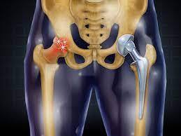
A hip replacement is a surgical procedure designed to alleviate severe hip pain and stiffness caused by hip arthritis, enhancing overall function. During this procedure, damaged portions of the hip joint are replaced with artificial implants.
Implants Used The implants utilized in hip replacement surgery consist of three crucial components:
The Stem: Crafted from metal, typically titanium or cobalt-chrome, it is inserted into the thigh bone for stability.
The Ball: Usually made of polished metal or ceramic, it securely rests on top of the stem.
The Socket: This component features a plastic liner combined with a cobalt-chrome or titanium backing.
Longevity of Hip Implants: Hip implants are designed to endure for approximately 13 to 15 years. Beyond this timeframe, patients may require a follow-up surgical procedure known as Hip revision surgery.
Reasons for Hip Replacement Surgery
Hip Replacement Surgery is most commonly performed on individuals aged 50 to 80 years old, often to address arthritis that has affected the hip joint.
Arthritis damages the cartilage, which acts as a cushion between bones, and when this cartilage is compromised, it results in bones rubbing against each other, leading to significant joint pain
Hip replacement surgery is typically used to treat
three main types of arthritis
Rheumatoid Arthritis
This is a chronic inflammatory disorder that can affect multiple joints, including those in the hands and feet.
Osteoarthritis
Osteoarthritis occurs when the flexible tissue at the ends of bones deteriorates, causing joint pain and stiffness.
Traumatic Arthritis
This type of arthritis results from joint damage caused by an injury or trauma.
Hip replacement might be recommended in situations where
Types of Hip Surgery
There are three main types of hip replacement surgeries, each suited for different situations
Total hip replacement, also known as total hip arthroplasty, is a common and effective procedure for alleviating hip pain, improving mobility, and restoring normal activities. In this surgery, the damaged hip bone and cartilage are removed and replaced with artificial components.
The damaged femoral head is replaced with a metal stem inserted into the femoral Canal and a metal or ceramic ball is attached to the upper part of the stem.
Damaged Cup is resurfaced and Reshaped into hemispherical shape and artificial component is attached. Two primary surgical approaches are used for total hip replacement: the more common posterior approach and the anterior approach.
Diagnosis Before Hip Replacement Surgery
Diagnosis is based on symptoms like pain, limited mobility, and daily life difficulties. Special tests like X-rays, CT scans, and MRI are used to assess the extent of damage. Hip Replacement Surgery Procedures:
Two methods: Traditional and Minimally-Invasive.
Anesthesia, incision, muscle dissection, removal of damaged joint, attachment of artificial joint, preparation of hipbone surface, reattachment of muscles, and closure.
Risks Associated with Hip Replacement Surgery
Potential risks include bleeding, infection, blood clots, nerve injury, fracture, and dislocation.
Recovery After Hip Replacement Surgery:
Why Choose Us for Hip Replacement Surgery at Gadge Hospital, Nagpur?
Total hip replacement is a widely performed and highly advantageous procedure. At Gadge Hospital, Nagpur, our surgical techniques evolve with the times. Dr. Swapnil Gadge, with over 8 years of experience, is one of the finest hip replacement surgeons in the region. He continually enhances his skills and remains at the forefront of the latest advancements in hip replacement surgery.
0 notes
Last Seen Blogs

pigmentosdecadmio
Pigmentos de Cadmio

catboymoments
nonbinary lemonade lover

waywardsan
San

dj-school
scratch-o hey!

neptunein1st
blow a wish