#endarch
Explore tagged Tumblr posts
Visit Tumblr Blog
Explore Tumblr blogs with no restrictions, modern design and the best experience.
Last Seen Tumblr Blogs


Fun Fact
Tumblr was acquired by Yahoo for $1.1B in 2013.
Text
IA Prep: Botany (sem 4)
Anomalous Secondary Growth ---
What is it?
Anomalous Secondary Growth is the growth resulting
Boerhaavia
A transverse section shows epidermis, collenchymatous hypodermis, parenchymatous cortex, endodermis and single layered pericycle.
The primary vascular bundle are conjoint collateral, endarch, and open.
These bundles occur in 3 concentric rings.
Inner most ring consists of 2 large vascular bundles present in the pith, opposite to each other.
Middle ring consists of 6-14 small vascular bundles.
The outer ring consists of 15-20 smaller vascular bundles, these are the normal vascular bundles.
Middle and inner rings of bundles are considered medullary bundles.
Secondary Growth
During secondary growth, the two central bundles and the middle ring of bundles increase in size due to the activity of fasicular cambium.
Inter-fasicular cambium do not develop.
The outer ring of vascular bundles are involved in the secondary growth.
The intra and interfasicular cambial strips join to form complete cambial ring.
This complete cambial ring forms the secondary xylem to the inner side and secondary phloem to towards the outside only in fasicualar region.
In between the bundles the cambium produces parenchymatous cells (thick walled) called conjuctive tissue.
After sometime this cambium ring stops activity.
Then a new cambial ring is formed external to the secondary phloem.
The cells in pericycle is involved in the formation of second cambial ring. This is called as accessory cambium.
This also produces secondary xylem to the inner side and secondary phloem to the outer side.
After a period of activity this cambium also stops its function. Another ring of cambium arises outside which behaves in the same pattern.
In this way 4-5 rings are produced and embedded in the conjuctive tissue.
Periderm- protective covering in outer stelar region.
Bignonia
Bignonia is a woody climber
A young stem shows ridges and furrows.
Epidermis is the outermost layer with thick cuticle.
Collenchyma is prominent below the ridges.
The cortex is made up of layer of parenchyma cells.
Endodermis is not very distinct
Pericycle is in the form of sclerenchymatous patches.
The vascular bundles are conjoint, collateral, endarch and open.
They are arranged in the form of ring around the periphery of the
pith.
Secondary Growth:
The inter and intra fasicular cambial strips become active to form a normal ring of cambium.
In the initial stages, the cambial behavior is normal, it makes more of secondary xylem and less secondary phloem.
Subsequently, at four corner points the cambium begins to produce more of secondary phloem and relatively small amount of secondary xylem.
Resulting in the formation of 4 deep wedges of phloem projecting into secondary xylem.
Initially the number of phloem furrows are 4 in number, but later additional furrows may be formed in older stems.
In this way, the xylem cylinder is broken into vertical plates, separated by furrows of secondary phloem.
Periderm is produced in cortex region due to the activity of phellogen.
---
Wood Structure
Teak
Scientific: Tectona grandis
Telugu: Adaviteeku
Hindi: Sagun
Family: Verbenaceae
One of the most important timber plants in the world, well known for both durability and strength
Occurrence:
- Native to India, Java, Sumatra, and Indonesia
- In India, teak forests mostly found in M.P, Maharashtra, Gujarat, and Karnataka.
- Other states include A.P, Rajasthan, U.P, Tamil Nadu, and Orissa.
Morphology:
- Tree height: 30m
- Girth: 2-4m
- Leaves: Opposite, simple, elliptical to obovate, 25-50cm long.
- Flowers: Small, arranged in panicles.
- Bractiate, Bisexual, and Hypogynous.
- Fruit: Hard, 4 lobed nut, 1-2 seeds.
Cultivation:
- Quick growing, temp requirement: 26-30˚C, annual rainfall: 130-300 cm.
- Growth is stunted in nitrogen deficient soil.
- Plantation time of seedlings: April-May.
- Good quality timber obtained from: 80 years old trees.
Prop of wood:
- Scented oil in wood acts as its preservative.
- Sapwood white in color, susceptible to termites and fungi
- Heartwood is golden yellow -(air)-> dark brown and resistant to fungal attacks.
- Ring is porous, presence of vessels.
- Tyloses are present.
Uses of Teak:
- Timber is durable, hard and resists decay, used in furniture and plywood.
- Used to make agricultural implements.
- Wood waste is raw material for paper pulp.
- Wood oil: used to treat eczema
- Flowers: relieves kidney problems
- Root bark: used for coloring mats.
Red Sanders
Scientific: Pterocarpus santalinus
Telugu: Erra chandanamu
Hindi: Lal chandan
Family: Fabaceae
This tree is valued for the rich red color of its wood, not aromatic.
Occurrence:
- Grows in southern India.
- Common in Cuddapah, Nellore and southern Kurnool districts.
Morphology:
- Grows in height to 10-11m.
- Leaves imparipinnate, leaflets pulvinous, 3 or rarely 5.
- Flowers bright yellow in short racemes.
- Pedicellate, Bisexual, and Zygomorphic.
- Flowering occurs during April-June
- Pods are winged with 1-2 seeds.
Props of wood:
- Bark is blackish-brown, wood is hard close grained.
- Differentiated into sap wood and heart wood. Sap wood is white and heart wood is black in color.
- Pores are medium sized and scattered
- Medullary rays are numerous and equidistant.
Uses of wood:
- Used in making agricultural implements.
- Gives a blood red colored die on distillation.
- Extensively used in carvings, statues, and picture frames.
Neem
Scientific: Azadirachta indica
Telugu: Yepa
Tamil: Vepa
Family: Meliaceae
Occurrence:
- Native to India, grows wildly.
- Also grows in Nepal, Bangladesh, Pakistan, and Sri Lanka.
Morphology:
- A fast growing tree which can reach a height of 15-20m.
- Evergreen, but in severe drought it may shed most or all of its leaves.
- Leaves: opposite pinnate with 20-30 medium dark green leaflets.
- White and fragrant flowers, arranged in more-or-less drooping axillary panicles.
- Each inflorescence has 150-250 flowers, they are protandrous and bisexual.
- Fruit: smooth olive-like drupe, it encloses one, rarely two or three,
elongated seeds (kernels)
Properties of wood:
- Bark: grey with numerous scattered tubercles.
- Sapwood is grey while heartwood is red.
- Pores are large/moderate sized and oval shaped; medium to coarse textured.
- Medullary rays are numerous and prominent.
- Wood is scented.
Uses of wood:
- Construction of cart axles and wheels, agricultural implements, and furniture.
- Bark contains tannins which are used in tanning, dyeing etc.
- Compounds extracted from the bark are used in production of some dental-care products like toothpaste.
- The bark acts as an insect repellent and it also has anti-bacterial
properties.
- Wood has been used as firewood and charcoal for a long time.
-------------------------------------------------
Stomatal Types ---
Stomata are minute pores which occur in the epidermis.
Stomata are used for the exchange of gases from the plant and the atmosphere, each stoma opens into a sub-stomatal chamber or a respiratory cavity to facilitate this function.
Evaporation of water takes place through the stomata.
Each stoma is surrounded by two kidneys shaped guard cells.
The stomata may occur on any part of a plant except the roots.
The epidermal cells bordering the guard cells are called accessory cells or subsidiary cells
There are 4 types of stomata based on the accessory cells and their arrangements:
- Anomocytic
- Anisocytic
- Paracytic
- Diacytic
Anomocytic Stomata:
Also called Type-A, Ranunculaceous type, or irregular-celled type stomata
No accessory cells are present
The stoma remains surrounded by ordinary epidermal cells which are arranged irregularly
Commonly found in dicotyledons such as Tridax.
picture
Anisocytic Stomata:
Also called Type-B, Cruciferous type, or unequal-celled type stomata.
The stomata is surrounded by three accessory cells.
Of the three accessory cells, one of them is distinctly smaller than the other two cells.
Commonly found in genera such as Brassica.
picture
Paracytic Stomata:
Also called Type-C, Rubiaceous type, or parallel-celled type stomata.
At least two accessory cells are present.
The accessory cells lie parallel to the long axis of the pore and guard cells
Commonly found in the members of Rubiaceae.
picture
Diacytic Stomata:
Also called Type-D, Caryophyllaceous type, or cross-celled type.
Each stoma is surrounded by a pair of accessory cells.
The common walls of each accessory cell remains at a right angle to the long axis of the guard cell.
Commonly found in Ocimum.
picture
There are two more types of stomata:
- Gramineous
- Coniferous
Gramineous Stomata:
The stoma have guard cells where in which the middle portions are much narrower than the ends, making the cell appear to be dumb-bell shaped
Commonly found in Gramineae
pic?
Coniferous Stomata:
The stomata are sunken and appear as if the accessory cells are suspended and arching over them.
In the median parts, the guard cells are have narrow lumina and the section is elliptical in shape.
At the ends, they lumina is wider and the section is triangular.
The walls of these guard cells and the accessory cells are partly lignified and partly non-lignified.
---
Simple Tissues ---
Simple permanent tissues are tissues which are composed of identical cells which together perform a common function.
Simple tissues can be divided into three groups:
- Parenchyma
- Collenchyma
- and Sclerenchyma.
=====
Parenchyma is made up of living cells.
Each cell is either spherical, ovular, rectangular, polygonal, elongated, or irregular in shape.
The cell wall is thin, made up of cellulose, hemicellulose, and pectin.
Young parenchyma cells are loosely arranged
There is intercellular space present in parenchyma
Parenchymatous cells can store reserve food material
Parenchymatous cells are found in all parts of plant such as cortex, pith, palisade, mesophyll, flower, seed, etc.
Parenchyma is also found in vascular tissues.
-
There are three types of parenchyma based on structural modifications and specialized functions:
- Prosenchyma
- Aerenchyma
- Chlorenchyma
Prosenchyma are thick-walled fiber-like elongated cells which provide rigidity and strength to the plant, ex: pericycle
Aerenchyma have large intercellular spaces filled with air, it helps in the buoyancy of the plant, ex: cortex of hydrophytes
Chlorenchyma are cells which have chloroplasts and perform photosynthesis, ex: palisade of leaves
-
Functions of parenchyma:
Photosynthesis - chlorenchyma has chloroplasts which perform photosynthesis
Storage - parenchyma can store food in the form of starch, proteins, oils, and fats
Buoyancy - aerenchyma helps aquatic plants float
Transportation - Parenchyma of xylem and phloem helps in the transport of nutrition and water
Mechanical support - prosenchyma provides mechanic support
diagram
=====
Collenchyma is a living tissue
The shape of each collenchyma cell is somewhat elongated
Collenchyma cells have a thick wall due to the deposition of hemicellulose and pectin in intercellular spaces
Intercellular spaces may or may not be present in collenchyma
-
There are three types of collenchyma based on the deposition of hemicellulose and pectin:
- Angular collenchyma
- Lacunar collenchyma
- Plate or lamellar collenchyma
Angular collenchyma have a thick cell wall at the corner of the cell, does not have intercellular space.
Lacunar collenchyma have a thick cell wall at the border of the cell, has intercellular space.
Plate or Lamellar Collenchyma have a thick cell wall at tangential wall, does not have intercellular space.
-
Functions of collenchyma:
Collenchyma provides mechanical support and elasticity to the stems of dicots.
Collenchyma has chloroplasts, therefore it can carry out photosynthesis.
Provides strength and flexibility to the plant body.
diagram
=====
Sclerenchyma is made up of dead cells.
The shape of sclerenchyma cells are elongated and pointed at both ends.
The cell wall of sclerenchyma is thick and lignified, it encloses an empty cavity called 'Lumen'
Sclerenchyma cells lack protoplasm.
Sclerenchyma provides strength and rigidity to the plant body.
-
There are two types of sclerenchyma:
- Fibers
- Sclereids
Fibers are thick walled, elongates, spindle shaped dead cells with pointed ends.
Cell walls enclose narrow lumen with simple round pits and lignified secondary wall.
Present in xylem, phloem, cortex and pericycle.
They made up the covering of fruits.
Fibers provide mechanical support.
There are three types of fibers:
- Surface Fibers: On fruit wall + seed coat (ex: coconut)
- Wood fibers: associated w xylem
- Bast fibers: associated w phloem, cortex, and pericycle.
Sclereids are very thick walled cells with either spherical, ovular, or dumbbell shape.
They are also called stone cells.
Sclereid cell wall has simple pits.
Present in hard part of plants and the pulp of fruit.
Provide local mechanical support.
5 Types based on shape:
- Stone cells: Isodiametric, similar to parenchyma in shape.
- Macrosclereids: Rod like shape
- Osteo sclereids: Bone like shape
- Astro sclereids: Star like shape
- Filiform sclereids: Hair like elongated cell with branches
-
Functions of Sclerenchyma:
Sclerenchyma is made up of dead and lignified cells which provides mechanical support.
Provides hardness to stony fruits like nuts, coconut, almond etc.
---
Complex Tissues ---
Complex permanent tissues are composed of 2 or more types of cells
All the cells contribute to a common function.
They are aka vascular tissues.
Vascular tissues are primarily associated with conduction of water, minerals, and food in plants.
There are 2 types of vascular tissues: xylem and phloem
===
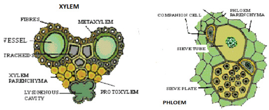
Xylem
Primary function of xylem is to conduct water and minerals from the roots to leaves.
Additional function involves providing mechanical support. (secondary xylem)
-
Two types of xylem: primary and secondary.
Primary xylem:
- Xylem in primary plant body
- Developed from pro-cambium
- Organized as bundles along with phloem
- Main function: water and mineral conduction
- 2 types: protoxylem + metaxylem
Secondary xylem:
- Xylem in secondary plant body
- Developed from vascular cambium
- Main function is the same
- Additional function: provide mechanical support.
-
Elements of Xylem: Tracheids, Vessels, Xylem fibers, and Xylem parenchyma.
Tracheids:
- Elongated tube like cells occurring along the long axis of the plant.
- Cells are devoid of protoplast, and hence is a dead component.
- Has a cavity, an empty lumen, and tapering ends.
- Round or polyhedral in cross-section.
- Average cell length is 5-6mm
- The most primitive and fundamental element in xylem element, found in the fossils of seed-plants.
- In modern plants they occur predominately in lower vascular plants, pteridophytes and gymnosperms
- Has secondary cell wall which is hard, thick and extremely lignified.
- Secondary walls are deposited in different patterns:
- annular: rings, most primitive
- spiral: helical spirals
- scalariform: ladder like
- reticulate: net like
- pitted: pits evenly distributed all over
Vessels:
- Aka trachea
- short dead cells devoid of protoplast
- have hard and lignified cell-wall
- forms a row of cylindrical cells arranged in longitudinal series like a tube.
- Each cell has perforation at the end walls, rarely occur on the lateral walls
- Distinct ‘perforate’ bodies makes translocation of solutes easy.
- Perforations remain either mostly in parallel series like bars called scalariform perforation, or in a network known as reticulate perforation, or even may form a group of circular holes called foraminate perforation.
- The perforation occurs in form of a single large circle, referred to as simple perforation
- Has various types of secondary thickenings such as:
- annular, spiral, scalariform, reticulate or pitted.
- The pits are mostly of bordered types.
- Present in angiosperms, usually absent in pteridophytes and gymnosperms.
- Does not occur in some parasitic and aquatic plants.
Xylem Fibers:
- Also called xylary fibers
- Fibers are very much elongated, dead cells with thick lignified cell walls.
- Primarily give mechanical support.
- Two types: fiber traeheids and libriform fibers.
- Fiber tracheids are intermediate forms between fibers and tracheids, have bordered pits.
- Libriform fibers are narrow with highly thickened secondary wall, have simple pits.
- Gelatinous fibers in tension wood are a type of xylem fiber.
Xylem Parenchyma:
- Living cells w cytoplasm + prominent nucleus
- Cells are thin-walled, lignified secondary wall absent.
- Parenchyma in secondary xylem rarely undergoes secondary growth.
- Two types: Axial parenchyma + Ray parenchyma
- Meant for storage of starch and fatty food, tannins, crystals, etc.
- They are involved in conduction of water and solutes and mechanical support.
===
Phloem
Primary function is to transport food to the different parts of the plant body
2 types of phloem: Primary (dev from pro-cambium during 1' growth) + secondary (dev from vascular cambium during 2' growth).
4 elements: Sieve elements, companion cells, phloem parenchyma, phloem fibers and sclereids.
-
Sieve Elements: divides into sieve tubes and sieve cells.
Sieve Cells:
- Less specialized and more primitive than tubes
- Occur in lower vascular plants and gymnosperms
- Narrow elongated cells with steeply inclined end walls
- Sieve areas are located on the lateral walls and very rarely at the end walls.
Sieve Tubes:
- More specialized and advanced than cells.
- Occurs in angiosperms
- Long tube-like structures formed arranged in longitudinal series.
- end-walls of Sieve tube are perforated in a sieve-like manner.
- The perforated end-walls are called the sieve plates.
- Through sieve plates, cytoplasm connections are established between adjacent cells.
Companion Cells:
- Occurs abundantly in angiosperms, particularly in the monocotyledons, absent in some primitive dicotyledons.
- Pro-cambial mother cell divides longitudinally into two daughter cells, one of which serves as the sieve element and the other one becomes the companion cell.
- Hence companion cell is considered as sister cell to sieve tube.
- Companion cells remains associated with the sieve tubes of angiosperms
- These are smaller cells, having dense cytoplasm and prominent nuclei without starch grains.
- The wall between the sieve tube and companion cell is thin and provided with primary pit fields.
- Function: controls the activity of sieve tube.
Phloem Parenchyma:
- Occurs in both primary and secondary phloem
- Thin walled living cells
- Have primary pit fields
- Some parenchyma cells store starch.
- Two types: Axial and Ray parenchyma
Phloem Sclerenchyma:
- Aka Phloem fibers or Bast fibers
- Sclerenchyma of phloem
- Dead cells with lignified cell wall
- Have simple or slightly bordered pits.
- Septate fibers occurs in Vitis
- In some plants, phloem fibers are very long.
- rare in pteridophytes and some spermatophytes
- Provides mechanical strength to the plant.
- Used for the manufacture of ropes and cords.
2 notes
·
View notes
Photo

New Post has been published on http://ramneetkaur.com/vascular-tissue-system/
Vascular tissue system

Vascular tissue system
Constitute the vascular bundles.
Procambial strands of plerome of apical meristem forms the vascular tissue.
It consists of xylem & phloem.
Xylem(wood) is made of tracheids, vessels, xylem parenchyma & xylem fibres.
Phoem(bast) is made of sieve elements, companion cells, phloem parenchyma & phloem fibres.
Early formed xylem & phloem are referred as protoxylem & protophloem.
Later formed are referred as metaxylem & metaphloem.
VASCULAR BUNDLES:
Are of 2 types:
RADIAL:
Xylem & phloem form separate bundles present on different radii.
Xylem is exarch i.e., protoxylem towards periphery & metaxylem towards the center.
Found in roots.
CONJOINT:
Xylem & phloem together form a bundle.
Xylem is endarch i.e., protoxylem towards center & metaxylem towards the periphery.
Found in stem & leaves.
Conjoint vascular bundles are of 3 types:
Collateral: xylem towards inner side & phloem towards outer side.
It’s of 2 types: open & closed.
Open: cambium is present in between xylem & phloem. Seen in dicot stem.
Closed: cambium is not present between xylem & phloem.
Seen in monocot stem.
Bicollateral: cambium & phloem are present on both sides of xylem. Seen in family Cucurbitaceae, Solanaceae.
Concentric: in this one surrounds the other.
It’s of 2 types:
Amphivasal: xylem surrounds phloem. E.g., Yucca, Dracena, Aloe.
Amphicribal: phloem surrounds xylem. E.g., ferns.

STELE: it is the region inner to the endodermis which includes the vascular tissue, xylem & phloem. Stele originated & evolved in Pteridophytes.
Different types of stele are:
Protostele: most primitive stele. It has an inner core of xylem surrounded by phloem.
Siphonostele: it is protostele with central pith.
Dictyostele: the circular stele breaks into smaller parts due to the formation of leaf gaps.
Eustele: vascular tissue form vascular bundles, and they are arranged in rings. E.g., dicot stem.
Atactostele: vascular tissue exists as vascular bundles that lie scattered. E.g., monocot stem.

#aipmt#aipmt 2017#Aloe#amphivasal#ampnhicribal#atactostele#bicollateral#biology#biology mnemonic#closed vascular bundles#collateral#concentric#conjoint vascular bundles#dicot stem#dictyostele#dracena#endarch#eustele#exrch#MCAT#metaxylem#monocot stem#neet#neet 2017#neet biology#NEET MCQs#neet mnemonic#NEET questions#neet ug#open vascular bundles
0 notes
Photo

endarch tapetis by Jared Haer Tempests Unresistedness Study #picoftheday #color #paint #instamood #iphonesia #GIMP
0 notes
Text
Mod 3: Gymnosperms
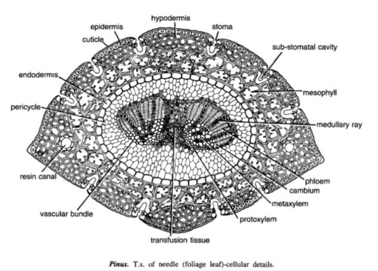
Pinus Needle T.S.
It is circular in outline in P. monophylla, semicircular in P. sylvestris and triangular in P. longifolia, P. roxburghii, etc.
Outermost layer is epidermis, which consists of thick-walled cells. It is covered by a very strong cuticle.
Many sunken stomata are present on the epidermis.
Each stomata opens internally into a substomatal cavity and externally into a respiratory cavity or vestibule.
Below the epidermis are present a few layers of thick-walled sclerenchymatous hypodermis. It is well developed at ridges
In between the hypodermis and endodermis is present the mesophyll tissue.
Cells of the mesophyll are polygonal and filled with chloroplasts. Many peg-like infoldings of cellulose also arise from the inner side of the wall of mesophyll cells.
Few resin canals are present in the mesophyll, adjoining the hypodermis. Their number is variable but generally they are two in number.
Endodermis is single-layered with barrel-shaped cells and clear casparian strips.
Pericycle is multilayered and consists of mainly parenchymatous cells and some sclerenchymatous cells forming T-shaped girder, which separates two vascular bundles. Transfusion tissue consists of tracheidial cells.
Two conjoint and collateral vascular bundles are present in the center. These are closed but cambium may also be present in the sections passing through the base of the needle.
Xylem lies towards the angular side and the phloem towards the convex side of the needle.
Idk where to find xeric nature info
Williamsoniaceae
Occurrence of Williamsonia
Williamsonia belongs to family Williamsoniaceae of Bennettiales.
It has been reported from Upper Triassic period but was more abundant in Jurassic.
This was earlier discovered under the name Zamia gigas by Willamson in 1870 but has now been named as Williamsonia.
Professor Birbal Sanhi (1932) described W. sewardiana from Rajmahal Hills of Bihar (India).
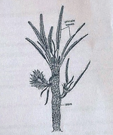
External Features of Williamsonia
Williamsonia resembled Cycas in appearance, but its best-knows species is W. sewardiana. The plant had an upright, branched, and stout stem covered by persistent leaf bases.
A terminal crown of pinnately compound leaves was present. For the stem genus Bucklandia, Sharma (1991) opined that features of leaf bases such as their shape, size and arrangement pattern are of taxonomic significance.
He observed that leaves in Williamsoniaceae show syndetocheilic stomata with rachis possessing collateral endarch vascular bundles.
Reproduction in Williamsonia
The fructifications of Williamsonia were large and attained a diameter of about 12 cm.
They were borne on a peduncle.
Many spirally arranged bracts were present around the base of the floral axis.
In W. gigas the cones were present among the crown of leaf bases while in W. sewardinia they were present on the short lateral branches.
Williamsonia plants were unisexual.
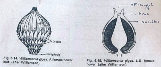
Female Flower
The female 'cones' of W. gigas and W. sewardiana have been investigated in detail. Instead of 'strobili' or 'cones', Sporne (1965) proposed to use the term 'flower'.
The conical receptacle was surrounded by many perianth-like bracts. The ovules were stalked.
The apex of the receptacle was naked and sterile. The nucellus was surrounded by a single vascularize integument, which was fused with the nucellus. The nucellus had a well-marked beak and a pollen chamber. In young ovules the micropylar canal was long and narrow.
In mature ovules, the canal widened because of the formation of nucellar plug and disappearance of interlocking cells. In the apical part of the endosperm, Sharma (1979) observed 2 or more archegonia.
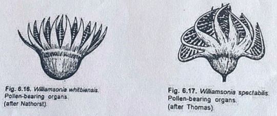
Male Flower
Male flowers consisted of a whorl of microsporophyll's which were united to form a more or less cuplike structure. In majority of the investigates species the sporophylls were un-branched but in some species they were also pinnately branched.
Sitholey and Bose discovered W. santalensis from Upper Gondwana, and observed that microsporophyll's in the species were bifid.
One of the branches of microsporophyll was fertile while the other was sterile. The fertile part has finger-like structures called synangia. Each synangium had two rows of chambers enclosing microsporangia.
The fertile branch of the bifid sporophyll possessed many purse-like capsules, in each of which there were present many monocolpate pollen grains.
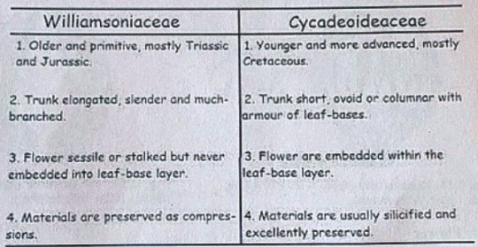
Cycadeoideaceae
Classification:
Division - Cycadeoidophyta
Order - Cycadeoideales
Family - Cycadeoideaceae
Genus - Cycadeoidea
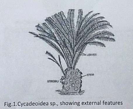
Introduction:
Cycadeoidea is the only genus of family Cycadeoidaceae, represented by thirty species. They are entirely extinct and resemble cycads in the outward stumpy appearance of trunk and an apical crown of pinnate compound leaves. This fossil group of plants flourished during the Triassic to Cretaceous periods of the Mesozoic era. They are reported from various places in the world, in India the Cycadeoidales are found in Rajmahal Hills in Bihar. The petrified trunks of C. entrusca are the oldest fossil ever collected by man.
External Features:
The genus Cycadeoidea had a short, branched, or unbranched spherical, conical, or irregular trunk. The diameter of the trunk is 50cm and the highest rarely reached a meter except in C. jenneyana, it attended the height of several meters. These trunks are covered by rhomboidal leaf bases having multicellular hairs in between. Crown of 10ft long pinnate compound leaves are present at the top.
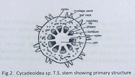
Anatomy of Stem:
The transverse section of the stem shows roughly a circular outline. The epidermis is not very distinct due to the presence of heavy armor of leaf bases. The cortex is parenchymatous and traversed by mucilage canals and numerous leaf traces. The primary vascular structure consists of a ring of endarch, collateral, conjoint, and open vascular bundles encircling the pith. Pith is wide and parenchymatous. A ray-like extension passes between the vascular bundles that make their appearance discrete.
There is a cambium ring with a thin zone of secondary wood. The secondary wood encircles the primary xylem and consists of tracheids with scalariform and bordered pits. The secondary medullary rays traverse the secondary xylem and secondary phloem.
The C-shaped leaf traces arise singly from the primary vascular strand and entering the cortex divided into several masarch strands and enters straight into the leaf.
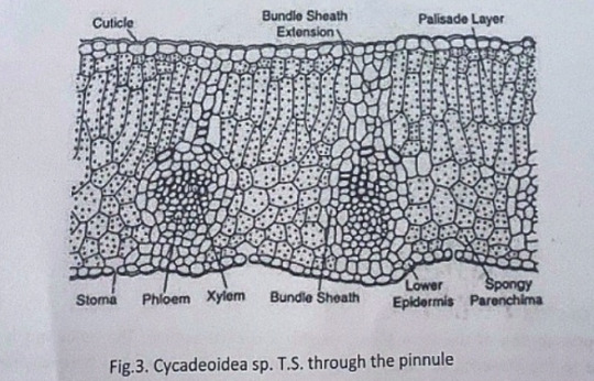
Anatomy of Leaf:
The pinnules show xerophilous structure. The upper and lower epidermis is heavily cutinized and thick walled. The mesophyll cells are distinguished into palisade and spongy parenchyma. The vascular bundles are mesarch and surrounded by bundle sheath.
Reproduction:
The reproductive structure is represented by flowers. In most of the species, the flowers are bisexual and arise in the axil of each leaf.
Structure of Flower:
The flowers are bisporangiate, stalked, and partially sunken in the leaf base armor. Rach such mature flower is 5-10cm in diameter and 10cm long. From the base of such flowers about 100 to 150 hairy bracts arise in close spiral little below the apex. These bracts formed a perianth like structure and protect the megasporangiate and microsporangiate parts of a flower. The microsporophyll or androecium forms a whorl united at the base into a sheath. The megasporophyll or gynaecium consists of numerous stalked ovules born around a central receptacle. Between the ovules, interseminal scales with expanded tips are present. These expanded tips fused to form a continuous surface with pores, through which the micropyle of ovules extended. The vascular supply of flowers consists of many branches from leaf traces.
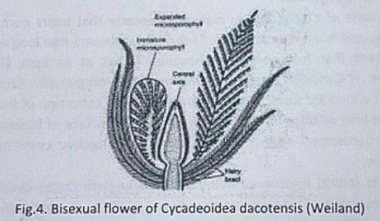
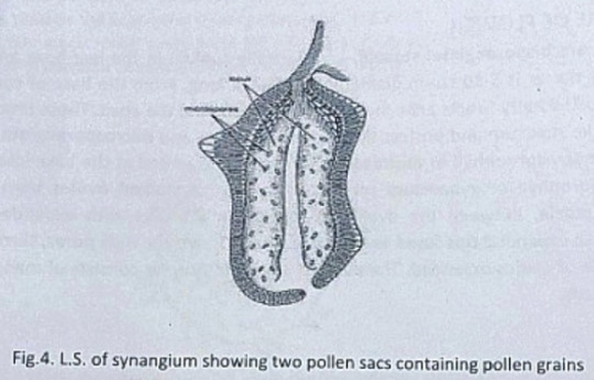
Microsporophyll or Androecium:
The microsporophyll is 10-12cm long, consists of a central rachis bearing numerous pinnae. The pinnae bear two rows of bean-shaped shortly stalked pollen capsules or synangia. These pollen capsules are born on the trabeculae within the fertile region of microsporophyll. A line of dehiscence is also visible at the base of each microsporophyll. This suggest that the entire microsporophyll might have been shed as a unit. The pollen capsule or synangia measures about 3.5x2.5mm and its wall is several layers thick, the outer layer made up of palisade like cells, and the inner layer is made up of thin-walled cells followed by a tapetum. The tapetum was not demarcated. A ring of microsporangia arranged around the periphery of each synangium. The microsporangia dehisce longitudinally and release the microspores into the synangial cavity. At maturity, the synangia liberate these microspores outside by an apical opening that splits into two valves. The liberated microspores or pollens are oval, measures up to 68µ that represents the male gametophytes. Pollen grains of Cycadeoidea are multicellular.
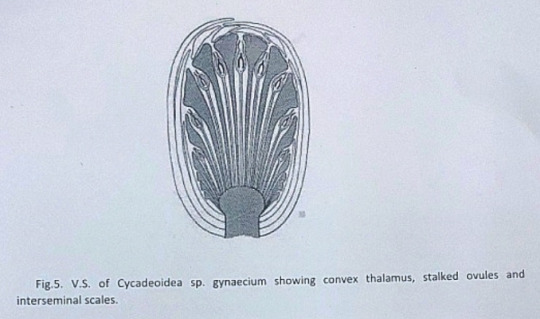
Megasporophyll or Gynoecium
The gynoecium consists of a spherical or conical receptacle that bears numerous stalked orthotropous ovules and interseminal scales. Each ovule is about 1mm long and consists of the single integument that fused with the nucellus except at its apex.
According to Lignier, in C. morieri, nucellus is free from the integument. Each ovule has a pollen chamber and a nucellar beak. This nucellar beak is the extension of the integument. The ovules also have long micropyle, extended from the flat surface of interseminal scales. The fused tips of interseminal scales form an external protective covering or pericarp surrounding the seeds.
Crepet and Delevoryas discovered many of bisporangiate cones from the Cretaceous of black hills. They studied the structure of these ovules in detail. These ovules are urn-shaped and resemble with the ovules of C. wellsii. According to them the micropyle of these ovules are funnel-shaped due to the constriction below the flaring. The inner wall of the micropyle is lined with large cells, considered to be epidermal cells. The integument has three distinct layers. The outer fleshy layer of radially elongated cells, the middle stony layer made up of thick-walled cells, and the inner layer is fleshy.
The young nucellus is made up of thin-walled cells. The cells at the micropylar end are much elongated (80µ long) in comparison to the cells of the chalazal end. The cell at the nucellar tip is pointed up tp whereas cells on either side are bend outward to give the nucellus a distinct shape.
Crepet and Delevoryas reported a linear tetrad or row of three cells in the center of the nucellus.
The seeds are somewhat elongated or oval and possessed two cotyledons.
#exam season#send help#biology#notes#science#botany#gymnosperms#pines#pine trees#pine needles#plants#plant science#plant biology#nature#long post
8 notes
·
View notes
Photo

New Post has been published on http://ramneetkaur.com/vascular-tissue-system/
Vascular tissue system

Vascular tissue system
Constitute the vascular bundles.
Procambial strands of plerome of apical meristem forms the vascular tissue.
It consists of xylem & phloem.
Xylem(wood) is made of tracheids, vessels, xylem parenchyma & xylem fibres.
Phoem(bast) is made of sieve elements, companion cells, phloem parenchyma & phloem fibres.
Early formed xylem & phloem are referred as protoxylem & protophloem.
Later formed are referred as metaxylem & metaphloem.

VASCULAR BUNDLES:
Are of 2 types:
RADIAL:
Xylem & phloem form separate bundles present on different radii.
Xylem is exarch i.e., protoxylem towards periphery & metaxylem towards the center.
Found in roots.
CONJOINT:
Xylem & phloem together form a bundle.
Xylem is endarch i.e., protoxylem towards center & metaxylem towards the periphery.
Found in stem & leaves.
Conjoint vascular bundles are of 3 types:
Collateral: xylem towards inner side & phloem towards outer side.
It’s of 2 types: open & closed.
Open: cambium is present in between xylem & phloem. Seen in dicot stem.
Closed: cambium is not present between xylem & phloem.
Seen in monocot stem.
Bicollateral: cambium & phloem are present on both sides of xylem. Seen in family Cucurbitaceae, Solanaceae.
Concentric: in this one surrounds the other.
It’s of 2 types:
Amphivasal: xylem surrounds phloem. E.g., Yucca, Dracena, Aloe.
Amphicribal: phloem surrounds xylem. E.g., ferns.

STELE: it is the region inner to the endodermis which includes the vascular tissue, xylem & phloem. Stele originated & evolved in Pteridophytes.
Different types of stele are:
Protostele: most primitive stele. It has an inner core of xylem surrounded by phloem.
Siphonostele: it is protostele with central pith.
Dictyostele: the circular stele breaks into smaller parts due to the formation of leaf gaps.
Eustele: vascular tissue form vascular bundles, and they are arranged in rings. E.g., dicot stem.
Atactostele: vascular tissue exists as vascular bundles that lie scattered. E.g., monocot stem.

#aipmt#aipmt 2017#Aloe#amphivasal#ampnhicribal#atactostele#bicollateral#biology#biology mnemonic#closed vascular bundles#collateral#concentric#conjoint vascular bundles#dicot stem#dictyostele#dracena#endarch#eustele#exrch#MCAT#metaxylem#monocot stem#neet#neet 2017#neet biology#NEET MCQs#neet mnemonic#NEET questions#neet ug#open vascular bundles
0 notes