#hydronephrosis in child treatment delhi
Text
Hydronephrosis Treatment In Delhi
Many people have never heard of the term hydronephrosis. That’s because it is only prevalent in around 1% of the general population according to a research paper published by Science Direct. Hydronephrosis can affect both children and adults. In fact, it can even affect babies in the womb; this can be found via prenatal ultrasound. The same study by Science Direct observed that 1 in 100 to 200 fetuses suffered from hydronephrosis. Because of this, finding out that you suffer from hydronephrosis and require surgery might seem daunting. But don’t worry about it. You can find hydronephrosis treatment in Delhi without breaking a sweat!
What exactly is it?
Hydronephrosis is a condition wherein one or both of the kidneys swell up. This happens either because of some blockage in drainage system of urineor urine refluxing back in the kidneys which can eventually damage the kidney of child.
Therefore, it is essential that you consult doctor once hydronephrosis has been diagnosed. Once the diagnosis is made child needs to be evaluated in detail. Not all hydronephrosis requires surgical intervention but needs to be monitored closely to avoid any renal damage.
Signs of Hydronephrosis
Here are some of the most commonly known signs and symptoms of hydronephrosis.
Antenatal diagnosis on ultrasound scan.
Urinary tract infection
Pain and lump in the back and the sides.
Urinary symptoms like frequent urination, crying during urination etc.
These signs are particularly useful to suspect hydronephrosis in children. Infants, in particular, can have failure to thrive. If you have suspicion that your child may be suffering from hydronephrosis or has all the signs mentioned above, it might be a good idea to consult a doctor.
Causes of Hydronephrosis
As mentioned above, hydronephrosis is a condition that prevents urine draining from the kidneys, which causes the kidneys to swell up. Hydronephrosis usually develops because of two main causes:
Obstruction in Urinary System
Blockage in the upper ureter (Pelvi-ureteric Junction) or lower ureter (Uretero-vesical junction) or Bladder oulet (Posterior urethral valve) can cause hydronephrosis on one side or both sides.
One of the commonest cause is blockage at ureteropelvic junction. This is essentially the very point (or junction) where the ureter and kidney meet.
Posterior Urethral valves are seen in boys and usually causes bilateral hydronephrosis. This is treated by endoscopic resection surgery. If not treated timely it can cause significant morbidity.
Vesicoureteral Reflux
Another cause of hydronephrosis is the vesicoureteral reflux where the urine flows backward from the bladder to the kidneys via the ureter. This condition is unique because usually the urine should only flow from the kidneys to the bladder- not the other way around.
Hydronephrosis treatment in Delhi
If you’re looking for hydronephrosis treatment in Delhi, then you’ll be happy to know that there’s plenty of options available. Your doctor who, after examination, ask for a few tests. This may include the following:
Blood test
Urine test
Ultrasound Imaging
Voiding Cystourethrogram
Renal Scan (DTPA or DMSA scan)
Combined, these tests examine your kidneys, bladder, Urethra and checks if they’re working fine. The kind of treatment you’ll receive for hydronephrosis depends strictly on how severe the condition is.
Some of the causes are self-limiting and may need just close observation with regular testings. Hydronephrosis causing recurrent urinary infections or deterioration of renal functions might require surgical intervention.
However, we do not recommend that you go with this approach as it can even lead to your mild case developing into a severe case of hydronephrosis, which will need surgery. Hydronephrosis surgery cost depends solely on how critical the situation is.
However, you should not look at hydronephrosis surgery costs when looking to treat the disease. As we mentioned, it’s not life-threatening at the same time living with hydronephrosis can severely impact your quality of living. So don’t wait it out!
#cause of hydronephrosis#hydronephrosis surgury#hydronephrosis treatment in delhi#signs of hydronephrosis
1 note
·
View note
Text
Expertise - Pediatric Nephrotic syndrome, Renal dysplasia, Chronic Kidney disease, Neurogenic bladder - Pediatricnephrologyindia
Dr. Sethi at Medanta, The Medicity is the leading Pediatric Nephrologist providing diagnostic and treatment services for children with conditions of the kidney especially Nephrotic syndrome, Glomerular disorders, rare tubular disorders and Chronic Kidney disease. Our team is the leading dialysis and renal transplant service centre for children. Dr. Sethi & team believes in personalized care that focuses on individual patient and family needs. Our team understands the challenges many of our families face when trying to access the care they need. Our caring goes beyond diagnostics and treatment. Our team is nationally recognized for their innovation, experience, dedication and expertise. We have the newest dialysis technology inpatient and outpatient, and provides comprehensive kidney transplant care to children of all ages.
Dr. Sethi leads in care of children with the following disorders:
Pediatric Nephrotic syndrome
Complex recurrent urinary tract disorders
Glomerular disorders
Blood or protein in the urine
Glomerulonephritis
Hemolytic uremic syndrome
Hydronephrosis
Hypertension
Rare tubular disorders
Polycystic kidney diseases
Neurogenic bladder
Renal dysplasia
Renal tubular acidosis
Systemic lupus erythematosus
Vesicoureteral reflux
Acute Kidney Injury
Chronic Kidney disease
Kidney Transplantation- Blood group compatible and incompatible; transplantation in complex renal anomalies
Tags - Nephrotic syndrome Specialist in Delhi, Child Kidney Specialist in Gurgaon, Pediatric Nephrology India
For more information link - www.pediatricnephrologyindia.com
#Nephrotic syndrome Specialist in Delhi#Child Kidney Doctor in Gurgaon#Pediatric Nephrology India#Dr. Sidharth Kumar Sethi
0 notes
Text
Hypospadias Surgery In Delhi - Dr. Prashant Jain
Hypospadias Surgery
Hypospadias is a birth defect in boys in which the opening of the urethra is not located at the tip of penis. In these cases the tube that carries urine from the bladder to outside of the body or the urethra does not end at the tip of the penis, rather ends on the underside of the penis. It can me mild or severe, in severe cases , the urethra opens at the middle of the penis or in some cases even behind the scrotum.
The treatment is aimed to facilitate the following.
Urine should pass in a forward way
The penis should be straight when erect
The penis should look as normal as possible
The surgery for this condition is known as Urethroplasty. If chordee is present then this is corrected to straighten the penis. The success of the operation and the ‘normality’ that can be achieved depends on the severity of the hypospadias.
Why is Hypospadias Surgery needed?
Hypospadias Surgery is performed generally when boys are 1 Year to 2 years old. If the hypospadias is mild, with the opening of the urethra just a little down from normal and with no bending of the penis, in that case treatment may not be needed. However, in the most cases an operation is required to correct the hypospadias. This can usually be done in one operation. However, if the hypospadias is more complicated, two operations may be necessary.
Preparation before the Surgery:
As mentioned earlier, the surgery isn’t a major one and does not require very specific pre-surgery preparations. The child’s health care provider may ask for the medical history of the child and suggest for a complete medical checkup before the surgery.
The procedure of the surgery:
The procedure is pretty simple and direct. The tissue extracted from the foreskin or some other part is used to make a tube. This tube is used to increase the length of the urethra. The extension is done so that the urethra opens at the tip of the penis. In many cases, during the surgery, an external tube called a Catheter is sewn or fastened to the head of the penis. This is done to keep it in place and help it retain a proper shape. It is usually removed after 1 – 2 weeks of the surgery. In many cases of Hypospadias, boys have a non-uniform or underdeveloped foreskin. This foreskin is circumcised at the end of the surgery. Most of the stitches used in this surgery are of a special type that dissolves on their own after the surgery and need not be removed manually.
Post surgery Care:
After the surgery, the child’s penis is taped to his belly so that there is a minimal movement to prevent any casualties. Often, the penis is protected by doing bulky dressing around the surgical area. A urinary Catheter is placed so that the urine can flow out of the dressed area into the diaper. The child is encouraged to drink fluid so that he will urinate more, this prevents pressure from building up in the urethra. The child is given medicines to help relieve the pain. In most of the cases, the child is discharged within a day. A urinary catheter may be needed for 6-14 days after the surgery. The penis of your child may be seen swollen and bruised but it’s nothing to worry about. The whole healing process takes around 6-7 weeks.
What are the risks of Hypospadias repair ?
All surgery carries a small risk of bleeding during or after the operation. For about one in ten boy, the original hole opens up again (fistula), and the patient passes urine through two holes. If this happens, the patient will need the operation again.
Tag =hydronephrosis in child treatment delhi, best pediatric urologist in delhi,best pediatric urologist in india, Undescended Testis In Children,
#hydronephrosis in child treatment delhi#best pediatric urologist in delhi#best pediatric urologist in india#Undescended Testis In Children
1 note
·
View note
Text
Antenatal Hydronephrosis, Hydronephrosis in Child Treatment, Delhi - Dr. Prashant Jain
Antenatal Hydronephrosis
Introduction:
With easy availability of ultrasound screening and improvement in expertise, hydronephrosis is now a very frequently diagnosed problem reported in 1 to 5% of all pregnancies. This has enabled us to have a better understanding of the natural course of the problem and early intervention before it results in permanent renal damage.
The distinction between urinary tract obstruction and dilatation remains a challenging problem for clinicians. Still there are no definite guidelines and protocols for evaluation of antenatal hydronephrosis (ANH).
Defining antenatal Hydronephrosis and grading of hydronephrosis:
Currently, the measurement of Antero-posterior diameter (APD) of the renal pelvis as visualized in transverse plane on ultrasound scan (USG) is most commonly used objective parameter for assessing ANH in utero and during post natal follow up. The measurement allows to classify the severity of hydronephrosis into mild, moderate and severe type. There is near uniform agreement that value of 4-5 mm is considered as cut off for abnormal APD. A value of more than 15 mm is considered as severe hydronephrosis. The severity of ANH can be graded in second and third trimester on the basis of APD.The mild form of hydronephrosis is seen in about 50 to 90 percent of cases while severe form is seen in 2 to 15 % of cases.
Relation of APD with Post natal Renal pathology:
A definite relation has been seen between the increasing severity of hydronephrosis and the risk of post natal pathology requiring some form of intervention. Based on the large systemic review of the current literature, the risk of postnatal pathology is 11.9% for mild, 45.1 % for moderate and 88.3 % for severe hydronephrosis.
Etiology of Antenatal Hydronephrosis:
Most children with antenatal history of renal pelvis dilatation ultimately resolve their hydronephrosis. The etiology of this finding may be related to a narrowing of ureteropelvic junction (UPJ) or natural kinks and folds that occur early in development that resolve as the patient matures. The differentiation between transient hydronephrosis versus clinically significant UPJ obstruction remains one of the most controversial challenges in modern pediatric urology. Nevertheless the incidence of transient hydronephrosis ranges from 41 to 88%. Most children with a pelvic dilatation less than 6mm diagnosed during the 2nd trimester or less than 8 mm diagnosed during the 3rd trimester have transient hydronephrosis.
Transient Hydronephrosis:
Majority of the children with history of ANH will show improvement. The etiology of this finding may be related to a narrowing of the uretero-pelvic junction (UPJ) or natural kinks and folds that occur early in development that resolve as the patient matures. The differentiation of transient hydronephrosis from pathological hydronephrosis is always a clinical challenge. The incidence will vary from 41 to 88%.
Most children with pelvic dilatation of less than 6 mm diagnosed during the 2nd trimester or less than 8 mm diagnosed during 3rd trimester have transient hydronephrosis. In contrast, the incidence of transient hydronephrosis is only 40% in children with an APD less than 10-12mm detected during the 3rd trimester.
Uretero Pelvic Junction Obstruction(UPJ):
Dilatation of pelvi-calyceal system without ureteral dilatation should raise the suspicion of UPJ obstruction. These are the patients with moderate to severe hydronephrosis which will show deterioration on USG and renal scans and then require surgical intervention. The challenge is to pick up the problem and intervene early before it causes permanent renal impairment.
Vesicoureteric Reflux(VUR):
The finding of variable dilatation on USG and the presence of hydroureter should the suspicion of VUR.Especially infants with UTI and older children with recurrent UTI should be investigated to rule out the possibility of VUR.
Ureterovesical junction obstruction/Megaureters
This is a problem associated with grossly dilated ureters but most of these (72%) will resolve on follow up. MCU study will rule the possibility of reflux in these cases.
Posterior Urethral Valve:
The findings of bilateral hydronephrosis, dilated ureters, thickened dilated bladder that fails to empty and dilated posterior urethra on antenatal scan should raise the suspicion of posterior urethral valves. This is a problem which can have high morbidity and poor prognosis depending on the severity of renal damage. One should have very high suspicion of this problem, so that earliest intervention to relieve the obstruction can be done.
Prenatal Evaluation of ANH:
The evaluation will depend on the gestational age, severity of dilatation, bilaterality, dilatation of ureter, amniotic fluid index and suspicion of posterior urethral valve.
The suspicion of posterior urethral valve warrants monitoring throughout the pregnancy. In such cases, in the presence of oligohydramnios fetal interventions such as vesicoamniotic shunt may be offered.
Imaging Modalities in evaluation of ANH:
Renal/Bladder Ultrasound:
This is most commonly used modality because of its easy availability and absence of radiation.It gives god anatomical details and is used to monitor the parenchymal status and the status of dilatation during follow up.
Its limitation being it does not give objective information about the functional status of kidney and is a poor independent predictor of those patients that will need surgical intervention.
Voiding Cystourethrogram(VCUG):
This test gives you information about the lower urinary tract and is important to rule out vesicoureteric reflux and posterior urethral valve. It is usually indicated in moderate to severe hydronephrosis, mild hydronephrosis with recurrent UTI, presence of hydroureter and suspected posterior urethral valve.
Renal scintigraphy:
Dynamic renal scintigraphy or DTPA scan is performed to assess the differential functions and to know the severity and level of obstruction. It is usually performed after 6 wks to allow for renal maturity.
Differential function <40% with impaired renal drainage (with T1/2 > 20min) or worsening renal function is often the indication of pyeloplasty. Also it is useful for follow up and post surgical assessment.
Post natal Evaluation of ANH:
Post natal evaluation requires detailed work up especially in case of moderate to severe hydronephrosis, hydroureter and in case with suspected posterior urethral valve. The aim of imaging should be to identify the underlying cause of dilatation, differentiate from transient hydronephrosis, to estimate the renal functions and to take decision regarding intervention.
The post natal evaluation begins with an ultrasound KUB, usually done after 48-72 hrs except in cases where posterior urethral valve is suspected or there is presence of bilateral hydronephrosis or hydronephrosis in a solitary kidney. The patients with moderate to severe hydronephrosis, hydroureter, posterior urethral valve should be started on prophylactic antibiotics. In cases of posterior urethral valve the complete evaluation and treatment needs to be done before dischrge.
The patients with mild hydronephrosis can be judtfollowed with serial ultrasounds. These patients usually donot require chemoprophylaxis and renal scintigraphy and are only evaulated further in case of increasing dilatation or recurrent UTI’s.DTPA/MAG3 scan is done after 45 days of life and when everPelviureteric junction obstruction is suspected.
DMSA scan is performed once a diagnosis of vesico-ureteric reflux and posterior urethral valve is made. Depending on the clinical, renal scintigraphy can be repeated as and when required.The patients with isolated hydronephrosis and retained renal function require long term follow up, although appropriate length of surveillance is yet to be determined.
The following chart depicts the algorithm for the management of antenatal hydronephrosis.
Antenatal Hydronephrosis
Post Natal USG at 48 – 72 hrs
Tag = hydronephrosis in child treatment delhi, best pediatric urologist in delhi, best pediatric surgeon in delhi
For more information = http://www.pedsurgerydelhi.com/
#hydronephrosis in child treatment delhi#best pediatric urologist in delhi#best pediatric surgeon in delhi
0 notes
Text
Expertise - Pediatric Nephrotic syndrome, Chronic Kidney disease, Pediatric Dialysis, Renal Transplantation | pediatricnephrologyindia |
Dr. Sidharth Sethi at Medanta, The Medicity is the leading Pediatric Nephrologist providing diagnostic and treatment services for children with conditions of the kidney especially Nephrotic syndrome, Glomerular disorders, rare tubular disorders and Chronic Kidney disease. Our team is the leading dialysis and renal transplant service centre for children. Dr. Sethi & team believes in personalized care that focuses on individual patient and family needs. Our team understands the challenges many of our families face when trying to access the care they need. Our caring goes beyond diagnostics and treatment. Our team is nationally recognized for their innovation, experience, dedication and expertise. We have the newest dialysis technology inpatient and outpatient, and provides comprehensive kidney transplant care to children of all ages.
Dr. Sethi leads in care of children with the following disorders:
Pediatric Nephrotic syndrome
Complex recurrent urinary tract disorders
Glomerular disorders
Blood or protein in the urine
Glomerulonephritis
Hemolytic uremic syndrome
Hydronephrosis
Hypertension
Rare tubular disorders
Polycystic kidney diseases
Neurogenic bladder
Renal dysplasia
Renal tubular acidosis
Systemic lupus erythematosus
Vesicoureteral reflux
Acute Kidney Injury
Chronic Kidney disease
Kidney Transplantation- Blood group compatible and incompatible; transplantation in complex renal anomalies
Tag = Pediatric Nephrology India, Child Kidney Specialist in Delhi, Nephrotic syndrome Specialist in India
For more information = http://www.pediatricnephrologyindia.com/
0 notes
Text
Pelvi Ureteric Junction Obstruction
What is Pelviureteric Junction Obstruction ?
It is a blockage or narrowing between the kidney pelvis and the ureter. PUJO impairs drainage of urine and this causes the urine to remain collected in the kidney causing swelling (Hydronephrosis). If it increases progressively then it causes back pressure on kidneys and subsequently affects the renal functions and may lead to a non functioning kidney.
How common is PUJ obstruction?
About 1 in 1500 childran have PUJ obstruction from birth (congenital). PUJ obstruction is one of the conditions that can cause hydronephrosis, which is the most common condition found in prenatal ultraound.
What are the symptoms of PUJ obstruction?
Sometimes there aren’t any outward symptoms of PUJ obstruction and it is only found when an ultrasound shows that the kidneys are swollen. This is called hydronephrosis. Some children may experience back or side (flank) pain, or a urinary tract infection (UTI). Some children experience pain that comes and goes.
How diagnosis of PUJ obstruction is confirmed ?
The diagnosis of PUJ obstruction is confirmed by a study called a DTPA scan. A DTPA scan shows how well the kidneys are working and also about the severity of blockage.
How is PUJ obstruction treated ?
The treatment for a PUJ obstruction depends on severity of the blockage. Blockages that are mild, appear to be stable or are improving over the time, will be monitored with ultrasound.
Blockage that are more severe or worsening can cause permanent kidney damage. These obstruction require a surgery to remove the portion of the blocked ureter. The surgery is called as pyeloplasty.
What is Pyeloplasty?
Pyeloplasty is the pocedure of choice for PUJ obstruction. The surgery involves removing the PUJ obstruction and joining the kidney pelvis onto the ureter (pyeloplasty). It has good results. This can be acheived through a traditional surgery (‘open pyeloplasty’) or by keyhole surgery (‘laparoscopic pyeloplasty’).
What can I expect after surgery?
The child needs to stay in the hospital for 1 to 2 days and then is called after 5 days for removal of the dressing. A tube called stent is placed in the ureter at the time of surgery to keep the ureter open and draining while it heals. Stents are temporary and need to be removed after 4-6 weeks after the surgery. Removing the stent is a day care surgery and is removed with a scope passed through the urethra into the bladder.
Tags- best pediatric urologist in delhi best pediatric urologist in india best pediatric surgeon in india
#best pediatric urologist in delhi#best pediatric urologist in india#best pediatric surgeon in india
0 notes
Text
Ayurvedic treatment for creatinine
Creatinine is a breakdown product of protein-type creatine. It is a natural protein that the liver produces and transfers to the different organs and parts of the body through bloodstreams. Creatine is an energy supplier that the human body needs to perform functioning. Generally, the consumption of protein is done on a regular basis by metabolic activities. As these activities slow down the amount of creatinine (a type of waste) increases. As these levels increase the need for Ayurvedic treatment for creatinine also increases.
Having high levels of creatinine can be an alarming sign of the unhealthy condition of your kidneys. It generally happens because of the damage done to the bunch of Glomeruli in kidneys. This bunch of Glomeruli is known as Glomerulus. These are the filtering units that have the responsibility of keeping the good and converting the waste in the form of urine.
Ayurvedic treatment for creatinine levels can help in eradicating all these disturbing factors.
There are a few factors that help in the elevation of such a disorder in the human body, that are:-
Dehydration
Too much exercise practice
Heavy blood loss
Chronic diseases
Hypertension
Not only these factors are responsible for high creatinine levels but also other supporting factors that you can find here.
Things and events that make you realize that you are suffering from such a problem are:-
Short of breath
Urinary problems
Dry and itchy skin
Fatigue
Weakness
Sleeping disorders
The symptoms of having high creatinine in the blood can vary from one person to another that you can see by clicking in this link. Ayurvedic treatment for creatinine can remove these causes and symptoms with herbal medicines and practices.
Creatinine is present in the blood and urine both, the normal levels of creatinine are:-
In blood:-
Men: 0.6 to 1.2 mg/Dl
Women: 0.5 to 1.1 mg/Dl
Teenager: 0.5 to 1.0 mg/Dl
Child: 0.3 to 0.7 mg/Dl
In Urine:-
Men: 107 to 139 mL/min
Women: 87 to 107 mL/min
Having high creatinine levels can provide a pathway to attack the functioning of kidneys. One such kidney disease is hydronephrosis. Hydronephrosis is a medical condition of the inflammatory kidney that happens because of the urine retention in one kidney. Initially, it affects only one kidney but when there is no treatment chosen for this, it can affect the other kidney as well.
In a recent case study of Karma Ayurveda, Mr. Ankit Kumar belongs to Noida, Delhi-NCR; was suffering from kidney disease. He was wandering here and there in search of a perfect treatment. He was getting constant dissatisfaction from the allopathic treatment and then he further explained that “while searching over Facebook and YouTube, I found out the genuine reviews and testimonials of real people talking and expressing their heart out. It was the time when I decided that it was worth giving a shot.”
We are in the making of history to serve kidney patients with the best of the Ayurvedic treatment to control creatinine levels. Where people like Mr. Nissar are the motivation to do better and better. He was provided with a renal diet chart and guidelines that described a little change in lifestyle and dietary habits.
For your convenience, here are the changes those were advised him to make:-
Consuming fresh fruits & vegetables only
Consuming the right herbs & shrubs
Consuming fresh juices
Avoiding the consumption of alcohol and drugs
Abstaining from the consumption of painkillers and antibiotics.
All it took him to consider and follow the guidelines that were advised and today he is living the life he has ever wanted to live.
If you are also searching for a treatment that can prevent you from further complications and diseases then seek the advice of the best Ayurvedic nephrologist ‘Dr. Puneet Dhawan’. He has been serving his expertise in the world for 8 years and taking a step forward with his grandfathers’ legacy. He is also following their same vision and same mission ‘Stop Kidney Dialysis’ and replacing the artificial methods with Ayurvedic treatment for kidney diseases.
#Ayurvedic treatment for kidney disease#karma ayurveda#Ayurvedic treatment for creatinine#treatment for creatinine
0 notes
Link
Hydronephrosis is a condition wherein one or both of the kidneys swell up. If you’re looking for hydronephrosis treatment in Delhi, Consult Dr. Prashant Jain, pediatric surgeon.
#Hydronephrosis in Child Treatment Delhi#best pediatric surgeon in delhi#pediatric laparoscopic surgeon in delhi
0 notes
Link
For More Info.(http://www.pedsurgerydelhi.com/)
Tag = hydronephrosis in child treatment delhi, best pediatric urologist in india, best pediatric laparoscopy surgeon in delhi
#hydronephrosis in child treatment delhi#best pediatric urologist in india#best pediatric laparoscopy surgeon in delhi
0 notes
Text
Antenatal Hydronephrosis Treatment In Delhi, India - Dr Prashant Jain
What Is Antenatal Hydronephrosis?
Antenatal hydronephrosis is the condition that occurs in the fetus during pregnancy. The condition is characterized by enlargement of the kidney due to the accumulation of fluid. Antenatal hydronephrosis indicates various renal disorders in the fetus. found more in males as compared to females. The condition is It is found in 0.5 percent of females and 1 percent in males. Fortunately, in almost all the case, other organs are not affected due to antenatal hydronephrosis
How Is Antenatal Hydronephrosis Diagnosed?
Antenatal hydronephrosis is diagnosed through various methods. Some diagnostic techniques involve advanced equipment and may not be available at al the centers for diagnosing this condition. Most cases of antenatal hydronephrosis are found during a routine ultrasound at around 20 weeks gestation period. Following are the methods to diagnose antenatal hydronephrosis:
Laboratory testing: Evaluating the urine sample of the fetus may help in identifying kidney dysfunction or renal dysplasia. Through the ultrasound-guided technique, the urine sample of the fetus is obtained. In the case of a healthy fetus, the urine so formed is hypotonic. However, in a diseased condition, the urine obtained is isotonic. Increased level of calcium, sodium, Microglobulin, and chloride indicates possible renal dysplasia.
Ultrasonography: Ultrasonography was the first diagnostic method that helped in identifying hydronephrosis in the fetus. It also helps in identifying the possible cause of accumulation of fluid in the kidney.
Magnetic Resonance Imaging: Magnetic resonance imaging during pregnancy provides more detailed condition and provide important insight into the severity of the disease. Once the severity is identified, optimum medical interventions can be designed.
Other additional procedures: The procedures that can help in diagnosis include amniocentesis, chromosomal analysis, maternal serum biochemistry, and chorionic villus sampling.
What Are The Various Grades Of Antenatal Hydronephrosis?
The grades of antenatal hydronephrosis are determined by the Antero-posterior diameter (APD) of the renal pelvis. The diameter is evaluated through ultrasonography. The grades or classification of antenatal hydronephrosisis done as mild, moderate and severe. Following are the various grades for antenatal hydronephrosis:
GRADING OF ANHII TRIMESTERIII TRIMESTER
Mild4-< 7 mm7 – < 9 mm
Moderate7 – ≤ 10 mm9 – ≤ 15 mm
Severe>10 mm>15 mm
Almost 57 – 88% of the antenatal hydronephrosis is mild while 10 to 30 % of the cases are of moderate grade. 2-13% of the cases of antenatal hydronephrosis are severe.
What Is The Etiology Of Antenatal Hydronephrosis?
Antenatal hydronephrosis is caused due to the following conditions:
Ureteral obstruction or blockage: This obstruction may be either Ureteropelvic junction obstruction (UPJ) or ureterovesical junction obstruction (UVJ) or megaureter. The UPJ obstruction is indicated when there is a dilation of the pelvic-calyceal system without any ureteral dilation.
Renal anomalies: Generally, only a single ureter drains the urine from a kidney. However, in almost 1 % of the humans, there are two ureters originated from a kidney. This duplication does not cause any complications in the majority of patients. In approximately 1 in 1500 infants, there is an obstruction in the upper tube.
Urethral obstruction: Urethral obstruction in the fetus may also lead to antenatal hydronephrosis.
Vesicoureteral reflux: When there is the backflow of urine from the ureter and bladder towards the kidney, the urine does not flow properly and gets accumulated.
Polycystic Kidney: Due to the complete obstruction of the ureter, one of the kidneys is not normally developed. The other kidney functions normally and the baby usually born with a multicyclic kidney.
What Are The Possible Complications Of Antenatal Hydronephrosis?
If there is a prolonged obstruction of urine and increased pressure, this may cause a progressive reduction in kidney function. Medical interventions may reduce the pressure and allow the kidney to function but may not be able to regain the lost function.
What Are The Treatment Options For Antenatal Hydronephrosis?
No intervention is required in antennal hydronephrosis due to various reasons such as lack of technology for accurate diagnosis, non-identification of the definite reason for the fluid accumulation, and no strong data corresponding to safety and efficacy of medical/surgical interventions. However, a follow-up is required during the post-natal period in infants with varying degrees of antenatal hydronephrosis.
How Postnatal Management Of Hydronephrosis Is Done?
Post-natal management of infants with moderate to severe hydronephrosis is done by identifying the cause of the condition and designing a treatment strategy. KUB ultrasound is done usually 48-72 hours after birth. Antibiotics are administered as prophylactic therapy. Before discharge, complete diagnosis, evaluation, and treatment should be provided to the infant.
What Is The Prognosis Of Antenatal Hydronephrosis?
Most fetuses with antenatal hydronephrosis have an excellent prognosis. The condition resolves on its own in many cases. The morbidity and mortality depend upon various factors such as underlying cause, or whether one or both the kidneys are affected.
For More Info.(http://www.pedsurgerydelhi.com/)
Tag = pediatric laparoscopic surgeon in delhi, hypospadias surgery in delhi, vesicoureteral reflux surgery child in delhi, hirschsprung disease treatment in india, hydronephrosis in child treatment delhi
#pediatric laparoscopic surgeon in delhi#hypospadias surgery in delhi#vesicoureteral reflux surgery child in delhi#hirschsprung disease treatment in india#hydronephrosis in child treatment delhi
0 notes
Text
Hydronephrosis Treatment In Delhi - Dr Prashant Jain
Hydronephrosis Surgery
Many people have never heard of the term hydronephrosis. That’s because it is only prevalent in around 1% of the general population according to a research paper published by Science Direct. Hydronephrosis can affect both children and adults. In fact, it can even affect babies in the womb; this can be found via prenatal ultrasound. The same study by Science Direct observed that 1 in 100 to 200 fetuses suffered from hydronephrosis. Because of this, finding out that you suffer from hydronephrosis and require surgery might seem daunting. But don’t worry about it. You can find hydronephrosis treatment in Delhi without breaking a sweat!
What exactly is it?
Hydronephrosis is a condition wherein one or both of the kidneys swell up. This happens either because of some blockage in drainage system of urineor urine refluxing back in the kidneys which can eventually damage the kidney of child.
Therefore, it is essential that you consult doctor once hydronephrosis has been diagnosed. Once the diagnosis is made child needs to be evaluated in detail. Not all hydronephrosis requires surgical intervention but needs to be monitored closely to avoid any renal damage.
Signs of Hydronephrosis
Here are some of the most commonly known signs and symptoms of hydronephrosis.
Antenatal diagnosis on ultrasound scan.
Urinary tract infection
Pain and lump in the back and the sides.
Urinary symptoms like frequent urination, crying during urination etc.
These signs are particularly useful to suspect hydronephrosis in children. Infants, in particular, can have failure to thrive. If you have suspicion that your child may be suffering from hydronephrosis or has all the signs mentioned above, it might be a good idea to consult a doctor.
Causes of Hydronephrosis
As mentioned above, hydronephrosis is a condition that prevents urine draining from the kidneys, which causes the kidneys to swell up. Hydronephrosis usually develops because of two main causes:
Obstruction in Urinary System
Blockage in the upper ureter (Pelvi-ureteric Junction) or lower ureter (Uretero-vesical junction) or Bladder oulet (Posterior urethral valve) can cause hydronephrosis on one side or both sides.
One of the commonest cause is blockage at ureteropelvic junction. This is essentially the very point (or junction) where the ureter and kidney meet.
Posterior Urethral valves are seen in boys and usually causes bilateral hydronephrosis. This is treated by endoscopic resection surgery. If not treated timely it can cause significant morbidity.
Vesicoureteral Reflux
Another cause of hydronephrosis is the vesicoureteral reflux where the urine flows backward from the bladder to the kidneys via the ureter. This condition is unique because usually the urine should only flow from the kidneys to the bladder- not the other way around.
Hydronephrosis treatment in Delhi
If you’re looking for hydronephrosis treatment in Delhi, then you’ll be happy to know that there’s plenty of options available. Your doctor who, after examination, ask for a few tests. This may include the following:
Blood test
Urine test
Ultrasound Imaging
Voiding Cystourethrogram
Renal Scan (DTPA or DMSA scan)
Combined, these tests examine your kidneys, bladder, Urethra and checks if they’re working fine. The kind of treatment you’ll receive for hydronephrosis depends strictly on how severe the condition is.
Some of the causes are self-limiting and may need just close observation with regular testings. Hydronephrosis causing recurrent urinary infections or deterioration of renal functions might require surgical intervention.
However, we do not recommend that you go with this approach as it can even lead to your mild case developing into a severe case of hydronephrosis, which will need surgery. Hydronephrosis surgery cost depends solely on how critical the situation is.
However, you should not look at hydronephrosis surgery costs when looking to treat the disease. As we mentioned, it’s not life-threatening at the same time living with hydronephrosis can severely impact your quality of living. So don’t wait it out!
For More Info.(http://www.pedsurgerydelhi.com/)
Tag = best pediatric urologist in india, best paediatric surgeon in Delhi, hydronephrosis in child treatment delhi, vesicoureteral reflux surgery child in delhi
#best pediatric urologist in india#best paediatric surgeon in Delhi#hydronephrosis in child treatment delhi#vesicoureteral reflux surgery child in delhi
0 notes
Text
Pelvi Ureteric Junction Obstruction Treatment Delhi, Best Pediatric Urologist In India - Treatment by Dr. Prashant Jain
Pelvi Ureteric Junction Obstruction
What is Pelviureteric Junction Obstruction ?
It is a blockage or narrowing between the kidney pelvis and the ureter. PUJO impairs drainage of urine and this causes the urine to remain collected in the kidney causing swelling (Hydronephrosis). If it increases progressively then it causes back pressure on kidneys and subsequently affects the renal functions and may lead to a non functioning kidney.
How common is PUJ obstruction?
About 1 in 1500 childran have PUJ obstruction from birth (congenital). PUJ obstruction is one of the conditions that can cause hydronephrosis, which is the most common condition found in prenatal ultraound.
What are the symptoms of PUJ obstruction?
Sometimes there aren’t any outward symptoms of PUJ obstruction and it is only found when an ultrasound shows that the kidneys are swollen. This is called hydronephrosis. Some children may experience back or side (flank) pain, or a urinary tract infection (UTI). Some children experience pain that comes and goes.
How diagnosis of PUJ obstruction is confirmed ?
The diagnosis of PUJ obstruction is confirmed by a study called a DTPA scan. A DTPA scan shows how well the kidneys are working and also about the severity of blockage.
How is PUJ obstruction treated ?
The treatment for a PUJ obstruction depends on severity of the blockage. Blockages that are mild, appear to be stable or are improving over the time, will be monitored with ultrasound.
Blockage that are more severe or worsening can cause permanent kidney damage. These obstruction require a surgery to remove the portion of the blocked ureter. The surgery is called as pyeloplasty.
What is Pyeloplasty?
Pyeloplasty is the pocedure of choice for PUJ obstruction. The surgery involves removing the PUJ obstruction and joining the kidney pelvis onto the ureter (pyeloplasty). It has good results. This can be acheived through a traditional surgery (‘open pyeloplasty’) or by keyhole surgery (‘laparoscopic pyeloplasty’).
What can I expect after surgery?
The child needs to stay in the hospital for 1 to 2 days and then is called after 5 days for removal of the dressing. A tube called stent is placed in the ureter at the time of surgery to keep the ureter open and draining while it heals. Stents are temporary and need to be removed after 4-6 weeks after the surgery. Removing the stent is a day care surgery and is removed with a scope passed through the urethra into the bladder.
Tag = Pelvi Ureteric Junction Obstruction Treatment Delhi, Best Pediatric Urologist In India, best pediatric surgeon in india
For more information = http://www.pedsurgerydelhi.com/
#Plevi Ureteric Junction Obstruction Treatment Delhi#Best Pediatric Urologist in India#best pediatric surgeon in india
0 notes
Text
Pelvi Ureteric Junction Obstruction
What is Pelviureteric Junction Obstruction ?
It is a blockage or narrowing between the kidney pelvis and the ureter. PUJO impairs drainage of urine and this causes the urine to remain collected in the kidney causing swelling (Hydronephrosis). If it increases progressively then it causes back pressure on kidneys and subsequently affects the renal functions and may lead to a non functioning kidney.
How common is PUJ obstruction?
About 1 in 1500 childran have PUJ obstruction from birth (congenital). PUJ obstruction is one of the conditions that can cause hydronephrosis, which is the most common condition found in prenatal ultraound.
What are the symptoms of PUJ obstruction?
Sometimes there aren’t any outward symptoms of PUJ obstruction and it is only found when an ultrasound shows that the kidneys are swollen. This is called hydronephrosis. Some children may experience back or side (flank) pain, or a urinary tract infection (UTI). Some children experience pain that comes and goes.
How diagnosis of PUJ obstruction is confirmed ?
The diagnosis of PUJ obstruction is confirmed by a study called a DTPA scan. A DTPA scan shows how well the kidneys are working and also about the severity of blockage.
How is PUJ obstruction treated ?
The treatment for a PUJ obstruction depends on severity of the blockage. Blockages that are mild, appear to be stable or are improving over the time, will be monitored with ultrasound.
Blockage that are more severe or worsening can cause permanent kidney damage. These obstruction require a surgery to remove the portion of the blocked ureter. The surgery is called as pyeloplasty.
What is Pyeloplasty?
Pyeloplasty is the pocedure of choice for PUJ obstruction. The surgery involves removing the PUJ obstruction and joining the kidney pelvis onto the ureter (pyeloplasty). It has good results. This can be acheived through a traditional surgery (‘open pyeloplasty’) or by keyhole surgery (‘laparoscopic pyeloplasty’).
What can I expect after surgery?
The child needs to stay in the hospital for 1 to 2 days and then is called after 5 days for removal of the dressing. A tube called stent is placed in the ureter at the time of surgery to keep the ureter open and draining while it heals. Stents are temporary and need to be removed after 4-6 weeks after the surgery. Removing the stent is a day care surgery and is removed with a scope passed through the urethra into the bladder.
Tags- best pediatric urologist in delhi best pediatric urologist in india best pediatric surgeon in delhi
#best pediatric urologist in delhi#best pediatric urologist in india#best pediatric surgeon in delhi
0 notes
Text
Vesicoureteric Reflux
Vesicoureteral Reflux Causes, Symptoms And Treatments
Kidneys are important organ for urine formation. Normally, urine flows into the bladder through ureters. However, in some children, urine from the bladder flows back through the ureters. This condition is known as Vesicouretral reflux (VUR) and is common in infants and children. It can be unilateral or bilateral. This could cause infections and damage your kidneys. VUR affects about 10% of children. Although most can grow out of this condition, people who have severe cases may need surgery to protect their kidneys.
This condition should not be ignored as it damages the function of kidneys and can lead to high blood pressure later in life. The risk of kidney damage is greatest during the first 6 years of life.
Causes of VUR:
A flap valve is located where the ureter joins with the bladder. Usually, the valve allows only a one-way flow of urine from the ureters to the bladder. Sometımes, a defective flap valve allows urine to flow backward. This can affect one or both ureters. This is called “primary vesicoureteral reflux.”
Sometimes VUR can be because of blockage at the bladder outlet(Posterior urethral valve or abnormality of the bladder functions (Neurogenic bladder) that can causes urine to push back into the ureters.
This back flow of urine is responsible for recurrent urinary tract infections and damage of kidney (Renal scarring).
Symptoms:
Urinary tract infection is one of the commonest presentation seen in children younger than the age of 5.
Common symptoms are:
Foul smelling or cloudy urine
Fever
Burning or pain while urinating
Frequency and urgency of urine
Vomiting
Infants may have following symptoms.
Diarrhoea.
Poor feeding.
Fever
Increased irritability
Also ultrasound scan done during pregnancy showing swelling in kidneys can be because of VUR.
Diagnosis Of VUR:
VUR can often be suspected by ultrasound before a child is born or if child has urinary tract infection. Ultrasound may show dilatation of drainage system of kidney (Pelvi-calyceal system and ureter) called as hydronephrosis, but this does not prove that reflux is present.
VUR is diagnosed using an X-ray of the bladder known as voiding cystourethrogram (VCUG). In this procedure, a thin, soft tube (catheter) is placed in the bladder through the urethra. Dye is then introduced into the bladder through the tube. X-ray pictures are taken to see if the dye flows back into the ureters. Based upon the severity, VUR is categorized into five grades. Milder grade of VUR does not require any treatment. All infants with urinary tract infection and other older children with frequent urinary tract infections with or without hydronephrosis should a be considered for VCUG test.
Treatment Options For Management Of VUR:
There are 3 main options for managing or treating VUR. One should understand the risks, benefits, and follow-up of each treatment.
Antibiotic: It is used to prevent infections until VUR goes away by itself. This treatment may take several years, and children must take medication every day. These children need to be reassessed for VUR and renal damage on regular basis. However, long term treatment with antibiotics may cause the bacteria to become resistant, increasing the risks of recurrent infections.
Surgery: This type of treatment cures most children. Surgery is good option for high grade reflux. This can be performed by open or laparoscopic technique.
Endoscopic treatment (Deflux Injection): In this day care procedure, the medication is injected where the ureter joins the bladder. Deflux is a safe and effective treatment for VUR. A gel is introduced into the body where the ureters meet the bladder. This procedure is performed as a day care procedure. Deflux gel is placed at the spot where the ureters connect to the bladder with the help of a small camera called a cystoscope (a type of endoscope used to view the bladder). Eventually, new tissue grows around the gel, preventing the reflux of urine. Usually, there will be no pain after the procedure. Deflux is used for the treatment of all grades of VUR in children. Many children have success after one injection; while some may need more injection procedures. However lower the grade of VUR, the better it works. Also, the procedure works better for children who have reflux in only one ureter.
Tags- best pediatric urologist in india best pediatric surgeon in india hernia surgery for child in delhi
0 notes
Text
Treatment for renal duplex system delhi - Dr Prashant Jain
Introduction
The duplex renal system involves the presence of two ureters in a single kidney. The renal duplex system may be complete or incomplete. Many patients do not have symptoms. Most of these cases are diagnosed on antenatal scan or when they present with urinary tract infections. It is a congenital condition and may require various procedures to salvage the renal functions.
Duplex Renal System
The duplex renal system is also known as a duplicated collecting system or renal duplex. Urine formation takes place in the kidney. From kidneys, the urine flows to the urinary bladder through small tube-like structures. These tube-like structures are known as ureters. In normal case, one ureter from each kidney drains the urine to the bladder. However, in duplex system kidney gets duplicated forming two system with two ureters each. One ureter drains the urine from the upper part of the kidney while another drains urine from the lower part.
Types Of Duplex Renal System
The duplex renal system may be of the following two types:
Incomplete duplex renal system: In the incomplete duplex renal system, two ureters drain the urine from a kidney. However, the ureters join somewhere between the kidney and the bladder and enter the bladder as a single ureter.
Complete duplex renal system: In the complete duplex urinary system, two ureters drain urine from a kidney and enters the bladder at two different points.
Prevalence Of Duplex Renal System
Approximately 0.7% of healthy people may have a duplex renal system. The incidence of this condition increases to 2-4% in patients who already have urinary tract issues. The prevalence of incomplete duplex system is three times more in comparison to the complete duplex system. The condition is more common in females.
Symptoms Of Duplex Renal System
Till the urine from the duplex ureters drains normally into the bladder and is not refluxing, the person may not experience any symptoms. Sometimes the ectopic uretermay open outside the bladder like urethta or vaginal in fenmales causing urinary incontinent. Some of the common presentation and symptoms are :
Recurrent urinary tract infection
Urinary incontinence
Hydronephrosis
Vesicoureteral reflux
Causes Of Duplex Renal System
The condition of the renal duplex system is congenital i.e., it is present by birth. It may be due to abnormal development of the urinary tract system. What causes this in the womb remains unknown.
Diagnosis Of Duplex Renal System
Your doctor may diagnose the condition through the following methods:
Physical examination: The doctor may perform the physical examination and ask you various questions about your symptoms. The doctor may ask you about an alteration in urination, medical history of urinary tract infection, and if you have a family history of a renal duplex system.
Ultrasound: Ultrasound may help in determining the presence of the duplex urinary system and associated hydronephrosis.
MRI Urogram or CT Urogram: The doctor recommends scanning to determine the presence of duplex ureters through 3-D imaging.
VCUG (voiding cysto-urethrogram) : To know the presence of vesico-ureteric reflux which is commonly associated with duplex system.
Treatment Of Duplex Renal System
Most people do not require treatment for the duplex renal system.
Surgery is recommended in a symptomatic child or in case of deteriorating renal function.
In case the patients require treatment, the doctor may recommend the following procedures:
Ureteral reimplantation: In this procedure, obstructed or refluxing ureter is disconnected and again reimplanted in bladder.
Ureteroureterostomy: The obstructed or refluxing ureter is joined to normal ureter.
Hemi-Nephrectomy: The doctor does not commonly use this procedure. The doctor removes the part of kidney, which is not working properly. This helps in avoiding symptoms such as urinary tract infection and urinary incontinence.
For More Info.(http://www.pedsurgerydelhi.com/)
Tag = Treatment for renal duplex system delhi, best pediatric urologist in india, best pediatric surgeon in india
#Treatment for renal duplex system delhi#best pediatric urologist in india#best pediatric surgeon in india
0 notes
Photo

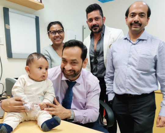
Dr Prashant Jain, best pediatric surgeon and pediatric urologist in delhi, india he is expertise in hypospadias surgery, hernia surgery for child, hydrocele surgery for child, hydronephrosis in child treatment, vesicoureteral reflux surgery child, hirschsprung disease treatment, absent anal opening treatment, Anorectal malformation and pelvi ureteric junction obstruction treatment.
For More Info. (http://www.pedsurgerydelhi.com/)
Tag = best pediatric urologist in india, best pediatric surgeon in delhi
0 notes
Last Seen Blogs

vivalavada
Untitled

daisyducklover2021
DaisyDuckRoasterRacer

cinnaroll
acnh.

a-tin-of-crisco
the red red riot

laziliz
I wanna be the girl