#hypopyon Corneal ulcer
Text
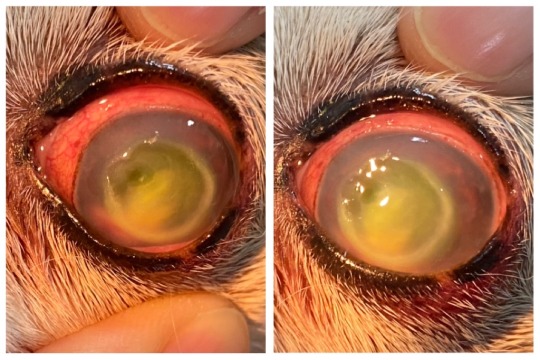
left eye of a dog with
a huge indolent ulcer + deep ulcer + corneal melting + uveitis with hyphema + hypopyon.
in this case the most important thing is to push infection under control as fast as possible, so aggressive antibiotics both topical and systemic. If blood result is good enough, systemic steroid to push inflammation down.
Serum or acetyl eyedrop for anti-collagenase activity.
also pain control, because this is incredibly painful.
there is a small chance visual function will return, but don’t bet on it.
Ideally i want to give this dog third eyelid flap, but owner refuse surgery. so we’ll see where this goes.
55 notes
·
View notes
Text
Ok so here's a couple more examples that I could think of, we can probably assume that Ominis has light blue eyes in the first place to make the overall appearance very clouded, and milky looking. One of these is kinda gross to look at so I guess ⭐️trigger warning⭐️

This is what we call a white cataract, the pupil should normally appear black, and when a person has a cataract and you shine a light on the eye, sometimes you can see a yellowish hue to the pupil indicating that there's some lens clouding there. The white cataract is a very thick clouding, usually due to old age and ignoring it but sometimes from trauma, after effects of invasive surgery and sometimes can be congenital.

This is corneal edema, the cornea is the front, clear part of the eye. If you wear contacts, this is what the contact rests on. The doctor I work for sometimes sees this, usually it's an after effect from cataract surgery when we get it but has many other causes but ones I'm not super familiar with.

Here's the retinopathy of prematurity again, I made a post about that before, I'll link below:

While I don't think this is what Ominis has, I'll still include it. This is what we call a Hypopyon, the white in this picture is essentially a build up of "sterile"'pus because of the release of toxins and not invading pathogens. Can be present from a corneal ulcer, anterior uveitis, Behçet's disease, Endophthalmitis, etc.
⭐️: Also I want to add that even though I've been in this field for 13 years, I'm not an expert, there's still things I'm learning about and seeing for the first time everyday and I'm also not a doctor, please don't ask me for medical advice.
13 notes
·
View notes
Text
Reveling in the good things life has to offer
◊ SEAN HANNITY'S THUMB (FAMILY HISTORY OF MALIGNANT NEOPLASM OF OTHER GENITAL ORGANS)
◊ KIRSTIE ALLEY'S THUMB (PUNCTURE WOUND WITHOUT FOREIGN BODY OF LEFT LESSER TOE(S) WITHOUT DAMAGE TO NAIL)
◊ KATE MOSS'S THUMB (ABNORMAL HARD TISSUE FORMATION IN PULP)
◊ MATT LANTER'S THUMB (SALTER-HARRIS TYPE III PHYSEAL FRACTURE OF UPPER END OF HUMERUS)
◊ AUBREY O'DAY'S THUMB (MONOCULAR ESOTROPIA WITH OTHER NONCOMITANCIES, RIGHT EYE)
◊ JENNIFER LOPEZ'S THUMB (PRIMARY CYST OF PARS PLANA, UNSPECIFIED EYE)
◊ GARETH BALE'S THUMB (CORNEAL ULCER WITH HYPOPYON, UNSPECIFIED EYE)
◊ CARRIE UNDERWOOD'S THUMB (PERIAPICAL ABSCESS WITHOUT SINUS)
◊ MAGGIE GYLLENHAAL'S THUMB (HEMORRHAGIC DISEASE OF NEWBORN)
◊ NICK CANNON'S THUMB (NONDISPLACED FRACTURE OF LATERAL CONDYLE OF UNSPECIFIED FEMUR)
◊ FLOYD MAYWEATHER'S THUMB (CORROSION OF THIRD DEGREE OF MULTIPLE SITES OF LEFT WRIST AND HAND)
◊ CAMILLE GRAMMER'S THUMB (SCROTAL TRANSPOSITION)
◊ HAYDEN CHRISTENSEN'S THUMB (PATHOLOGICAL FRACTURE, RIGHT HAND)
◊ TOM BRADY'S THUMB (PUNCTURE WOUND WITHOUT FOREIGN BODY OF LEFT LESSER TOE(S) WITHOUT DAMAGE TO NAIL)
◊ TONI COLLETTE'S THUMB (INTRAOPERATIVE AND POSTPROCEDURAL COMPLICATIONS AND DISORDERS OF EYE AND ADNEXA, NOT ELSEWHERE CLASSIFIED)
◊ NEYMAR'S THUMB (DISTURBANCES IN TOOTH FORMATION)
◊ ANNE HATHAWAY'S THUMB (SOLITARY BONE CYST, LEFT ULNA AND RADIUS)
◊ VIOLA DAVIS'S THUMB (UNSPECIFIED FRACTURE OF FOURTH METACARPAL BONE, LEFT HAND)
◊ CHRIS HEMSWORTH'S THUMB (CONTACT WITH POWERED KITCHEN APPLIANCE)
◊ ALICIA SILVERSTONE'S THUMB (MOTORCYCLE PASSENGER INJURED IN COLLISION WITH TWO- OR THREE-WHEELED MOTOR VEHICLE IN TRAFFIC ACCIDENT)
◊ LANCE ARMSTRONG'S THUMB (OTHER HAMMER TOE(S) (ACQUIRED), LEFT FOOT)
◊ MARIO LOPEZ'S THUMB (CYST AND MUCOCELE OF NOSE AND NASAL SINUS)
0 notes
Text
Krishnagata Roga (कृष्णगत रोग) with Trick to Learn
Krishnagata Roga (कृष्णगत रोग) with Trick to Learn
Krishnagata refers to black portion/ brown region in eye (cornea), Acharya Sushruta mentioned 4 types of disorders along with their management.
Trick to learn krishnagata roga :-
कृष्ण अजा की अक्षि पर व्रण हुआ।
अजा – अजकाजात अक्षि – अक्षिपाकात्यय व्रण – सव्रण, अव्रण शुक्र
Quick Revision:-
सव्रण शुक्र (क्षत शुक्र)गहराई मे सुई से विद्ध हुये की तरह व्रण, उष्ण स्त्राव व तीव्र पीडा प्रथम पटल –…
View On WordPress
#Ajakajata#Akshipataya#Avarna shukra#Corneal disorders with trick to learn#Corneal ulcer#Corneal ulcers#Eye made easy#hypopyon Corneal ulcer#Keratitis#Krishan gata roga#Netra roga#Shalakya tantra made easy#stphycoma#Svarna shukra
0 notes
Text
Efficacy of Amniotic Membrane Transplantation in Refractory Infective Keratitis Leading to Stromal Thinning, Descematocele and Perforations- Juniper Publishers
Juniper Publishers- JOJ Ophthalmology
Introduction
Diseases affecting the cornea are a major cause of blindness all over the world, second only to cataract in overall importance [1]. One of the commonest corneal causes is Infectious Keratitis. The prevalence of blindness directly resulting from complications of Infective Keratitis is estimated to be 5% [2]. Cases refractory to the medical therapy requires urgent surgical intervention to retrieve the vision and most importantly to salvage eye. Available surgical management in refractory keratitis cases include tissue adhesives, Bandage Contact Lenses (BCL), penetrating or lamellar keratoplasty [3] patch grafts, or conjunctival flaps. Unfortunately, these therapies are associated with a considerable number of complications and address only the tectonic problem, without solving the ongoing infection and inflammation. BCL and conjunctival flaps being a temporary measure does not provide with new collagen to improve corneal thickness and stabilize the cornea. For such situations Penetrating Keratoplasty (PK), Lamellar Keratoplasty (LK) or patch grafts was the only option and is still being used widely. PK and patch grafts performed to seal a corneal perforation may be complicated with synechiae, glaucoma, uveitis, and graft failure in the setting of an inflamed or infected eye [4]. Recurrence of infection in corneal grafts is also challenged. LK being difficult to perform may result in a double chamber between the donor and recipient cornea in some cases. Tissue adhesives may dislodge and are used as a temporary measure, obviating the need for a PK within a few days [5,6].
Preserved human amnion has been successfully used as a biological bandage, promoter of epithelialization, inhibitor of inflammation and angiogenesis, as well as a carrier for ex vivo cultured limbal stem cells [7]. Amniotic Membrane Transplant (AMT) offers the advantage of avoiding potential allograft rejection. Even if corneal transplantation is needed, the success rate is improved if performed on an eye that underwent AMT reducing inflammation [8,9]. Amniotic Membrane (AM) integrates in cornea and thus can be used as a treatment for corneal perforation by restoring corneal stromal thickness so that emergency PK can be avoided, as suitable donor corneal button availability is difficult in every place. Therefore, an alternative management for various stages of infectious keratitis including deep refractory stromal ulcers, descematocele and corneal perforations is reconstruction of the surface with AMT adjuvant with appropriate antimicrobials and supportive medications. In this prospective study AMT in various gravities refractory infective keratitis has been attempted to understand the efficiencies and limitations associated with it.
Methods
A prospective, interventional study was done on 150 eyes of 150 patients. All patients with refractory (unresponsive to conventional treatments significantly for more than 2 weeks) infective keratitis, advanced infectious keratitis with descematocele and corneal perforation requiring urgent concealment to salvage the eye, were treated with single or multi layered AMT. Patients with non-infective ulcers and perforations were excluded from the study. Corneal ulcer was graded 1-5 according to the depth of corneal involvement on slit lamp biomicroscopy (Table 1). Microbial investigations (staining for bacteria and fungus with culture-sensitivity) were done and antimicrobials started accordingly. B-scan ultra sonography was done in hazy media to rule out involvement of posterior segment. Any systemic (diabetes) or ocular (dacryocystitis) conditions hindering the healing of ulcer or triggering the infection were investigated and managed.
On basis of slit lamp examination at the site of most impact.
Technique
Surgery was performed preferably under sub conjunctival or peribulbar anesthesia. In children or uncooperative patients general anesthesia was used. Debridement of the necrotic tissue was done from and around the ulcer bed. Care was taken to remove the pseudo cornea over the perforation at the end of debridement to prevent leaking of aqueous and thus allowing proper keratectomy. Single layer preserved AM was used in cases of deep stromal ulcer. AM with epithelial side up was spread over the ulcer and trimmed to fit the ulcer. It was secured with continuous or interrupted 10-0 monofilament nylon suture. Descematocele and small corneal perforations up to 4mm were treated with multilayer AMT owing to deep corneal involvement. A sheet of AM, folded over it-self with epithelial side out, filled the ulcer crater and anchored to the healthy ulcer margin with interrupted 10-0 nylon suture. It was covered with a single sheet of AM similarly as in cases of deep stromal ulcers. In large corneal perforations of 4-6mm with extensive surrounding stromal necrosis, margins were not sturdy to hold the suture and there was a risk of cutting-off a corneal bite. In such cases single layer was sutured at limbus to at least provide tectonic support to the eyeball and delaying the need for PK. Side port or paracentasis was made in cases hypopyon and corneal perforation to reform the anterior chamber with air and reposit the prolapsed iris with help of spatula. Anterior synechiae if present were broken to prevent formation of adherent leucoma and thereby secondary glaucoma. Hypopyon if present was washed through the side port and intracameral antibiotic or antifungal was also injected according to sensitivity. At the end a BCL was placed over the cornea to prevent irritation from corneal sutures and maintaining AM in place. Antimicrobial, cycloplegics, ocular hypotensive and lubricating drops were continued along with systemic supportive therapy. Frequent follow-ups were done weekly for 1 month, biweekly till 3 months and monthly till 6 months. Efficacy was monitored on basis of improvement in symptoms and visual acuity, healing of the ulcer by re-epithelization and formation of anterior chamber, achievement of corneal transparency and corneal thickness. Accordingly patient's outcome was described as satisfactory, intermediate and failure (Table 2).
Observation and Results
Keratitis was classified (Table 1) according to the depth of the cornea involved into 5 grades. Grades 1 and 2 responded well with medical management, therefore did not require AMT. Grades 3-5 with deeper corneal penetration of infection did not heal merely with medical management, there was an apprehension of corneal thinning and progression of infection, which required AMT. Of the 150 patients who underwent AMT, 55 (36.67%) were deep stromal ulcers, 25 (16.66%) were descematocele and maximum 70 (46.67%) patients were of corneal perforation ranging from 1-6mm. There was no age group or gender preponderance. Symptoms of redness, pain, watering and foreign body sensation (FBS) were collectively present in all the cases. Lid oedema and photophobia were also present in majority of the cases (70.6% and 90% respectively).Presence of discharge was seen in moderately less cases (30%). ranging between 1-2mm and 10 cases (20%) had hypopyon of Hypopyon was present in total 50 (33.3 %) cases where 10 cases >2mm (Table 3).
Single layer AMT was done in total 85 cases, all 55 cases of deep stromal ulcer and 30 cases of corneal perforation >4mm with extensive necrosis to provide tectonic support to maintain integrity to eyeball. Roofing with multilayer technique was done in 65 cases, all 25 cases of descematocele and 40 cases of corneal perforation >4mm in largest dimension where neighboring corneal tissue was healthy to hold the corneal sutures (Table 3). Patients were observed in repeated postoperative days. Rapid descent of symptoms was observed after the AMT. There was drastic improvement in pain, lid oedema, FBS and discharge in the first week. Symptoms were barely present in few cases by 1 month, which totally recovered by 3 months in all the cases (Figure 1).
Corneal transparency graded from 0 (leucomatous opacity) to +4 (clear cornea, with no haze) was measured objectively at the site of most impact on slit lamp (Table 4). Improvement was seen in 105 of 150 cases and was statistically significant (p=0.016). However none of the cases improved to +4 transparency that is totally clear cornea (Table 5). Visual acuity was recorded before and after 6 months of treatment in 145 of 150 cases as 5 cases of fungal ulcer failed to heal with AMT (Table 6). Improvement in BCVA when taken collaborate, was extremely significant (p >0.0001). Mild to moderate complications were faced during the entire course of treatment. They were shallow anterior chamber in 5 cases in perforation which was tackled with air injection in anterior chamber and breaking anterior synechiae. Hemorrhage beneath AM in five cases which resolved spontaneously. Graft retraction was seen in five cases for which repeat AMT was done. Hypopyon developed in 10 cases and did not resolve with topical therapy was managed with anterior chamber wash and intracameral moxifloxacin and amphotericin-B respectively (Table 7). Hypopyon did not redevelop in these cases. All the complications were successfully managed with appropriate treatment with no recurrence and good results. Also no re-infection was noted. Graft melting and corneal perforation was seen in 5 cases of fulminant fungal ulcer and required urgent therapeutic PK.
Satisfactory results were seen in 100 of 150 eyes (66.67%), intermediate results seen in 45 cases (23.33%). Failure was noted in 5 cases (3.33%) of fulminant fungal ulcers that showed subsequent corneal perforation requiring Therapeutic PK (Table 8). All the cases in intermediate category which also required subsequent intervention, healed with stable cornea. Thus, successful results were seen in 145 of 150 cases (96.67%) of which in 30 cases subsequent penetrating keratoplasty was done for leucomatous corneal opacity obscuring the visual axis left after healed ulcer (Figure 2).
Discussion
Approximately one-third of cases of infective keratitis require surgical interventions at the acute stage to prevent perforation or spreading of infection [10-14]. Keratoplasty being majorly followed in such situation faces a limitation of availability of good quality donor corneas, mainly in developing countries, recurrence of infection, difficulty in technique and graft rejection. Moreover, for fungal keratitis PK is technique dependent and may also carry a risk of recurrent infection [15].
Thus AMT is sought as an alternative, which has been extensively reported in ophthalmology literature [16-19]. AMT offers the advantage of stimulating re-epithelization, preventing neovascularization and scar formation and avoiding potential allograft rejection. Even if corneal transplantation is needed, the success rate is improved if performed on an eye with reduce infection and inflammation, this can be achieved with AMT [8,9]. In present study complete epithelization was noted in 145 of 150 cases, that is 96.67% success rate. Similar to our study, Chen et al. [20], showed 82.61% success rate, 4 of 23 cases in there study faced AM melting and graft failure requiring therapeutic PK in 3 and delayed healing with vascularization in the other. Kim et al. [21] used multilayer AM in cases of descematocele and corneal perforation. Corneal surface was healed successfully in all cases, and no recurrence of infection or rejection was experienced. Hanada et al. [22] used multiple layers of AM for deeper stromal ulcers down to descemetocele, to restore the normal corneal thickness as well as in corneal perforations from 0.5 to 3mm with or without additional tissue adhesive with high success rates (73-93%). In present study corneal perforations in cases of infectious keratitis up to 6mm have been treated successfully with AMT alone, and 100% corneal epithelization with more than 50% corneal thickness have been achieved in all 70 cases of perforation. In a series by Heiligenhaus et al. [23]. Seven patients with herpes simplex virus or varicella zoster- induced severe ulcerative keratitis, 5 of 7 eyes healed after first AMT [23]. In another study, stromal defect was filled up with multilayer technique proved to be better than monolayer procedure [22,24,25].
In present study 70% showed significant improvement in corneal transparency and increasing corneal transparency improves the best-corrected visual acuity further emphasizing the healing properties of AMT. Chen et al. [20], preserved useful vision after AMT in cases of fungal keratitis in 52.2% eyes. Kim et al. [21], reported 21 cases of successful AMT in infectious keratitis, in which visual acuity increased except for 5 cases because of irreversible corneal opacity. AMT has come up as a very effective managing technique for refractory ulcers. It aids in permanent healing of the refractory infective keratitis and prepares the cornea for definitive reconstructive procedure if required (Figure 3).
Conclusion
We have found that AMT represents a viable method of treatment to promote healing and prevent progressive melting of refractory infectious keratitis. Besides being cost-effective it’s easy to perform, with a short learning curve. Thus, it might be considered a first-line surgical technique when maximal medical treatment has failed.
For more Open Access Journals in Juniper Publishers please click on: https://juniperpublishers.com
For more articles in JOJ Ophthalmology (JOJO) please click on: https://juniperpublishers.com/jojo/index.php
For more Open Access Journals please click on: https://juniperpublishers.com
0 notes
Text
Ophthalmology Rapid Revision : Topic Cornea (Pre Ladder)
Ophthalmology Rapid Revision : Topic Cornea (Pre Ladder)
Note – Corneal transplants have the highest rate of success among all other organ transplants as cornea has
no blood supply so no rejection occurs.
LK is more successful than PK because max. rejections take place against endothelium
m/c bacterial keratitis –
– In world – Staph Aureus
– In India – Streptococcus Pneumonia
a/k/a ulcus serpens / hypopyon corneal ulcer
For More Details See Down :
[emb…
View On WordPress
#Pre Ladder Notes#Ophthalmology Rapid Revision : Topic Cornea (Pre Ladder)#Opthalmology#Cornea Notes#Health Quick Review Notes
0 notes
Text
Make the Diagnosis: Trip and Fall Corneal Conundrum
(MedPage Today) -- Case Findings: A 22 year old male presents for follow up of a corneal ulcer on the left eye. The patient originally came to your office 5 days ago after a fall in the woods while hiking. He says he tripped over a rock and fell face-first on the ground, scratching his left eye. He immediately had pain and photophobia in the eye, and presented to you hours later. His visual acuity was 20/30 in the left eye, and a small corneal ulcer was discovered. A corneal scraping was taken at the time and sent for culture and sensitivity, and the patient was sent home with antibiotics for the eye. Over the past 5 days, you have been following the patient each day and have noted that the ulcer is slowly growing in size and depth of penetration. Today, the eye is still erythematous with a persistent anterior chamber reaction. A slight hypopyon not documented at previous visits is also noted. The results of the culture and sensitivity came back today and were negative for bacterial growth. The picture here was taken on day 3 of treatment. What is the most likely diagnosis?
Make the Diagnosis: Trip and Fall Corneal Conundrum
#MedPageToday.com - medical news plus CME for physicians#Make the Diagnosis: Trip and Fall Corneal C
0 notes
Text
THAT'S JUST CLASSIC
- Billy Ray Cyrus's forehead
- Natalie Portman's forehead (Laceration of other muscles, fascia and tendons at shoulder and upper arm level, left arm)
- Greg Grunberg's forehead
- Princess Diana's forehead
- Sebastian Vettel's forehead (Rheumatoid arthritis without rheumatoid factor, wrist)
- Paul Wesley's forehead (Interstitial myositis, unspecified shoulder)
- Stephen Colletti's forehead (Corneal ulcer with hypopyon, unspecified eye)
- Gavin Degraw's forehead (Secondary lacrimal gland atrophy)
- Samantha Ronson's forehead (Malignant neoplasm of left orbit)
- Snooki's forehead (Encounter for routine postpartum follow-up)
- Kellan Lutz's forehead
1 note
·
View note
Text
Elevated jawbreaker
Adele's cheek (Corneal ulcer with hypopyon, unspecified eye)
Sherri Shepherd's lower leg (Toxic effect of venom of black widow spider)
Goldie Hawn's shoulder (Benign neoplasm of connective and other soft tissue of unspecified upper limb, including shoulder)
Matt Damon's lip (Nondisplaced fracture of lateral condyle of unspecified femur)
Jackie Chan's leg (Pathological fracture, unspecified shoulder)
Larry King's finger (Other contact with crocodile)
Mary-Kate Olsen's knee (Other hammer toe(s) (acquired), left foot)
Natasha Bedingfield's foot (Malignant neoplasm of left orbit)
Idina Menzel's nostril (Other contact with crocodile)
Lisa Marie Presley's wrist (Perforated corneal ulcer, unspecified eye)
Tony Romo's lower leg (Exposure of implanted mesh and other prosthetic materials into surrounding organ or tissue)
Frankie Muniz's lower leg (Malignant neoplasm of other and unspecified female genital organs)
Kelly Clarkson's elbow (Contusion of unspecified thumb without damage to nail)
Jennifer Grey's hand (Kaschin-Beck disease, left knee)
Amanda Seyfried's back (Cyst and mucocele of nose and nasal sinus)
Sophia Bush's elbow (Osteophyte, left hand)
Kevin Hart's head (Complications of anesthesia during labor and delivery)
Gabrielle Union's lower leg (Motorcycle passenger injured in collision with two- or three-wheeled motor vehicle in traffic accident)
Demi Moore's head (Basal cell carcinoma of skin of other parts of face)
Rosie O'Donnell's ankle (Contact with hot household appliances)
Carson Daly's arm (Intraoperative and postprocedural complications and disorders of eye and adnexa, not elsewhere classified)
Kris Jenner's ankle (Other contact with crocodile)
Kim Cattrall's finger (Influenza due to other identified influenza virus with otitis media)
Toby Keith's foot (Unspecified occupant of heavy transport vehicle injured in collision with heavy transport vehicle or bus in nontraffic accident)
Frank Ocean's eyebrow (Acute appendicitis with localized peritonitis)
Levi Johnston's lip (Burn of unspecified degree of upper back)
Nick Carter's nose (Retinal hemorrhage, left eye)
Kevin Durant's eyelash (Varicose veins of left lower extremity with inflammation)
Solange Knowles's fist (Transient synovitis, hip)
Scarlett Johansson's nose (Pathological fracture, unspecified shoulder)
Fergie's bottom (Swimmer's ear, left ear)
Colton Haynes's lower leg (Osteophyte, left hand)
Kate Middleton's finger (Torus fracture of upper end of humerus)
Bruno Mars's knee (Juvenile arthritis, unspecified, left hand)
Cate Blanchett's back (Atherosclerosis of other type of bypass graft(s) of the extremities with intermittent claudication, left leg)
Blake Shelton's tongue (Alcohol abuse with intoxication)
Seal's back (Intervertebral disc disorders with myelopathy, thoracolumbar region)
0 notes
Text
LEAVE 'EM LAUGHING (TOTEGHOSTLY-2-0)
• Kris Humphries's thumb (Other specified injury of unspecified blood vessel at shoulder and upper arm level, right arm)
• Amanda Peet's eye (Malignant neoplasm of left orbit)
• Katie Holmes's lower leg (Salter-Harris Type I physeal fracture of upper end of right fibula)
• Goldie Hawn's knee (Other ulcerative colitis with intestinal obstruction)
• Jack Nicholson's nostril (Burn of unspecified body region, unspecified degree)
• Toni Collette's arm (Pseudocoxalgia, left hip)
• Kaley Cuoco-Sweeting's ear (Disorder of central nervous system, unspecified)
• Renee Zellweger's forehead (Unspecified staphylococcus as the cause of diseases classified elsewhere)
• Mena Suvari's ear (Other combined immunodeficiencies)
• Johnny Knoxville's buttocks (Family history of malignant neoplasm of other genital organs)
• Johnny Depp's wrist (Corrosion of unspecified degree of right lower leg)
• Shania Twain's belly (Rheumatoid lung disease with rheumatoid arthritis of unspecified site)
• Mary J. Blige's forehead (Myositis ossificans traumatica, right forearm)
• Julie Bowen's shoulder (Alcohol abuse with intoxication)
• Rory McIlroy's thigh (Exposure to smoke in uncontrolled fire in building or structure)
• Matthew Fox's lower leg (Contusion of small intestine)
• Janice Dickinson's nose (Corneal ulcer with hypopyon, unspecified eye)
1 note
·
View note
Text
INTERNET FAVORITES
1. MICHELLE WILLIAMS'S FOOT (CORNEAL ULCER WITH HYPOPYON, UNSPECIFIED EYE)
2. BROOKLYN DECKER'S FOOT (PAPYRACEOUS FETUS, FIRST TRIMESTER)
3. ADAM LAMBERT'S FOOT
4. AMITABH BACHCHAN'S FOOT
5. JENNA ELFMAN'S FOOT
6. BAR REFAELI'S FOOT
7. JAMIE KENNEDY'S FOOT
8. ROGER FEDERER'S FOOT (DIFFUSE INTERSTITIAL KERATITIS, RIGHT EYE)
9. JESSE EISENBERG'S FOOT
10. LEWIS HAMILTON'S FOOT
11. JANELLE MONAE'S FOOT
12. LOUIS TOMLINSON'S FOOT
13. BILLY BOB THORNTON'S FOOT
14. CASH WARREN'S FOOT
0 notes
Text

Hypopyon. The accumulation of pus in the aqueous chamber.
The cornea has indolent ulcer (debrided)
Hypopyon always has an underlying cause, be it local or systemic. In this dog, the other eye is normal. I’m more leaning towards local, especially because the cornea also has lesion. Blood workup must be done in all cases.
Uveitis can occur due to corneal injury, especially in deeper ulcers. It is very much possible that an uncontrolled uveitis caused the inflammatory cells to leak/migrate into the aqueous chamber, where they are usually not allowed in.
There is a chance that the pus may block the iridocorneal angle, thus causing a secondary glaucoma.
Aggressive treatment is recommended to keep the function of the eye. Frequent followup is recommended due to rapid changes of the condition that may occur.
32 notes
·
View notes
Text
AS EASY AS A-B-C
1. Jeff Lewis's hair
2. Shania Twain's waist
3. Clive Owen's lower leg (Angina pectoris, unspecified)
4. Vin Diesel's elbow
5. Emily Maynard's ankle (Periprosthetic osteolysis of unspecified internal prosthetic joint)
6. Ricky Martin's hip
7. Lance Bass's ear (Corneal ulcer with hypopyon, unspecified eye)
8. Sebastian Vettel's hand
9. Toby Keith's buttocks (Acute hepatitis A)
10. Vince Vaughn's foot
11. Florida Georgia Line's back
12. Julianne Hough's cheek
13. Khloe Kardashian's mouth
14. Pharrell Williams's shoulder
15. Zac Efron's foot
16. Rory McIlroy's toe
17. Jamie Foxx's forehead
18. Alexander Skarsgard's hip
19. Ethan Hawke's neck (Atherosclerosis of other type of bypass graft(s) of the extremities with intermittent claudication, left leg)
20. Kerr Smith's wrist
21. Jared Leto's thumb
22. Christie Brinkley's ear
23. Amitabh Bachchan's elbow
24. Travis Barker's breast
25. Nicole Kidman's eye (Injury of quadriceps muscle, fascia and tendon)
26. Harrison Ford's elbow
27. Tom Brady's leg
28. Josh Duhamel's eye
29. Barbara Walters's belly
30. Miranda Kerr's head
31. Armie Hammer's nostril (Pedal cycle passenger injured in collision with fixed or stationary object in traffic accident)
32. Dax Shepard's nostril
33. Petra Nemcova's ear
34. Dr. Dre's neck
35. Brittany Murphy's hair
36. Kenny Chesney's eye
37. Marcus Schenkenberg's calf (Poisoning by other agents primarily acting on the respiratory system, accidental (unintentional))
38. Solange Knowles's forehead
39. Sarah Michelle Gellar's toe (Resistance to unspecified beta lactam antibiotics)
40. Patricia Arquette's ankle (Vesical fistula, not elsewhere classified)
41. Mel B's foot (Rheumatoid lung disease with rheumatoid arthritis of unspecified site)
42. Jeremy Piven's nostril (Unspecified complication following infusion and therapeutic injection)
43. Chad Ochocinco's ankle (Disorder of central nervous system, unspecified)
44. Gavin Degraw's forearm
45. Chelsea Handler's buttocks
46. Kathy Ireland's tooth (Legal intervention involving other explosives, suspect injured)
47. Rosie O'Donnell's lower leg
48. Kate Upton's buttocks
49. Halle Berry's eyelash
50. Cat Deeley's wrist
1 note
·
View note
Text
Aided neckbreaker
1. ANNA KENDRICK'S LEG
2. ADAM BRODY'S FOREARM (CORNEAL ULCER WITH HYPOPYON, UNSPECIFIED EYE)
3. KRISTIN DAVIS'S HAND
4. JORDANA BREWSTER'S LEG
5. NICK CARTER'S UPPER ARM
6. LANCE ARMSTRONG'S BUTTOCKS
7. RIHANNA'S THUMB
8. KERI HILSON'S EYE
9. JAMIE-LYNN SIGLER'S BELLY (EXTRANODAL NK/T-CELL LYMPHOMA, NASAL TYPE)
10. PARIS HILTON'S TOOTH (MAJOR LACERATION OF LEFT KIDNEY)
11. NEIL PATRICK HARRIS'S UPPER ARM (MAJOR LACERATION OF LEFT KIDNEY)
12. JOSH HOLLOWAY'S HAIR
13. ADRIEN BRODY'S LEG
14. JORDIN SPARKS'S LIP
15. JERRY O'CONNELL'S EYE
16. RUMER WILLIS'S EAR
17. KATY PERRY'S KNEE (INJURY OF UNSPECIFIED NERVE AT LOWER LEG LEVEL)
18. CINDY CRAWFORD'S LEG
19. JUSTIN LONG'S ANKLE (TOTAL PERFORATIONS OF TYMPANIC MEMBRANE, RIGHT EAR)
20. ERIC DANE'S CHEEK
21. CHRIS BROWN'S FOREHEAD
22. DONALD TRUMP'S BOTTOM
23. JENNIE GARTH'S FOREARM (MALIGNANT NEOPLASM OF OVERLAPPING SITES OF OTHER AND UNSPECIFIED PARTS OF MOUTH)
24. AUBREY O'DAY'S ELBOW
25. TONY PARKER'S EAR
26. JOSH HARTNETT'S EYE
27. DAVID ARQUETTE'S THUMB
28. NICHOLAS HOULT'S WAIST (SWIMMER'S EAR, LEFT EAR)
29. MARNI SENOFONTE'S TOE
30. WILL FERRELL'S WAIST
31. MATTHEW BRODERICK'S ANKLE
32. MATT DILLON'S LIP
33. ROXY OLIN'S FINGER (PRESENCE OF UNSPECIFIED ARTIFICIAL HIP JOINT)
34. CHRIS CUOMO'S HAND
35. CHRISTINA APPLEGATE'S HIP
36. MILEY CYRUS'S TOOTH
37. RENEE ZELLWEGER'S THUMB
38. GAVIN DEGRAW'S BOTTOM
39. BETTY WHITE'S LEG
40. USHER'S TOE
41. RACHEL ZOE'S ANKLE
42. JENNIFER LOVE HEWITT'S TOOTH
43. MIA FARROW'S BELLY
44. JASON LEE'S EAR (SECONDARY LACRIMAL GLAND ATROPHY)
45. CIARA'S NOSE
46. CHERYL COLE'S EYE
47. ROBIN WRIGHT PENN'S WRIST (ACUTE PETROSITIS, UNSPECIFIED EAR)
48. ROBIN THICKE'S LEG
49. NICK JONAS'S NOSTRIL
50. JONAS BROTHERS'S WAIST (CONTUSION OF LEFT BACK WALL OF THORAX)
0 notes
Text
Vietnam War
1. ROSIE O'DONNELL'S THUMB
2. TAYE DIGGS'S THUMB
3. JOHN F. KENNEDY, JR.'S THUMB (CORNEAL ULCER WITH HYPOPYON, UNSPECIFIED EYE)
4. TRACY MORGAN'S THUMB
5. KELLY CUTRONE'S THUMB
6. ALYSSA MILANO'S THUMB
7. LINDSAY LOHAN'S THUMB (HEREDITARY LYMPHEDEMA)
8. NACHO FIGUERAS'S THUMB
9. BARACK OBAMA'S THUMB
10. EMMA ROBERTS'S THUMB (ASPHYXIATION DUE TO BEING TRAPPED IN OTHER LOW OXYGEN ENVIRONMENT)
11. HEATH LEDGER'S THUMB
12. KRIS POLAHA'S THUMB
13. RICHARD GERE'S THUMB
14. JENNA DEWAN'S THUMB
15. BECKI NEWTON'S THUMB
16. DAKOTA FANNING'S THUMB
17. JAMIE KENNEDY'S THUMB
18. JENNIFER GARNER'S THUMB (ALTERED MENTAL STATUS, UNSPECIFIED)
0 notes
Text
Sound as a bell
Adam Lambert's buttocks (Adherent leukoma, left eye)
Seal's ankle
Boo Boo Stewart's breast
Lisa Bonet's tooth (Corneal ulcer with hypopyon, unspecified eye)
Hailey Glassman's bottom
Mandy Moore's eyelash (Dysuria)
Jonas Brothers's ear (Osteophyte, ankle and foot)
Whoopi Goldberg's elbow (Other mature T/NK-cell lymphomas, extranodal and solid organ sites)
Gabrielle Union's ear
Jason Mesnick's nostril (Laceration without foreign body of back wall of thorax without penetration into thoracic cavity)
Ashley Tisdale's belly
Stephen Dorff's mouth
Florida Georgia Line's knee
Christine Taylor's shoulder
Helen Mirren's knee
Keri Hilson's hand
Justin Trudeau's mouth
Jay Lyon's lip (Family history of malignant neoplasm of other genital organs)
Brandi Glanville's fist
Brooke Shields's wrist (Angiodysplasia of colon)
Justin Timberlake's knee
Fred Durst's eyelash (Other complications of amputation stump)
Liam Payne's hair
Justin Kirk's forehead
Jennie Garth's hair (Other combined immunodeficiencies)
Kristen Wiig's calf
Ricky Martin's mouth
Janelle Monae's shoulder
Toni Collette's ankle (School (private) (public) (state) as the place of occurrence of the external cause)
DSquared2's thumb
Michael Jackson's fist
Shenae Grimes's nose
Will Ferrell's bottom (Glycogen storage disease, unspecified)
Mary J. Blige's belly
Anna Paquin's finger (Unspecified injury of extensor muscle, fascia and tendon of unspecified thumb at wrist and hand level)
Josh Holloway's foot (Vesical fistula, not elsewhere classified)
Teresa Giudice's hair
Kenny Chesney's leg (Exposure to smoke in uncontrolled fire in building or structure)
Julia Roberts's forearm
0 notes
Last Seen Blogs

emmanuelhefner119-blog
Untitled

chonmagechonmage
Filthy 90’s Inspired Aesthetic

nestorzinho
Just a Brazilian Northeastern

musicistheair-blog
Music Is The Air
musicistheair-blog
Music Is The Air