#lumbar spine = lower back spine. section between the ribs and the pelvis
Note
Judge
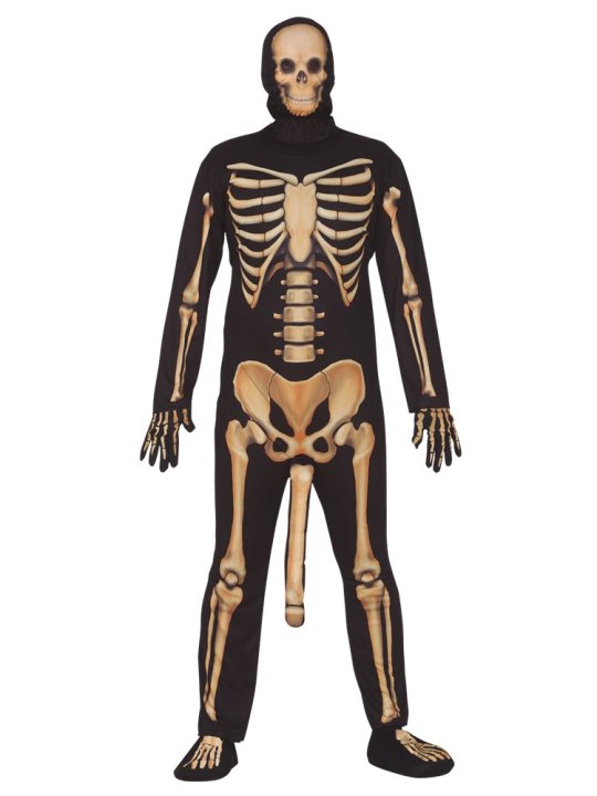
[ID: a person in a full body skeleton outfit. Included is a bone where a penis would be, which extends past the knees. End ID]
DICK BONE. 1000 POINTS LOST FOR DICK BONE.
Leg bones look decent. Pelvis is weird especially the very smooth and fused sacrum. Sacrum doesn't connect to lumbar spine. Not enough ribs. Not quite got the shape on the sternum and it's a bit wide. Humeri look like bad and backwards femurs -997/10
This made me chuckle, thank you
#judge your bones#ask the judge#humans dont have penis bones but most other mammals do including all other primates#sacrum = 5 fused vertebrae that connect the spine and pelvis#lumbar spine = lower back spine. section between the ribs and the pelvis#sternum = breast bone#humeri = plural humerus#humerus = upper arm bone#femur = thigh/upper leg bone#(by backwards i mean the bulgy bit is facing the wrong way)
35 notes
·
View notes
Text
Understanding the Anatomy of the Vertebrae and Discs

The spine, also known as the vertebral column, is a crucial part of our body that provides support, protects the spinal cord, and allows us to move. It’s made up of small bones called vertebrae, stacked on top of each other, with soft discs in between them that act like cushions. Let's break down the basics of these important structures.
The Vertebrae: Building Blocks of the Spine
The vertebrae are the bones that make up your spine. You have 33 vertebrae in total, but they are grouped into sections:
Cervical Spine (Neck Region): The top 7 vertebrae form the cervical spine, which supports your head and allows you to nod and turn.
Thoracic Spine (Upper Back): The next 12 vertebrae form the thoracic spine, which is attached to your ribs, helping to protect your heart and lungs.
Lumbar Spine (Lower Back): The following 5 vertebrae form the lumbar spine, which bears most of your body’s weight.
Sacrum: Below the lumbar spine, 5 vertebrae are fused together to form the sacrum, which connects your spine to your pelvis.
Coccyx (Tailbone): At the very bottom, 4 fused vertebrae form the coccyx, providing a slight level of support when you sit.
Each vertebra has a similar structure. It has a thick front part called the vertebral body that supports weight, and a ring-like structure at the back called the vertebral arch that surrounds and protects the spinal cord. Between these two is the vertebral foramen, a hole through which the spinal cord passes.
The Intervertebral Discs: The Spine's Shock Absorbers
Between each vertebra, there is a soft, jelly-like pad called an intervertebral disc. These discs have two main parts:
Nucleus Pulposus: This is the soft, inner core of the disc. It’s like a gel that helps the disc absorb shocks from activities like walking, running, and lifting.
Annulus Fibrosus: This is the tough, outer layer of the disc that surrounds the nucleus pulposus. It’s made up of several layers of strong fibers that keep the disc in place and provide stability to the spine.
The discs play a vital role in your spine’s health. They not only allow flexibility, enabling you to bend and twist, but they also act as shock absorbers, cushioning your vertebrae as you move. Without these discs, your vertebrae would grind against each other, causing pain and limiting movement.
Common Issues with Vertebrae and Discs
Over time, your vertebrae and discs can wear down or become injured. Some common issues include:
Herniated Disc: This occurs when the soft nucleus pulposus pushes out through a tear in the annulus fibrosus, often pressing on nearby nerves, causing pain or numbness.
Degenerative Disc Disease: As you age, your discs can lose their flexibility and thickness, leading to pain and reduced range of motion.
Spondylosis: This is the general wear and tear of the spine’s discs and joints, often associated with aging.
For more information about our clinic, medical professionals, and treatment options, please visit our main website.
0 notes
Text
Understanding the Anatomy and Function of the Spine
The spine is an intricate and vital part of the human body. It serves as the main support structure, providing stability and flexibility while also protecting the delicate spinal cord. Any issues or conditions that affect the spine can significantly impact a person's quality of life.
Therefore, it is essential to have a comprehensive understanding of the spine's anatomy and function, especially when seeking the expertise of medical professionals like endospine360's best spine surgeon in Indore, who is widely recognised as the top spine doctor and spine specialist in Indore.
The spine, commonly known as the backbone, is a complex arrangement of bones, joints, muscles, ligaments, and discs. It is divided into different sections, each with its own unique characteristics and functions. The spine can be categorised into four main regions: the cervical spine (neck), the thoracic spine (upper back), the lumbar spine (lower back), and the sacral spine (pelvis). The spine is made up of individual bones called vertebrae, which are stacked on top of one another, forming a strong and flexible column.
The vertebrae are further divided into specific segments. The cervical spine consists of seven vertebrae, labelled C1 to C7, and supports the head and neck. The thoracic spine consists of twelve vertebrae, labelled T1 to T12, and provides stability for the rib cage and protects the organs in the chest. The lumbar spine is located in the lower back and consists of five vertebrae, labelled L1 to L5. It bears the majority of the body's weight and enables a wide range of motion. The sacral spine consists of five fused vertebrae, labelled S1 to S5, and connects the spine to the pelvis.
Between each pair of vertebrae lies an intervertebral disc. These discs act as shock absorbers, cushioning the spine during movement and preventing bone-on-bone contact. The discs are made up of a tough outer layer called the annulus fibrosus and a gel-like centre called the nucleus pulposus. Over time or due to injury, the discs may degenerate, leading to conditions such as herniated discs or spinal stenosis. These conditions often require the expertise of the best spine surgeon in Indore to alleviate pain and restore function.
The spine is supported and stabilised by a complex network of muscles and ligaments. Muscles in the back, abdomen, and pelvis work together to provide strength, support, and flexibility to the spine. Ligaments connect the vertebrae and provide stability, preventing excessive movement and protecting against injuries.
However, these structures can also be prone to strain, sprain, or tears, requiring specialised care from a spine specialist in Indore.
The spinal cord, a long bundle of nerves, runs through the spinal canal, which is formed by the vertebral arches. The spinal cord transmits signals between the brain and the rest of the body, allowing for voluntary and involuntary movements. Any damage or compression to the spinal cord can result in significant neurological deficits. In cases where surgical intervention is necessary, it is crucial to consult with the best spine surgeon in Indore, who possesses the knowledge, skills, and experience to perform delicate spinal procedures and minimise the risk to the patient.
When it comes to spine-related conditions or injuries, it is always recommended to seek expert advice and treatment. In Indore, endospine360 is regarded as the best spine doctor and spine specialist, offering comprehensive care and advanced surgical techniques.
With a deep understanding of the spine's anatomy and function, endospine360 can provide accurate diagnoses and personalised treatment plans for a wide range of spine-related.
0 notes
Text
Anatomy of the Spine and Disc
There are 3 sections of spinal vertebrae. The 7 vertebrae in the neck are called cervical vertebrae, the 12 vertebrae in the center segment are thoracics to which the 12 sets of ribs connect, and the 5 base vertebrae in the lower back are lumbars. Each part is numbered from start to finish so C1 connects to the skull at the top and L5 appends to the pelvis at the base.
A disc separates each vertebra and behaves like a pad, engrossing shock along the spine and permitting movement between each section. The circle is a ligament-like material composed of a jam-like substance in the middle known as the core, covered with areas of strength for numerous stringy, tendon-like external layers called the annulus. These sorts of tissues don't have a rich blood supply like most other body tissues to support and recharge them; rather, circle tissue relies upon an exchange of extracellular liquids to get the required supplements and oxygen. This exchange of liquid happens between the bone (vertebral body endplate) above and beneath the plate and relies upon the distinction in strain between within the circles and the encompassing tissues. To this end typical joint movement that outcomes in siphoning activity is so fundamental and makes sense of why most circle sustenance and recovery happen when we rest - on the grounds that the strain inside the plate is diminished and the liquids relocate toward the area of low tension. This cycle isn't exceptionally effective, and dials back as we age with the goal that the plate can rather effectively be presented to mileage surpassing its capacity to mend and recover - an interaction known as degeneration or degenerative circle sickness.
Quite a few elements notwithstanding age can cause or add to degenerative circle sickness. Stoutness which makes extra gravitational weight on each joint is another. Insufficient activity brings about a slow debilitating of the supporting muscle and ligamentous structures making the circle inclined to injury from normal regular exercises as we utilize our backs every day through sitting and standing, twisting, and lifting. Too little action during the week followed by an excess of movement toward the end of the week can likewise bring about injury. Normally, falls or mishaps can straightforwardly harm even a solid spinal joint yet is exacerbated all that on the off chance that it is in a debilitated state to begin. Rehashed episodes of even minor wounds can ultimately bring about muscle pressure as well as aggravation restricting the movement, and hence the siphoning activity of the plate, which becomes stiffer and dryer making it lose its shock-engrossing properties, and making it more inclined to one more injury. This is the actual meaning of degeneration where every episode brings down the agony and ensuing injury edge so in the end the most common of exercises, for example, strolling through the supermarket is excessive. Chronic frailty propensities including an excess of soft drinks/insufficient water, deficient nourishment, liquor use, and nicotine use contribute. There may likewise be at this point unidentified hereditary element making a few people be bound to foster this condition. The Encoris Anatomy Model leads the industry in innovation and quality.
Encoris
180 Roost Ct
Holland, MI
49424
USA
1 note
·
View note
Photo

The Sections of the Spinal Column
Cervical Spine
5 Vertebrae (C1 - C5)
Cervical Collars (C-Collars) are so named because they immobilize this section.
Most vulnerable section of the spinal column. Thoracic, Sacrum, and Coccyx sections support by other bony structures, protecting from over extension or flexion that causes vertebrae injuries.
Supports head, which weighs 17 lbs, is relatively poorly supported by muscles/other anatomy, and head and neck move independently, increasing susceptibility to injury.
C1 and C2 (Known as Atlas and Axis, respectively) are very unique vertebrae. The Atlas is just under the head. The axis has what is called the odontoid process (its anterior surface is an oval or nearly circular facet for articulation) about which the atlas rotates. The joint between the atlas and axis is a pivot type of joint. It allows the head turn from side to side.
Thoracic Spine
12 Vertebrae (T1 - T12)
Built for stability, plays an important role in holding the body upright and providing protection for the vital organs in the chest.
The rib cage is connected to thoracic spine. One rib is connected firmly on each side of each thoracic vertebra. T1-T10 connect anteriorly to the Sternum, while T11 and T12 do not (free floating), but do protect the Kidneys.
Lumbar Spine
5 Vertebrae (L1 - L5)
Comes from the Latin word “Lumbus,” meaning “Lion.”
Built for both power and flexibility - lifting, twisting, and bending. The five vertebrae of the lumbar spine are the biggest unfused vertebrae in the spinal column, enabling them to support the weight of the entire torso.
Like the Cervical Spine the Lumbar is not well supported by other anatomical structures and bears the most weight of any spinal section, leaving it susceptible to injury.
The spinal cord ends at the point of connect between T12 and L1. At that point numerous nerve roots from the spinal cord continue down and branch out, forming the "cauda equina.” These nerves extend to the lower extremities (buttocks, legs and feet). Because the spinal cord does not run through the lumbar spine, it is quite rare that lower back injuries would result in spinal cord damage or paralysis.
Sacrum Spine
Triangular-shaped bone and consists of five segments (S1-S5) that are fused together.
As part of the pelvic girdle, the sacrum forms the back wall of the pelvis and also forms joints at the hip bone.
A healthy sacral region is rarely fractured except in instances of serious injury.
Coccyx Spine
Also known as the “Tailbone”
Depending on an individual’s development, the coccyx may consist of three to five vertebrae connected by fused—or semi-fused—joints. It’s generally assumed to be 4 vertebrae.
Although the tailbone is considered vestigial (or no longer necessary) in the human body, it does have some function in the pelvis. It is one part of a three-part support for a person in the seated position. Weight is distributed between the bottom portions of the two hip bones (or ischium) and the tailbone, providing balance and stability when a person is seated.
Injury to the Coccyx is more common in women. Women tend to place more weight on the coccyx when sitting and during childbirth there can be acute damage as the baby moves over the tailbone.
#ems#emsstudent#emt#emtstudent#anatomy#physiology#spine#spinalcolumn#cervical#thoracic#lumbar#sacrum#coccyx
14 notes
·
View notes
Text
PREPARING FOR THE MAT

PILATES IS MORE THAN EXERCISE—it’s a healthy life style choice that requires a commitment. In this chapter, we guide you through the process of getting started,including what to wear, how to create a comfortable workout space, how to choose a mat, and more. We also set you up for success in understanding and memorizing the exercises, transitions, and—most importantly—the language of Pilates.
MAKE A COMMITMENT TO PILATES
We know you’re busy. We totally get it! But it’s easier than you think to fit Pilates into your busy schedule. Consistency is the key to success, so all we’re asking as you begin is that you commit to practicing for 20 to 30 minutes a day, four to five times per week. Studies show that people who commit to a morning exercise regime are much more likely to stick to it. We suggest setting your alarm 30 minutes earlier than you usually do, with the intent of practicing Pilates. If a morning workout isn’t possible, carve out four to five time slots each week that you can dedicate to Pilates. Do your best to stick with this schedule for two to three months.
You’ll likely find that after three months, your 20- to 30-minute sessions naturally turn into 30- to 40-minute sessions, which is
great! We encourage you to build your practice as it becomes available.
You may want to block off time in your calendar so there are no scheduling conflicts, and to help you avoid procrastination. Make an appointment with yourself, and keep it. A Pilates journal is a great way to monitor your progress and hold yourself accountable. Remember, Pilates is a marathon, not a sprint. Dedication and consistency are essential.
WHAT YOU NEED TO BEGIN
Pilates is a convenient and affordable way to begin exercising. You don’t need fancy equipment, and all the moves can be executed at home in a relatively small space. Here are some essentials to consider before you get started.
What to Wear:
Choose comfortable clothing that allows for a wide range of movement. You don’t need any fancy fitness wear, but if that motivates you, then go for it! Choose clothing that doesn’t restrict your movement in any way and allows heat to escape. Loose clothing may be more comfortable, but avoid items that are so baggy the seams start to twist or you find yourself constantly adjusting your clothing. We prefer form-fitting workout wear in a blend of Supplex, Lycra, nylon, and spandex.
The material should breathe, have moisture-wicking attributes, and not be so tight that your range of motion is inhibited. As Pilates exercises work on movement of the foot and ankle in addition to the whole body, footwear isn’t needed. If you’re practicing at home, barefoot is great. You can also wear socks with treads on the bottom, which provide traction to execute the exercises.
Choosing a Mat
When selecting a Pilates mat, choose one that’s approximately 6 feet long, 2 feet wide, and ¼ to ½ inch thick. While you can use a yoga mat, they are typically much thinner (less than ⅛ inch thick), and therefore don’t provide the same level of comfort needed to perform certain Pilates exercises. If you’re exercising on carpet, you may not need a mat (but you may still want one, as carpet fibers can get itchy and stick to your clothing or hair). If you have allergies, then you may opt for a bamboo mat.
Making Space
When creating a space for your Pilates practice, choose an area that you find quiet and calming. Make sure your space is clear of furniture and obstacles. When gauging the size of the space, be mindful of the fact that Pilates requires room for your arms and legs to move in all directions. While it can be extremely soothing to make a space of your own for your practice, we admit that we’ve
been known to exercise in front of the TV while someone’s watching golf. It’s really a matter of personal preference. And don’t forget your pets! Your furry friend might think you’re playing, and you don’t want to inadvertently kick or roll on Fido or Kitty while practicing your Pilates.
WHAT TO EXPECT
Pilates is a comprehensive system with specific choreography of movement and breath for each exercise. As you practice, it’s important to remember the choreography of each exercise and stay mindful of the coordination of breath and movement.
Memorization
The first step of Pilates is learning and memorizing the choreography for each exercise. However, rather than sitting down with this book and memorizing with your brain alone, we recommend that you do the exercises as you memorize so that you learn with your body and your brain. You don’t need to master each
individual exercise before moving on to the next, but we do recommend that you master all of the exercises in each section before moving on to the next level.
Fluid Transitions
Keeping with the concept of mindfulness is the theme of fluid transitions. Rather than executing individual exercises, you want to move with purpose from one exercise to the next. In other words, when you complete an exercise, try to maintain your form, posture, and intent before fluidly moving into the next. As you work through the programs in this book, stay mindful of your body during all transitions: Your breathing should remain fluid and calm, and movements should be deliberate and controlled.
LEARN THE LANGUAGE
Even if you’re an avid exerciser, if you jump into a Pilates class without any briefing, you’ll hear some unfamiliar terms. We want to provide you with the Pilates vocabulary you need to get the most out of your practice. All Pilates terms are based on movement principles. As you grow accustomed to pairing the terms with the movements, they’ll become second nature. The common cues and terms below are used throughout this book, but this is by no means
an exclusive list.
All fours:
A common starting position in Pilates, this is the cue to get onto your hands and knees (“all fours”) with your wrists directly under your shoulders and your knees under your hips. Your pelvis and spine should be neutral and hips flexed to 90 degrees. Pull your shoulders down and draw your belly button to your spine.
Chin to chest:
With Pilates exercises that start by lying on you back, the first cue before lifting your head and shoulders off the mat will usually be “chin to chest.” This is meant to prompt a slight tilt of the chin, just enough to protect your neck—there’s no need to jam your chin down into your chest.
Close the ribs:
Because the abdominal muscles connect to the rib cage, if the ribs are relaxed, or “popping,” that means the abdominal connection is lost. “Close the ribs” is a cue to engage your abdominal muscles around the rib cage. This “closing” occurs with the right and left sides of the rib cage coming closer together.
Ears back:
When standing or sitting in Pilates, proper head placement entails positioning the center of your ear over the center of your shoulder. Most of us tend to tilt our heads forward past our shoulders (especially when leaning over a screen!), so the cue “ears back” is helpful in assuming proper alignment.
Imprint position:
When performing certain Pilates exercises on your back, engaging the abdominal muscles makes the difference between getting stronger and getting injured. The term “imprint” is often used to cue moving the hip bones closer to the rib cage and pressing the lower back into the mat (leaving an “imprint”). However, for certain body types, this is difficult or even impossible to achieve. Actually imprinting your spine is not vital. What’s important is creating a strong abdominal connection by closing the distance between the ribs and the hips.
Lateral breathing:
This refers to breathing into the sides and back of the lungs as opposed to allowing the chest to elevate and the belly to inflate, as is common in yoga. This is the preferred breathing technique for Pilates.
Lengthen:
The image of lengthening is used extensively in Pilates, whether to “lengthen the back of your neck” or “lengthen your spine.” This is an image, meaning that no change in the length of bones or muscles will actually occur. But cues to lengthen can help you tap in to the proper muscles to improve overall posture and form.
Neutral pelvis:
This refers to an anatomical state when the pelvis is level in both the horizontal and vertical planes. When standing, imagine your pelvis as a basin of water. You want the water in the basin to be level and balanced so that it cannot spill out the front or back. If you tilt your pelvis forward, lowering your hip bones toward the floor, then the water would spill out the front. If you tuck your tailbone under too much, lifting the hip bones, water would spill out the back. When lying on the mat, make sure that the triangle created by your hip bones and pubic bone is parallel to the floor, and that you feel your tailbone on the mat (not lifted).
Neutral spine:
Contrary to popular belief, the spine is not meant to be perfectly straight. The spine has three natural curves, which are important for proper movement of the spine and shock absorption in the body. The three curves occur in the neck region (cervical spine), the rib cage region (thoracic spine), and the lower back (lumbar spine). The spine in the neck region naturally curves forward, the spine in the rib cage region naturally curves toward the rear, and the spine in the lower back naturally curves forward again. The spine is considered “neutral” when these natural curves are present and not exaggerated (as they are when you are sitting hunched over).
0 notes
Last Seen Blogs

on-my-throne
Buster Mcthunderstick

on-fire-fandom
“you passed certifiable three off ramps ago”

on-li-----ne
Untitled

9anon4dz
9ANON4DZ

sharkblogging
sharks!!!