#apdss
Explore tagged Tumblr posts
Visit Tumblr Blog
Explore Tumblr blogs with no restrictions, modern design and the best experience.
Last Seen Tumblr Blogs


Fun Fact
130K people were victims of a chain letter scam that affected Tumblr in May 2011.
Text
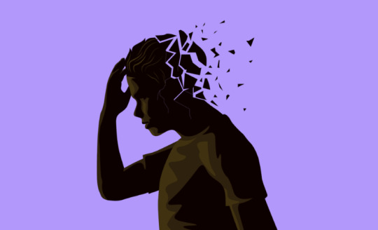
Understanding Elderly Depression: Causes, Symptoms, and Treatment

As our population ages, understanding the mental health challenges faced by the elderly becomes increasingly important. Depression is a significant concern among older adults, affecting their overall well-being and quality of life. In this article, we will explore the causes, symptoms, and treatment options for elderly depression, providing valuable insights for caregivers, family members, and healthcare professionals.
What is Elderly Depression?
Elderly depression is a mental health disorder characterized by persistent sadness, loss of interest in activities, and a range of physical and emotional symptoms. It is not a normal part of aging but a medical condition that requires attention and treatment.
Causes of Depression in the Elderly
Physical Health Issues: Chronic illnesses such as heart disease, diabetes, and arthritis can contribute to depression. The physical pain and limitations these conditions impose can lead to feelings of hopelessness and despair.
Loss and Bereavement: The death of a spouse, friends, or family members can trigger profound grief and loneliness, increasing the risk of depression.
Social Isolation: Reduced social interactions due to retirement, mobility issues, or the loss of loved ones can lead to feelings of isolation and depression.
Medication Side Effects: Some medications commonly prescribed to older adults can have side effects that contribute to depressive symptoms.
Genetics: A family history of depression can increase the likelihood of developing the condition.
Life Transitions: Major life changes, such as moving to a nursing home or adjusting to a new living situation, can be stressful and contribute to depression.
Symptoms of Elderly Depression
Recognizing depression in the elderly can be challenging as symptoms may differ from those in younger individuals. Common symptoms include:
Persistent sadness or anxiety
Loss of interest in activities once enjoyed
Fatigue and lack of energy
Changes in appetite and weight
Difficulty sleeping or oversleeping
Feelings of worthlessness or guilt
Difficulty concentrating and making decisions
Physical symptoms such as aches and pains without a clear cause
Thoughts of death or suicide
Treatment Options for Elderly Depression
Psychotherapy: Cognitive-behavioral therapy (CBT) and interpersonal therapy (IPT) are effective in treating depression by addressing negative thought patterns and improving coping strategies.
Medication: Antidepressants can be prescribed to help balance brain chemicals. It’s crucial to work closely with a healthcare provider to find the right medication and dosage, considering potential side effects and interactions with other medications.
Lifestyle Changes: Encouraging physical activity, a healthy diet, and regular sleep can improve mood and overall health.
Social Support: Maintaining strong social connections through family, friends, and community activities can help alleviate feelings of isolation and depression.
Support Groups: Joining a support group for older adults can provide a sense of belonging and understanding.
Medical Care: Managing chronic health conditions and ensuring proper medication management can reduce the physical factors contributing to depression.
Preventing Depression in the Elderly
Stay Connected: Encourage regular social interactions and participation in community activities.
Promote Physical Health: Encourage regular exercise and a balanced diet.
Provide Emotional Support: Be attentive and supportive, recognizing the emotional needs of older adults.
Monitor Medications: Be aware of potential side effects and interactions of prescribed medications.
Seek Professional Help: If signs of depression are present, seek the advice of healthcare professionals promptly.
Remember, if you need further guidance or support, don’t hesitate to reach out to your mental health professional or contact us for assistance.
#health#medicine#pain management#back pain#mental health#chiropractic#apdss#neckpain#depressionhelp#neurostar
2 notes
·
View notes
Note
like what? -cat kat cat
...
#and i meow meow and i meow meow meow || cat kat cat....#ooc: soery for no writing . ik about fo apdss out .
2 notes
·
View notes
Photo

#prontofalei (em Goiânia, Brazil) https://www.instagram.com/p/CCBQg-ApDss/?igshid=bgewoiuxx3l2
0 notes
Photo

Don’t you know I’m locoooooo !!!! #pachucostyle Chapeau en feutre, chemise «en coton, 70’s, Plume de faisan faites main, pince à col , épingle à cravate « trombone », veste longue « Canda », pantalon 50’s, bretelles à boutons, boutons « Bertelles », chaînes à goussets, montre à gousset « L.Roskopf », gants pilotes en cuir, edelweiss en ivoire, spectator en daim et tissus. #pachuco #heritage #zootsuit #1940s #oldfashioned #sartorialist #dressup #dapperstyle #suitstyle #mensfashion #barondxl #style #class #grandandytisme #gentleman #suit #ptoman #wiwt #ootd #dandy #style #chic #menwithstyle #frenchteush #jacket #cabcalloway #hat #barondxl #panamagomina #lesBaronsduGranDandytisme . https://www.instagram.com/p/B701L-apdSS/?igshid=upwue5lvk9nq
#pachucostyle#pachuco#heritage#zootsuit#1940s#oldfashioned#sartorialist#dressup#dapperstyle#suitstyle#mensfashion#barondxl#style#class#grandandytisme#gentleman#suit#ptoman#wiwt#ootd#dandy#chic#menwithstyle#frenchteush#jacket#cabcalloway#hat#panamagomina#lesbaronsdugrandandytisme
0 notes
Text
Children at risk of depression are associated with body dissatisfaction.
A recent longitudinal study led by UCL researchers reveals a connection between body dissatisfaction at age 11 and an elevated risk of depression by age 14. The study, published in The Lancet Psychiatry and supported by Wellcome, focused on 13,135 participants from the Millennium Cohort Study—a nationally representative birth cohort study spanning those born between 2000 and 2002.
The findings indicate that concerns about body image significantly contribute to the link between body mass index (BMI) and depression in children, particularly in girls. High BMI at age seven is associated with increased depressive symptoms by age 14, along with greater body dissatisfaction at age 11. Notably, body dissatisfaction explains 43% of the association between BMI at age seven and subsequent depressive symptoms.
The study highlights that all three associations are more pronounced in girls compared to boys. Lead author Dr. Francesca Solmi emphasizes the need for a nuanced approach to childhood weight management, considering potential mental health impacts and avoiding stigmatization. The study did not explore other factors contributing to the association between high BMI and depressive symptoms but suggests biological (e.g., inflammation) or environmental (e.g., bullying) pathways could play a role.
Emma Blundell, the first author and a trainee clinical psychologist at UCL Psychology & Language Sciences, raises concerns about public health strategies that may inadvertently foster feelings of guilt or shame. While promoting healthy diet and exercise is essential, interventions should prioritize not increasing body dissatisfaction and harming children’s mental health.
The researchers propose that targeting body image concerns in early adolescence may prevent depression, especially in girls, and suggest exploring interventions like psychological approaches or media literacy training. They emphasize the importance of further research to effectively address body image concerns in young people. The collaborative study involved researchers from UCL Great Ormond Street Institute of Child Health, UCL Institute of Epidemiology & Health Care, MRC Unit for Lifelong Health & Ageing at UCL, and Imperial College London. The Millennium Cohort Study is based at the UCL Centre for Longitudinal Studies in the IOE, UCL’s Faculty of Education & Society.
Remember, if you need further guidance or support, don’t hesitate to reach out to your mental health professional or contact us for assistance.
#chiropractic#apdss#neckpain#neurostar#depressionhelp#back pain#pain management#medicine#mental health#health
4 notes
·
View notes
Text
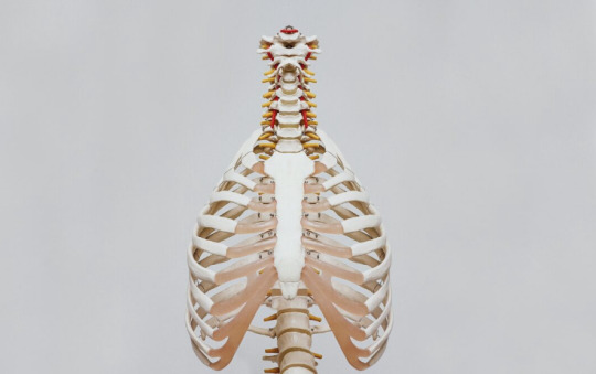
The link between depression and chronic back pain
According to the DSM-IV (1994), a diagnosis of major depression requires the presence of the following symptoms occurring daily for a minimum of two weeks, with at least five of the following being experienced:
Persistent mood characterized by feelings of depression, sadness, hopelessness, low mood, or irritability, which may be accompanied by occasional episodes of crying.
Significant change in appetite resulting in either noticeable weight loss or gain, or a noticeable increase or decrease in appetite.
Disturbances in sleep patterns, which may manifest as excessive sleep (hypersomnia) or insufficient sleep (hyposomnia).
Restlessness (agitation) or feeling of being slowed down (fatigue or low energy).
Loss of interest or pleasure in activities that were previously enjoyable or engaging.
Decreased sexual desire.
Feelings of worthlessness and/or guilt.
Difficulty concentrating or experiencing problems with memory.
Recurrent thoughts of death, suicide, or a desire to be deceased.
It is important to note that these symptoms must be consistently present and significantly impairing an individual’s daily functioning in order to meet the criteria for a major depressive episode.
Health professionals frequently encounter chronic pain and depression, yet only a limited number of studies have explored the relationship between these conditions in the general population (Currie and Wang, 2004) [2].
Individuals with chronic back pain are believed to be four times more likely to experience major depression compared to the general population (Sullivan, Reesor, Mikail & Fisher, 1992) [3]. Moreover, research conducted on chronic low back pain patients seeking treatment at pain clinics reveals even higher rates of depression. Between 32 to 82 percent of patients exhibit some form of depression or depressive symptoms, with an average prevalence of 62 percent (Sinel, Deardorff & Goldstein, 1996) [4].
Recent findings demonstrate a direct correlation between pain severity and the incidence of major depression, with a progressively higher rate of depression observed as pain intensity increases (Currie and Wang, 2004) [2]. Furthermore, the combination of chronic back pain and depression is associated with greater disability compared to experiencing either depression or chronic back pain alone.
Depression is a common occurrence among individuals dealing with chronic back pain compared to those with acute, short-term pain.
The development of depression in such cases can be attributed to a range of symptoms frequently experienced by individuals with chronic back pain or other spine-related discomfort.
Firstly, the pain itself often disrupts sleep, resulting in fatigue and daytime irritability. Additionally, the limitations imposed by back pain make patients move cautiously and slowly, leading to reduced social interaction and a lack of engagement in enjoyable activities, which can contribute to feelings of social isolation.
Furthermore, the inability to work due to chronic back pain may cause financial difficulties that impact not only the individual but also their entire family. Additionally, the use of anti-inflammatory medications can lead to gastrointestinal distress, while pain medications can induce a general sense of mental dullness, further exacerbating the challenges faced by individuals.
The persistent pain also serves as a distraction, making it difficult to concentrate and affecting memory function. Moreover, the decreased interest in sexual activity due to the pain adds additional stress to the patient’s relationships.
Understandably, these symptoms accompanying chronic back pain or neck pain can lead to feelings of hopelessness, despair, and other symptoms associated with major or clinical depression.
A recent study conducted by Strunin and Boden (2004) [5] examined the family consequences of chronic back pain. The findings revealed that patients experienced various limitations in their family and social roles. Physical constraints hindered their ability to perform household chores, take care of children, and engage in leisure activities with their spouses. Consequently, spouses and children often assumed the responsibilities previously handled by the individual with back pain. These changes in family dynamics frequently led to depression and anger among the individuals experiencing back pain and resulted in stress and strain within family relationships.
At Advanced Pain Diagnostic & Solutions, we can help with your chronic back pain and with your depression. Contact us to learn more!
#health#medicine#mental health#pain management#back pain#neurostar#depressionhelp#apdss#chiropractic
3 notes
·
View notes
Text
Discovery of new genetic links offers potential for the prevention and treatment of a common form of inflammatory arthritis.

A groundbreaking genetic study has identified two key genes linked to calcium pyrophosphate deposition (CPPD) disease, a painful and common form of inflammatory arthritis also known as pseudogout. Published in the Annals of the Rheumatic Diseases, this first-ever genome-wide association study (GWAS) highlights ENPP1 and RNF144B as major contributors to the development of CPPD in people of both European and African ancestry.
What Is CPPD Disease?
Calcium pyrophosphate deposition disease (CPPD) is a type of crystal-induced arthritis caused by the buildup of calcium pyrophosphate crystals in the joints. Often mistaken for gout, pseudogout leads to episodes of severe joint pain, inflammation, and swelling. CPPD is most common in adults over 60 and is seen in up to 30% of people over the age of 80.
In many cases, CPPD also coexists with osteoarthritis, and while the connection is not fully understood, the condition contributes significantly to joint damage and reduced mobility in older adults.
Key Findings: ENPP1 and RNF144B Identified as Genetic Drivers of CPPD
This large-scale genetic study was conducted through the Million Veteran Program, which includes health data from over 550,000 U.S. Veterans. Researchers analyzed every gene in the human genome to uncover genetic links to CPPD arthritis, and the results were clear: ENPP1 and RNF144B are strongly associated with the disease across multiple ethnic groups.
ENPP1 encodes a protein responsible for producing inorganic pyrophosphate, a critical component in CPP crystal formation.
RNF144B, though less understood, appears to play a role in inflammatory signaling pathways relevant to arthritis.
A Major Breakthrough in Understanding and Treating Pseudogout
Dr. Tony R. Merriman, lead researcher from the University of Alabama at Birmingham and University of Otago, explains:“Our discovery of ENPP1 is a game-changer. This gene directly impacts the chemical pathways that lead to CPP crystal buildup. It provides a clear and actionable target for new therapies.”
Dr. Sara K. Tedeschi, a rheumatologist at Harvard Medical School, adds:“This is an exciting time. ENPP1 inhibitors, already under development for other diseases, could be repurposed to finally offer an effective treatment for CPPD disease—something patients have needed for decades.”
Why This Study Matters: Unmet Need for CPPD Treatment
Currently, CPPD disease treatment options are limited to managing inflammation with NSAIDs, colchicine, or corticosteroids. There are no therapies that target the root cause—crystal formation.
Dr. Josef Smolen, Editor-in-Chief of Annals of the Rheumatic Diseases, emphasizes:“This landmark GWAS not only identifies potential drug targets but also brings us closer to precision medicine for inflammatory arthritis like pseudogout.”
What’s Next?
Because ENPP1 inhibitors are already in development for cancer and infectious diseases, they may soon be tested in clinical trials for CPPD arthritis. This could rapidly accelerate the timeline for delivering effective, disease-modifying treatments.
Key Takeaways for Patients and Healthcare Providers
CPPD (pseudogout) is a common inflammatory arthritis in older adults caused by calcium pyrophosphate crystals.
New genetic research identifies ENPP1 and RNF144B as key contributors.
ENPP1 inhibitors may represent a future treatment for CPPD disease.
Current CPPD treatments only manage symptoms, not the underlying cause.
If you or someone you know suffers from pseudogout or unexplained joint pain, stay informed about new genetic research and treatment options. Subscribe to our newsletter for the latest updates on CPPD disease and arthritis breakthroughs.
For more information about our clinic, medical professionals, and treatment options, please visit our main website.
#health#medicine#pain management#mental health#apdss#back pain#chiropractic#neckpain#depressionhelp#neurostar
0 notes
Text
Spending time in nature may assist individuals with chronic back pain in managing their condition.

Introduction: A Natural Approach to Chronic Pain Relief
Chronic lower back pain affects millions worldwide and is often difficult to manage through medication or physical therapy alone. But according to a groundbreaking study published in The Journal of Pain, nature may offer a powerful and underutilized tool for relief.
Researchers from the University of Plymouth and the University of Exeter have discovered that time spent in natural environments can significantly improve physical and emotional well-being for people living with chronic lower back pain—some for nearly 40 years. This is the first study to specifically explore how people with this condition use nature as part of their pain-coping strategies.
Key Findings: How Nature Helps Chronic Pain Sufferers
The study, based on interviews with ten individuals who had experienced chronic lower back pain for between five and 38 years, uncovered several consistent benefits of being in nature:
1. Social Connection Reduces Isolation
Many participants reported that outdoor environments provided opportunities for social interaction, helping them feel less isolated compared to staying indoors.
2. Nature Offers a Healthy Distraction
Being in natural surroundings created a sense of escapism. The visual beauty, sounds of water, and fresh air helped divert attention away from persistent pain.
3. Gentle Exercise in a Pleasant Setting
Participants preferred walking or light physical activity outdoors over indoor environments like gyms, as the natural setting felt more enjoyable and soothing.
4. Calming Sensory Effects
The sights, sounds, and smells of nature promoted feelings of calm and reduced the anxiety often associated with chronic pain. Natural elements such as greenery, flowing water, and birdsong helped create a tranquil mental state.
Accessibility Barriers Limit Nature’s Healing Power
Despite the clear benefits, many participants faced physical challenges that limited their access to outdoor spaces. These included:
Unstable or uneven terrain
Lack of seating
Limited mobility or difficulty leaving the home
These barriers reduced the frequency and enjoyment of time spent outdoors, especially for those with severe mobility issues.
Recommendations: How to Make Nature-Based Pain Relief More Accessible
The researchers emphasize that natural spaces should be made more accessible for individuals with chronic pain. Recommended improvements include:
Installing smoother, wheelchair-friendly paths
Providing resting benches at regular intervals
Offering shade and shelter to accommodate different weather conditions
They also suggest that healthcare professionals consider nature exposure as part of a holistic pain management plan.
Virtual Reality: Bringing Nature to Those Who Can’t Go Outside
For individuals who are unable to access outdoor environments, the research team is developing virtual reality (VR) technologies to simulate the experience of being in nature. This innovative approach could bring the calming, therapeutic benefits of forests, lakes, and gardens into the homes of people living with chronic pain.
According to Dr. Sam Hughes, Senior Lecturer in Pain Neuroscience at the University of Exeter, VR could play a crucial role in improving inclusivity in chronic pain care:“Immersive technologies can offer people the benefits of nature without the physical challenges of navigating outdoor terrain.”
Final Thoughts: Nature as a Powerful Pain Management Tool
As modern medicine shifts toward more holistic and accessible treatment methods, this study highlights the powerful role of nature in managing chronic lower back pain. Whether through physical access to green spaces or the use of virtual environments, reconnecting with nature may become a key strategy in chronic pain relief.
If you or someone you know suffers from chronic pain, consider adding nature walks, outdoor mindfulness, or VR-based natural experiences to your routine. The healing power of nature might be closer—and more effective—than you think.
For more information about our clinic, medical professionals, and treatment options, please visit our main website.
Recent Posts
Spending time in nature may assist individuals with chronic back pain in managing their condition.
How Stem Cell Therapy is Revolutionizing Chronic Pain Management
Legal Mind in Medicine: Pain Medicine by Kayvan D. Haddadan, MD.
Knee Osteoarthritis: Causes, Symptoms, and the Best Treatments to Relieve Pain
Spondylolisthesis: Causes, Symptoms, Diagnosis, and Treatment
#health#medicine#pain management#mental health#apdss#back pain#chiropractic#neurostar#neckpain#depressionhelp
0 notes
Text
How Stem Cell Therapy is Revolutionizing Chronic Pain Management

Introduction: A New Frontier in Pain Relief
Millions of people worldwide suffer from chronic pain — a condition that can severely impact quality of life. Traditional treatments like opioids, steroid injections, and surgery often provide temporary relief or come with significant side effects. Fortunately, advances in regenerative medicine have introduced a promising alternative: stem cell therapy for chronic pain.
Stem cell therapy harnesses the body’s natural healing abilities to repair damaged tissues and reduce inflammation. This innovative treatment is helping many patients find lasting relief from conditions such as osteoarthritis, back pain, joint pain, and tendon injuries. In this article, we’ll explore how stem cell therapy works, its benefits, and why it is becoming a game-changer in chronic pain management.
What Is Stem Cell Therapy?
Stem cells are the body’s master cells — capable of developing into various cell types such as cartilage, muscle, tendon, or nerve cells. In stem cell therapy, these cells are harvested (commonly from the patient’s bone marrow or adipose tissue), processed, and then injected into the site of pain or injury.
Once introduced, the stem cells can:
Modulate inflammation
Promote tissue repair and regeneration
Reduce pain signals
Improve function and mobility
Benefits of Stem Cell Therapy for Chronic Pain
1. Natural Healing, Not Just Symptom Management
Unlike pain medications that simply mask symptoms, stem cell therapy addresses the underlying cause of pain. By promoting tissue regeneration and reducing inflammation, it facilitates the body’s ability to heal itself.
2. Minimally Invasive Alternative to Surgery
Many patients with joint or back pain face the prospect of invasive surgeries like joint replacements or spinal fusion. Stem cell injections offer a less invasive option with shorter recovery times and fewer risks.
3. Reduces Dependence on Pain Medications
Long-term use of opioids and NSAIDs can lead to side effects and dependency. Stem cell therapy may reduce or eliminate the need for these medications by providing lasting pain relief.
4. Effective for Multiple Chronic Pain Conditions
Stem cell therapy is being successfully used to treat a wide range of pain conditions, including:
Osteoarthritis of the knee, hip, and shoulder
Degenerative disc disease and chronic back pain
Rotator cuff injuries
Tendonitis and ligament injuries
Neuropathic pain
5. Low Risk of Side Effects
Because most procedures use the patient’s own stem cells (autologous), there is a low risk of rejection or allergic reactions. Side effects are generally minimal, such as temporary soreness at the injection site.
The Science Behind It: How Stem Cells Relieve Pain
Stem cells release bioactive molecules like growth factors, cytokines, and extracellular vesicles that:
Reduce inflammation in damaged tissues
Stimulate the growth of new blood vessels (angiogenesis)
Recruit other cells needed for tissue repair
Modulate the immune response to prevent further tissue damage
This biological cascade leads to pain reduction and improved function over time.
What Does the Research Say?
Growing clinical evidence supports the use of stem cell therapy in pain management:
A 2022 review in Pain Physician Journal reported significant pain reduction and improved joint function in osteoarthritis patients treated with stem cell injections.
Studies on degenerative disc disease show improved pain scores and spinal disc height restoration following stem cell treatment.
Athletes and active individuals have returned to sport faster with stem cell therapy after tendon or ligament injuries.
While more large-scale studies are underway, early results are highly encouraging.
Who Is a Good Candidate?
Stem cell therapy may be ideal for patients who:
Have chronic pain unresponsive to conventional treatments
Want to avoid surgery or are poor surgical candidates
Prefer a natural, regenerative approach to healing
Suffer from joint, spine, or soft tissue pain
Consulting with a specialist in regenerative medicine is key to determining eligibility and expected outcomes.
Conclusion: The Future of Pain Management Is Here
Stem cell therapy represents an exciting advancement in the treatment of chronic pain. By tapping into the body’s inherent healing power, it offers the potential for long-lasting relief, improved function, and a better quality of life — without the downsides of surgery or long-term medication use.
If you’re struggling with chronic pain, consider speaking to a qualified provider about whether stem cell therapy might be right for you.
For more information about our clinic, medical professionals, and treatment options, please visit our main website.
#health#medicine#pain management#mental health#apdss#chiropractic#back pain#neckpain#neurostar#depressionhelp
0 notes
Text
Knee Osteoarthritis: Causes, Symptoms, and the Best Treatments to Relieve Pain

Knee osteoarthritis (OA) is one of the most common causes of knee pain worldwide, affecting millions of people—especially those over the age of 50. As the cartilage in the knee joint wears away, bones begin to rub together, causing pain, stiffness, and swelling. Whether you’re newly diagnosed or have been living with OA for years, this guide offers a comprehensive look at knee osteoarthritis—its causes, symptoms, and top treatments available today.
What Is Knee Osteoarthritis?
Knee osteoarthritis is a degenerative joint disease that results from the breakdown of articular cartilage in the knee. Cartilage acts as a cushion between bones. When it deteriorates, movement becomes painful and less fluid.
Common Causes and Risk Factors
Understanding what causes knee osteoarthritis can help with prevention and early treatment. Here are some of the most common knee OA risk factors:
Age: Risk increases significantly after 45 years old.
Obesity: Excess weight puts more stress on the knee joint.
Previous knee injuries: Sports injuries or accidents can accelerate cartilage damage.
Genetics: Family history may play a role.
Repetitive stress: Jobs or sports that involve frequent kneeling or squatting.
Symptoms of Knee Osteoarthritis
People with knee osteoarthritis often report:
Knee pain that worsens with activity but improves with rest
Stiffness, especially in the morning or after sitting
Swelling and tenderness around the knee
Grinding or popping sounds during movement (crepitus)
Reduced range of motion and difficulty walking or climbing stairs
Stages of Knee Osteoarthritis
Knee OA is often classified in five stages (0–4):
Stage 0: Normal knee with no signs of OA.
Stage 1 (Minor): Minor wear and tear, possible bone spur growth.
Stage 2 (Mild): More noticeable bone spurs, slight joint space narrowing.
Stage 3 (Moderate): Cartilage damage, frequent pain, swelling.
Stage 4 (Severe): Major loss of cartilage, chronic pain, very limited mobility.
Top Treatments for Knee Osteoarthritis
There is no cure for osteoarthritis, but many knee OA treatment options can help relieve symptoms and slow progression.
1. Lifestyle Changes
Weight loss: Reduces joint pressure and improves mobility.
Exercise: Low-impact activities like swimming, walking, and cycling strengthen muscles around the knee.
Physical therapy: Improves joint function and flexibility.
2. Medications
NSAIDs: Ibuprofen or naproxen reduce inflammation and pain.
Acetaminophen: Useful for mild to moderate pain.
Topical analgesics: Creams like capsaicin applied to the skin.
3. Injections
Corticosteroid injections: Provide short-term pain relief.
Hyaluronic acid injections: Lubricate the joint for smoother movement.
Platelet-rich plasma (PRP): Experimental, may promote healing.
4. Assistive Devices
Knee braces: Provide support and alignment.
Canes or walkers: Help reduce pressure on the knee.
5. Surgical Options
When conservative treatments fail:
Arthroscopy: Minimally invasive, but less common now.
Osteotomy: Bone reshaping for better alignment.
Total knee replacement (arthroplasty): Most effective long-term solution for severe OA.
Natural Remedies for Knee Osteoarthritis
In addition to medical treatments, some natural options may help:
Turmeric (curcumin): Anti-inflammatory properties.
Omega-3 fatty acids: Found in fish oil, help reduce joint inflammation.
Glucosamine and chondroitin: Supplements that may protect cartilage.
Note: Always consult your healthcare provider before starting new supplements.
Can You Prevent Knee Osteoarthritis?
While not always preventable, you can lower your risk with these strategies:
Maintain a healthy weight
Stay physically active
Avoid knee injuries
Strengthen leg muscles
Practice good posture and body mechanics
Final Thoughts: Living Well with Knee Osteoarthritis
Knee osteoarthritis can affect your quality of life, but early diagnosis and proactive treatment can make a huge difference. By combining lifestyle changes, physical therapy, medications, and sometimes surgery, many people find significant relief.
If you’re experiencing chronic knee pain, talk to your healthcare provider to explore the best options for managing knee OA and getting back to doing what you love.
Frequently Asked Questions
Q: What is the best exercise for knee osteoarthritis? A: Low-impact exercises like swimming, walking, and stationary cycling help reduce stiffness and improve joint function.
Q: Is walking good for knee osteoarthritis? A: Yes, regular walking helps keep the knee joint mobile and reduces stiffness.
Q: Can knee osteoarthritis be reversed? A: It cannot be reversed, but treatments can significantly reduce symptoms and improve joint function.
Q: When is knee replacement necessary? A: When conservative treatments no longer relieve pain and your mobility is severely affected.
For more information about our clinic, medical professionals, and treatment options, please visit our main website.
#health#medicine#pain management#mental health#apdss#back pain#chiropractic#neckpain#neurostar#depressionhelp
0 notes
Text
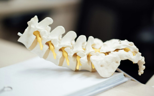
Spondylolisthesis: Causes, Symptoms, Diagnosis, and Treatment

What Is Spondylolisthesis?
Spondylolisthesis is a spinal condition in which one vertebra slips forward over the one beneath it. This misalignment can cause pain, nerve compression, and mobility issues. The condition is most commonly seen in the lower back (lumbar spine) but can also affect other areas of the spine.
Types of Spondylolisthesis
There are several types of spondylolisthesis, classified based on their underlying causes:
Congenital Spondylolisthesis – A birth defect in the spine that predisposes individuals to vertebral slippage.
Isthmic Spondylolisthesis – Caused by a small stress fracture (spondylolysis) in the pars interarticularis, a region of the vertebra.
Degenerative Spondylolisthesis – Occurs due to aging and wear-and-tear changes in the spine, leading to instability.
Traumatic Spondylolisthesis – Results from a sudden injury or trauma to the spine.
Pathologic Spondylolisthesis – Develops due to diseases that weaken the spinal structures, such as tumors or osteoporosis.
Post-Surgical Spondylolisthesis – A rare occurrence following spinal surgery.
Causes and Risk Factors
Spondylolisthesis can develop due to various factors, including:
Genetics – Some individuals are born with spinal abnormalities that increase their risk.
Repetitive Stress – Athletes, such as gymnasts, football players, and weightlifters, are more prone due to repeated stress on the spine.
Aging – Degeneration of the spinal discs and joints can lead to instability.
Injury or Trauma – Sudden forceful impact can displace a vertebra.
Spinal Conditions – Diseases such as osteoporosis or spinal tumors may weaken the vertebrae.
Symptoms of Spondylolisthesis
Symptoms vary depending on the severity and location of the slippage. Common signs include:
Lower Back Pain – Worsens with activity and prolonged standing.
Stiffness in the Back – Reduced flexibility, especially in the lumbar spine.
Muscle Tightness or Spasms – Particularly in the hamstrings and lower back.
Leg Pain (Sciatica) – Due to nerve compression, leading to tingling, numbness, or weakness in the legs.
Difficulty Walking or Standing for Long Periods – Pain relief often occurs when bending forward or sitting.
Loss of Bladder or Bowel Control (Rare but serious) – May indicate severe nerve compression requiring immediate medical attention.
How Is Spondylolisthesis Diagnosed?
A doctor typically diagnoses spondylolisthesis through:
Physical Examination – Evaluating posture, movement, and pain levels.
X-rays – Detects vertebral misalignment and determines the grade of slippage.
MRI (Magnetic Resonance Imaging) – Identifies nerve compression and soft tissue involvement.
CT Scan (Computed Tomography) – Provides detailed images for a more precise assessment.
Spondylolisthesis Grading
Spondylolisthesis is classified into grades based on the percentage of vertebral slippage:
Grade I: 1-25%
Grade II: 26-50%
Grade III: 51-75%
Grade IV: 76-100%
Grade V (Spondyloptosis): The vertebra has completely fallen off the one below it.
Treatment Options for Spondylolisthesis
The treatment approach depends on the severity of symptoms and the degree of vertebral slippage. Options include:
Non-Surgical Treatments
Rest and Activity Modification – Avoid high-impact activities to prevent worsening symptoms.
Physical Therapy – Strengthening core and back muscles to provide better spinal support.
Pain Management – Over-the-counter NSAIDs (ibuprofen, naproxen) or prescription pain relievers.
Bracing – Helps stabilize the spine in younger patients or those with mild slippage.
Epidural Steroid Injections – Reduces inflammation and relieves nerve pain.
Surgical Treatments
Surgery is considered if non-surgical methods fail, or if symptoms are severe. Common surgical options include:
Spinal Fusion – Fusing affected vertebrae to prevent further movement.
Laminectomy (Decompression Surgery) – Removes part of the bone or tissue pressing on nerves.
Instrumented Fusion – Using screws, rods, or cages to stabilize the spine.
Can You Prevent Spondylolisthesis?
While not all cases are preventable, you can reduce your risk by:
Maintaining Good Posture – Reducing strain on the lower back.
Regular Exercise – Strengthening core and back muscles for better spinal support.
Avoiding High-Impact Activities – Especially if you have a history of back pain.
Using Proper Lifting Techniques – Lifting with your legs instead of your back to minimize strain.
When to See a Doctor
Seek medical attention if you experience:
Persistent lower back pain not relieved by rest.
Leg weakness, numbness, or tingling.
Difficulty walking or standing for prolonged periods.
Loss of bladder or bowel control (emergency sign).
Conclusion
Spondylolisthesis can significantly impact daily life, but with proper diagnosis and treatment, most individuals can manage their symptoms effectively. Whether through physical therapy, medications, or surgery in severe cases, early intervention is key to maintaining spinal health and mobility. If you suspect spondylolisthesis, consult a healthcare professional for an accurate diagnosis and personalized treatment plan.
For more information about our clinic, medical professionals, and treatment options, please visit our main website.
#health#medicine#pain management#mental health#apdss#chiropractic#neckpain#back pain#neurostar#depressionhelp
0 notes
Text
Spondylolisthesis: Causes, Symptoms, Diagnosis, and Treatment
What Is Spondylolisthesis?
Spondylolisthesis is a spinal condition where one vertebra slips forward over the one beneath it. This displacement can cause significant discomfort, nerve compression, and mobility issues. The severity ranges from mild to severe, with some cases requiring surgical intervention.
Types of Spondylolisthesis
There are several types of spondylolisthesis, classified based on their cause:
Congenital Spondylolisthesis – Present at birth due to abnormal spinal formation.
Isthmic Spondylolisthesis – Caused by a small fracture in the pars interarticularis, commonly seen in athletes.
Degenerative Spondylolisthesis – Resulting from aging and spinal degeneration, usually affecting older adults.
Traumatic Spondylolisthesis – Occurs due to injury or trauma that weakens spinal structures.
Pathologic Spondylolisthesis – Caused by diseases such as osteoporosis or tumors affecting the spine.
Post-Surgical Spondylolisthesis – A complication from previous spinal surgery.
Causes and Risk Factors
Spondylolisthesis can develop due to various factors, including:
Genetic predisposition – Some individuals are born with structural weaknesses in the spine.
Repetitive stress – High-impact activities such as gymnastics, football, and weightlifting.
Aging – Wear and tear over time leading to spinal instability.
Injury – Sudden trauma or fractures to the vertebrae.
Degenerative conditions – Arthritis and disc degeneration contribute to spinal instability.
Symptoms of Spondylolisthesis
Symptoms vary based on severity and affected spinal level, including:
Lower back pain – The most common symptom, worsened by activity.
Sciatica – Pain radiating down the legs due to nerve compression.
Tingling or numbness – Especially in the legs and feet.
Weakness in the lower limbs – Difficulty walking or standing for long periods.
Muscle tightness – Particularly in the hamstrings.
Postural changes – Increased curvature of the lower spine (lordosis or swayback).
Diagnosis
To diagnose spondylolisthesis, doctors use:
Physical examination – Evaluating flexibility, pain levels, and nerve function.
X-rays – Identifying vertebral slippage.
MRI or CT scans – Providing detailed images of spinal nerves and soft tissues.
Grade Classification – Measured using the Meyerding scale:
Grade I: 0-25% slippage
Grade II: 26-50% slippage
Grade III: 51-75% slippage
Grade IV: 76-100% slippage
Grade V (Spondyloptosis): Complete vertebral slippage
Treatment Options
Treatment depends on severity, symptoms, and patient lifestyle.
Non-Surgical Treatments
Physical Therapy – Strengthening core muscles to support the spine.
Pain Management – NSAIDs, muscle relaxants, and epidural steroid injections.
Bracing – Wearing a lumbar brace to stabilize the spine.
Lifestyle Modifications – Avoiding heavy lifting and high-impact activities.
Surgical Treatments
Surgery is recommended for severe cases or when conservative treatments fail. Procedures include:
Spinal Fusion – Fusing two vertebrae to prevent further slippage.
Laminectomy – Removing part of the vertebra to relieve nerve pressure.
Instrumented Fixation – Using screws and rods for stability.
Prevention Tips
Maintain a healthy weight – Reducing pressure on the spine.
Exercise regularly – Strengthening core and back muscles.
Use proper lifting techniques – Preventing spinal injuries.
Stretching routines – Enhancing spinal flexibility and mobility.
For more information about our clinic, medical professionals, and treatment options, please visit our main website.
#health#medicine#pain management#mental health#apdss#back pain#chiropractic#neckpain#depressionhelp#neurostar
0 notes
Text
The brain experiences unexpected pain more intensely.
Pain perception varies significantly, with some instances feeling more intense than expected and others less so. This variability suggests that our experience of pain is influenced by expectations and uncertainty.
Two main hypotheses explain how the brain perceives pain. The Estimate Hypothesis suggests that the brain predicts pain intensity based on prior expectations, while the Surprise Hypothesis proposes that pain perception is driven by the difference between prediction and actual experience—known as prediction error.
In this study, researchers investigated the underlying mechanisms of pain perception. Participants were exposed to painful thermal stimuli while viewing either painful or non-painful visual cues in virtual reality. They then reported their perceived pain intensity. The findings revealed that pain was perceived more strongly when the prediction error was large, supporting the Surprise Hypothesis as a more accurate model of pain processing. Additionally, unexpected events were shown to amplify pain perception.
For individuals with chronic pain, persistent uncertainty and anxiety about pain may further increase perceived intensity. Reducing the discrepancy between pain expectation and reality—minimizing “surprise”—could help manage pain more effectively. A deeper understanding of pain perception may lead to improved treatments for chronic pain and trauma recovery.
For more information about our clinic, medical professionals, and treatment options, please visit our main website.
#apdss#back pain#chiropractic#neckpain#mental health#neurostar#pain management#medicine#health#depressionhelp
0 notes
Text
Healthy lifestyle and back pain

Low back pain is a top cause of disability worldwide, with many treatments, such as medications, offering little lasting relief. However, a groundbreaking study from the University of Sydney’s Centre for Rural Health, published in JAMA Network Open, reveals that integrating lifestyle changes into back pain care could be the key to reducing disability and enhancing overall quality of life.
Lifestyle-Focused Care vs. Standard Treatment
The study involved 346 Australians with chronic low back pain and at least one lifestyle risk factor, such as obesity, poor diet, smoking, or inactivity. Participants were randomly assigned to either the “Healthy Lifestyle Program (HeLP)” or standard physiotherapy-based care.
The HeLP group received comprehensive support from physiotherapists, dietitians, and health coaches. These professionals helped participants identify lifestyle habits affecting their back pain—such as lack of exercise, poor sleep, or smoking—and provided evidence-based advice to address these issues over six months.
Key Results: Improved Disability and Weight Loss
The results showed clear benefits of lifestyle-integrated care. Compared to standard treatment, HeLP participants experienced reduced disability, scoring an average of 1.3 points lower on the Roland Morris Disability Questionnaire (where higher scores indicate more severe disability). They also lost an average of 1.6 kg more than those in the standard care group.
Why Back Pain Care Needs a Paradigm Shift
Associate Professor Chris Williams, the study’s lead investigator, emphasized the need to rethink back pain management: “Resolving back pain requires more than just focusing on the spine. Our bodies are complex ecosystems where many factors interact. Comprehensive care that addresses lifestyle factors can make all the difference.”
Williams also highlighted that issues like bulging discs or joint degeneration are rarely the main causes of long-term back pain. Yet, many patients are still referred for unnecessary surgeries or prescribed medications that may do more harm than good.
Empowering Patients Through Lifestyle Changes
Lead author Dr. Emma Mudd stressed the real-world impact of this approach: “Many people with chronic back pain feel abandoned, often receiving high-cost, ineffective treatments while missing out on self-management strategies. By focusing on simple lifestyle changes, we empower patients to take control of their pain, improve their symptoms, and enhance their overall quality of life.”
Beyond Pain Relief: Broader Health Benefits
The researchers believe that integrating lifestyle support into back pain care could also reduce the risk of other chronic conditions. However, they note that global back pain guidelines have yet to fully embrace this approach.
“This research has the potential to influence future updates to back pain treatment guidelines,” said Dr. Mudd. “Patients value holistic care, and the results speak for themselves.”
Takeaway for Clinicians and Patients
Clinicians are encouraged to incorporate lifestyle support into their treatment plans for back pain. “There’s no single ‘right’ way to do this,” said Associate Professor Williams, “but listening to patients and involving them in decision-making is key.”
By addressing lifestyle factors, treatments like the HeLP program not only improve back pain outcomes but also empower patients to lead healthier, more fulfilling lives. This innovative approach offers hope for millions living with chronic back pain worldwide.
For more information about our clinic, medical professionals, and treatment options, please visit our main website.
#health#medicine#pain management#mental health#apdss#back pain#chiropractic#neckpain#neurostar#depressionhelp
1 note
·
View note
Text

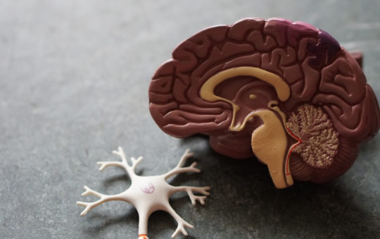
Neural stem cell transplantation holds potential as a treatment for chronic spinal cord injuries.
A Phase I clinical trial conducted by researchers at the University of California San Diego School of Medicine has confirmed the long-term safety and feasibility of neural stem cell transplantation for chronic spinal cord injuries. These injuries, which often cause partial or complete paralysis, currently have no cure. Over a five-year period, the study observed four patients with chronic spinal cord injuries, two of whom showed sustained neurological improvement following neural stem cell implantation. Improvements included increased motor and sensory scores, enhanced electromyography (EMG) activity, and, in some cases, better pain management.
Neural stem cell transplantation, an emerging treatment for neurological disorders and injuries, involves implanting human-derived stem cells into damaged areas of the nervous system. This innovative approach leverages the regenerative potential of these cells to repair damaged tissue and integrate effectively with the existing nervous system.
All participants in the trial tolerated the treatment well. While the primary goal was to assess safety and tolerability, the findings indicate potential therapeutic benefits for chronic spinal cord injuries. Encouraged by these outcomes, the researchers aim to launch a Phase II clinical trial to evaluate the treatment’s efficacy.
The study, published in the December 17 edition of Cell Reports Medicine, was led by Dr. Joseph Ciacci, a professor in the Department of Neurological Surgery at UC San Diego School of Medicine and a neurosurgeon at UC San Diego Health, and Dr. Joel Martin, a former neurological surgery resident at UC San Diego and now a neurosurgeon at Orlando Health. The research received support from the California Institute of Regenerative Medicine (CIRM) UC San Diego Alpha Stem Cell Clinic and the Sanford Stem Cell Clinical Center within the Sanford Stem Cell Institute.
For more information about our clinic, medical professionals, and treatment options, please visit our main website.
#medicine#health#pain management#mental health#apdss#back pain#chiropractic#neckpain#depressionhelp#neurostar
0 notes
Text
PRP and Chronic Pain Treatment: A Comprehensive Guide

Chronic pain is a debilitating condition that affects millions of individuals worldwide, significantly impacting their quality of life.
While traditional treatments like medication, physical therapy, and surgery have long been the mainstays of care, regenerative medicine has emerged as a promising frontier. Among these innovative treatments, Platelet-Rich Plasma (PRP) therapy has gained considerable attention for its potential to manage chronic pain effectively. This article delves into the science behind PRP, its applications, and what patients can expect during treatment.
Understanding PRP Therapy
PRP therapy utilizes the body’s natural healing mechanisms to repair damaged tissues. Platelet-rich plasma is derived from the patient’s own blood, processed to concentrate platelets, which are rich in growth factors and other bioactive proteins that promote tissue repair and regeneration.
How Is PRP Prepared?
Blood Collection: A small amount of blood is drawn from the patient.
Centrifugation: The blood is placed in a centrifuge, separating its components to isolate platelets and plasma.
Injection: The concentrated PRP is injected into the targeted area, such as a joint, tendon, or muscle.
Mechanism of Action
PRP works by stimulating cellular repair processes. Platelets release growth factors like transforming growth factor-beta (TGF-β) and vascular endothelial growth factor (VEGF), which:
Encourage collagen synthesis.
Enhance blood flow to the injured area.
Reduce inflammation.
Promote cellular regeneration.
This mechanism makes PRP particularly effective for conditions where traditional treatments have limited success, especially in chronic pain syndromes.
Applications of PRP in Chronic Pain Management
1. Osteoarthritis (OA)
PRP injections are widely used for managing pain and improving function in patients with knee, hip, and shoulder osteoarthritis. Studies suggest that PRP can delay the progression of OA by restoring cartilage integrity and reducing inflammation.
2. Tendon Injuries
Conditions like tennis elbow (lateral epicondylitis) and Achilles tendinitis often respond poorly to conventional therapies. PRP has been shown to accelerate tendon healing, offering pain relief and improved function.
3. Back Pain
Chronic back pain due to degenerative disc disease or facet joint arthritis is another area where PRP has shown promise. By addressing inflammation and promoting tissue repair, PRP can alleviate discomfort and improve mobility.
4. Muscle Injuries
Athletes and active individuals with muscle injuries may benefit from PRP to speed up recovery and minimize downtime.
5. Ligament Injuries
PRP is increasingly used for partial tears in ligaments, such as those in the knee (e.g., ACL) or shoulder, helping to strengthen the tissue and reduce pain.
Benefits of PRP Therapy
Minimally Invasive: PRP therapy involves simple injections, eliminating the need for surgery.
Low Risk: Since PRP is derived from the patient’s own blood, the risk of adverse reactions is minimal.
Natural Healing: PRP harnesses the body’s innate healing mechanisms, making it a holistic treatment option.
Long-Lasting Relief: Many patients report significant and sustained pain relief following PRP therapy.
The PRP Procedure: What to Expect
Consultation: The provider evaluates the patient’s condition to determine if PRP is appropriate.
Preparation: On the day of treatment, blood is drawn and processed.
Injection: Using ultrasound guidance, PRP is precisely injected into the affected area.
Post-Treatment: Patients may experience mild swelling or discomfort, which usually subsides within a few days. Physical activity is often limited for a short period to optimize healing.
For more information about our clinic, medical professionals, and treatment options, please visit our main website.
#health#medicine#pain management#mental health#apdss#back pain#chiropractic#neckpain#neurostar#depressionhelp
0 notes