#medical radiation technologist
Text
The Importance of Radiologic Technology in Early Diagnosis and Treatment
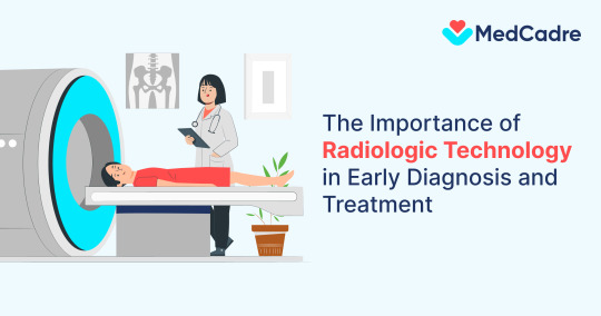
In the ever-evolving landscape of healthcare, technological advancements play a pivotal role in enhancing diagnostic capabilities and treatment outcomes. Among these, radiologic technology stands out as a cornerstone in early diagnosis and treatment planning. This article explores the significance of radiologic technology in healthcare, highlighting its contributions to timely and accurate diagnoses, treatment decisions, and patient care.
The evolution of radiologic technology
Radiologic technology has witnessed remarkable advancements since the discovery of X-rays by Wilhelm Roentgen in 1895. From the early days of basic radiography to the sophisticated modalities available today, including computed tomography (CT), magnetic resonance imaging (MRI), ultrasound, and nuclear medicine, the field has transformed the way healthcare professionals perceive and address medical conditions.
Early diagnosis saves lives
One of the primary advantages of radiologic technology lies in its ability to facilitate early diagnosis. Early detection of diseases such as cancer, cardiovascular disorders, and neurological conditions significantly improves the chances of successful treatment and enhances patient outcomes. Imaging modalities like mammography, CT scans, and MRIs empower healthcare providers to identify abnormalities at their nascent stages, allowing for prompt intervention.
Precision medicine and treatment planning
Radiologic technology not only aids in the early identification of diseases but also plays a crucial role in treatment planning. Advanced imaging techniques enable healthcare professionals to visualize the internal structures of the body with unparalleled clarity, facilitating precision medicine. This precision ensures that treatment strategies are tailored to the unique characteristics of each patient, maximizing effectiveness and minimizing side effects.
Minimally invasive interventions
Interventional radiology has emerged as a game-changer in the field of medicine, offering minimally invasive alternatives to traditional surgical procedures. Procedures such as angioplasty, embolization, and radiofrequency ablation are guided by real-time imaging, reducing recovery times and enhancing patient comfort. Radiologic technology has thus expanded the scope of treatment options available to patients across various medical disciplines.
Radiologic technologists: The heroes behind the screens
The individuals responsible for operating and interpreting radiologic technology are radiologic technologists. Their expertise is critical in ensuring the accuracy of diagnostic images and the safety of patients. Radiologic technologists play a vital role in collaborating with other healthcare professionals to provide comprehensive and timely healthcare solutions.
Join MedCadre for a rewarding radiologic technology career
For those aspiring to embark on a fulfilling career in radiologic technology, partnering with a top healthcare staffing agency is paramount. MedCadre, a leading healthcare staffing agency in the USA, connects qualified healthcare professionals with rewarding job opportunities across the nation. Whether you are an experienced radiologic technologist or a recent graduate, MedCadre can help you find the perfect placement to kickstart or advance your career.
Conclusion
In conclusion, the importance of radiologic technology in early diagnosis and treatment cannot be overstated. From its humble beginnings with X-rays to the sophisticated modalities available today, radiologic technology continues to revolutionize healthcare. As we navigate the complexities of modern medicine, the role of radiologic technologists remains central to ensuring accurate diagnoses, personalized treatment plans, and ultimately, improved patient outcomes. Take the first step towards a rewarding career in radiologic technology – join MedCadre and unlock a world of opportunities to make a difference in healthcare.
Read more about radiologic technologists: https://blog.medcadre.com/radiologic-technologist-professional-development-tips/
Visit our site and submit your CV: https://medcadre.com/careers
#radiologic technologist#radiologic technologist jobs#radiology technician#medical radiation technologist#medcadre
0 notes
Text
ah, the things your sibling texts you when you are a radiation safety officer.
The dose was elsewhere given in layperson's terms as "ten x-rays per hour" which, using the RADAR Medical Procedure rough estimate, and a high-dose procedure (lumbar spine), gives me something on the order of 7 mSv/hour (700 mrem/hour). This is, in fact, pretty bad; for reference, the NRC's goal for the general public is 5 mSv (500 mrem) per year, and as a radiation worker, if I were to get about half that dose (that is to say, the dose from being in close proximity with this capsule, unshielded, for an hour) this would trigger an investigation into what I was doing.
With all that said, dose limits are intentionally fairly conservative, and you would need to handle this for quite a long time continuously to get a fatal dose. The Guardian reports it as being 19 Bq (about 0.5 nCi), incidentally, which is a pretty standard Cs-137 test source. Like, if you know a nuclear medicine technologist, they probably handle a very similar source regularly, but for brief periods at a time.
Anyway, if you're in Western Australia, do pay attention to this and if you see a weird metal thing on the road, matching this description, don't touch it! Follow the instructions of authorities! But, that said, the good thing about radiation is that your dose drops off steeply if you keep a distance. Unless you are in close proximity for an extended period of time, you are not at risk.
(On the other hand, from that same safety officer standpoint, I am screaming into a pillow. No one checked this en route? No one ensured this was in a secure container? No one checked this on delivery? This is in violation of like 5 separate parts of HAZMAT training in the US, and probably in Australia too.)
#welcome to cursed physics knowledge with me#also yes. all of radiation physics in the us anyway is done in both metric and imperial. it sucks.#i'm more comfortable with sieverts/gray for dose but also all activities are still given in curies so 19 bq was hard to conceptualize
359 notes
·
View notes
Text
https://www.mobilexraychennai.com/?utm_source=google&utm_medium=organic&utm_campaign=BL&utm_id=divya+ag
X-ray Services at Home: Practical Guide for Patients
X-ray Comfort: A Patient's Guide to Home
Services
In recent years, healthcare has experienced a significant shift towards providing more personalized and convenient care. One notable advancement is the availability of X-ray services at home. This innovation has made diagnostic imaging more accessible and less stressful for patients who may find it difficult to visit traditional healthcare facilities. This comprehensive guide aims to provide patients and caregivers with practical insights into X-ray service at home, highlighting the benefits, process, safety, and considerations to ensure a smooth and effective experience.
Benefits of X-ray Services at Home
1. Convenience and Comfort
One of the most significant benefits of X-ray services at home is the convenience it offers. Patients no longer need to endure the hassle of traveling to a medical facility, which can be particularly challenging for those with mobility issues or severe health conditions. Instead, they can receive the same high-quality diagnostic care in the comfort of their own home.
2. Reduced Exposure to Infections
Hospitals and clinics are places where patients are often exposed to various infections. By receiving X-ray services at home, patients can minimize their risk of exposure to contagious diseases, including COVID-19, which remains a concern in healthcare settings.
3. Time Efficiency
Home X-ray services can save considerable time for patients and caregivers. There is no need to spend time commuting, waiting in crowded waiting rooms, or navigating through large healthcare facilities. The entire process is streamlined to fit the patient's schedule, making it more time-efficient.
4. Personalized Care
In-home X-ray services provide a more personalized experience. Technicians can focus entirely on the patient, ensuring that they feel comfortable and informed throughout the procedure. This one-on-one attention can enhance the overall quality of care.
The Process of X-ray Services at Home
1. Scheduling an Appointment
The first step in utilizing X-ray services at home is to schedule an appointment. Patients or caregivers can contact the service provider via phone or online to arrange a suitable time for the visit. It is essential to provide accurate information about the patient's condition and any specific requirements to ensure proper preparation.
2. Preparation for the X-ray
Before the technician arrives, patients should prepare a suitable space for the procedure. This area should be clean, well-lit, and easily accessible. It is also helpful to have any previous medical records or doctor's prescriptions available for the technician.
3. The Technician’s Visit
On the day of the appointment, a qualified radiologic technologist will arrive at the patient’s home with portable X-ray equipment. These devices are compact and designed to perform high-quality imaging in various settings. The technician will explain the procedure, answer any questions, and ensure the patient is comfortable.
4. Performing the X-ray
The X-ray procedure itself is quick and painless. The technician will position the patient and the X-ray machine correctly, ensuring optimal image quality. Protective measures, such as lead aprons, may be used to minimize radiation exposure. The images are typically captured digitally and can be reviewed immediately for clarity.
5. Post-Procedure Care
After the X-ray is completed, the technician will provide any necessary post-procedure instructions. The images are then sent to a radiologist for interpretation. Results are usually available within a short period and can be shared with the patient’s primary care physician for further evaluation.
Safety Considerations
1. Radiation Exposure
One common concern with X-rays is radiation exposure. It is important to note that modern X-ray machines use low doses of radiation, and the benefits of accurate diagnosis far outweigh the minimal risk. Additionally, technicians are trained to use the lowest effective dose of radiation to achieve the necessary imaging results.
2. Equipment Sterilization
To prevent any risk of infection, X-ray equipment used in home services is thoroughly sterilized before and after each use. Technicians follow strict hygiene protocols to ensure patient safety.
Choosing a Reliable Service Provider
When selecting a provider for X-ray services at home, it is crucial to consider several factors to ensure quality care:
1. Accreditation and Certification
Ensure that the service provider and their technicians are accredited and certified by relevant medical boards and authorities. This guarantees that they adhere to industry standards and regulations.
2. Experience and Expertise
Choose a provider with a proven track record of delivering high-quality in-home X-ray services. Experienced technicians are more likely to provide accurate imaging and handle any challenges that may arise during the procedure.
3. Patient Reviews and Testimonials
Reading patient reviews and testimonials can provide valuable insights into the reliability and quality of the service provider. Positive feedback from previous patients is a good indicator of satisfactory service.
X-ray services at home represent a significant advancement in patient-centered care, offering numerous benefits such as convenience, reduced infection risk, and personalized attention. By understanding the process, safety considerations, and how to choose a reliable provider, patients and caregivers can make informed decisions to ensure the best possible care. As technology continues to evolve, the availability and quality of home X-ray services are set to improve, making healthcare more accessible and efficient for all.
0 notes
Text
X-Ray Technician: A Quick Guide to a Vital Healthcare Role
What Is an X-Ray Technician?
An X-ray technician, also known as a radiologic technologist, operates imaging equipment to capture internal body images, aiding in the diagnosis of medical conditions. These professionals are key in healthcare, providing essential support to doctors and radiologists.
Key Responsibilities
Patient Preparation: Explain procedures and position patients for accurate imaging.
Image Capture: Operate X-ray machines to create clear diagnostic images.
Safety: Use protective measures to minimize radiation exposure.
Image Processing: Analyze images and assist in diagnosing conditions.
Equipment Maintenance: Ensure imaging machines are in good working order.
Educational Path and Certification
Education
To become an X-ray technician, an Associate’s degree in Radiologic Technology is typically required, though some pursue a Bachelor’s degree for advancement.
Certification
Certification involves passing a comprehensive exam. Licensing requirements vary by state but generally include continuing education.
Skills and Qualities Needed
Technical Skills: Proficiency in operating complex imaging equipment.
Attention to Detail: Accuracy in capturing high-quality images.
Communication: Ability to explain procedures and interact with patients.
Physical Stamina: Capability to stand for long periods and assist patients.
Opportunities for Advancement
Specializations: Technicians can specialize in areas like CT or mammography for higher salaries.
Further Education: Continuing education and additional certifications can lead to advanced roles in management or specialized fields.
Challenges in the Field
Radiation Exposure: Managing and minimizing exposure risks is crucial.
Physical Demands: The job requires significant physical effort.
Technological Advances: Staying updated with new technology and procedures is necessary for career longevity.
Conclusion
Becoming an X-ray technician offers a rewarding career with a strong job outlook and opportunities for growth. This role is essential in providing high-quality patient care and supporting the medical field.
#XRayTechnician#RadiologicTechnologist#MedicalImaging#HealthcareCareer#XRayTech#Radiology#ImagingTech#RadiologyTechnologist#MedicalDiagnostics#TechInHealthcare#MedicalTechnician
1 note
·
View note
Text
Comprehensive Radiology Clinic Services in Kuwait
Radiology clinics play a pivotal role in modern healthcare by providing essential diagnostic imaging services that aid in the early detection and treatment of various medical conditions. Radiology clinic services in Kuwait have evolved significantly, incorporating advanced technology and highly skilled professionals to deliver precise and reliable diagnostic results.
One of the primary services offered by radiology clinics in Kuwait is Digital X-ray diagnostics in Kuwait. This advanced imaging technique uses digital sensors instead of traditional photographic film, resulting in enhanced image quality, reduced radiation exposure, and faster processing times. Digital X-rays are crucial in diagnosing a wide range of conditions, from bone fractures and infections to tumors and other internal issues.
In addition to digital X-rays, radiology clinics in Kuwait offer a comprehensive suite of imaging services, including MRI (Magnetic Resonance Imaging), CT (Computed Tomography) scans, ultrasound, and mammography. MRI is particularly valuable for imaging soft tissues, such as the brain, muscles, and ligaments, providing detailed images that help in diagnosing neurological conditions, musculoskeletal injuries, and more. CT scans, on the other hand, offer cross-sectional images of the body, aiding in the detection of abnormalities like tumors, blood clots, and internal injuries.
Ultrasound imaging is another critical service provided by radiology clinics. It uses high-frequency sound waves to produce real-time images of the body's internal structures, making it indispensable for monitoring pregnancies, diagnosing abdominal and pelvic disorders, and guiding certain medical procedures. Mammography, a specialized type of X-ray imaging, is essential for early detection and diagnosis of breast cancer, significantly improving patient outcomes.
Radiology clinics in Kuwait are equipped with state-of-the-art technology and staffed by experienced radiologists and technologists who ensure accurate and timely diagnoses. These clinics adhere to strict quality standards and protocols to maintain patient safety and comfort throughout the imaging process. The integration of advanced imaging technologies and expert medical professionals ensures that patients receive the best possible care.
In conclusion, Radiology clinic services in Kuwait are comprehensive and advanced, providing critical diagnostic imaging that supports effective medical treatment and care. Digital X-ray diagnostics in Kuwait and other imaging services offered by these clinics play a vital role in the healthcare system, enabling early detection and intervention for various medical conditions. With their commitment to excellence and patient-centered care, radiology clinics in Kuwait continue to enhance the quality of healthcare services available to the population.
0 notes
Text
Why X-ray Clinics Are Essential for Diagnosis
Introduction
X-ray clinics play a vital role in the field of healthcare by offering essential diagnostic imaging services that aid in the detection and diagnosis of various medical conditions. At Stethcare Diagnostic Centre, we recognize the significance of X-ray imaging in facilitating accurate medical assessments and treatment decisions. In this blog, we delve into the importance of X-ray clinics for diagnosis and highlight the benefits of choosing Stethcare Diagnostic Centre for your diagnostic needs.
Understanding X-ray Technology
Principles of X-ray Imaging
X-ray imaging utilizes electromagnetic radiation to create detailed images of the internal structures of the body. X-rays pass through soft tissues, such as muscles and organs, but are absorbed by denser materials, such as bones and tumors, resulting in contrasted images that reveal abnormalities or injuries.
Types of X-ray Examinations
X-ray technology can be used to examine various parts of the body, including the chest, abdomen, extremities, and skeletal system. Different types of X-ray examinations, such as radiography, fluoroscopy, and computed tomography (CT), serve specific diagnostic purposes and provide valuable information to healthcare providers.
Importance of X-ray Clinics in Diagnosis
Early Detection of Medical Conditions
X-ray imaging plays a crucial role in the early detection of medical conditions, allowing healthcare providers to identify abnormalities or injuries before symptoms manifest. Early detection enables timely intervention and treatment, improving patient outcomes and prognosis.
Diagnostic Accuracy
X-ray clinics provide essential diagnostic tools that help healthcare providers accurately assess the extent and severity of medical conditions. X-ray images provide detailed information about the internal structures of the body, allowing for precise diagnosis and treatment planning.
Treatment Guidance
X-ray imaging is instrumental in guiding medical procedures and interventions, such as fracture reduction, joint injections, and placement of medical devices. Real-time imaging provided by fluoroscopy or CT scans helps healthcare providers navigate complex anatomical structures and ensure optimal treatment outcomes.
Benefits of Choosing Stethcare Diagnostic Centre
State-of-the-Art X-ray Technology
Stethcare Diagnostic Centre is equipped with state-of-the-art X-ray technology, including digital radiography and advanced fluoroscopy systems. Our cutting-edge equipment produces high-quality images with minimal radiation exposure, ensuring patient safety and diagnostic accuracy.
Experienced Radiology Team
Our team of experienced radiologists and radiologic technologists at Stethcare Diagnostic Centre possesses expertise in interpreting X-ray images and providing comprehensive diagnostic assessments. Their knowledge and skills enable accurate diagnosis and effective communication of findings to referring healthcare providers.
Efficient and Patient-Centered Care
At Stethcare Diagnostic Centre, we prioritize efficiency and patient-centered care in our X-ray clinics. From streamlined appointment scheduling to prompt imaging studies and timely reporting of results, we strive to minimize wait times and ensure a seamless diagnostic experience for our patients.
FAQ’s
1. What is an X-ray clinic, and why are they essential for medical diagnosis?
X-ray clinics are healthcare facilities equipped with X-ray technology used to create images of the internal structures of the body. These clinics are essential for medical diagnosis because X-ray imaging aids in detecting abnormalities, injuries, and medical conditions that may not be visible externally.
2. What types of medical conditions can be diagnosed using X-ray imaging?
X-ray imaging is versatile and can be used to diagnose a wide range of medical conditions, including fractures, pneumonia, lung cancer, dental problems, gastrointestinal issues, and skeletal abnormalities, among others. X-ray images provide valuable insights into the structure and function of various organs and tissues.
3. Are X-rays safe, and what precautions are taken to minimize radiation exposure?
While X-rays involve exposure to ionizing radiation, the amount of radiation used in diagnostic X-ray imaging is relatively low and considered safe for most patients. At Stethcare Diagnostic Centre, we prioritize patient safety by employing state-of-the-art X-ray technology and adhering to strict radiation safety protocols to minimize radiation exposure.
4. How long does it take to undergo an X-ray examination at Stethcare Diagnostic Centre?
The duration of an X-ray examination may vary depending on the type of exam being performed and the specific area of the body being imaged. In general, most X-ray exams are completed within a few minutes, with minimal discomfort to the patient.
5. Can X-ray imaging be used to monitor treatment progress or assess the effectiveness of therapies?
Yes, X-ray imaging can be used to monitor treatment progress and assess the effectiveness of therapies for certain medical conditions. For example, X-rays may be used to evaluate the healing of fractures or the placement of medical devices, such as pacemakers or joint prostheses.
6. How can I schedule an appointment for an X-ray examination at Stethcare Diagnostic Centre?
Scheduling an appointment for an X-ray examination at Stethcare Diagnostic Centre is simple and convenient. You can contact us via phone or through our online portal to book an appointment at your preferred date and time. Our friendly staff will assist you in scheduling your exam and answering any questions you may have.
7. What should I expect during an X-ray examination at Stethcare Diagnostic Centre?
During an X-ray examination at Stethcare Diagnostic Centre, you will be asked to remove any metal objects or clothing that may interfere with the imaging process. You will then be positioned appropriately by a radiologic technologist, who will capture the X-ray images using our state-of-the-art equipment.
Conclusion
In conclusion, X-ray clinics play an indispensable role in medical diagnosis by providing essential imaging services that aid in the detection, assessment, and treatment of various medical conditions. Stethcare Diagnostic Centre leverages advanced X-ray technology and experienced healthcare professionals to deliver accurate and timely diagnostic services for our patients. By choosing Stethcare Diagnostic Centre for your diagnostic needs, you can trust that you will receive high-quality care and reliable diagnostic assessments that contribute to improved health outcomes
0 notes
Text
Expiratory Chest Radiographs: Purpose, Procedure, and Interpretation - A Blog By Prognosys Medical System
Expiratory chest radiographs play a crucial role in the field of diagnostic imaging, offering valuable insights into the dynamics of lung function and pulmonary pathology. This article delves into the purpose, procedure, and interpretation of expiratory chest radiographs, shedding light on their clinical significance and practical applications. Understanding the nuances of obtaining and analyzing expiratory chest radiographs is essential for healthcare professionals involved in respiratory care, radiology, and pulmonology. By exploring the nuances of this specialized imaging technique, we can enhance our ability to diagnose and manage a wide range of respiratory conditions effectively.
Introduction to Expiratory Chest Radiographs
Definition and Purpose:
Expiratory chest radiographs are specialized x-ray images taken while the patient is actively exhaling. The purpose of these images is to evaluate lung function and detect conditions such as air trapping, hyperinflation, and dynamic airway collapse.
Historical Perspective:
The concept of expiratory chest radiographs dates back to the early 20th century when doctors observed the changes in lung volumes during breathing. Over time, advancements in imaging technology have made it possible to capture these dynamic changes with greater precision.
Purpose and Clinical Significance
Diagnostic Uses:
Expiratory chest radiographs are valuable in diagnosing conditions like chronic obstructive pulmonary disease (COPD), asthma, and bronchiolitis. They help identify abnormalities that may not be visible on standard chest x-rays taken at rest.
Role in Pulmonary Function Testing:
These radiographs play a crucial role in assessing lung function and monitoring the effectiveness of treatment in respiratory diseases. They provide vital information for pulmonary function testing and treatment planning.
Procedure for Obtaining Expiratory Chest Radiographs
Patient Positioning: Patients are often positioned in a standing or seated position for expiratory chest radiographs to ensure optimal lung expansion and clear imaging of the airways.
Breathing Instructions: Patients are instructed to take a deep breath in and then exhale forcefully while the image is being captured. This dynamic breathing process helps reveal any abnormalities in lung function.
Image Acquisition Techniques: Radiologic technologists use specialized techniques to capture images at specific points during exhalation to visualize changes in lung volumes and airflow dynamics accurately.
“The PRORAD 2FC – Floor mounted tube column design is the first of its kind in India, designed specifically for the high density patient population markets and to accommodate the space constraints, without compromising on Quality, Convenience and Safety.”
Patient Preparation and Safety Considerations
Pre-procedure Instructions: Patients may need to remove any metal objects or clothing that could interfere with the imaging process. They are also briefed on the breathing instructions to ensure successful image acquisition.
Radiation Safety Measures: Radiation exposure during chest radiographs is minimal but essential safety measures are followed to minimize risks. Lead shielding and precise imaging protocols are used to ensure patient safety during the procedure.
“Prognosys Medical Systems design every x-ray product to be the safest product with lowest or negligible radiation (Radiation Conscious).”
Normal vs. Abnormal Findings:
When examining expiratory chest radiographs, it's crucial to differentiate between normal and abnormal findings. Normal findings may include a slightly flattened diaphragm and minor airway narrowing. On the other hand, abnormal findings could indicate conditions such as atelectasis, pneumothorax, or underlying lung pathology.
Key Anatomical Structures to Assess
During the interpretation of expiratory chest radiographs, several key anatomical structures should be carefully assessed. These structures include the lungs, diaphragm, airways, and pleura. Changes in the position, shape, or density of these structures can provide valuable insights into potential abnormalities.
Common Findings and Abnormalities
1. Atelectasis
Atelectasis refers to the partial or complete collapse of a lung or a lobe of the lung. On expiratory chest radiographs, atelectasis may appear as a volume loss in lung tissue, commonly seen as opacities or consolidations. The affected area may also show signs of mediastinal shift towards the affected side.
2. Pneumothorax
A pneumothorax occurs when air collects in the pleural space, leading to lung collapse. On expiratory chest radiographs, a pneumothorax may present as a visible air-fluid level or a visceral pleural line without lung markings beyond it. The affected lung may appear smaller in size compared to the contralateral side.
Advantages and Limitations of Expiratory Chest Radiographs
Comparison with Inspiratory Views: Expiratory chest radiographs offer unique advantages when compared to inspiratory views. They can help detect dynamic airway collapse, assess air trapping, and identify subtle abnormalities that may not be visible on inspiratory images. However, it's essential to consider that expiratory views may not always be feasible in all patients or imaging settings.
Clinical Utility in Specific Conditions
Expiratory chest radiographs play a significant role in the evaluation of specific conditions such as asthma, chronic obstructive pulmonary disease (COPD), and bronchiolitis. By capturing the lung at maximum exhalation, these images can provide valuable information on hyperinflation, bronchial wall thickening, and other dynamic changes that aid in the diagnosis and management of these respiratory disorders.In conclusion, expiratory chest radiographs serve as a valuable diagnostic tool in evaluating respiratory function and pathology. By familiarizing ourselves with the purpose, procedure, and interpretation of these radiographs, healthcare professionals can enhance their ability to provide accurate diagnoses and tailored treatment plans for patients with lung-related conditions. Continued research and advancements in imaging technology will further refine the role of expiratory chest radiographs in clinical practice, ensuring improved patient outcomes and quality of care in the field of respiratory medicine.
Click the links to Know More about Prognosys Medical Systems Radiology Product Range and Request for Quote.
Contact us for more information:
– Content Team
Prognosys Medical Systems
[email protected]
0 notes
Text
https://www.mobilexraychennai.com/our-services/?utm_source=google&utm_medium=organic&utm_campaign=BL&utm_id=divya+ag
Safety Measures and Protocols in Home X-ray Services
Radiation Safety at Your Doorstep: Home X-ray Service Protocols Unveiled
In the realm of modern healthcare, convenience and accessibility are increasingly valued by patients. Home X-ray services in Adyar cater to this need, offering diagnostic imaging capabilities right at patient’s doorsteps. However, alongside convenience, ensuring safety remains paramount in these mobile healthcare settings. This article explores the stringent safety measures and protocols involved in home X-ray services, highlighting their importance in maintaining patient well-being and ensuring accurate diagnostic outcomes.
Home X-ray services in adyar provide a valuable alternative to traditional hospital visits, especially for patients with limited mobility, elderly individuals, or those requiring immediate diagnostic imaging. In Adyar, a bustling neighbourhood in Chennai, these services have gained popularity due to their convenience and the ability to deliver timely medical assessments in familiar surroundings.
Healthcare and Diagnostic Imaging
According to recent trends:
Increased Demand: There has been a noticeable increase in the demand for home healthcare services, driven by factors such as convenience, personalized care, and improved patient outcomes.
Technological Advancements: Advances in portable diagnostic equipment have made it feasible to conduct complex medical tests, including X-rays, outside traditional healthcare settings.
Patient Preferences: Many patients prefer the comfort and privacy of receiving healthcare services at home, contributing to the popularity of home-based medical solutions.
Safety Measures in Home X-ray Services
Ensuring safety during home X-ray services in adyar involves adherence to rigorous protocols designed to mitigate risks associated with radiation exposure and ensure accurate imaging results. Key safety measures include:
Qualified Personnel: Trained radiologic technologists or radiographers with expertise in mobile imaging perform X-ray procedures. They are responsible for setting up equipment correctly and ensuring patient positioning for optimal imaging while minimizing radiation exposure.
Equipment Safety Checks: Before each home visit, equipment undergoes thorough safety checks to ensure proper functioning and calibration. This includes verifying radiation shielding, exposure settings, and image quality parameters.
Radiation Protection: Lead shielding and collimation techniques are employed to focus X-ray beams precisely on the targeted area, reducing unnecessary radiation exposure to surrounding tissues and limiting scatter radiation.
Patient Shielding: Patients are provided with lead aprons or shields to protect sensitive organs from direct exposure to X-rays during imaging procedures.
Distance and Time Minimization: Radiographers adhere to the ALARA (As Low As Reasonably Achievable) principle, optimizing imaging parameters to minimize radiation dose without compromising diagnostic quality. They also limit the duration of exposure and maintain a safe distance from the radiation source.
Quality Assurance: Strict quality control measures ensure that images produced during home X-ray services meet diagnostic standards. Radiographers review images for clarity and accuracy before transmitting them to healthcare providers for interpretation.
Protocols in Home X-ray Services
Protocols in Home X-ray services in adyar encompass comprehensive guidelines that govern every aspect of the imaging process, from patient preparation to post-imaging procedures. These protocols include:
Patient Screening: Before scheduling a home visit, patients undergo screening to assess their medical history, current health status, and any potential contraindications to X-ray exposure.
Informed Consent: Patients or their caregivers are informed about the risks and benefits of X-ray imaging, including radiation exposure considerations. Informed consent is obtained before proceeding with the procedure.
Procedure Documentation: Detailed documentation of the imaging procedure, including exposure parameters, patient positioning, and any pertinent observations, ensures traceability and accountability in healthcare delivery.
Emergency Preparedness: Contingency plans are in place to handle unforeseen emergencies or complications during home X-ray services. This includes access to emergency medical assistance if needed.
Advantages of Home X-ray Services in Adyar
The benefits of home X-ray services extend beyond convenience to encompass:
Patient Comfort: Conducting X-ray examinations in a familiar environment reduces patient anxiety and improves cooperation during the procedure.
Accessibility: Home X-ray services cater to patients who may find it challenging to travel to healthcare facilities due to physical limitations or medical conditions.
Timely Diagnosis and Treatment: The prompt availability of diagnostic imaging results facilitates faster medical interventions and treatment planning, potentially improving patient outcomes.
Home X-ray services in Adyar represent a significant advancement in healthcare delivery, offering a blend of convenience and safety for patients requiring diagnostic imaging. By adhering to stringent safety measures and protocols, healthcare providers ensure that these mobile services uphold the highest standards of patient care and diagnostic accuracy. As the demand for home healthcare continues to grow, particularly in urban centers like Adyar, the role of home X-ray services in enhancing accessibility and improving healthcare outcomes cannot be overstated.
By prioritizing safety and quality in every aspect of Home X-ray services in adyar, healthcare providers contribute to the well-being and satisfaction of patients, ultimately fostering a more patient-centric approach to medical diagnostics and treatment.
0 notes
Text
Get Ultrasound Sonographer Canada

For the Ultrasound Sonographer program in Canada, get in touch with us. Upon graduation and successful completion of the Generalist credentialing examinations (Sonography Canada) and entrance into the College of Medical Radiation Technologists of Ontario, graduates can choose to find employment as a generalist sonographer or specialize in a particular field. For more information, you can call us at 613.726.CNIH (2644) or 1.866.726.CNIH (2644).
0 notes
Text
MSC Medical Imaging Technology | Masters in Medical Science and Technology
The field of medical science and technology is rapidly advancing, with innovative tools and techniques revolutionizing healthcare delivery. One such groundbreaking area is MSC Medical Imaging Technology, a specialized branch within the broader domain of medical science and technology. Through advanced imaging modalities such as MRI, CT scans, and ultrasound, professionals in this field play a crucial role in diagnosing diseases, guiding medical interventions, and monitoring treatment responses.
A Masters in Medical Science and Technology with a focus on MSC Medical Imaging Technology equips students with the theoretical knowledge and practical skills needed to excel in this dynamic field. The curriculum typically covers a range of subjects including anatomy, physiology, imaging physics, image interpretation, and advanced imaging techniques. Students delve into the intricacies of different imaging modalities, learning how to optimize image quality while minimizing radiation exposure or other risks to patients.
Upon completion of the program, graduates are prepared to pursue various career paths in healthcare settings, research institutions, or medical device companies. They may work as radiologic technologists, MRI technologists, CT technologists, or pursue roles in medical imaging research and development. With the demand for skilled imaging professionals on the rise, a Masters in Medical Science and Technology specializing in MSC Medical Imaging Technology offers promising career prospects and opportunities for making significant contributions to the field of healthcare. Kindly visit https://www.sihs.edu.in/msc-medical-technology to know more.
0 notes
Text
The Power of Visual Diagnosis: Exploring Images Diagnostic Centers
In the realm of modern healthcare, advancements in technology have revolutionized the way medical conditions are diagnosed and treated. Among these innovations, images diagnostic centers stand out as pivotal hubs where cutting-edge imaging technologies converge with expert medical analysis to provide accurate and timely diagnoses. In this blog, we delve into the role of images diagnostic centers, their significance in healthcare, and the array of imaging modalities they offer.
The Essence of Images Diagnostic Centers: Images diagnostic centers serve as the cornerstone of diagnostic medicine, offering a diverse range of imaging services to aid in the detection, diagnosis, and management of various medical conditions. These centers house state-of-the-art equipment and employ highly skilled radiologists and technologists who specialize in interpreting medical images with precision and accuracy.
Exploring Imaging Modalities: Images diagnostic centers utilize a variety of imaging modalities, each offering unique insights into different aspects of the human body. These modalities include:
X-Ray: X-ray imaging remains one of the most commonly used modalities for visualizing bones and detecting fractures, tumors, infections, and other abnormalities.
Computed Tomography (CT): CT scans utilize X-rays to generate detailed cross-sectional images of internal structures, providing valuable information about organs, tissues, and blood vessels.
Magnetic Resonance Imaging (MRI): MRI employs powerful magnets and radio waves to produce high-resolution images of soft tissues, such as the brain, spinal cord, joints, and organs, without the use of ionizing radiation.
Ultrasound: Ultrasound imaging uses sound waves to create real-time images of internal organs, blood flow, and fetal development, making it a valuable tool for obstetric, cardiac, and abdominal examinations.
Nuclear Medicine: Nuclear medicine techniques involve the use of radioactive tracers to visualize and assess organ function, metabolism, and disease processes, such as cancer staging and cardiac imaging.
The Significance of Visual Diagnosis: Visual diagnosis plays a crucial role in modern medicine, enabling healthcare providers to accurately identify and evaluate a wide range of medical conditions. Images diagnostic centers facilitate visual diagnosis by providing physicians with detailed images and diagnostic reports, guiding clinical decision-making and treatment planning.
The Benefits of Centralized Imaging Services: By centralizing imaging services in dedicated images diagnostic centers, healthcare systems can streamline patient care, improve diagnostic accuracy, and enhance overall efficiency. Centralization allows for standardized protocols, quality control measures, and access to specialized expertise, leading to better patient outcomes and satisfaction.
Empowering Patients Through Education: In addition to serving as diagnostic facilities, images diagnostic centers play a vital role in patient education and empowerment. Through educational initiatives, outreach programs, and online resources, these centers strive to raise awareness about the importance of early detection, preventive screening, and imaging safety.
Conclusion: In conclusion, images diagnostic centers serve as indispensable pillars of modern healthcare, providing essential imaging services that facilitate accurate diagnosis, treatment planning, and patient care. With their advanced technologies, expert staff, and commitment to excellence, these centers continue to drive innovation and advancement in diagnostic medicine, ultimately improving outcomes and enhancing the quality of life for patients worldwide.
0 notes
Text
CT scans provide detailed information for precise diagnosis.
Introduction to CT Scan
CT scan, short for Computed Tomography, is a medical imaging technique that creates detailed cross-sectional images of the body using X-rays. It's valuable for diagnosing various conditions like injuries, tumors, and infections. Though it's quick and provides high-resolution images, it involves radiation exposure, so doctors use it judiciously.

Types of CT Scans
There are several types of CT scans, each tailored to specific medical purposes:
Head CT Scan: This focuses on imaging the brain and skull, commonly used to diagnose conditions such as head trauma, stroke, tumors, and infections.
Chest CT Scan: It examines the organs and structures within the chest, including the heart, lungs, and major blood vessels. It helps diagnose conditions like lung cancer, pneumonia, and pulmonary embolism.
Benefits of CT Scans
CT scans offer several benefits in medical diagnosis and treatment:
Detailed Imaging: CT scans provide detailed cross-sectional images of the body, allowing healthcare providers to visualize internal organs, tissues, and structures with exceptional clarity. This enables accurate diagnosis of various medical conditions, including tumors, fractures, and infections.
Rapid Results: CT scans are relatively quick to perform and provide immediate results, making them valuable in emergencies where timely diagnosis is critical for prompt treatment.
Versatility: CT scanning can be used to examine a wide range of body parts and systems, including the head, chest, abdomen, pelvis, and musculoskeletal system. This versatility allows for a comprehensive evaluation of different medical conditions.
Non-invasive: In most cases, CT scans are non-invasive procedures that do not require incisions or the insertion of instruments into the body. This reduces the risk of complications and discomfort associated with invasive diagnostic techniques.
Pain-Free: CT scans are generally painless for patients. Unlike certain procedures, such as endoscopy or surgery, CT scanning does not cause discomfort or require sedation in most cases.
Guidance for Treatment Planning: The detailed images provided by CT scans assist healthcare providers in planning and executing various treatment modalities, including surgery, radiation therapy, and chemotherapy. CT scans help identify the precise location and extent of abnormalities, facilitating targeted interventions.
Monitoring Disease Progression: CT scans are valuable for monitoring the progression of chronic diseases, such as cancer or pulmonary fibrosis. Serial CT scans over time can track changes in disease status and response to treatment, guiding adjustments in management strategies.
Minimized Radiation Exposure: Modern CT technology incorporates dose-reduction techniques to minimize radiation exposure while maintaining image quality. This ensures patient safety and reduces the potential long-term risks associated with radiation exposure.
Choosing the Right CT Scan Facility
Choosing the right CT scan facility is essential to ensure accurate diagnosis, patient safety, and quality care. Here are some factors to consider when selecting a CT scan facility:
Accreditation and Certification: Look for facilities accredited by recognized organizations, such as the American College of Radiology (ACR) or the Radiological Society of North America (RSNA). Accreditation indicates adherence to strict quality and safety standards.
Expertise and Experience: Choose a facility with experienced radiologists and technologists who specialize in CT imaging. They should have expertise in interpreting CT scans accurately and efficiently, ensuring reliable diagnostic results.
State-of-the-Art Equipment: Opt for a facility equipped with modern CT scanners capable of producing high-quality images with minimal radiation exposure. Advanced technology, such as multi-slice or dual-energy CT scanners, can enhance diagnostic accuracy and patient comfort.
Safety Measures: Inquire about the facility's safety protocols to minimize radiation exposure and ensure patient comfort during the scanning process. This includes proper shielding, dose optimization techniques, and adherence to radiation safety guidelines.
Accessibility and Convenience: Consider the location of the facility and its accessibility, especially if you have mobility issues or require transportation assistance. Choose a facility with convenient scheduling options and flexible appointment times to accommodate your needs.
Referral Network and Collaboration: If you're being referred for a CT scan by a healthcare provider, inquire about the facility's collaboration with other medical professionals and specialists. Seamless communication and coordination between providers can facilitate timely diagnosis and appropriate follow-up care.
Patient Reviews and Recommendations: Research patient reviews and recommendations to gauge the quality of care provided by the facility. Positive feedback from previous patients can provide valuable insights into the facility's service quality, professionalism, and patient-centered approach.
Insurance Coverage and Cost: Verify whether the facility accepts your health insurance plan and inquire about any out-of-pocket costs associated with the CT scan procedure. Understanding insurance coverage and financial considerations upfront can help you make informed decisions about your healthcare options.
Cost of CT Scan Services in Delhi
The cost of CT scan services in Delhi can vary depending on various factors, including the location, facility, type of CT scan, and whether contrast dye is used.
Factors influencing the cost of CT scan services include:
Type of CT Scan: Different types of CT scans, such as head, chest, abdominal, or full-body scans, may have varying costs due to differences in the complexity of imaging and interpretation.
Use of Contrast: If contrast dye is required to enhance visualization of certain structures or abnormalities, the cost of the CT scan may increase due to the additional materials and expertise needed.
Facility Fees: The pricing structure of CT scan services can vary between healthcare facilities, including hospitals, imaging centers, and private clinics. Hospital-based facilities tend to have higher costs compared to independent imaging centers.
Geographic Location: The cost of CT scan services may vary based on regional differences in healthcare costs, facility overhead expenses, and market competition.
Insurance Coverage: Health insurance coverage can significantly impact out-of-pocket expenses for CT scan services. Patients with insurance may be responsible for copayments, deductibles, or coinsurance, depending on their plan's coverage and network.
It's essential to check with your healthcare provider or insurance company to understand your coverage for CT scan services and any associated costs. Some facilities may offer discounted rates or financial assistance programs for uninsured or underinsured patients.
Conclusion
In conclusion, CT scans are valuable diagnostic tools used in modern medicine to obtain detailed images of internal organs and structures. These scans play a crucial role in diagnosing various medical conditions, guiding treatment decisions, and monitoring disease progression. When choosing a CT scan facility, factors such as accreditation, expertise, equipment quality, safety measures, accessibility, and cost should be carefully considered to ensure optimal care and accurate diagnosis.
Frequently Asked Questions (FAQs)
What is a CT scan?
A CT scan, or computed tomography scan, is a medical imaging procedure that uses X-rays and computer processing to create detailed cross-sectional images of the body.
How does a CT scan work?
During a CT scan, X-ray beams rotate around the body, capturing multiple images from different angles. These images are processed by a computer to generate detailed cross-sectional views of internal organs and tissues.
What are CT scans used for?
CT scans are used to diagnose and evaluate a wide range of medical conditions, including injuries, tumors, infections, and abnormalities in organs and tissues such as the brain, chest, abdomen, pelvis, and musculoskeletal system.
Are CT scans safe?
While CT scans are generally safe, they involve exposure to ionizing radiation, which carries a small risk of radiation-induced cancer. However, modern CT technology incorporates dose-reduction techniques to minimize radiation exposure while maintaining image quality.
How long does a CT scan take?
The duration of a CT scan procedure varies depending on the type of scan and the body part being imaged. Most CT scans can be completed within a few minutes to half an hour.
Do I need to prepare for a CT scan?
In some cases, you may be asked to refrain from eating or drinking for a certain period before the scan, especially if contrast dye is used. It's essential to follow any preparation instructions provided by your healthcare provider or the CT scan facility.
What should I expect during a CT scan?
During a CT scan, you will lie on a table that moves into a doughnut-shaped machine called a CT scanner. You may be asked to hold your breath briefly during the scan to minimize motion artifacts. The procedure is painless, and you will be able to resume normal activities afterward.
Are there any risks associated with CT scans?
While CT scans are generally safe, there is a small risk of allergic reactions to contrast dye, as well as potential risks associated with radiation exposure. However, the benefits of CT scans in diagnosing and managing medical conditions often outweigh the risks.
0 notes
Text
Discover Authentic Sonography Job Opportunities in Canada
Finding a sonography career in Canada might be difficult. Still, with the right resources, individuals can make their application process run more easily and improve their chances of landing the ideal position.
This article puts forward three reliable platforms that can assist you in your search for sonography jobs in Canada:
Job Bank Canada: Job Bank is a government-supported platform for work searchers in Canada. It provides a broad database of job postings, including openings within the healthcare segment, such as sonography. The site permits you to filter employment offers based on area, salary, and work profile, making it simple to discover significant positions in your preferred area. Job Bank is known for its up-to-date and precise postings, guaranteeing that you can access the most recent openings on sonography jobs in Canada.
Canadian Association of Medical Radiation Technologists (CAMRT): CAMRT is an affiliation that underpins medical radiation technologists and is an important resource for job searchers. The association's website highlights a dedicated job board that caters particularly to medical radiation experts, including sonographers. This stage offers curated job listings from legitimate employers, providing a focused and targeted approach to your job lookout. Also, CAMRT often collaborates with healthcare institutions, guaranteeing that you get access to authentic and high-quality work opportunities within the field of sonography.
Indeed Canada: Indeed, is a widely used global platform for finding jobs, and its Canadian adaptation is no exception. The platform aggregates work postings from different sources, counting company websites and recruitment agencies. By utilizing Indeed, you can explore a diverse range of sonography Canada jobs openings posted by employers nationwide. The user-friendly interface lets you set up work alarms, upload your resume, and apply directly through the platform. Furthermore, you can read company surveys and gain experience in the work culture of potential employers, helping you make informed decisions about your job applications.
Final Thoughts:
In conclusion, utilizing authentic platforms like Job Bank Canada, CAMRT, and Indeed Canada provides a reliable approach to finding sonography Canada jobs and increases your chances of landing a successful career in healthcare.
To know more about sonography canada jobs please visit the website.
0 notes
Text
Paramedical Courses: Shaping the Next Generation of Healthcare Professionals

The healthcare industry is witnessing an unprecedented expansion, driven by technological advancements and an increasing demand for specialized healthcare services. Within this burgeoning sector, paramedical courses are emerging as a cornerstone, equipping students with the skills and knowledge to become proficient healthcare professionals. These courses cover a wide array of specializations, including Medical Lab Technology, Radiology & Medical Imaging Technology, Operation Theatre Technology, Cardiac Care Technology, and Dialysis Technology. Each of these fields plays a vital role in the diagnosis, treatment, and care of patients, making paramedical professionals indispensable to the healthcare industry.
Medical Lab Technology
Medical Lab Technology is a critical field that focuses on the analysis of body fluids and tissues. Medical Laboratory Technicians and Technologists perform complex tests that help in the diagnosis and treatment of diseases. The curriculum for this course includes studying blood banking, clinical biochemistry, microbiology, and pathology. Graduates can pursue careers as Medical Laboratory Technicians, Clinical Laboratory Technicians, and Biochemists, among others.
Radiology & Medical Imaging Technology
Radiology & Medical Imaging Technology is at the forefront of diagnostic healthcare, providing detailed images of the body's internal structures. This specialization trains students in the operation of advanced imaging equipment such as X-rays, CT scans, and MRIs. The course curriculum covers anatomy, patient care, radiation physics, and imaging techniques. Career opportunities in this field include Radiographers, MRI Technicians, CT Technicians, and Ultrasound Technologists.
Operation Theatre Technology
Operation Theatre Technology focuses on the preparation and management of operation theatres for surgical procedures. Students learn about sterilization processes, surgical instruments, anesthesia equipment, and patient care before, during, and after surgery. Graduates can work as Operation Theatre Technicians, Surgical Assistants, and Anesthesia Technicians, playing a crucial role in the success of surgical operations.
Cardiac Care Technology
Cardiac Care Technology is a specialized field that deals with the care of patients with heart diseases. The course includes studying cardiac diagnostics, cardiovascular technology, and patient care management. Professionals in this field operate sophisticated equipment to perform tests like ECG, echocardiography, and stress tests, which are vital for diagnosing heart conditions. Career paths include Cardiac Technologists, ECG Technicians, and Cardiac Rehabilitation Specialists.
Dialysis Technology
Dialysis Technology trains students to perform dialysis on patients with kidney failure. The curriculum covers nephrology, dialysis equipment, and patient care during dialysis sessions. Dialysis Technicians are responsible for operating dialysis machines, monitoring patients during treatment, and ensuring the effective functioning of the equipment. This specialization offers career opportunities in hospitals, dialysis centers, and clinics.
Career Opportunities and Course Details
Paramedical courses offer a plethora of career opportunities in hospitals, diagnostic labs, research centers, and healthcare institutions. These courses range from diploma and certificate programs to bachelor's and master's degrees, with durations varying from one to four years depending on the level of study. The demand for trained paramedical professionals is on the rise, not only in India but globally, due to their critical role in healthcare delivery.
The healthcare industry's reliance on paramedical professionals underscores the importance of these courses in shaping the next generation of healthcare workers. With a focus on practical skills, technological proficiency, and patient care, paramedical courses prepare students for rewarding careers, contributing significantly to the health and well-being of society. As the healthcare landscape continues to evolve, the role of paramedical professionals will become even more pivotal, making now an opportune time to pursue a career in this field.
#best paramedical courses in noida#medical diploma in noida#medical lab technology#radiology medical lab in noida#bsc.voc in medical lab technology#cardiac care technology#b. voc in radiology medical lab
0 notes
Text
An Early Look at Radiology Technician School in Agra?

Introduction
Are you considering a career as a radiology technician in Agra? Pursuing education in this field can open up numerous opportunities in the healthcare industry. If you're eager to learn more about what radiology technician school in Agra has to offer, this guide will provide you with an early look at what to expect.
1. Understanding the Role of a Radiology Technician
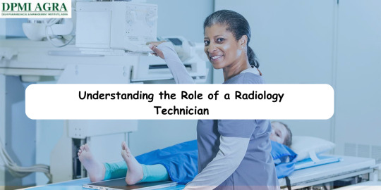
Before diving into the details of radiology technician school, it's essential to understand the role of a radiology technician. These professionals operate imaging equipment such as X-ray machines, CT scanners, and MRI machines to assist physicians in diagnosing and treating medical conditions.
2. Researching Schools in Agra
Start by researching the various schools in Agra that offer radiology technician programs. Look for institutions that are accredited and have a strong reputation for producing competent graduates. You can gather information online, visit campus websites, or even reach out to admissions counsellors for more details.
3. Curriculum Overview
Once you've identified potential schools, take a closer look at the curriculum offered in their radiology technician programs. Radiology Technician Course In Agra A typical curriculum may include courses in anatomy, medical terminology, patient care, radiographic procedures, and radiation protection. Make sure the program aligns with your career goals and interests.
4. Hands-on Training Opportunities
In addition to classroom instruction, hands-on training is a crucial aspect of any radiology technician program. Find out if the schools you're considering offer clinical internships or externships where you can gain practical experience working alongside experienced radiology technologists in real healthcare settings.
5. Faculty and Resources
Consider the qualifications and experience of the faculty members who will be teaching in the radiology technician program. Radiology Technician Course In Agra It's also important to assess the availability of resources such as state-of-the-art imaging equipment, simulation labs, and academic support services to enhance your learning experience.
6. Admission Requirements

Take note of the admission requirements for each school, including prerequisites, application deadlines, and any entrance exams that may be required. Be sure to start the application process early and submit all required documents to increase your chances of acceptance into the program.
7. Financial Aid Options
Explore the financial aid options available to help cover the cost of tuition, fees, and other expenses associated with attending radiology technician school. Radiology Technician Course In Agra This may include scholarships, grants, loans, or work-study programs. Be proactive in researching and applying for financial assistance to alleviate the financial burden of your education.
8. Career Opportunities and Outlook
Finally, consider the career opportunities and job outlook for radiology technicians in Agra. With the growing demand for medical imaging services, graduates of radiology technician programs can pursue employment in hospitals, clinics, diagnostic imaging centers, and other healthcare facilities.
By taking these factors into account and conducting thorough research, Radiology Technician Course In Agra you can gain valuable insight into what to expect from radiology technician school in Agra and make informed decisions about your education and future career path. Whether you're just starting your journey or looking to advance your existing career, investing in a quality education in radiology technology can pave the way for a rewarding and fulfilling profession in healthcare.
FAQs
Q1. What is the role of a radiology technician?
Ans. Radiology technicians operate imaging equipment such as X-ray machines, CT scanners, and MRI machines to assist physicians in diagnosing and treating medical conditions.
Q2. How can I find schools offering radiology technician programs in Agra?
Ans. You can research online, visit campus websites, or contact admissions counselors to gather information about schools offering radiology technician programs in Agra.
Q3. What courses are typically included in a radiology technician program curriculum?
Ans. A typical curriculum may include courses in anatomy, medical terminology, patient care, radiographic procedures, and radiation protection.
Q4. Are there hands-on training opportunities in radiology technician programs?
Ans. Yes, many programs offer clinical internships or externships where students can gain practical experience working alongside experienced radiology technologists in real healthcare settings.
Q5. What are the admission requirements for radiology technician programs?
Ans. Admission requirements vary by school but may include prerequisites, application deadlines, and entrance exams. It's important to check the specific requirements of each program.
0 notes
Text
The Importance of Hands-On Ultrasound Training for Sonographers
Introduction
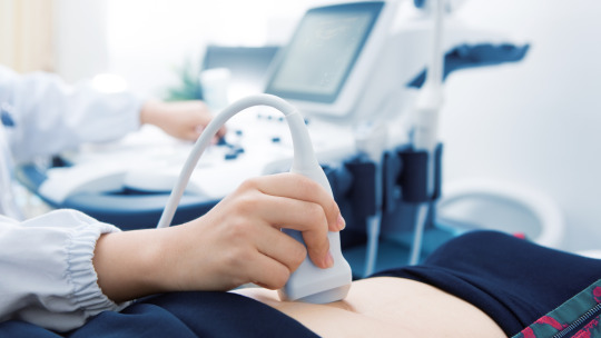
Sonography, a non-invasive medical imaging procedure that uses sound waves to visualize the substructure of the body, is an integral part of today's healthcare system. It aids doctors in identifying and diagnosing a wide range of conditions.
The Growing Field of Ultrasound and Its Significance in Medical Diagnosis
The medical field has seen a growing dependency on ultrasound due to its simplicity, cost-effectiveness, and safety. Unlike other imaging techniques, ultrasound does not involve radiation, making it a suitable choice for various examinations, including maternal and fetal assessments.
• Ultrasound aids in the quick detection of illnesses.
• It is widely used due to its non-invasive nature.
• Ultrasound does not expose patients to harmful radiation.
Importance of Hands-On Training for Sonographers
To become proficient in this tool, sonographers must receive proper training. The efficiency of an ultrasound highly depends on the expertise of the sonographer, making his/her training critical to patient outcomes. Ultrasound hands-on training refines the operator's skills, enhances their understanding of the technology, and enables them to perform precise diagnostics. It is indeed the bedrock on which their diagnostic expertise rests.
The Role of Sonographers in Medical Diagnosis
In fulfillment of an integral role within the healthcare industry, sonographers, or ultrasound technologists, are indispensable when it comes to providing reliable diagnostic imaging services.
Definition and Responsibilities of a Sonographer
Sonographers employ high-frequency sound waves, or ultrasound, to capture images of the body's internal structures. Their role includes:
• Conducting patient screening for medical history,
• Applying and adjusting the ultrasound equipment,
• Analyzing and interpreting ultrasound imagery,
• Preparing preliminary reports for physicians,
These multifaceted tasks necessitate a solid foundation in both theoretical knowledge and practical skills.
Importance of Accurate and Precise Imaging in Medical Diagnoses
When attempting medical diagnoses, accurate and precise ultrasound images can influence the physician’s decisions, and ultimately, patient outcomes. It's here that a well-trained sonographer makes the difference, reducing diagnostic errors and improving the patient’s treatment course. Investing in hands-on sonography training, therefore, not only optimizes clinical proficiency but also ensures quality patient care.
The Limitations of Theoretical Training in Sonography
Theoretical knowledge is undoubtedly an essential part of any sonographer's education and training program. However, it alone does not fully equip a sonographer to navigate the complexities and challenges of the real world.
Differences Between Theoretical and Practical Knowledge in Sonography
Theoretical knowledge in sonography provides an understanding of the basic principles, techniques, and terminologies. But it does not fully prepare one for practical applications such as manipulating the ultrasound transducer, interpreting images, or communicating findings to patients and physicians. Hands-on training, in contrast, allows for practical experience under professional guidance.
Challenges Faced by Sonographers Without Proper Hands-On Training
Sonographers lacking adequate hands-on training often encounter many obstacles. They include:
- Difficulty interpreting complex images
- Inattentiveness to subtle abnormalities
- Inefficiency during emergency situations
- Poor patient communication due to lack of confidence and experience
These challenges highlight why a balance between theoretical knowledge and practical experience is indispensable in ultrasound training for doctors and other sonographers.
Benefits of Hands-On Ultrasound Training for Sonographers
Hands-on ultrasound training equips sonographers with essential skills and knowledge that lead directly to enhanced patient care. Apart from gaining theoretical knowledge, sonographers especially benefit from practical, hands-on learning experiences. Here are the major benefits of hands-on ultrasound training for sonographers.
Enhanced Understanding of Ultrasound Technology and Equipment
Hands-on training provides a deeper understanding of ultrasound technology and equipment. It involves the use of real ultrasound machines, thereby ensuring students comprehend their functionality, operation and maintenance. This training facilitates:
- Familiarization with different ultrasound machines
- Understanding of machine settings and interpretations
- Proficiency in troubleshooting common machine problems.
Ability to Perform Accurate Ultrasound Scans
Being able to deliver accurate scans is a pivotal skill in the sonographer's role. Hands-on training cultivates precision in:
- Patient positioning
- Use of transducers
- Operation of ultrasound machine to obtain accurate images.
Skill Development in Interpretation and Communication of Results
Interpreting ultrasound imaging results and communicating them effectively with other healthcare providers is crucial. Through hands-on training, sonographers:
- Develop remarkable image interpretation skills
- Gain competence in report writing
- Enhance their communication skills with other medical practitioners.
Indeed, the importance of hands-on training for a sonographer cannot be overemphasized. It prepares them for real-life scenarios, fosters professionalism, and ultimately, enhances healthcare delivery quality.
The Best Ultrasound Training Programs
The selection, pursuit, and completion of an ultrasound training program is a critical phase that garners categorical consideration.
Criteria to Consider When Selecting an Ultrasound Training Program
Some factors to consider when choosing an ultrasound training program include program accreditation, program length, curriculum comprehensiveness, internships and hands-on training opportunities, cost, faculty expertise, and job placement success rates. A reliable program will typically have:
- A CAAHEP accreditation or equivalent
- A comprehensive and in-depth curriculum covering theoretical and practical aspects of ultrasound imaging
- Ample opportunities for hands-on ultrasound training
- A high job placement rate upon graduation
Overview of the Top-Rated Sonography Training Programs
Sonographers should consider top-rated programs such as the Mayo Clinic Sonography Program and the Johns Hopkins Sonography Program, which lay significant emphasis on hands-on ultrasound training, thereby enabling students to excel in various sonographic procedures.
Testimonials from Sonographers Who Have Completed Hands-On Training
Insights from professionals who have experienced hands-on ultrasound training underline its importance. One sonographer noted, "Hands-on training gave me the confidence to handle real patient scenarios. I could not have asked for a better preparation for my job."
The Impact of Hands-On Training on Sonographer Performance
Understanding and interpreting ultrasound images sufficiently requires a combination of technical know-how and clinical acumen, which are both provided during hands-on ultrasound training.
Improved Confidence and Efficiency in Performing Ultrasound Examinations
Through hands-on training, sonographers gain practical skills and experiences that build their confidence - leading to increased efficiency in conducting ultrasound examinations. They understand the broad range of variables that can affect ultrasound images, and how to adjust the equipment settings to get the best possible image.
Positive Outcomes and Patient Satisfaction Related to Hands-On Training
Sonographers who have completed extensive hands-on ultrasound training tend to achieve more accurate diagnoses. This increased accuracy directly translates to improved patient outcomes and higher patient satisfaction scores.
The Role of Hands-On Training in Career Advancement for Sonographers
Additionally, sonographers with comprehensive hands-on training often experience better career advancement opportunities. Employers highly value the practical skills and experiences these professionals bring, making them ideal candidates for positions of greater responsibility or specialised areas within the field.
The Future of Hands-On Ultrasound Training
The future of hands-on ultrasound training is dynamic and progressive. With advancements in technology, the face of sonography training is receiving a profound transformation.
Advancements in Ultrasound Technology and their Impact on Training Methods
Continuous improvements in ultrasound technology introduce new methodologies and procedures, consequently shaping the ultrasound training programs. Advances in 3-D and 4-D imaging, fusion imaging, and elastography, for instance, require tailored strategies to ensure proficient utilization. These include:
• Dedicated practical sessions
• Case-based learning
• Simulated scenarios
The Need for Ongoing Hands-On Training to Keep up with Technological Advancements
Given the fast-paced advancements in this field, there's an enduring necessity for ongoing hands-on ultrasound training. It empowers sonographers to preserve the currency of their skills and uphold top-notch service delivery in line with the latest technologies. Sonographers need to stay up-to-date, continually learning as technology evolves to ensure accurate diagnosis and effective patient care.
Conclusion
Recap of the importance of hands-on ultrasound training for sonographers
In sum, hands-on ultrasound training holds fundamental importance in the career of a sonographer. Key reasons include developing a solid foundation of ultrasound knowledge, mastering psychomotor skills needed for effective scanning, safety measures, and better patient care. These relevant skills include:
- Properly using ultrasound machines and interpreting the data generated
- Accurately distinguishing between normal and abnormal anatomy
- Safely utilizing ultrasound technology to prevent any possible harm to patients or the sonographer themselves
- Enhancing better patient outcomes by increasing diagnostic accuracy
#ultrasound hands on training#ultrasound training#hands on ultrasound course#sonography training program#sonographer training#best ultrasound course#ultrasound training centre#ultrasound training for doctors#best ultrasound training centre
0 notes
Last Seen Blogs

anti-josiesaltzman
anti josie saltzman

kaaulitzmwxo
Marley W

cobra197
dreams of a older man.

fluffycat-fluffy
Fluffy
fluffycat-fluffy
Fluffy