#orthopaedic in Janakpuri
Text
Dr. Rahul Agrawal is the epitome of excellence in orthopaedic care, recognized as the best orthopaedic doctor in Rohini, Delhi. With a focus on providing top-notch treatment, Dr. Agrawal is renowned for his expertise in orthopaedic surgery, making him the go-to choice for patients seeking the best knee, hip, and shoulder replacement surgeon in Delhi. As a leading arthroscopic surgeon in Rohini, Delhi, Dr. Rahul Agrawal's commitment to delivering exceptional care is unmatched. If you're searching for an orthopaedic doctor near you, look no further than Dr. Agrawal for unparalleled expertise and compassionate care.
#bestorthopedicdoctornearme#orthopedicdoctorinrohini#BestKneeReplacementSurgeoninDelhi#BesthipReplacementSurgeoninDelhi
0 notes
Text

0 notes
Text

Dr. Rahul Agrawal is the epitome of excellence in orthopaedic care, recognized as the best orthopaedic doctor in Rohini, Delhi. With a focus on providing top-notch treatment, Dr. Agrawal is renowned for his expertise in orthopaedic surgery, making him the go-to choice for patients seeking the best knee, hip, and shoulder replacement surgeon in Delhi. As a leading arthroscopic surgeon in Rohini, Delhi, Dr. Rahul Agrawal's commitment to delivering exceptional care is unmatched. If you're searching for an orthopaedic doctor near you, look no further than Dr. Agrawal for unparalleled expertise and compassionate care.
#bestorthopedicdoctornearme#orthopedicdoctorinrohini#BestKneeReplacementSurgeoninDelhi#BesthipReplacementSurgeoninDelhi
0 notes
Text
All About Knee Arthroscopy

What is knee arthroscopy?
Arthroscopy in Delhi is a surgical technique that allows you to directly see the inside of the knee joint and work inside it, without having to open it. Only two small incisions or cuts are made in the skin, about one centimetre each (which is why it is called a mini-invasive technique).
Arthroscopy in Delhi is considered the best current technique for meniscal injuries, adhesions, plica, loose bodies, cartilage injuries (chondroplasty) and reconstruction of cruciate ligaments, explains the orthopaedic in Delhi.
How is knee arthroscopy done?
The orthopaedic surgeon in Delhi, in order to see the inside of the joint well and avoid tissue injury with his manoeuvres, fills it with sterile pressure serum, which has the effect of inflating a balloon; and at the same time, it allows continuous joint washing, eliminating blood residues, excised tissue fragments, etc.
The patient lies on his back on the operating table. No system is necessary to pull the joint. You only need to lock the position of the thigh and the surgeon or assistant mobilizes the leg, opening the joint space.
Through an incision, a micro camera is introduced that illuminates and amplifies the interior of the joint, viewing the image on a television monitor. On the other hand, work instruments are introduced, such as probes, hand grippers and motorized smoothing devices.
The anaesthesia used is spinal anaesthesia (patient conscious but asleep from the waist down). Some sedation may be associated with this procedure to be calmer during the surgical act. General anaesthesia is reserved for special cases.
A tourniquet is used on the thigh to prevent bleeding from the knee during the operation, thus promoting vision through the camera.
Although it is a surgical act and requires the same aseptic conditions (cleanliness and sterility to avoid infection) as any other operation, the hospital stay is usually very short. In most cases, the patient can be discharged on the same day, when the anaesthetic effect has worn off. These operations can therefore be included in the program of major outpatient surgery, explains the orthopaedic surgeon in Delhi.
What does knee arthroscopy in Delhi show?
The appearance of synovialfluid (viscous fluid that lubricates the joint), which may be cloudy, contain blood or loose bodies, usually cartilage. Synovial fluid can be analysed to determine its composition in special cases.
• The synovial membrane(the sac that lines the joint inside and produces synovial fluid). In certain cases, a sample (biopsy) is taken for analysis under a microscope.
• The cartilage that lines the articular surfaces of the femur, tibia, and patella. It is palpated with a special hook to see the consistency and it is observed if it has injuries: wear (osteoarthritis), fissures, chondromalacia …
• The menisci (internal and external): observed and palpated with the probe hook. Breaks, tearing, wear are detected …
The cruciate ligaments(anterior and posterior): they are seen and touched to determine partial or total tears, laxity, function … The collateral ligaments are not seen with this technique.
• The way the patellamoves when the knee is bent and stretched, as well as the friction surfaces.
In which cases should an arthroscopy in Dwarka be performed?
Less and less to diagnose, as advances in ultrasound, CT (scanner) and nuclear magnetic resonance resolve it more and more frequently, although they are not infallible.
However, in cases of doubt or when a major intervention on the knee is planned, an arthroscopy can be performed beforehand, which will make it possible to confirm the diagnosis, rule out other injuries and decide the best possible treatment, which is also sometimes arthroscopic. Thus, in the same surgical act it is diagnosed and treated. In addition, there are patients who have contraindicated MRI (due to claustrophobia, or prosthetic heart valves), in those cases, diagnostic and therapeutic arthroscopy would be indicated by the orthopaedic doctor in Delhi.
Another diagnostic utility is to allow a synovial biopsy in certain diseases.
Currently, the main indications for performing a knee arthroscopy in Dwarka are:
Meniscal injuries: remove broken fragments, suture certain tears, regeneration techniques and meniscal reimplantation
cruciate ligament reconstruction: avoid opening the knee as before
cartilage injuries: cleaning, regenerative techniques (platelet growth factors, mosaicplasty)
removal of intra-articular loose bodies: fragments of detached cartilage or meniscus
removal of synovial plica or synovitis (synovial membrane hypergrowth)
cleaning on knees with osteoarthritis (wear) before reaching the total knee replacement
Recommendations at hospital discharge:
They are usually quite simple since it is a mini-invasive technique.
A compression bandage is placed, which the patient will remove at home after 48 hours. Then the first treatment is carried out, which consists of painting the two small wounds with Betadine and covering them with two adhesive dressings.
From there, the treatment will be repeated every day until the stitches fall out (about 2 to 4 weeks). It can be made to coincide with the shower as long as two rules are met:
quick shower: the less time the wounds are wet the better
WITH the dressings on: so that soap, shampoo, dirty water do not get into the wounds
After showering, the wet dressings are removed, the wounds are thoroughly dried with sterile gauze, painted with Betadine, and new dressings are placed.
From the moment the mobility of the legs recovers after anaesthesia, it is advisable to start walking. At first helped with crutches and following the indications of your orthopaedic surgeon in Dwarka regarding load (partial or complete). Usually, the patient leaves the hospital the same day walking with the help of two crutches.
An anti-inflammatory treatment is usually recommended at discharge for the first days.
It is advisable to apply ice locally for 10-15 minutes about 3-4 times a day to help reduce inflammation.
Depending on the diagnosis and treatment carried out, a specific physiotherapy may or may not be prescribed, with the recovery times greatly varying. The time in which you can return to sports or hard work depends on the injury: from 1 month to several months.
In the event of residual effusion (usually due to the persistence of the arthroscopic lavage fluid and more rarely due to bleeding into the joint, which is the hemarthros), an evacuating puncture may be necessary: the area is punctured, and the excess fluid is extracted with a syringe. This procedure should only be performed by an orthopaedic surgeon in Delhi.
#arthroscopy in Delhi#arthroscopy in Dwarka#arthroscopy in West Delhi#best knee surgeon in west Delhi#orthopaedic surgeon in Delhi#orthopaedic surgeon in Dwarka#orthopaedic surgeon in west Delhi#orthopaedic in Delhi#orthopaedic in Dwarka#orthopaedic in janakpuri#orthopaedic in west Delhi#orthopaedic clinic in Delhi#orthopaedic doctor in Delhi
0 notes
Text
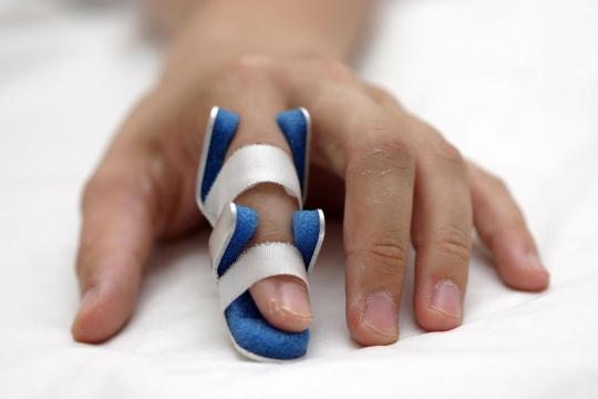
Everything We Need To Know About Finger Sprain
The sprain of the finger occurs when a bone from its formation (a phalanx) moves out of the joint. Injuries resulting from sports activities, falls or accidents can cause dislocation of the fingers.
Dislocation or sprained ness of a finger can be extremely painful, but not life-threatening. However, it is very important to receive medical support from orthopaedic in Dwarka as quickly as possible.
Symptoms of finger sprain
The symptoms of finger sprain are not numerous, but they successfully manage to indicate the condition. Thus, the affected finger changes its shape and looks crooked, but it is also particularly painful.
Other signs that may indicate finger sprain include:
- numbness or tingling;
- bruising or discoloration in the skin;
- functional impotence.
What are the causes of finger sprain
Normally, the thumb contains two phalanges, and the rest of the fingers contain three. Thus, there are two joints in the case of the thumb and three joints for the other fingers.
Ligaments are strips of fibrous tissue that help keep the joints integrated. Dislocation occurs when a significant force overcomes the opposite resistance of ligaments and causes the bones out of the joint.
Injuries suffered from sports are the most common causes of finger sprains. Thus, it is estimated that over 50% of the injuries found in the sport target fingers. Sports with the highest rates of injuries to the hands include football, gymnastics, basketball and wrestling.
Other possible causes that may lead to a sprain of the finger are:
- exaggerated extension of the finger;
- applying a strong blow to the tip of the fingers;
- falls;
- the existence of other health conditions that can weaken the joints and ligaments.
First measures in case of finger sprain
People who suspect their finger is sprained should seek medical attention from orthopaedic in Delhi immediately. To relieve inflammation and immediate pain, ice can be applied, but with great care, since the injured finger should not be moved.
The affected person or the people around you should not attempt to place the bone back in the joint. The only people who can do this are those who have medical training in this field.
Trying to re-encase the bone by unprepared people can seriously aggravate the situation, causing additional pain and inflammation. In addition, moving their own bones back to the original place comes with a very high risk of permanently harming the surrounding structures (tendons, ligaments, nerves, blood vessels, joint cartilage).
Diagnosis of finger sprain
After arrival at the orthopaedic in Janakpuri, it will examine the affected finger. The orthopaedic in West Delhi should also be informed when the trauma occurred and what were the circumstances of the production.
Further investigations may be requested after primary evaluation to confirm the diagnosis or to assess the severity of the lesion. The necessary imaging investigations are:
- X-rays are used to observe the internal structures of the body, in this case the bones. X-rays can help confirm the diagnosis of luxuryness, but also check if there are certain associated fractures;
- magnetic resonance imaging – MRI scanning uses strong magnetic fields and radio waves. With their help, detailed images of the tissues inside the body are obtained. In most cases, this method is required if significant damage to the tissue adjacent to the lysed joint is suspected.
Treatment options
Treatment options vary depending on the location and severity of the sprain. Thus, the methods of treatment are:
Reduction of sprain
The first step in treating a dislocated finger is to carefully handle the bone and relocate it into the joint. The procedure is known as a discount.
Before performing local anesthetics can be used to relieve pain, and after the procedure has been completed, x-rays can be performed to check its effectiveness.
Immobilisation
After the reduction, the patient is recommended to wear an immobilization soften. It protects and secures the wounded finger as it heals.
Also, wearing the band stops the patient from moving the finger and prevents further dislocation and injury.
Wearing softening by patients is necessary for about a few weeks.
However, immobilization for prolonged periods can cause permanent rigidity and reduced finger mobility.
Fixing
Depending on the type or severity of injury, some people may suffer alongside sprains even bone fractures. Fractures are defined as staples or interruptions in a bone.
Bone fractures also require reduction and immobilization, but fixation may also be needed. Fastening is carried out with K brooches, these are thin metal rods that surgeons attach to stabilize bone fragments.
Surgery
Dislocated fingers involving torn ligaments and complex bone fractures may require surgical procedures known as open cuts. Like other treatments, surgeries are aimed at reducing, stabilizing and restoring the mobility of the affected finger, without damaging the surrounding structures.
Recovery
According to orthopaedic surgeon in Delhi, finger luxations usually heal in about 4-6 weeks.
The factors affecting the recovery time are as follows:
- severity and location of dislocation;
- the existence of ligament damage or tendons;
- the existence of bone fractures;
- the need for surgery.
As a result of reduction and immobilization, certain patients may require physical or occupational therapy. The physiotherapist in Delhi can help and instruct the patient how to speed up his healing and how to expand his range of movements.
There are also certain tips to be followed at home:
- maintenance of skins, but also clean and dry steas;
- maintaining the affected finger at a higher level than the heart to reduce inflammation;
- avoiding the request of the affected finger;
- application of cold compresses and ice packets to reduce pain and inflammation;
- administration of medicines against pain and inflammation, such as ibuprofen or acetaminophen;
- performing exercise just as recommended by the specialist.
It should be known that a finger recovered from a sprain presents a high risk of further injury in the future. Thus, caution is recommended when physical activities are carried out, even wearing protective equipment and avoiding risk sports.
Conclusions
Dislocation of a finger can cause panic and extremely much pain. However, it should be known that finger sprain is not life-threatening, but requires prompt medical assistance.
No attempt should be made to reduce home sprain by the patient or his close friends. In this way complications can occur, and healing can be hampered, sometimes there is a risk of permanent injuries.
Visit to the orthopedic in Delhi is mandatory, it can correctly assess the located to provide adequate treatment. After treatment, healing usually lasts a few weeks. However, if the sprain is accompanied by bone fractures or damage to adjacent tissues, the recovery time may be prolonged.
#orthopaedic surgeon in Delhi#orthopaedic in Delhi#orthopaedic in Dwarka#orthopaedic in Janakpuri#orthopaedic in West Delhi
0 notes
Text
Everything You Need To Know About Shoulder Subluxation
Shoulder subluxation refers to the partial dislocation of the shoulder joint. This happens when the upper part of the humerus (humeral head) partially exits the glenoid cavity of the shoulder blade. In a complete dislocation, the humerus is completely removed from the joint cavity.
The shoulder is the most mobile joint of the human body. This joint contains a series of bones, ligaments, and muscles that work together to maintain a stable shoulder. However, due to the high mobility, the shoulder is very susceptible to dislocation.
Thus, a subluxation is often the result of trauma or injury. Also, another cause can be the stroke, because it can weaken the muscles in that area.
Clinical picture
A subluxation may be more difficult to identify than a complete dislocation. However, in some cases, the partially dislocated humerus may be visible under the skin.
Also, the bone can easily feel palpable and can cause discomfort and pain. The most common symptoms of subluxation include:
The possibility to observe the deformation of the shoulder;
Pain;
Inflation;
Sensation of numbness or tingling in the arm (paresthesia);
Functional impotence;
Sensation of shaking or interrupting movements during the activity.

Causes
Because the shoulder can move in several directions, it can move forward, backward or downward. This is also true for subluxations.
When the dislocation is partial, the joint capsule may be affected, including rupture, which can lead to different complications.
Typically, strong hits or falls can cause a shoulder dislocation. Also, the extreme rotation of the arm can cause the humerus to be pulled out of the glenoid cavity.
Once a shoulder has been dislocated, the joint may become unstable and prone to future dislocations or subluxations.
Shoulder subluxation is mostly caused by:
Trauma - subluxation can result from accidents or injuries that affect the joints or other structures that provide stability to the shoulder. The most common examples are falls and road accidents.
Sports accidents - contact sports, but not only these, can often cause shoulder subluxation. Gymnastics and skiing are other sports with a high degree of risk, due to frequent falls.
Stroke - this neurological condition can cause muscle weakness. Thus, the destabilization of the shoulder joint can be complicated by a subluxation.
A small study reported that 80% of stroke participants also suffered a shoulder subluxation.
Young men, as well as groups of extremely physically active people, have the highest risk of shoulder subluxation.
Treatment options
Proper diagnosis is essential for adopting the best treatment option. Thus, very good diagnostic methods consist of ultrasonography of the shoulder joint or radiography.
The treatment aims to reposition the humerus back to the original place and ensure that it stays in position. Treatment options include:
- Closed reduction - this intervention involves easy handling of the bone back into position. In most cases, severe pain disappears almost immediately after reducing maneuvers.
- Surgical intervention - it is recommended by orthopaedic in Delhi especially when the dislocations are repeated. This is also the preferred treatment when the nerves, blood vessels or ligaments of the shoulder have been damaged.
- Orthosis - some people may need to wear orthotics for a few days or weeks to prevent the shoulder bones from moving. The length of time this device should be worn depends on the severity of the trauma.
- Medicines - after surgery, but before, the orthopaedic surgeon in Delhi may recommend a muscle relaxant and an anti-inflammatory drug. In this way, pain and swelling can be reduced.
- Rehabilitation - After surgery, the orthopaedic in Dwarka may indicate rehabilitation programs. Their purpose is to restore the range of motion, endurance, and shoulder joint.
The physiotherapist in Delhi may recommend certain exercises that can help you recover faster. These should be done correctly and under supervision, as in certain circumstances, they may aggravate the symptoms. Also, massage and ice applications may be required.
Recovery and potential complications
When an injured person has a subluxation without major injury to the nerves and tissues, the simple repositioning of the humerus should rapidly improve the patient's condition.
However, if a rest period is not respected and normal activities returned, it is possible to dislocate the shoulder later.
After surgery for a subluxated shoulder, the orthopaedic in West Delhi will recommend, most of the time, that the person in question should wear shoulder support for several weeks.
The gradual introduction of physiotherapy can help a person regain his or her power and movements. However, it is not indicated the intense movements of the shoulder and the activities not recommended by the orthopaedic in Janakpuri. Thus, subsequent complications can be prevented.
The shoulder contains strong tissues, muscles, and ligaments, all helping to maintain the humerus in its place. However, these structures can be injured, complicating subluxation.
Thus, the most common complications of a shoulder subluxation include:
- Affecting blood vessels and shoulder nerves;
- Temporary or permanent loss of movement and flexibility;
- Instability at the shoulder level, followed by recurrent subluxations;
- Other lesions in the tissues of the affected shoulder.
Differential diagnosis
Shoulder subluxation presents some common symptoms with other similar lesions. For this reason, there may be errors in the diagnosis. A subluxation can be confused with:
Biceps tendonitis - the condition refers to inflammation of the biceps tendon. Due to the position of this tendon, its damage can usually lead to shoulder pain.
Clavicular injuries - fractures or traumas of the clavicle can cause pain in the shoulder, but also in the hindrance of movements at this level.
Rotator cuff injuries - this structure covers much of the shoulder joint capsule. His injuries can cause symptoms like those of the shoulder subluxation. Also, minor injuries can heal on their own, while severe injuries may require surgery.
Shoulder dislocation - dislocation involves the complete separation of the humerus from the joint cavity. The symptoms are like those of subluxation, and the correct differentiation of these conditions can only be made by an orthopedic in Delhi.
“Swimmer's Shoulder” - Shoulder pain in professional swimmers is often referred to as this.
Because swimming requires a high level of shoulder flexibility and a wide range of movements, swimmers are often likely to have an increased risk of a shoulder injury.
Perspective
When a person requests medical attention quickly and receives a correct diagnosis, shoulder subluxation is completely treatable.
However, when no surgery is required, it may take several months for the person to resume normal activity.
The recovery time varies depending on the extent of the subluxation and whether or not the affected person has undergone surgery.
Also, following a subluxation, the person concerned must avoid intense activity, to prevent relapse and to speed up recovery.
#orthopaedic surgeon in Delhi#orthopaedic in Delhi#orthopaedic in Dwarka#orthopaedic in Janakpuri#orthopaedic in West Delhi
0 notes
Text
INJURIES AND DISEASES OF THE HIP

Introduction
The hip or coxofemoral joint relates the coxal bone (pelvis) to the femur (thigh), thus joining the trunk with the lower limb. Hips are ball joints is classified as ball and socket of diartrosis type, characterized in that the two articular surfaces involved are spherical or nearly spherical, one concave and one convex, allowing great mobility.
When they are healthy, it takes a lot of strength to hurt them. However, sometimes sports, running, excessive use or falls can lead to hip injuries. These injuries include:
Strains
Bursitis
Dislocations
Fractures
Some diseases can also lead to injuries or problems with the hips. Osteoarthritis can cause pain and limit movements. Osteoporosis in the hip causes the bones to weaken and break easily. Both pictures are common in the elderly.
Treatment of hip problems may include rest, medicine, physiotherapy or surgery, including hip replacement in Delhi.
Hip Bursitis
Synovial bags are gelatinous sacs that are found throughout the body, including around the shoulder, elbow, hip, knee and heel. These bags contain a small amount of fluid, and are located between the bones and soft tissues, acting as pads or cushions to help reduce friction.
Bursitis is the inflammation of the synovial bag. There are two major bags in the hip that typically suffer from irritation and inflammation. A bag covers the bony bump of the hip bone, called the greater trochanter. The inflammation of this bag is called trochanteric bursitis.
Another bag, the psoas-iliac bag, is located on the inside (side of the groin) of the hip. When this bag becomes inflamed, the condition is also called hip bursitis, but the pain is located in the groin area. This condition is not as common as trochanteric bursitis, but it is treated similarly.
Symptom
Pain at the point of the hip and usually extends to the outside of the thigh area. In the early stages, pain is usually described as stabbing and intense. Later, the pain can become more of a neuralgia and spreads into a larger area of the hip. You may notice more when getting up from a chair or bed, when sitting for a long time and sleeping on the affected side.
Usually, acute bursitis arises within hours or days. Chronic bursitis can last from a few days to several weeks, and may disappear and reappear again. Acute bursitis can become chronic if it reappears or if a hip injury occurs.
Over time, the bag may become thick, which can make swelling worse. This can cause limited movement and cause the muscles to weaken (called atrophy) in the area.
Causes and Risk Factors
Several factors can cause hip bursitis, including the following:
Repeated excessive use or hip stress.
Rheumatoid arthritis.
Hip injury
Infection with bacteria, such as Staphylococcus aureus (or a staph infection).
Spinal problems, such as scoliosis.
Irregular leg length.
Bone spurs (bone growths above the normal bone) in the hip.
Diagnosis and tests
Orthopaedic in Delhi will examine you and ask about your symptoms. Sometimes, certain tests may be needed to rule out other conditions that may cause similar symptoms. These tests may include x-rays and magnetic resonance imaging (MRI).
Treatment
Usually, treatment for bursitis involves having the joint at rest as much as possible. Non-steroidal anti-inflammatory drugs (NSAIDs), such as ibuprofen (brands: Advil, Motrin) or naproxen (brand: Aleve) can be used to relieve pain and swelling. You may also want to use an ice pack on the area to reduce swelling. During this time, be sure to avoid activities that may make the symptoms worse.
Orthopaedic in Janakpuri may recommend exercising the area once your pain subsides. This helps prevent muscle atrophy. Ask your orthopaedic in Tilak Nagar about the exercises to help you strengthen the area. If bursitis affects your ability to function normally, you may need physical therapy to help you move again. This is especially true for people who have chronic bursitis.
If these treatments do not help, you may need to remove fluid from the bag or receive corticosteroid injections to reduce pain and swelling. Usually, corticosteroid injections are very effective in treating bursitis. You may need another injection after a few months.
Surgery is rarely needed to treat bursitis. It is only used when all other treatments fail. For people who need surgery, this is a simple procedure. The orthopaedic surgeon in Delhi removes the hip bag. The hip can function normally without the bag. Usually, the surgery does not require a long hospital stay and the recovery period is short.
Prevention
You can prevent bursitis by avoiding too much effort with your hips. Avoid activities that are particularly difficult or painful, and take breaks for the hips to rest. When exercising, remember to preheat the muscles and then stretch to prevent injuries. If you are overweight, losing weight can help reduce pressure on your joints, including your hips.
Strengthening your hips with an approved physical activity routine can greatly reduce your chances of developing bursitis. Ask your orthopaedic in Najafgarh what types of exercises are best for you.
Questions to ask your doctor
What could have caused my symptoms?
What is the best treatment option for me?
How long will it take until I can wait for my symptoms to get better?
Is it possible for my symptoms to return?
Is it safe for me to exercise? What type of exercise should I do?
#hip replacement in Delhi#orthopaedic surgeon in Delhi#orthopaedic in Delhi#orthopaedic in Najafgarh#orthopaedic in janakpuri#orthopaedic in tilak nagar
0 notes
Text
ORTHOPAEDIC SURGEON AND TYPES OF ORTHOPAEDIC TREATMENTS

Orthopaedics is the medical specialty that focuses on diseases and injuries of the musculoskeletal system in the human body. This complex system includes joints, bones, tendons, ligaments, muscles and nerves that allow one to move, work and be active.
An orthopaedic surgeon in Delhi treats patients of all ages, from new-borns with club foot to young athletes who require arthroscopy in Delhi and older people with arthritis. Most of us would have contacted him in case of an injury due to a bad fall or a car accident or an accidental torsion of one of our limbs, which would cause sprains or muscle tears.
Your orthopaedic surgeon
Orthopaedic surgeon in West Delhi treats musculoskeletal system problems. This involves:
Diagnosis of your disorder or injury.
Treatment with exercise, casting, medication, surgery or other options. Surgery performed by an orthopaedic surgeon in Dwarka with special tools and instruments.
Rehabilitation recommending physiotherapy in Dwarka or exercises to restore movement, function and strength.
Prevention with treatment plans and information to prevent injuries or delay the progression of a disease.
While orthopaedic in Delhi is familiar with all the characteristics of the musculoskeletal system, several orthopaedists specialize in certain areas, such as the foot and ankle, shoulder, spine, hip, hand, or knee. They can also choose to focus on specific fields such as trauma, paediatrics or sports medicine. Some orthopaedic in West Delhi may specialize in many areas.
Visit and medical treatment
Your orthopaedic surgeon in Delhi will take a history of your illness or injury and perform a physical exam. This can be followed by diagnostic studies, such as blood or X-ray tests or CT scans / MRI.
Depending on the diagnosis, the orthopaedic in Najafgarh develops the best treatment plan so that he can lead a functional and active life.
Non-surgical treatment
Orthopaedic in Janakpuri treats various musculoskeletal conditions without surgery using exercises, medications and other rehabilitation or alternative therapies.
For most orthopaedic injuries and diseases, there is more than one form of treatment. The orthopaedic in Tilak Nagar may recommend the option of surgical treatment in cases where non-surgical options do not help.
Surgical treatment
Orthopaedic surgeons perform various types of surgeries. The common procedures are:
Arthroscopy: A procedure that uses special cameras and orthopaedic equipment to visualize, diagnose and treat problems within a joint.
Fusion: A “welding” process by which bones are fused with internal devices (such as metal rods) and bone grafts to heal in a single solid bone.
Internal fixation: A method to keep broken bone pieces in a suitable place with metal plates, pins or bone screws while the bone is healing.
Joint replacement surgery in Delhi (partial, total and revision): When a damaged or arthritic joint is removed and replaced by an artificial joint called a prosthesis.
Osteotomy: The correction of bone deformity by cutting and repositioning bone.
Soft tissue repair: Repair of soft tissues, such as torn ligaments or tendons.
#arthroscopy in Delhi#orthopaedic surgeon in Delhi#orthopaedic surgeon in dwarka#orthopaedic surgeon in west Delhi#orthopaedic in Delhi#orthopaedic in Najafgarh#orthopaedic in janakpuri#orthopaedic in tilak nagar#orthopaedic in west Delhi#joint replacement surgery in Delhi
0 notes
Text
Everything You Need To Know About Hip Replacement Surgery in Delhi
During the hip replacement surgery in Delhi, a hip joint that has been damaged or worn is replaced, usually by arthritis or injury. In the hip revision (or repetition of hip replacement) an artificial hip joint that has become loose, infected or worn is replaced.
You will meet with the orthopaedic in Delhi who will perform the procedure to discuss your treatment. It may be different from what is described in this document because it will be designed to fit your needs.

ABOUT HIP REPLACEMENT
The hip is an enarthrosis joint. Normally, the ball at the upper end of the thigh bone (femur) moves smoothly in the pelvic cavity (hip) over a cartilage lining. Cartilage prevents bones from rubbing. If the cartilage wears out, the underlying bone is exposed and causes pain and stiffness in the joint. As a result, walking and moving causes pain.
A new hip joint can improve your mobility and decrease pain.
TYPES OF ARTIFICIAL HIPS
The artificial parts of hips can be metal, ceramic or plastic. The hip joints can be fixed in place by a special substance called 'bone cement'. Alternatively, it can be designed so that its bone grows on the metal. These 'uncemented' hips can be covered with a type of bone mineral (hydroxyapatite) or they can be made of a material with many tiny holes (porous coating). This stimulates the bone to grow into the artificial joint and snaps into place.
HIP REVISION SURGERY
During the original hip replacement in Delhi, the hip joint is replaced with artificial hip pieces. Generally, they last between 10 and 20 years, until they need to be replaced.
Renewing an artificial hip joint is more complicated than the original operation because the artificial hip joint should be removed before placing a new one. If the hip has loosened, it may not be so difficult for the orthopaedic in South Delhi, but if it is still attached to the bone, removing the old components can be a challenge.
Although there will be a great improvement, you may feel that the new joint does not improve your life like the original hip operation. This may be due to the fact that the muscles may take longer to recover from the reconstruction of scar tissue and the repetition of the operation.
WHAT ALTERNATIVES ARE THERE FOR HIP REPLACEMENT?
Generally, hip replacement surgery in South Delhi is recommended only if nonsurgical treatments, such as analgesics (eg, acetaminophen), anti-inflammatories (eg, ibuprofen), or using physical assistance such as a cane, no longer help reduce pain or improve Mobility.
The hip surface prosthesis is a better option for people with stronger bones. In this operation, the surfaces of the enarthrosis are covered with metal covers.
PREPARATION FOR THE OPERATION
The orthopaedic in Dwarka will explain how to prepare for the operation. For example, if you smoke, you will be asked to stop smoking, as this increases your risk of infection in the chest and wound, which may delay your recovery.
Normally, the operation will be performed under general anaesthesia. This means that you will be asleep during the operation. As an alternative, you will undergo surgery under epidural anaesthesia or lumbar [add link to epidurals for surgery and pain relief]. This completely blocks the sensitivity of the waist down and you remain awake during the operation. Your orthopaedic in Janakpuri will tell you which type of anaesthesia is best for you. Often, they are placed combined, so that people remain asleep, but epidural / lumbar anaesthesia will calm any pain immediately after surgery.
If you are going to be given general anaesthesia, you will be asked to fast. It means that you should not eat or drink, normally about six hours before. However, it is important to follow the advice of your anaesthetist.
Your orthopaedic in Uttam Nagar will explain what will happen before, during and after the procedure, and about any pain you may feel. This is your opportunity to understand what will happen, and you can help yourself and prepare questions about the risks, benefits and other alternatives to the procedure. This will help you to be informed, so that you can give your consent by signing a consent form, to carry out the procedure.
You may be asked to wear compression stockings to prevent blood clots from forming in the veins of the legs (deep vein thrombosis, DVT).
ABOUT THE OPERATION
A hip replacement in South Delhi, usually, lasts two hours.
The surgeon will make an incision (20 to 30 cm long) on your hip and thigh. Then, it will separate the enarthrosis (hip joint).
The head will be removed from the upper end of the femur (the femoral head) and a head will be inserted on a replacement rod in the femur. The cavity of the hip will be emptied to make a shallow cup and an artificial cavity will be placed. Then the two halves of the hip joint are joined (the head is placed inside the cavity).
The orthopaedic surgeon in Delhi will close the incision in the skin with stitches and clips and cover it with a bandage.
It may be possible for the orthopaedic in RK Puram to make a smaller incision on the hip and thigh. This type of operation (minimally invasive hip replacement) is performed with specially designed surgical instruments. It is not suitable for everyone; Consult your orthopaedic in Malviya Nagar if it is an option for you.
WHAT SHOULD I EXPECT AFTER
You should rest until the anaesthesia has passed. After epidural or lumbar anaesthesia, you may not be able to feel or move your legs for a few hours. You may need painkillers to help with any discomfort when the effects of anaesthesia subside.
They can place a special pillow between the legs so that the hip joint is still and to prevent it from dislocating.
You may be given medicine (injections or tablets), such as rivaroxaban or dabigatran, to prevent DVT. They will be given shortly after surgery, and you may need to take them for a few weeks.
A physiotherapist (a health professional who specializes in movement and mobility) will visit you daily to guide you in exercises designed to help your recovery.
You will remain in the hospital until you can safely walk with the help of a cane or crutch. Normally it's five days. However, if the general line is in shape and feels good, the surgeon can suggest that you perform an accelerated rehabilitation program, where you start walking the day of the operation and you are discharged one to three days later.
Before you go home, the nurse will give you advice on how to take care of your hip and a date for a follow-up visit.
Most stitches or clips should be removed after 12 to 14 days. The absorbable points do not need to be removed.
RECOVERY FROM A HIP REPLACEMENT SURGERY
If you need pain relief, you can take an over-the-counter pain reliever, such as acetaminophen or ibuprofen. Always read the information for the patient that comes with your medication and if you have any questions, check with your pharmacist.
The exercises recommended by the physiotherapist are an indispensable part of your recovery, so it is essential that you continue doing them.
During the first six weeks you should not make certain movements. For example, do not cross your legs or turn your hips in or out. This is to reduce stress on the scar and to decrease the risk of dislocation. The physiotherapist will give you more tips on how to protect your hips.
Must be able to move around your house and up and down stairs. For a few weeks, you will feel that some routine activities, such as shopping, are difficult to perform and you should ask for help. You must use crutches for four to six weeks.
Generally, you can do a light job again after six weeks. But if you must stop and sit continuously at work, you may need to stay at home longer.
Follow your surgeon's instructions about driving, since the time before you can drive again will depend on several factors, including which leg was operated on and if your car is automatic.
WHAT ARE THE RISKS?
Hip replacement in Delhi is a common and generally safe practice. However, to make an informed decision and give your consent, you need to be aware of possible side effects and the risk of complications.
Side effects
Side effects, although unwanted, are mostly temporary effects that you may have after the procedure.
Your hip will hurt for several weeks, and you may have temporary pain and swelling in your thigh and ankle.
Complications
Complications are problems that arise during or after the operation. Most people who are operated on the hip are not affected. The possible complications of any operation are unexpected reactions to anaesthesia, excessive bleeding, or the formation of clots, usually in a vein of the leg (deep vein thrombosis, DVT).
Specific complications of hip replacement in South Delhi are not common, but may include:
Infection: You will be given antibiotics during and after surgery to prevent it.
Joint dislocation: it is more likely to happen immediately after surgery, and you may need another surgery to treat it.
Difference in leg length: your leg may be slightly shorter or longer and you may need to wear a high shoe on the shorter side to correct the balance.
Hip fracture: the bone can undergo small cracks while the new joint is placed. It usually heals, but sometimes the bone can break and require surgery.
Unstable joint: the hip joint may loosen and may require surgery to correct it.
Damage to the nerves: can often result in insensitivity around the scar, but rarely the sciatic nerve can stretch and weaken the foot (it is usually temporary).
Generally, the artificial hip joint lasts between 10 and 20 years, after which time you may need to replace it.
The exact risks are individual and vary according to the patient, so we have not included statistics here. Consult the surgeon to explain how the risks apply to you.
#orthopaedic in delhi#orthopaedic surgeon in delhi#orthopaedic in south delhi#orthopaedic in dwarka#orthopaedic in janakpuri#orthopaedic in uttam nagar#orthopaedic in rk puram#orthopaedic in malviya nagar#hip replacement surgery in Delhi#hip replacement surgery in South Delhi#hip replacement in South Delhi#hip replacement in Delhi
0 notes
Link
#arthroscopy in dwarka#orthopaedic in dwarka#orthopaedic in janakpuri#orthopaedic in uttamnagar#orthopaedic in green park#orthopaedic in saket#orthopaedic in rk puram#orthopaedic in malviya nagar#ligament surgery in dwarka#joint replacement in dwarka#knee replacement in dwarka
0 notes
Text
Several specialists treat fibromyalgia. Many people who have the condition can work with a team of health professionals to alleviate the many typical symptoms. Best Orthopaedic Surgeon in Dwarka, Delhi

The majority of people who have fibromyalgia get treated by primary care physicians. Sometimes, one specialist acts as the lead doctor and coordinates care with other providers. A family doctor, a rheumatologist or internist may serve as the lead doctor.
What is a Rheumatologist?
Fibromyalgia, which is a rheumatic disorder, can be diagnosed by a rheumatologist. Rheumatologists are specialists in pain conditions that affect the joints, muscles and ligaments. A rheumatologist may refer a patient to a specialist if not the primary doctor. The National Fibromyalgia Association survey revealed that almost half of respondents had been diagnosed by a doctor.
Treatment of Fibromyalgia Symptoms and Other Conditions
Other fibromyalgia symptoms include fatigue, stiffness, pain, and sleep problems. Numerous people have fibromyalgia and other severe conditions, including systemic lupus, ankylosing spine, Rheumatoid Arthritis, and irritable bowel syndrome.if need a specialist then here Sarwarpro provide Best Orthopaedic Surgeon in Dwarka, Delhi Book appointment now.
To treat fibromyalgia symptoms or co-existing conditions, the lead doctor might recommend one or more of these specialists:
Alternative medicine practitioners, such as massage therapists and an acupuncturist, are also available.
Chiropractor
Nutritionist
Specialist in pain
Physical medicine and rehabilitation specialist, physiatrist
Therapists who are physical and occupational
Psychologist
Specialist in sleep
Arthritis Specialists
Depending on the patient’s needs, one or more of these specialists can provide treatment.
Arthritis Treatment
In some areas, there are pain management clinics and fibromyalgia clinics. These clinics provide the benefit of multiple specialists under one roof.
Advertisement
Fibromyalgia Treatment: How to Choose a Doctor
Fibromyalgia is a complex condition that can present multiple symptoms and require multiple treatment options. Finding the best medication, diet, exercise and therapy is often a trial-and-error process. we are provide all delhi orthapaedic doctor like -
Best Orthopaedic Surgeon in Janakpuri,
Best Orthopaedic Surgeon in Abul Fazal Enclave, Delhi
Best Orthopaedic Surgeon in Adarsh Nagar, Delhi
Best Orthopaedic Surgeon in Alaknanda, Delhi
Best Orthopaedic Surgeon in Aman Vihar, Delhi
Best Orthopaedic Surgeon in Amar Colony, Delhi
Best Orthopaedic Surgeon in Amar Vihar, Delhi
Best Orthopaedic Surgeon in Ambedkar Nagar, Delhi
Best Orthopaedic Surgeon in Anand Parbat, Delhi
Best Orthopaedic Surgeon in Anand Vihar, Delhi
Best Orthopaedic Surgeon in Ashok Vihar, Delhi etc.these are multiple area here are provide otherapedic doctor in Delhi.
Multi-Specialty Fibromyalgia Treatment
When choosing a doctor, it is essential to consider working together to relieve pain and improve function. These questions can help you choose the right doctor for you.
What percentage of people who have fibromyalgia were treated by a doctor?
Are you a good listener and a good doctor?
What is the doctor’s opinion on alternative medicine?
Is physical therapy or chiropractic care something the doctor recommends?
What is the doctor’s view of the patient’s role?
Is the doctor compassionate?
Other Specialists for Arthritis Treatment
After selecting a doctor, it is essential to plan to get the most out of each visit. It can be helpful to note down the most important symptoms and bring them with you to your next appointment.
Mind-Body Techniques to Help with Fibromyalgia
This helps organize the symptoms and prevents any misunderstandings during the office visit.
Fibromyalgia: Is it real? 6 Conditions You Need to Know
A doctor might ask the patient to fill out questionnaires to help diagnose the condition and track progress over time. This will help determine the effectiveness of treatment. These tools are handy when dealing with patients who have fibromyalgia. For more information Visit Sarwarpro.com
0 notes
Text
Knee Replacement Clinic in Delhi

The specialized kneeling services offered by orthopaedic in Delhi is a comprehensive approach to the care and management of knee injuries and diseases. Orthopaedic in West Delhi uses many of the new techniques and technologies in this field. The team of specialists offers cutting-edge experience in the evaluation and treatment of a wide range of diseases and injuries that can occur at any stage of life and that affect both adolescents and the elderly. Our service is among the most experienced in Delhi.
The services include the following:
Evaluation of diseases and injuries of the knees.
Conservative treatments to preserve or correct the knee in order to relieve pain and restore mobility, including injection therapy.
Minimally invasive procedures, arthroscopy in Delhi.
Latest techniques in sports injury surgeries, including the reconstruction of the ligament complex.
Advanced care of the knee fracture.
Knee replacement surgery in Delhi, including complex revision surgery.
Rehabilitation, advanced therapy.
Our program is unique in its kind in Delhi: it offers vast experience and treatment options available for the evaluation and management of knee problems. Our orthopaedic doctor in Dwarka experience-based care to treat knee problems in people of any age. Whether it’s the professional athlete or the weekend warrior, the orthopaedic in Najafgarh can offer you the best knee care in Delhi.
Best knee surgeon in Delhi works with a team to devise a comprehensive and personalized treatment and rehabilitation plan for our patients. In addition to other doctors, the team has nurses, physiotherapists, occupational and recreational therapists and social workers.
The team of orthopaedic in Janakpuri provides comprehensive care, considering the individual needs of the patient and always for the benefit of the patient.
Our team of specialists is at the forefront of technology and minimally invasive surgery (including arthroscopic procedures for knee surgery).
Orthopaedic in Palam pioneered many of the innovative techniques currently used, emphasizing patient safety and long-term mobility.
Thanks to its vast experience and research carried out, Orthopaedic in Tilak Nagar can offer alternatives to patients, including innovative ways to minimize the need for invasive procedures as well as prevent joint replacement.
#joint replacement in delhi#knee replacement in delhi#best knee surgeon in delhi#orthopaedic in delhi#orthopaedic in malviya nagar#orthopaedic in uttam nagar
0 notes
Last Seen Blogs

ayyo-this-is-my-love-song
Full of myself, but still hungry.

aiopsconsulting
Advanced Analytics, Generative AI, ML, Ops

adultra
GiNuTailYa

abcholiday
In Between半熟戀人

pickcpodcast
When in Doubt, Pick C