#orthopaedic in Dwarka
Text
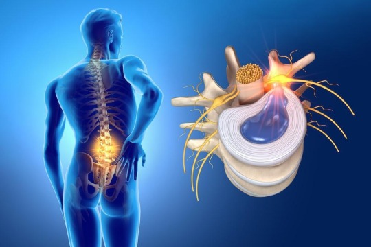
Disc Herniation
What is
Disc herniation occurs when an intervertebral disc degenerates and deteriorates, causing the inner nucleus to leak into a weakened area on the outside of the disc.
The weak point in the outer nucleus of the intervertebral disc is directly below the spinal nerve root, so a herniation in this area can put direct pressure on nearby nerves or the spinal cord.
Therefore, herniated discs are sometimes a cause of radiculopathy, which encompasses any disease that affects the nerve roots of the spine.
Dr Ashu Consul, best orthopaedic in Dwarka, consultant at Venkateshwara Hospital, adds that, initially, herniated discs can be confused with the following pathologies: “piriformis syndrome, facet arthropathies, deep gluteal syndrome, peripheral neuropathies, muscle trigger points and, in more severe cases, tumors”.
Causes
The vertebrae of the spine are separated by discs that cushion movement and leave space between the vertebrae. In the same way, they allow their movement, which makes it possible to bend down or stretch out.
In addition, the vertebrae of the spine protect the spinal cord that comes from the brain and runs down the back to the lower back. The discs fulfill a very important function of cushioning and distribution of loads and any damage to them can be serious if not treated quickly.
The disc can move out of place, that is, herniate, or rupture due to injury or stress. This can cause excess pressure on the spinal nerves resulting in pain, numbness or weakness in the patient.
Normally, herniated discs are located in the lumbar region, with the second most affected area being the cervical discs (the neck).
Symptoms
A cervical disc herniation can cause pain in the neck, which in turn can radiate to the arm, shoulder, or can cause numbness or tingling in the arm or hand. Sometimes the pain can be dull, constant, and difficult to locate.
In addition to this pain, the symptoms of herniated discs are the following:
The first sign that the patient has a herniated disc is pain in the arms and neck. If numbness or tingling occurs it may indicate that the problem is more serious.
Typically, the patient complains of sharp, cutting pain, and in some cases, there may be a prior history of episodes of localized pain, present in the back and radiating down the leg.
The episode of pain may come on suddenly or be heralded by a tearing or snapping sensation in the spine.
When the pain starts slowly, it can worsen after the patient spends a long time sitting, standing, at night, when sneezing, coughing or laughing.
Weakness is also a common symptom that affects the leg or arm and may require excessive effort to move them.
Usually, the numbness or weakness goes away over a period of several weeks or months.
Prevention
According to Orthopaedic in Dwarka, “exercising regularly and appropriately is important. Also avoiding leading a sedentary lifestyle, being overweight and smoking helps prevent this type of back pathology. Finally, avoid unnecessary risks such as lifting heavy objects, improperly bending or twisting the lower back, or sitting or standing in the same position for many hours and in an unergonomic way.
Types
There are three degrees:
Disc protrusion: when the nucleus pulposus has not yet come out of the annulus fibrosus, it is therefore weaker and gives way in its structure. This is the first stage of a herniated disc.
Disc herniation: the material of the nucleus pulposus is ejected from the limits of the annulus fibrosus.
Disc extrusion: the exit of the disc material is violent and breaks the posterior common vertebral ligament, leaving free fragments in the vertebral canal.
Diagnosis
To diagnose a herniated disc, the orthopaedic doctor in Delhi will carry out a medical examination of the spine, arms and lower extremities. Depending on the region where the patient's symptoms are located, the orthopaedic in Delhi will look for possible numbness or loss of sensitivity.
In addition, he will test your muscle reflexes, which may have been affected and slowed down or even disappeared. He will also study the patient's muscle strength and the shape of the curvature of the spine.
On the other hand, the patient may also be asked to sit, stand or walk, bend forward, backward or sideways and move the neck, shoulders or hands.
Diagnostic tests that can verify the existence of a herniated disc are:
An electromyography that will determine which nerve root is affected and where it is compressed.
A myelography to specify the size and location of the hernia.
An MRI that will show if there is pressure on the spinal cord.
Finally, an X- ray of the spine may also be performed to rule out other injuries that cause cervical or back pain.
Treatments
The first treatment given to patients with this condition is short rest and pain medication, followed by a period of physiotherapy session with physiotherapist in Dwarka. In most cases, almost immediate recovery occurs, but in other cases medication or injections may be required.
In the case of corticosteroids, they are usually administered, above all, non-steroidal anti -inflammatory drugs to control pain and also muscle relaxants.
Injections into the area of your back where the herniated disc is located can help control pain for a few months. In addition, these injections greatly reduce the swelling of the disc.
The last option is microdiscectomy, considered as the surgery that is used to relieve pressure on the nerve root and allow the nerve to recover more effectively. This type of intervention does not entail great difficulty, since it is usually enough with a small incision and one night of admission.
Regarding the therapeutic approach to herniated discs, Orthopaedic in West Delhi states that "one of the most important advances has occurred in the increased precision of diagnostic tools, which has made treatment much more effective and specific, both in management conservative as in the surgical. From the surgical point of view, the trend is towards minimally invasive, what we commonly know as microsurgery, so that the tissues suffer the least negative impact after the surgical intervention”.
At what age do herniated discs usually appear?
“Disc herniation can appear at any age, since its causes are multifactorial. Although, it begins to be more frequent in the range of 30 to 50 years of age. And they are more prevalent after the age of 50, where it is estimated that more than 80% of the population begins to show disc degeneration”, says orthopedic in Delhi.
0 notes
Text
Best Surgery Hospital in Dwarka
Discover excellence in surgical care at the Best Surgery Hospital Maharaja Agrasen Hospital in Dwarka. Equipped with cutting-edge technology and staffed by skilled surgeons and medical professionals, we specialize in a wide range of surgical procedures. From minimally invasive techniques to complex surgeries, our hospital prioritizes patient safety, comfort, and recovery.
0 notes
Text
Joint doctors prescribe a hip replacement for conditions like osteoarthritis, rheumatoid arthritis, or hip fractures. The Bone Clinic is one of the best hospital for hip replacement in Delhi.
#hip replacement in delhi#hip surgery#orthopedic hospital in delhi#bone doctor in dwarka#bone doctor#best orthopaedic surgeon in delhi#knee replacement in dwarka
0 notes
Text
Robotic Knee Replacement by Best Orthopaedic in Delhi Dr Vibhore Singhal
Knee pain can be debilitating, affecting every step and every move. If you’re tired of living with knee pain, robotic knee replacement surgery might be the solution you need. Dr. Vibhore Singhal is one of the best orthopaedic surgeon in Dwarka for this procedure. This article will take you through everything you need to know about robotic knee replacement, the expertise of Dr. Vibhore Singhal, and why this procedure might be the best choice for you.
Introduction to Robotic Knee Replacement
Knee pain can affect your quality of life, making it hard to enjoy simple activities like walking or climbing stairs. For many, robotic knee replacement surgery offers a chance to regain mobility and live pain-free. But what exactly is robotic knee replacement, and why should you consider it?

Who is Dr. Vibhore Singhal?
Dr. Vibhore Singhal is a renowned orthopaedic in Dwarka. With years of experience and numerous successful surgeries, he has established himself as a leading expert in robotic knee replacement. His dedication to patient care and advanced surgical techniques make him a trusted choice for those seeking relief from knee pain.
What is Robotic Knee Replacement?
Robotic knee replacement is a cutting-edge procedure that uses robotic technology to assist surgeons in performing precise knee replacement surgeries. This technology allows for better alignment and positioning of the knee implant, leading to improved outcomes and a quicker recovery time.
Benefits of Robotic Knee Replacement
Robotic knee replacement offers numerous benefits over traditional surgery. These include:
Precision: The robotic system provides real-time feedback, helping surgeons make more accurate cuts and placements.
Customization: Each surgery is tailored to the patient’s unique anatomy, ensuring a better fit and function.
Less Pain: Patients often experience less pain post-surgery due to the minimally invasive nature of the procedure.
Faster Recovery: With less tissue damage, recovery times are typically shorter, allowing patients to return to their daily activities sooner.
The Procedure: Step by Step
Understanding the procedure can alleviate some of the anxiety associated with surgery. Here’s a step-by-step breakdown of what to expect:
Pre-Surgery Consultation: Your journey begins with a thorough consultation with Dr. Vibhore Singhal, where he will assess your condition and discuss your options.
Preoperative Planning: Using advanced imaging techniques, a personalized surgical plan is created.
Surgery Day: The robotic system assists Dr. Singhal in performing the surgery with high precision.
Post-Surgery Recovery: After the procedure, you’ll receive detailed instructions for recovery and rehabilitation.
Why Choose Dr. Vibhore Singhal?
Choosing the right surgeon is crucial for a successful outcome. Dr. Vibhore Singhal’s expertise in robotic knee replacement and his commitment to patient care make him an excellent choice. He stays updated with the latest advancements in orthopedic surgery, ensuring that his patients receive the best possible care.
Recovery and Rehabilitation
Recovery is a critical part of the surgical process. Dr. Singhal and his team will provide you with a comprehensive rehabilitation plan tailored to your needs. This plan typically includes physical therapy, exercises, and follow-up appointments to ensure your recovery is on track.
Potential Risks and Complications
While robotic knee replacement is generally safe, it’s important to be aware of potential risks and complications, such as:
Infection: As with any surgery, there is a risk of infection.
Blood Clots: These can form in the legs post-surgery.
Implant Issues: There might be rare instances where the implant does not function as expected.
Dr. Singhal will discuss these risks with you and take steps to minimize them.
Cost of Robotic Knee Replacement in Delhi
The cost of robotic knee replacement in Delhi can vary based on several factors, including the hospital, the complexity of the surgery, and the patient’s overall health. On average, you can expect to pay between INR 2,50,000 to INR 4,50,000. It’s important to discuss the costs and payment options during your consultation with Dr. Singhal.
Preparing for Surgery
Preparing for surgery involves several steps to ensure you are in the best possible health. This may include:
Medical Evaluations: Comprehensive health checks to assess your fitness for surgery.
Medications: Adjusting current medications and possibly starting new ones.
Lifestyle Adjustments: Making changes to your diet and exercise routine.
Post-Surgery Care
Post-surgery care is vital for a successful recovery. This includes:
Wound Care: Keeping the surgical site clean and dry.
Medication Management: Taking prescribed medications to manage pain and prevent infection.
Physical Therapy: Engaging in physical therapy to restore mobility and strength.
Frequently Asked Questions
1. What is the recovery time for robotic knee replacement?
Recovery time can vary, but most patients can expect to return to normal activities within 6 to 12 weeks. Dr. Singhal will provide a detailed recovery plan tailored to your needs.
2. Are there any age restrictions for robotic knee replacement?
There are no strict age restrictions, but the suitability of the procedure depends on individual health conditions. Dr. Singhal will evaluate your case to determine if you are a good candidate.
3. How long does the robotic knee replacement surgery take?
The surgery typically takes about 1 to 2 hours, depending on the complexity of the case.
4. Can both knees be replaced at the same time?
Yes, bilateral knee replacement is possible and can be done in one surgery. However, this decision is based on the patient’s overall health and Dr. Singhal’s recommendation.
5. Will I need physical therapy after the surgery?
Yes, physical therapy is an essential part of the recovery process. Physiotherapist in Dwarka helps in regaining strength, flexibility, and mobility in the knee.
Conclusion
Robotic knee replacement surgery with Dr. Vibhore Singhal in Delhi offers a promising solution for those suffering from chronic knee pain. With advanced technology, personalized care, and a focus on achieving the best outcomes, Dr. Singhal ensures that his patients can look forward to a pain-free and active life. If you’re considering knee replacement surgery in Delhi, Dr. Singhal’s expertise and the benefits of robotic assistance make a compelling case for taking the next step toward better knee health.
0 notes
Text
Liver Problem Treatment In Dwarka Under CGHS, CAP

Jeena Sikho Lifecare Ltd. Ayurveda & Panchkarma Clinic in ( Dwarka )
Jeena Sikho Lifecare Ltd in Dwarka is one of the leading businesses in the Ayurvedic Doctors. Also known for Ayurvedic Doctors, Ayurvedic Clinics, Ayurvedic Treatment Centres, Ayurvedic Doctors For Joint Pain, Ayurvedic Doctors For Orthopaedic, Ayurvedic Doctors For Kidney Treatment, Ayurvedic Doctors For Kidney Stone and much more.
Ayurvedic methods are well-known. Ayurveda has been effective in treating numerous diseases for a very long period in India. Many people still choose Ayurvedic medications over western therapies. Ayurvedic Doctors in Dwarka recommend medications that typically contain more herbs and have fewer negative effects. They address their patients’ conditions from the source of the issue, frequently providing a lengthy yet effective course of treatment.
If you are looking for ayurvedic treatment for your ailments, we advise you to consult Jeena Sikho Lifecare Ltd. in Dwarka.
You can Contact us on - 9650932223 - 7873578735
Or visit our Clinic in Dwarka - Address: RZ-6A, Basement, Raghu Nagar Pankha Road, Mod, above Sunshine Super Market, adjacent to World Brain Hospital, Dabri, Delhi, 110045
#CGHS Approved Hospital Dwarka#CGHS Ayurvedic Hospital Dwarka#CGHS Empanelled Panchkarma Center Dwarka#CGHS Panel Clinic Dwarka#CGHS PANEL HOSPITAL NEAR ME Dwarka#CGHS Panel Hospital Near Me Dwarka#CGHS CLINIC NEAR ME Dwarka#Ayushman Capf Daycare Center Dwarka#CGHS EMPANELLED HOSPITAL Dwarka#CGHS AYURVEDIC DAY CARE CENTRE Dwarka#CGHS EMPANELLED PANCHKARMA CLINIC Dwarka#capf panel hospital Dwarka#CGHS APPROVED CLINIC Dwarka#capf day care center Dwarka#CGHS TREATMENT NEAR ME Dwarka#AYUSHMAN CAPF PANEL Dwarka#CGHS PANEL Dwarka#CGHS APPROVED HOSPITAL Dwarka#CGHS AYURVEDIC HOSPITAL Dwarka#CGHS EMPANELLED PANCHKARMA CENTER Dwarka#Ayurvedic Hospitals in CGHS panel Dwarka#CAPF AYURVEDIC HOSPITAL Dwarka#CAPF Ayurvedic Hospital Near me Dwarka#CGHS Ayurvedic Hospital Near me Dwarka#CGHS Ayush Private Day Care Therapy Centres Dwarka#CGHS Panel Hospital in Delhi ncr Dwarka#CGHS Panel Hospital near me Dwarka#CGHS empanelled Ayurvedic Hospitals in Delhi Dwarka#List of CAPF approved Ayurvedic Hospitals in Delhi Dwarka#CAPF APPROVED CLINIC RDC Dwarka
0 notes
Text

Multispeciality Hospital in Dwarka
At the Maharaja Agrasen Multi-Specialty Hospital, you may find the best doctors in orthopaedics, neurology, cardiology, gynecology, dermatology, etc.
Visit us: https://www.mahdwarka.in/
Contact Now: 7042129624
#maharajaagrasenhospitaldwarka#bestmultispecialtyhospitaldwarka#bestsurgeryhospital#besthospitaldwarka#healthcare#besthearthospital#dwarka#hospital#patient#doctors
0 notes
Text
Ayushman Day Care Centre for Bone Grafting
What Is Bone Grafting?
Bone Grafting is a medical procedure that involves adding bone or bone-like material to a person's existing bone structure to promote bone growth and regeneration. It is commonly used to treat conditions that cause bone loss, such as periodontal disease, dental implants, and orthopaedic surgery.

What Is The Purpose Of Bone Grafting Treatment?
The purpose of bone grafting, also known as bone augmentation, is to promote bone growth and regeneration in an area where there is insufficient bone or where the bone has been lost or damaged. Bone grafting is commonly used to:
Provide Support For Dental Implants:- If a person has lost a tooth or several teeth, bone grafting may be required to build up the jawbone so that it can support dental implants.
Treat Periodontal Disease:- Periodontal disease can cause bone loss in the jaw, leading to tooth loss. Bone grafting can help rebuild the jawbone and provide support for the remaining teeth.
Repair Bone Fractures:- Bone grafting may be necessary to repair bones that have been fractured or damaged due to injury or disease.
What Are The Advantages Of Bone Grafting Treatment?
Here are some of the potential benefits of bone grafting at the Best Dental Care Centre.
Promotes Bone Growth And Regeneration:- Bone grafting stimulates the growth of new bone tissue, which can help restore strength and stability to the affected area.
Reduces The Risk Of Implant Failure:- In dental implants, bone grafting is often required to create a sturdy base for the implant to attach to. This can help reduce the risk of implant failure, as the implant will be more securely anchored in the jawbone.
Improves Bone Density:- Bone grafting can help improve bone density in patients with osteoporosis or other bone diseases, reducing the risk of fractures and other complications.
Minimizes The Need For Additional Surgeries:- By promoting bone growth and regeneration, bone grafting can help avoid the need for additional surgeries to correct problems that may arise due to insufficient bone.
Is Bone Grafting Treatment Safe?
Bone grafting is generally considered safe when performed by a qualified and experienced medical professional.
It is important for patients to discuss the potential risks and complications with their healthcare provider before undergoing bone grafting. Additionally, patients should follow their healthcare provider's instructions for aftercare and attend all follow-up appointments to monitor the healing process.
What To Do For Recovery After Treatment?
Here are some tips to take care of your treatment at Best Dental Care Centre In Dwarka Sector 12.
Follow Your Healthcare Provider's Instructions:- It is important to follow all of your healthcare provider's instructions for aftercare, including taking any prescribed medications and attending all follow-up appointments.
Rest And Avoid Physical Activity:- Depending on the type and location of the graft, your healthcare provider may recommend that you rest and avoid physical activity for a certain period of time following the procedure.
Apply Ice Packs:- Applying an ice pack to the affected area relieves swelling and discomfort.
Conclusion
In conclusion, Bone Grafting is a safe and effective medical procedure that can promote bone growth and regeneration in patients with bone loss or damage. The procedure has several advantages, including improving bone density, reducing the risk of implant failure, and minimizing the need for additional surgeries.
#ayushman dentistry & implantology centre in dwarka sector 12#ayushman dentistry & implantology centre in dwarka#ayushman dentistry & implantology centre#ayushman dentistry & implantology#best dental care centre in dwarka sector 12#best dental care centre in dwarka#best dental care centre
0 notes
Text
Achilles tendon rupture - treatment, surgery

What is Achilles tendon rupture?
An Achilles tendon rupture or rupture occurs when the tendon “ruptures or tears” leading to separation or discontinuity in the tissues that make up the tendon.
In terms of anatomy, the Achilles tendon, a sort of fibrous “ribbon” or “cord” that connects muscle to bone, is the largest and strongest tendon in the body that connects the calcaneus (heel bone) to the medial gastrocnemius muscles., lateral gastrocnemius and soleus muscles (popularly known as calf muscles or “leg belly” muscles). When the muscle contracts, it “pulls” the tendon, which in turn moves the foot.
The rupture often reflects the previous existence of tendinosis phenomena caused by sports microtraumas or degeneration (aging) of the tendon. In most cases, the tear occurs at the bottom of the tendon (just above the heel), but it can occur anywhere along the tendon. In the presence of a healthy tendon, the lesion can appear in the bone or muscle.
Total ruptures, partial ruptures
We can generically classify Achilles tendon ruptures into:
Total rupture - in the total rupture of the Achilles tendon, the tissues break completely, that is, the tendon is completely “separated”.
Partial rupture - in a partial rupture of the Achilles tendon, the tendon does not completely rupture, only an incomplete rupture occurs. Partial tears can vary greatly in severity according to the extent of the injury.
A total rupture is more frequent than a partial rupture. As a rule, a total rupture is a more serious injury when compared to a partial rupture and with more exacerbated symptoms. A partial tear can also present with pain that can range from moderate to severe and, if not recognized, it can quickly progress to a total tear.
See more information on treatments to better understand the different therapeutic approaches according to the extent of the lesion.
Achilles tendon rupture - causes
The Achilles tendon can lose elasticity and become “weak and thin” with age and lack of use. Then, it becomes prone to injury or breakage.
Rupture is more common in people with pre-existing Achilles tendonitis (tendon inflammation). Repetitive tendinitis and the consequent calcifications are a risk factor for tendon rupture.
Certain diseases (such as arthritis and diabetes) and medications (such as corticosteroids, for example) can also increase the risk of rupture.
Rupture occurs most often in the middle-aged male athlete. The injury usually occurs during recreational sports that require impacts with the ground, running, jumping, etc. Most of the time, these are football, athletics, tennis, basketball, among others. Injury can happen in these situations as a result of traumatic dorsiflexion when the muscle is strongly contracted causing it to tear.
Breaks can, however, also occur in everyday activities. For example, when falling from a significant height, when entering a pothole abruptly, traffic accidents, etc.
Achilles tendon rupture - symptoms
The signs and symptoms of Achilles tendon rupture are:
Sudden, severe pain may be felt in the “back” of the ankle or the “tummy of the leg”, often described as “being hit by a rock or shot” or “like someone stepping on the back of the ankle”;
A loud clicking sound can be heard;
A discontinuity (“gap”) or depression (void) can be felt and seen in the tendon above the calcaneus (heel bone);
Inability to stand on tiptoe on the affected side;
Initial pain, swelling (swelling) and stiffness may be followed by bruising and weakness (not being able to land on the foot, walking).
Achilles tendon rupture - diagnosis
The diagnosis is made by the orthopaedic doctor in Delhi after collecting the clinical history, performing the physical examination and some complementary means of diagnosis (MCDT).Bottom of Form
A simple test is to “stretch” the “calf or calf muscles” while lying on your stomach (Thompson test). In the impossibility of being able to elevate the foot, there is, most likely, a rupture in the tendon. This test isolates the connection between the “calf muscles” and the tendon and eliminates other tendons that may still allow poor movement.
The orthopaedic doctor in Dwarka may order the following tests to confirm the diagnosis and to know in greater detail the location and degree of severity of the lesion:
Plain radiography (XR) - not being a very useful exam, it can identify a bone fragment avulsion of the calcaneus;
Ultrasound or ultrasound - Ultrasound of the leg and thigh can help assess the possibility of deep vein thrombosis and can also be used to rule out a Baker's cyst (or cyst). Ultrasound can identify Achilles tendon rupture or signs of inflammation (tendinitis or tendinosis);
Magnetic Resonance Imaging (MRI) - MRI is extremely sensitive for diagnosis and allows you to determine if there is still a tendon in continuity. It allows other diagnoses such as tendinitis, tendinosis and bursitis.
Pain in the “back of the heel” is not always due to an Achilles tendon rupture. In the differential diagnosis, tendinitis (inflammation of the Achilles) and bursitis (inflammation of the bursae) should be considered, among the most frequent pathologies that cause pain in the Achilles region.
Achilles tendon rupture - treatment
The objective of the treatment is to restore the function of the tendon, for this, it is necessary that the tissues that make up the tendon heal “united” with each other. In this way, it will be possible for the patient to return to the same level of activity before the injury. Regaining Achilles tendon function after an injury is critical to making walking possible.
Treatment reflects a balance between tendon protection and initial movement. Protection is necessary to allow time for healing and to prevent further injury. Moving the foot and ankle is necessary to prevent stiffness and loss of muscle strength.
We can divide treatment options into surgical and non-surgical. Conservative (non-surgical) treatment consists of a set of therapeutic attitudes aimed at healing the tendon and restoring its function without resorting to any type of surgical intervention.
The choice between surgical and non-surgical treatment can be controversial in some cases. Both surgical and non-surgical treatment will require an initial period of approximately six weeks of immobilization. For most patients, both treatment options have good functional results.
Non-surgical treatment
Nonsurgical treatment is often used for non-athletes or for people with a low general level of physical activity who will not benefit from surgery. In the elderly and people with clinical complications, conservative (non-surgical) treatment should also be considered as a first option.
Initially, a cast below the knee is performed with the foot in equinus (foot in marked plantar flexion, “down”). Although it is not routine, it is possible to perform an MRI to verify that the tops of the tendon are in contact. The cast is changed, at intervals of two to four weeks, to slowly stretch the tendon back to its normal length. This treatment usually takes 8 to 12 weeks. During this period, global strengthening and flexibility exercises are taught.
Surgery (surgical treatment)
Surgery on Achilles tendon rupture is often indicated in healthy and active people who want to resume activities such as walking, running, cycling, etc. Even those who are less active may be candidates for surgical repair of the tendon. The decision to operate should be discussed with your orthopaedic surgeon in Delhi.
Surgery should not be performed if there is an active infection or damaged skin at or around the Achilles tendon rupture site. In addition, some diseases or lifestyle habits, such as diabetes, smoking habits, sedentary lifestyle, steroid use and inability to follow instructions after the operation, may be a contraindication for surgery.
Surgical intervention for an Achilles tendon rupture is usually performed on an outpatient basis. This means that the patient is operated on and goes home the same day.
We can identify two distinct surgical approaches:
The first is to perform the intervention percutaneously, allowing to perform a minimally invasive surgery, through small incisions. A kind of needles with attached sutures are passed, allowing the Achilles tendon to be sutured.
The second approach is the open approach (traditional method, where the surgeon makes an open incision to access the tendon). This starts with an incision made in the back of the leg, just above the calcaneus (heel bone). After the best orthopaedic in Dwarka finds the two ends of the torn tendon, these ends are sutured. The incision is subsequently closed.
The surgical technique will be previously determined by the orthopaedic in West Delhi, depending on the type and location of the rupture, among other factors. In the postoperative period, regardless of the surgical technique chosen, the patient is immobilized with an equinus foot.
Despite being a safe surgery, some complications can arise, such as risks associated with anesthesia, infection, damage to nerves and blood vessels and bleeding or blood clots, among others. A new rupture can also occur (recurrence).
Recovery after surgery
After the surgery, the patient is placed with a splint or a “plaster boot” from the foot to the knee. Usually, the patient cannot walk or put weight on the involved leg. Crutches, a walker or a wheelchair are used to allow the patient to remain mobile for the first few times. Patients are encouraged to keep the operated leg elevated above the level of the heart to decrease swelling (swelling) and pain.
Patients are usually seen in the office two weeks after surgery. The splint or cast is removed, and the surgical incision is evaluated. Stitches are usually removed at this point if necessary. After two to six weeks, depending on postoperative protocol and orthopedic in Dwarka preference, patients may be allowed to begin performing some force. For this, a “hiking boot” can be used. Ankle movement is often allowed and encouraged.
After six weeks, full-body strength is generally allowed. Physiotherapy in Dwarka will need to be started and is intended to restore ankle range of motion. Strengthening of the “calf muscles” and Achilles is gradually allowed as the tendon heals. It is usually possible to resume full activity after six months. Recovery time after surgery can extend up to a year, until the patient can achieve full rehabilitation.
Even in cases where surgery is performed, the above-mentioned therapeutic attitudes are included in the rehabilitation plan. See more information on conservative treatment.
#orthopaedic in Dwarka#orthopaedic doctor in Dwarka#orthopaedic doctor in Delhi#orthopaedic in West Delhi#orthopaedic surgeon in Delhi#Achilles tendon rupture
0 notes
Text
Avascular Necrosis (AVN) of Hip, Symptoms, and Treatment in Delhi

Avascular necrosis (AVN), also known as osteonecrosis, is a condition of hip joint, causing pain, limited mobility, and, if left untreated, leads to disability. As an ortho specialist with an experience of more than 2 decades Dr. Mahanta, treated many cases of AVN patients. In this article, he is explaining AVN of the hip, its causes, symptoms, and treatment in Ortho hospital in Dwarka, Delhi.
What is AVN of the Hip?
AVN occurs when there is a disturbance of the blood supply to the bone, leading to bone tissue death. In the cases of the hip joint, it is responsible for mobility and weight-bearing capability of a body. The femoral head, the spherical end of the femur that fits into the hip socket, is gets affected by AVN.
What are the Causes of AVN?
AVN of the hip can be caused by the following factors, including the common denominator being impaired blood flow to the femoral head:
Chronic Corticosteroid Use: Prolonged use of corticosteroid medicine or drugs for inflammatory conditions or autoimmune disorders, is a top cause of AVN due to its side-effects on blood vessel health.
Alcohol Abuse: Excessive alcohol consumption is can cause AVN, due to its toxic effects on bone and blood vessels.
Trauma: Hip injuries, such as fractures or dislocations, disrupt the blood supply to the femoral head, predisposing it to AVN.
Medical problems: Conditions such as sickle cell anemia, lupus, and HIV/AIDS increase the risk of AVN by compromising blood flow or causing inflammation in the body.
What are the Symptoms of AVN?
Symptoms of Avascular Necrosis develop gradually or suddenly and include following:
Hip pain is a main symptom of AVN. It gets worsen with exercise and weight activities and progresses over time.
As AVN progresses, stiffness and decreased mobility in the hip joint become more common, making normal life activities like walking or bending increasingly difficult.
Pain intensify during physical activity and subside with rest, but as the condition advances, it become persistent even at rest.
Diagnosis of AVN can be done by any ortho doctor, you can google “ortho specialist near me“ and get clinical assessment, X-rays, or CT scan, and possibly blood tests to rule out any cautious conditions.
AVN Treatment in Delhi
The optimal treatment for AVN depends stage of the disease, the extent of bone damage, and the patient’s overall health. Treatment options include following:
In the early stages of AVN, rest, activity modification, and pain reduction with nonsteroidal anti-inflammatory drugs (NSAIDs) suffice to decrease symptoms and slow disease progression.
Bisphosphonates, drugs that help preserve bone density, may be prescribed to reduce bone loss and potentially delay the need for hip surgery.
Bone Cell Therapy is another new treatment that can cure the AVN completely without surgery.
AVN Surgery in Delhi
The following type of surgery are performed when no medicine or ointment or other treatment is able to show results:
Core Decompression: Hip surgery involves drilling into the femoral head to relieve pressure, stimulate blood flow, and facilitate formation of new blood vessels and healthy bone tissue.
Bone Grafting: In advanced cases of AVN where bone loss has occurred, bone grafting surgery becomes necessary to replace damaged bone with healthy bone from another part of the body or a donor.
Total Hip Replacement (THR): For end-stage AVN or cases THR offer the best chance for long-term pain relief and restoration of function. During this hip surgery, the diseased femoral head and socket are replaced with artificial components.
Avascular necrosis of the hip is a complex hip condition. It can happen in old ages. The average age of patients ranges from 38 to 50. Dr. Mahanta heads the orthopedics hospital in Dwarka called the Bone Clnic where you can visit him or his team of best bone doctor in Delhi to consult regarding AVN or any bone problem.
#hip replacement in delhi#Hip replacement surgery in dwarka#orthopedic hospital in delhi#hip surgery#best orthopaedic surgeon in delhi#bone doctor in dwarka
0 notes
Text
Signs of tendonitis and how to cure it

Repetitive strain or motion is often the cause of tendonitis. We explain how this injury alerts you to be taken seriously because it could become chronic.
Muscles are attached to bones by long, fibrous structures called tendons, which are responsible for transmitting the necessary force from the muscle to the bone to generate movement.
When a tendon becomes inflamed, we speak of tendinitis. However, although it is painful, it is usually not given too much importance and we do not “take care” of the injury as we should. An error because it can be repeated and cause a degeneration of the tendon or tendinosis (chronic tendinitis) or even its rupture, explains the orthopaedic in Delhi.
THE SIGNS OF TENDONITIS
The symptoms that warn us that we suffer an injury of this type are the following:
Pain is the main symptom, either near the joint or along the course of the tendon.
The discomfort worsens with movement and is more intense at night.
Palpation or rubbing also hurts.
Sometimes the area is red, hot, and swollen.
When we go to the orthopaedic doctor in Delhi after suffering an injury of this type, in principle, it is enough for the specialist to carry out a physical examination to detect it. If there are doubts, then you can send complementary tests. An imaging test (X-ray, ultrasound, MRI or CT) is usually performed to make the diagnosis.
The pain is close to the joint and increases with movement
Tendinitis must be differentiated from a sprain, which would be an injury to the ligaments that support the joint. Of course, a badly healed sprain can end up in tendinitis, explains the orthopaedic in Dwarka.
WHO HAS MORE RISK OF SUFFERING IT?
Any tendon in the body can become inflamed, but the most common tendinitis affects the heel, shoulder, wrist, and elbow.
The most common that originates in athletes and young people due to repetitive efforts on an area of the body, especially when exercising, or due to overload due to repeated use of a tendon, for example if the computer mouse is used for hours with a position wrong hand, says the orthopaedic in Dwarka.
An overly sedentary lifestyle also favors tendonitis: the muscles are not in shape and can suffer at the slightest effort.
It can also appear in older adults, due to aging and natural wear of the tissues.
A repetitive stress or overload on the tendon causes it
Shoes that squeeze and materials that are not suitable for the foot or misuse of these (for example, running without sports shoes), can aggravate or cause Achilles tendinitis to appear.
On the other hand, certain systemic diseases, such as diabetes or rheumatoid arthritis, are capable of causing its appearance. It’s not common, but cholesterol drugs like statins can also cause it, says the orthopaedic in west Delhi.
5 TYPES OF TENDINITIS
Depending on the tendon that is injured, tendinitis adopts one name or another:
1. Achilles tendonitis occurs when the Achilles tendon is injured.
2. “Tennis elbow” or lateral epicondylitis appears due to inflammation of the tendons that are inserted into the lateral aspect of the elbow.
3. Golfer’s elbow or medial epicondylitis occurs when the tendons of the elbow inserted on the inside of the elbow are irritated.
4. Rotator cuff tendinitis is caused by inflammation of the tendons in the shoulder.
5. Lastly, “De Quervain’s tendinitis” is caused by inflammation of the tendons of the thumb.
PREVENTION AND TREATMENT
In reality, it is quite easy to prevent it: it is enough to avoid repetitive movements and joint overloads, maintaining adequate muscle tone and warming up before starting to exercise or work if we are going to carry out tasks that involve physical effort of any muscle group, suggests the orthopaedic doctor in West Delhi.
How is such an injury treated?
During the acute condition: rest, combined with anti-inflammatories and analgesics, is the main treatment, which is why the area is usually immobilized with plaster splints or prostheses.
Combining cold and heat also relieves. Thus, ice helps reduce inflammation in the first 48 hours after the onset of pain. Apply it to the area 3 or 4 times a day for 15 minutes. After that time, you will notice relief if you follow the same routine but applying heat.
In the most “stubborn” cases that are not resolved with rest and anti-inflammatories, it is advisable to do rehabilitation in a center or with the help of a physiotherapist.
And if the pain persists, it may be necessary to apply other techniques (such as local infiltration of corticosteroids) or even operate.
Muscles and joint flexibility should be exercised
There may be a greater predisposition to re-suffer tendinitis if the injury has not been properly healed, as the ligament is distended, making it more unstable. To prevent relapses, it is important to maintain and train joint mobility and flexibility, and strengthen the muscles that support the joint, says the orthopaedic doctor in Dwarka.
#orthopaedic in Delhi#orthopaedic in Dwarka#orthopaedic in west Delhi#orthopaedic doctor in Delhi#orthopaedic doctor in West Delhi#orthopaedic doctor in Dwarka
0 notes
Text
Panchkarma Center Near me For Kidney diseases .

Jeena Sikho Lifecare Ltd. in Dwarka .
Jeena Sikho Lifecare Ltd in Dwarka is one of the leading businesses in the Ayurvedic Doctors. Also known for Ayurvedic Doctors, Ayurvedic Clinics, Ayurvedic Treatment Centres, Ayurvedic Doctors For Joint Pain, Ayurvedic Doctors For Orthopaedic, Ayurvedic Doctors For Kidney Treatment, Ayurvedic Doctors For Kidney Stone and much more.
Ayurvedic methods are well-known. Ayurveda has been effective in treating numerous diseases for a very long period in India. Many people still choose Ayurvedic medications over western therapies. Ayurvedic Doctors in Dwarka recommend medications that typically contain more herbs and have fewer negative effects. They address their patients’ conditions from the source of the issue, frequently providing a lengthy yet effective course of treatment.
If you are looking for ayurvedic treatment for your ailments, we advise you to consult Jeena Sikho Lifecare Ltd. in Dwarka .
You Can Contact us on - 9650932223 - 7873578735
Or Visit Our Clinic in Dwarka - Address: RZ-6A, Basement, Raghu Nagar Pankha Road, Mod, above Sunshine Super Market, adjacent to World Brain Hospital, Dabri, Delhi, 110045
#CGHS Approved Hospital Dwarka#CGHS Ayurvedic Hospital Dwarka#CGHS Empanelled Panchkarma Center In Dwarka#CGHS Panel Clinic Dwarka#CGHS PANEL HOSPITAL NEAR ME#CGHS Panel Hospital Near Me#CGHS CLINIC NEAR ME Dwarka#Ayushman Capf Daycare Center Dwarka#CGHS EMPANELLED HOSPITAL Dwarka#CGHS AYURVEDIC DAY CARE CENTRE in Dwarka#CGHS EMPANELLED PANCHKARMA CLINIC in Dwarka#capf panel hospital in Dwarka#CGHS APPROVED CLINIC in Dwarka#capf day care center in Dwarka#CGHS TREATMENT NEAR ME in Dwarka#AYUSHMAN CAPF PANEL in Dwarka#CGHS PANEL in Dwarka#CGHS APPROVED HOSPITAL in Dwarka#CGHS AYURVEDIC HOSPITAL in Dwarka#CGHS EMPANELLED PANCHKARMA CENTER in Dwarka#CGHS EMPANELLED HOSPITAL in Dwarka#Ayurvedic Hospitals in CGHS panel Dwarka#CAPF AYURVEDIC HOSPITAL in Dwarka#CAPF Ayurvedic Hospital Near me Dwarka#CGHS Ayurvedic Hospital Near me Dwarka#CGHS Ayush Private Day Care Therapy Centres Dwarka#CGHS Panel Hospital in Delhi ncr#CGHS Panel Hospital near me Dwarka#CGHS empanelled Ayurvedic Hospitals in Delhi#List of CAPF approved Ayurvedic Hospitals in Delhi
0 notes
Text
Multispeciality Hospital in Dwarka
At the Maharaja Agrasen Multi-Specialty Hospital, you may find the best doctors in orthopaedics, neurology, cardiology, gynecology, dermatology, etc.
Visit us: https://www.mahdwarka.in/
Contact Now: 7042129624

#maharajaagrasenhospitaldwarka#bestmultispecialtyhospitaldwarka#bestsurgeryhospital#besthospitaldwarka#besthearthospital#dwarka#healthcare#hospital#patient#doctors#nephrologist
0 notes
Link
Common back pain will resolve itself with rest over a few days or weeks, coupled with some therapy but in case the pain has been present for six months and not improving with therapy and medication: could require disc replacement surgery to restore the ability to function properly. A disc replacement surgery will restore movement and relieve pain. Aakash ranks amongst the best orthopaedic hospitals in west Delhi and with some of the best orthopaedic doctors in Delhi onboard, has raised the bar of orthopaedic treatment and surgery in the city.
#best orthopaedic hospital in delhi#best orthopaedic hospital in dwarka#best ortho hospital in delhi
0 notes
Text
Best Hip and Knee Replacement Hospital in Delhi | Venkateshwar Hospital
Venkateshwar Hospital is one of India's leading Bone and Joint specialist’s Orthopedic hospital in Delhi, India, where we have a team of highly skilled experienced Doctors
For More - https://www.venkateshwarhospitals.com/orthopedics-and-joint-replacement.php

0 notes
Text
Orthopaedic in Gurgaon Cures Bone Problems for All Age Group
Orthopaedics is the medical specialty in which the doctor deals with the injuries and diseases related to bones, ligaments, joints, tendons, muscles, and nerves allowing a person to move and perform their daily activities. Orthopaedic in Gurgaon, Delhi, Dwarka are devoted to caring for the patients of all age from a newborn to young athlete and older people suffering from arthritis. The orthopaedic surgeon in Delhi are trained in diagnosing the injuries and diseases of musculoskeletal system through surgical and non-surgical treatments. A non-surgical method of the treatment includes exercises, medications, and other alternative therapies to cure the injury or disease. Whereas the surgical treatment includes:
Fusion- the surgery is used to heal a single solid bone through a welding process in which the bones are fused together with bone grafts and metal rods.
Arthroscopy in Delhi, Gurgaon- the process is used to examine the issue inside a joint through specialized cameras and equipment.
Internal fixation-when a bone is broken due to some serious collision or injury, a surgery is performed to hold the broken pieces of the bone in position with the help of metal plates, pins, and screws.
Osteotomy- this is performed to correct the deformity of the bone by cutting or repositioning the bone.
Soft tissue repair- Orthopaedic in Delhi, Gurgaon, Dwarka mends the soft tissues of the bone such as tendons or ligaments.
Joint replacement in Gurgaon, Delhi - when the other surgeries and medication don’t work to cure the damage, the joints is replaced with an artificial joint called as the prosthesis.
When do you need to consult an Orthopaedic in Gurgaon, Delhi, Dwarka?
Our human body is a framework of the skeleton system consisting of 206 bones and around 400 joints. Our body moves and works because the musculoskeletal system is performing properly. Even if the single bone is damaged, our body will suffer pain.
So let’s check when we really need to consult an Orthopaedic surgeon in Delhi, Gurgaon:
Difficulty in performing daily activities such as climbing the stairs
Unusual pain in muscles, tendons, or joints
Swelling around the joints
Inability to move the body part in full motion such as difficulty in turning the neck left or right.
Heat, inflammation, or redness in the joint areas.
#Orthopaedic in Gurgaon#best Orthopaedic in Gurgaon#Orthopaedic doctors in Gurgaon#top Orthopaedic in Gurgaon#Orthopaedic in Delhi#Orthopaedic in Dwarka#Orthopaedic Surgeon in Delhi#Orthopaedic Surgeon in Gurgaon#Arthroscopy in Delhi#Arthroscopy in Gurgaon#Joint Replacement in Gugaon#Joint Replacement in Delhi
0 notes
Last Seen Blogs

kasadiiia
Are You A Cool Cat Too?

ask-poltergeist-42
Pilot Poltergeist-42 of undead squadron

kweendestiny
👑Kween👑

lunaliora
Listen Loudly, Speak Softly
kasadiiia
Are You A Cool Cat Too?