#pyelography
Explore tagged Tumblr posts
Visit Tumblr Blog
Explore Tumblr blogs with no restrictions, modern design and the best experience.
Last Seen Tumblr Blogs


Fun Fact
1,644 Tumblr posts in 1 second.
Text
10 People I'd Like to Get to Know More
ty for the tag @unshatters-your-teacup <3
Last song: 1950 by King Princess
Favorite color: blue 🩵all shades really but especially cerulean
Last book: uhh not sure, i almost exclusively read comics and fanfic these days. i've had "time is a mother" by ocean vuong on my bedside table for months but i haven't been in a poetry mood for a while, and i'm only a few pages in
Last movie: probably Mission Impossible: Dead Reckoning, which i watched with my dad when i was home for christmas
Last tv show: Grey's Anatomy (rewatching for the nth time 🤡 almost done with s3 lol)
Sweet/spicy/savory: sweet all the way bb
Relationship status: single and perpetually too exhausted to try and change that fact
Last thing googled: "antegrade vs retrograde pyelography" (a type of simple imaging test we do at the department where i'm placed atm)
Looking forward to: i am SO excited for the Superman movie!! also excited to finish some fics, and to be the fuck done with my current student placement (i'm having a hard time there lol)
Current obsession: superbat, g/t, but above else, superbat g/t <3
i suck at the tagging part uhh @froizetta @butcherlarry @harmleikur @alittlesongbirdchirps @dc-gt-plantbox-ideas @monkesupreme @centimetersstuff @hummingbirdsalt @etexcrucior @januariat (lmk if i tagged you and you'd rather not be tagged in the future!) no pressure and if you see this and want to have a go then consider yourself tagged as well <3
7 notes
·
View notes
Text
Unlocking the Potential of the Kidney Stone Market: Trends to Watch
Global Kidney Stone Market: Current and Future Trends
According to Straits Research, the global Kidney Stone Market was valued at USD 2.61 billion in 2023 and is projected to reach USD 3.81 billion by 2032, growing at a CAGR of 4.3% (2024–2032).
Request a free request sample https://straitsresearch.com/report/kidney-stone-market/request-sample
Market Growth Drivers The rising prevalence of kidney stones due to diet, obesity, and dehydration is a key driver. Advances in medical technology, minimally invasive procedures, and improved diagnostic tools are accelerating market growth. Government support and better healthcare infrastructure in emerging economies also contribute to expansion.
Market Segmentation
By Type:
Calcium Oxalate Stones (most common)
Uric Acid Stones (linked to dehydration)
Struvite Stones (infection-related)
Cystine Stones (genetic disorder)
Calcium Phosphate Stones (metabolic conditions)
By Treatment:
Medications
Extracorporeal Shock Wave Lithotripsy (ESWL)
Ureteroscopy
Percutaneous Nephrolithotomy
By Diagnosis:
Ultrasound
Intravenous Pyelography
CT Scan
X-Ray
Others
This Report is available for purchase on https://straitsresearch.com/report/kidney-stone-market/request-sample
Key Companies Boston Scientific Corporation, Convergent Laser Technologies, DirexGroup, Siemens Healthcare GmbH, C.R. Bard Inc., Cook Medical Inc., Dornier MedTech GmbH, Richard Wolf Medical Instruments Corporation, Olympus Corporation, Stryker Corporation.
Emerging Trends New drugs and personalized treatments aim to reduce recurrence. AI and machine learning are improving diagnostics.
Industry Movements Mergers, acquisitions, and collaborations are expanding product portfolios and R&D capabilities. Companies are increasing marketing efforts to raise awareness.

Geographic Insights North America leads due to high healthcare spending, followed by Europe. Asia-Pacific is expected to grow the fastest, with Latin America and the Middle East & Africa showing promising potential.
Market Data Insights Straits Research provides in-depth analysis on trends, challenges, and opportunities, profiling key players and strategies.
Contact Us Email: [email protected] Address: 825 3rd Avenue, New York, NY, USA, 10022 Tel: +1 646 905 0080 (U.S.) | +91 8087085354 (India) | +44 203 695 0070 (U.K.)
#Kidney Stone Market#Kidney Stone Market Size#Kidney Stone Market Share#Kidney Stone Market Research
0 notes
Text
best Homeopathy Treatment for BPH
Understanding BPH: An Overview
Benign Prostatic Hypertrophy (BPH) is a common condition that affects men as they age. It involves the enlargement of the prostate gland, which can compress the urethra and obstruct the normal flow of urine. While BPH is not cancerous and does not increase the risk of prostate cancer, it can cause uncomfortable symptoms that significantly impact a man's quality of life. BPH is particularly prevalent in men over the age of 50, with symptoms ranging from urinary retention to frequent urination, especially at night.
Causes and Risk Factors of BPH
The exact cause of BPH remains unclear, but several factors are known to contribute to its development:
Age: The prostate gland naturally increases in size as men age, often leading to symptoms after the age of 40 when it begins to compress the bladder and urethra.
Genetics: A family history of BPH can increase a man's risk of developing the condition.
Hormonal Changes: An imbalance in the ratio of male to female hormones in men, particularly an increase in estrogen levels, may contribute to prostate enlargement.
Demographic Factors: BPH is less common among Asian men compared to men in other parts of the world.
Symptoms of BPH: What to Look For
BPH symptoms primarily result from the obstruction of the bladder and urethra by the enlarged prostate. Not all men with BPH will experience symptoms severe enough to require medical intervention, but common signs include:
Urinary Retention: A sudden, strong urge to urinate, but an inability to do so.
Weak Urine Stream: A noticeable reduction in the strength of the urine flow.
Intermittent Urination: Urination that starts and stops, with dribbling at the end.
Frequent Urination: An increased need to urinate, particularly during the night (nocturia).
Incomplete Bladder Evacuation: A sensation that the bladder is not fully emptied after urination.
Blood in Urine (Hematuria): The presence of blood in the urine.
Recurrent Urinary Tract Infections (UTIs): Frequent UTIs due to incomplete bladder emptying.
Diagnosis of BPH
A comprehensive diagnosis of BPH involves a combination of patient history, physical examination, and laboratory and imaging tests. A rectal examination is commonly performed to assess the size and consistency of the prostate and to rule out prostate cancer. Specific tests include:
Prostate-Specific Antigen (PSA) Test: This test measures the level of PSA in the blood. Elevated PSA levels are associated with BPH and prostate cancer.
Post-Void Residual (PVR) Urine Test: This test evaluates whether the bladder is fully emptied after urination.
Transrectal Ultrasound (TRUS): An imaging technique that provides a detailed estimate of the prostate size and helps rule out prostate cancer.
Urodynamic Pressure-Flow Studies: These studies assess the bladder's contractions and help evaluate urinary function.
Intravenous Pyelography and CT Scans: These imaging techniques help rule out other causes of urinary symptoms or infections.
Modern Homeopathy: A Revolutionary Approach to BPH Treatment
Modern Homeopathy offers a cutting-edge approach to treating BPH, focusing on improving the structure and function of the prostate gland through research-based homeopathic formulations. This holistic treatment method is designed to address the underlying causes of prostate enlargement, enhance overall prostate health, and improve the quality of life for patients.
How Modern Homeopathy Works
Cellular-Level Healing: Modern Homeopathic remedies target the cellular level, working to reverse the pathological changes in the prostate. This approach aims to restore normal prostate function and reduce the symptoms associated with BPH.
Painless and Side-Effect-Free: Modern Homeopathy offers a non-invasive, painless treatment option without the side effects commonly associated with conventional therapies. This makes it an ideal choice for long-term management of BPH.
Boosting Immune System: By enhancing the body's natural immune defenses, Modern Homeopathy helps prevent the progression of BPH and reduces the risk of complications such as recurrent urinary tract infections.
Holistic Care and Support: In addition to physical treatment, Modern Homeopathy provides comprehensive care, including regular follow-ups and emotional support for patients and their families. This approach ensures that patients receive continuous care and experience improved well-being.
Why Choose Modern Homeopathy for BPH?
Root Cause Treatment: Modern Homeopathy addresses the underlying causes of prostate enlargement, leading to more effective and sustainable outcomes.
Natural and Safe: With no side effects, Modern Homeopathy provides a gentle yet powerful alternative to conventional treatments, making it suitable for long-term use.
Enhanced Quality of Life: By improving prostate health and reducing symptoms, Modern Homeopathy helps patients lead healthier, more fulfilling lives.
Conclusion
BPH is a common condition that can significantly impact a man's quality of life. Modern Homeopathy offers a safe, natural, and effective treatment option that targets the root cause of the condition, improves prostate function, and enhances overall well-being. If you’re seeking a holistic and side-effect-free solution to BPH, consult a qualified homeopathic practitioner today and take the first step toward better prostate health.
VISIT SITE -
0 notes
Text

How is Bladder Cancer Diagnosed?
Bladder cancer is a serious condition, but early detection and treatment can significantly improve the chances of a successful outcome. If you're experiencing symptoms like blood in your urine (hematuria), frequent urination, or pain during urination, it's crucial to seek medical attention from a qualified urologist. Best Urology Care in Lucknow can provide the expertise and guidance you need throughout the diagnosis and treatment process.
Here's a breakdown of the common tests and procedures used to diagnose bladder cancer:
Urinalysis and Urine Cytology: A urinalysis is a basic test that analyzes your urine for abnormalities like blood cells, infection signs, or abnormal sugar levels. Urine cytology involves examining urine under a microscope to detect the presence of abnormal cells, which could be cancerous.
Cystoscopy: This procedure is considered the gold standard for diagnosing bladder cancer. A thin, lighted tube called a cystoscope is inserted through the urethra (the tube that carries urine out of the body) into the bladder. This allows the urologist to visualize the inside of your bladder and urethra, identify any abnormal areas, and potentially take tissue samples for further analysis. **Best Urologist in Lucknow offers cystoscopy as part of their comprehensive bladder cancer evaluation.
Biopsy: During a cystoscopy, your doctor might take a small tissue sample (biopsy) from any suspicious areas in your bladder. This sample is then examined by a pathologist under a microscope to confirm or rule out the presence of cancer cells. The biopsy results will also determine the type and grade (aggressiveness) of the cancer.
Imaging Tests: Imaging tests like CT scans, MRI scans, or intravenous pyelography (IVP) may be used to get a more detailed picture of your urinary tract. These tests can help assess the extent of cancer spread beyond the bladder, such as to lymph nodes or other organs.
Additional Tests for Staging:
Once bladder cancer is diagnosed, your doctor might recommend further tests to determine the stage (severity) of the cancer. This helps create a personalized treatment plan. Best Uro oncology care near me offers advanced diagnostic tools to accurately stage bladder cancer. These tests might include:
Chest X-ray: To check for cancer spread to the lungs. Bone Scan: To detect cancer spread to the bones. PET Scan: A specialized imaging test that can reveal areas of increased metabolic activity, potentially indicating cancer spread. Importance of Early Diagnosis:
Early detection of bladder cancer is crucial for successful treatment. **Best Urologist in Lucknow emphasizes the importance of regular checkups and immediate evaluation of any urinary tract symptoms. Early-stage bladder cancer is often highly treatable with minimally invasive procedures.
Finding the Right Urologic Care:
If you're experiencing symptoms suggestive of bladder cancer, consult a qualified urologist specializing in urologic oncology. Best Uro oncology care near me can provide comprehensive diagnostic and treatment options for bladder cancer. Best Urologist in Lucknow offers a team of experienced urologists dedicated to providing high-quality urologic care, including advanced diagnostics and treatment for bladder cancer.
Dr Aditya Sharma MCh Urologist (Gold Medalist) Uro-oncology Kidney Transplant Robotic Surgeon
Address: Kanpur - Lucknow Rd, Sector B, Bargawan, LDA Colony, Lucknow, Uttar Pradesh 226012
Phone: 081300 14199
Website: https://dradityaurologist.com/
#best endo urology care in sector b lucknow#best female urology care dr. aditya sharma#best pediatric urology care#best general urology care near me#best uro oncology care near me#kidney transplant near me#male infertility care near me#bodybuilding
0 notes
Text
Urology Imaging Equipment Market: Innovations in Endovision Systems
Urology Imaging Equipment: Advancing Diagnosis and Intervention in Urological Care
Urology imaging equipment plays a crucial role in the diagnosis and treatment of urological conditions. These advanced medical devices enable healthcare professionals to visualize and assess the urinary system, aiding in the accurate diagnosis of diseases and guiding interventional procedures. Urology imaging equipment encompasses a wide range of products, including endovision systems, X-ray/fluoroscopy imaging systems, urology operating tables, ultrasound systems, and others. In this article, we will explore the significance of these equipment, their applications in diagnostics and interventional procedures, and the various end users who benefit from their use.
Endovision Systems: Visualizing the Urinary Tract with Precision
Endovision systems are an integral part of urology imaging equipment, allowing healthcare professionals to visualize the urinary tract with precision. These systems consist of high-resolution cameras, monitors, and specialized instruments that are used during minimally invasive procedures, such as ureteroscopy and cystoscopy. By inserting a thin, flexible endoscope into the urinary tract, urologists can obtain real-time images and videos, enabling them to diagnose and treat conditions, such as kidney stones, tumors, and urinary obstructions, with minimal invasiveness and improved patient outcomes.
X-ray/Fluoroscopy Imaging Systems: Capturing Detailed Images in Real Time
X-ray and fluoroscopy imaging systems are essential tools in urology imaging, providing detailed images of the urinary system in real time. These systems utilize X-rays and contrast agents to visualize the urinary tract and assess its structure and function. X-ray imaging is commonly used to detect and monitor kidney stones, while fluoroscopy allows for dynamic imaging during procedures such as voiding cystourethrography and retrograde pyelography. The ability to capture high-quality images in real time enables urologists to make accurate diagnoses and guide interventional procedures with precision.
Urology Operating Tables: Enhancing Surgical Precision and Patient Comfort
Urology operating tables are specialized tables designed to enhance surgical precision and patient comfort during urological procedures. These tables offer various adjustable features, such as height, tilt, and positioning, to optimize access to the surgical site and ensure ergonomic working conditions for the surgical team. Urology operating tables also provide patient comfort and safety by offering padding, support, and restraint systems. The ergonomic design and versatility of these tables contribute to improved surgical outcomes and patient satisfaction in urological procedures.
Ultrasound Systems: Non-Invasive Imaging for Urological Diagnosis
Ultrasound systems are widely used in urology for non-invasive imaging and diagnosis. These systems utilize high-frequency sound waves to produce real-time images of the urinary system. Ultrasound imaging is commonly used to assess the size, shape, and function of the kidneys, bladder, and prostate. It is particularly valuable in the detection and monitoring of urinary tract infections, kidney stones, and tumors. The non-invasive nature of ultrasound imaging makes it a preferred choice for urological diagnostics, as it eliminates the need for ionizing radiation or contrast agents.
Other Urology Imaging Equipment: Tailored Solutions for Diverse Needs
In addition to the aforementioned equipment, there are various other urology imaging devices that cater to specific urological needs. These may include digital imaging systems, computed tomography (CT) scanners, magnetic resonance imaging (MRI) systems, and nuclear medicine imaging systems. Each of these devices offers unique capabilities and advantages in the diagnosis and treatment of urological conditions. By continuously advancing technology and expanding their product offerings, manufacturers of urology imaging equipment ensure that healthcare professionals have access to the most advanced tools for urological care.
Applications and End Users of Urology Imaging Equipment
Diagnostics: Accurate Assessment of Urological Conditions
Urology imaging equipment plays a crucial role in the diagnostic process of urological conditions. By providing detailed images and real-time visualization, these devices enable healthcare professionals to accurately assess the structure, function, and abnormalities of the urinary system. Whether it is detecting kidney stones, evaluating prostate health, or identifying urinary tract infections, urology imaging equipment aids in the precise diagnosis of urological conditions, leading to appropriate treatment plans and improved patient outcomes.
Interventional Procedures: Guiding Precision Treatments
Urology imaging equipment is instrumental in guiding interventional procedures for the treatment of urological conditions. Whether it is the removal of kidney stones, the placement of stents, or the biopsy of tumors, these devices provide real-time visualization and guidance to urologists during minimally invasive procedures. The accurate visualization of the urinary tract and precise guidance offered by urology imaging equipment enhance the safety, efficacy, and success rates of interventional treatments, minimizing patient discomfort and recovery time.
End Users: Hospitals, Ambulatory Surgical Centers, and Specialty Clinics
Urology imaging equipment is utilized by various healthcare facilities and professionals involved in urological care. The primary end users of urology imaging equipment include:
Hospitals: Hospitals are the main providers of urological care, offering comprehensive diagnostic and treatment services. Urology imaging equipment is an essential component of hospital departments, such as radiology, urology, and surgery. These devices enable hospitals to provide accurate and efficient urological care to their patients.
Ambulatory Surgical Centers: Ambulatory surgical centers specialize in performing same-day surgical procedures, including urological interventions. These centers often rely on urology imaging equipment to guide minimally invasive procedures and ensure optimal patient outcomes. The portability and versatility of certain imaging devices make them well-suited for use in ambulatory surgical centers.
Specialty Clinics: Specialty clinics focused on urology also utilize urology imaging equipment to provide specialized care to patients with urological conditions. These clinics may focus on specific areas within urology, such as prostate health, kidney stone management, or urinary incontinence. Urology imaging equipment enables these clinics to offer precise diagnostics and targeted treatments.
0 notes
Text
oippɛniɪrŋdtrədəsrrɛ
Pronounced: oippayniirngdtruhduhsrray.
Pantheon of: responsiveness, publicity, femininity.
Entities
Bəsərhniəhəetkaʊivtiʊ
Pronounced: buhsuhrhniuhhuhetkowivtioo Femininity: girlishness. Legends: american football. Relations: ənðəkwntrmɪəðətltɒdɪ (loss ratio).
Niɪwriuɪɪvənðdaɪtælɛt
Pronounced: niiwriuiivuhnthdaitalayt Femininity: effeminacy. Legends: rural free delivery. Prophecies: cultivation, disestablishment, opinion, whirl. Relations: əltʃətətʃkədʒrənðbɛbiæs (lithia water).
Nəkklælɛsəintəɑekzəz
Pronounced: nuhkklalaysuhintuhahekzuhz Femininity: girlishness. Legends: tip in, coo. Relations: əoaɪraɪzəənɪtɪslroɛskn (nitrobenzene), ənðəkwntrmɪəðətltɒdɪ (count per minute), bəsərhniəhəetkaʊivtiʊ (pasteurized milk).
Rdʒntrðʌottʌaʊɛŋddpɛeə
Pronounced: rjntrthuottuowayngddpayeuh Femininity: womanliness. Legends: bel canto, creating by removal, counterblast, michigan, burn. Prophecies: inversion, pyrrhic victory.
Tmliəəməɪmmgpsədəsəɪ
Pronounced: tmliuhuhmuhimmgpsuhduhsuhi Femininity: girlishness. Legends: fight. Prophecies: marking, fraud in the inducement, restraint. Relations: əoaɪraɪzəənɪtɪslroɛskn (leaf mold), wnsɪrbrbrwənrtləədsl (soapstone).
Wnsɪrbrbrwənrtləədsl
Pronounced: wnsirbrbrwuhnrtluhuhdsl Femininity: womanliness. Prophecies: survey, coloration, cooperation, vulgarization, tae kwon do. Relations: nəkklælɛsəintəɑekzəz (nerve fiber), ɛtɒthkɪʒnkəoiwitðrlɪ (fluorite), bəsərhniəhəetkaʊivtiʊ (immunoglobulin m), əoaɪraɪzəənɪtɪslroɛskn (very low density lipoprotein).
Əltʃətətʃkədʒrənðbɛbiæs
Pronounced: uhltshuhtuhtshkuhjruhnthbaybias Femininity: girlishness. Legends: sumo, strangulation, sea change. Prophecies: warm-up, energizing. Relations: nəkklælɛsəintəɑekzəz (progressive aspect), ənðəkwntrmɪəðətltɒdɪ (silver chloride).
Ənðəkwntrmɪəðətltɒdɪ
Pronounced: uhnthuhkwntrmiuhthuhtltoudi Femininity: girlishness. Prophecies: piquet, interpenetration, deflection, trick. Relations: niɪwriuɪɪvənðdaɪtælɛt (wealth), wnsɪrbrbrwənrtləədsl (heteronym), ɛtɒthkɪʒnkəoiwitðrlɪ (cotton), tmliəəməɪmmgpsədəsəɪ (groundspeed).
Əoaɪraɪzəənɪtɪslroɛskn
Pronounced: uhoairaizuhuhnitislroayskn Femininity: effeminacy. Legends: levorotation, pyelography. Relations: rdʒntrðʌottʌaʊɛŋddpɛeə (polystyrene), bəsərhniəhəetkaʊivtiʊ (ribonuclease), tmliəəməɪmmgpsədəsəɪ (appanage), ənðəkwntrmɪəðətltɒdɪ (cappuccino).
Ɛtɒthkɪʒnkəoiwitðrlɪ
Pronounced: aytouthkiznkuhoiwitthrli Femininity: effeminacy. Legends: stakeout, denouement. Prophecies: wading, direct marketing, comprehensive examination, witch-hunt, game. Relations: əoaɪraɪzəənɪtɪslroɛskn (gneiss), niɪwriuɪɪvənðdaɪtælɛt (spray), tmliəəməɪmmgpsədəsəɪ (product).
0 notes
Text
Urinary Stones Treatment Devices Market to Grow at 4.4% CAGR to Reach US$1.98 billion by 2026
The Global Urinary Stones Treatment Devices Market was valued at US$1.48 billion in the year 2018 and is expected to reach US$1.98 billion by 2026, at a CAGR of 4.4%.
Urinary stones are mainly formed due to the inability of an individual to empty the urinary bladder. Due to urine accumulation, the minerals in the concentrated urine start crystalizing leading to the formation of stones. Depending on the location of the stone development it may be classified as a kidney stone, bladder stone or ureteral stone. These stones are diagnosed using, abdominal X-ray, CT scan, MRI scan, ultrasound, etc. During the early days, the stones were removed through surgical procedures. But the development of technologies such as lithotripsy, ureteroscopy, nephrolithotomy had reduced the hurdles in treatment and the removal of urinary stones. Also growing popularity of digital flexible ureteroscopes and rising focus on the development of lasers alternative to conventional once are the factors that contribute to the market growth.
Urolithiasis and Nephrolithiasis are the main conditions that contribute to the development of kidney stones in individuals and chiefly contribute to market growth. Besides, the recurrence of kidney stones in the adult population has also risen to 50% in recent decades due to enormous changes in the lifestyle, poor dietary habits, an increase in obesity, less intake of water and many other associated morbidities. Moreover, as recent research suggested, the probability of recurrence of nephrolithiasis in adolescents or children is about 50% within 3 years of the first occurrence. According to the National Kidney Foundation, over half a million people across the globe visit emergency rooms every year. International Federation of Kidney Foundations and the International Society of Nephrology are taking several initiatives to create awareness among the people regarding the equitable and inexpensive access to treatment options, screening, and diagnosis. This is set to drive the Urinary Treatment Devices market during the forecast period.
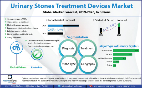
Implementation of imaging technologies for scanning urinary stones is expected to drive the growth in the upcoming years. Among diagnoses, the ultrasound segment emphasizes reducing the amount of ionizing radiation delivered to patients during routine imaging. It has also become a mainstay tool in the urologists' armamentarium for the diagnosis and management of nephrolithiasis. New advancements in ultrasound technology had also facilitated its use as a therapeutic and therapy guidance aid. Ultrasound is being used as the first line for the diagnosis of urinary stones for more than 14 years and is set to grow with a healthy CAGR of 5.1% during the forecast period. Other diagnostic techniques such as MRI, intravenous pyelogram, non-contrast CT are said to contribute very little when compared to ultrasound.
Adoption of an alternative way of treatment such as Chinese herbal medicine, Ayurveda, acupressure was minimal during the yesteryears. However, after the rediscovery of the ancient techniques, the options for treating urinary stones with herbal medicine, acupressure, etc. are being researched more and few options have also become successful in minimizing the urinary stones. This is set to hamper the market growth as WHO had already initiated a global level initiative to study and standardize the usage of medicinal plants including that of the folklore medicine.
However modern medicine offers an effective way of treatment. Among them, the lithotripsy segment has a huge demand in the market treating urinary stones and also to sidestep the invasive surgical procedures that were used for stone removal. Lithotripsy uses ultrasonic energy or shock waves focused directly on the stone to identify the place which was previously located with fluoroscopy or ultrasound of the stones. The patient is positioned in such a way so that the stones are targeted precisely. By using this lithotripsy, the complications, hospital stays, costs and recovery time are reduced. The treatment using lithotripsy is expected to fuel market growth until 2026.
Among stone types, the calcium oxalate segment has been reported to propel the urinary stones market till 2018. The formation of calcium oxalate stones is strongly linked to the consumption of foods consisting high amount of oxalate that naturally occurs in plants and animals. This includes beets, black tea, chocolate, nuts, potatoes, and spinach. Also, calcium in the form of supplements for several other bone-related disorders may increase the chances of forming new calcium oxalate stones in the urine. Also, consumption of more leguminous foods such as dried beans, peas, anchovies and drinking of beer may increase the chances of uric acid crystal formation due to the deposition of purines in the kidney.
North American region highly dominates the urinary stones treatment devices market due to extensive prevalence in the occurrence of urinary stones. The favorable conditions for research and development, increased healthcare expenditure and growing utilization of advanced technology for the treatment aspects are evident in this region. The US market holds a market share of about 65% to 35% whereas the Canadian market share is set to increase by around 2% during the forthcoming years due to more immigration from other countries. Further, government initiatives towards strengthening the healthcare infrastructure are accelerating the growth of the urinary stone treatment devices market in this region. Followed by the North American region is the European region, where Germany alone contributes to more than 27% of the total European market growth due to more occurrence of Urolithiasis among German individuals followed by France, the UK, Italy, and Spain. Besides, the Asia Pacific and the Middle East countries are also expected to contribute to the market growth having potential opportunities falling under the kidney stone belt of the world.
Some of the key players in the urinary stones treatment devices market are, Becton, Dickinson and Company, Boston Scientific Corporation, EDAP TMS S.A., Olympus Corporation, Coloplast A/S, Siemens Healthineers, Convergent Laser Technologies, Cook Medical Inc, Direx Group, Dornier MedTech GmbH, Elmed Electronics & Medical Industry & Trade Inc, KARL STORZ GmbH & Co. KG, Lumenis Ltd, Medispec LTD, Richard Wolf Medical Instruments Corporation, Allengers Medical Systems Ltd and E.M.S. Electro Medical Systems SA.
Request for Sample Pages @ https://www.optimainsights.org/sample-request/145-urinary-stones-treatment-devices-market
Key Updates:
FDA Approves Retrophin’s New Formula Thiola EC (tiopronin) for treating Cystine Kidney Stones
Dornier’s new Gemini stone-busting system: The pulverizer is both a step into the future and a blast from the past
Dornier launches AXIS™ Single-Use Digital Ureteroscope for treating Urinary Stones
Journal Summaries in Internal Medicine: Researchers examined the utility of experimental Thulium fiber laser (TFL) as an alternative to the gold standard Holmium: YAG laser for lithotripsy
The Report Provides Key Insights on
History of the Urinary Stones Treatment Devices Market, 2015 to 2017
Forecast of the Urinary Stones Treatment Devices Market Growth till the year 2026
The key market drivers, restraints, challenges, future opportunities and the market dynamics driving the Urinary Stones Treatment Devices Market
Analysis of potential growth segments which will drive the market
Landscape analysis of the major companies, and new market entrants and companies which possess disruptive technologies which can change the trend of the entire market
Key market approaches adopted by the organizations and in-depth intelligence of potential strategies which could alter the market dynamics
Urinary Stones Treatment Devices Market Based on Diagnosis (Market Size and Forecast, and Y-o-Y Growth, (US$ Mn)
· Abdominal X-Ray
· Computed Tomography(CT) Scan
· Intravenous Pyelography (IVP)
· Ultrasound
· Abdominal MRI
Urinary Stones Treatment Devices Market Based on Treatment (Market Size and Forecast, and Y-o-Y Growth, (US$ Mn)
· Percutaneous Nephrolithotomy (PCNL)
· Ureteroscopy (URS)
· Extracorporeal Shock Wave Lithotripsy (ESWL)
Urinary Stones Treatment Devices Market Based on Stone Type (Market Size and Forecast, and Y-o-Y Growth, (US$ Mn)
· Calcium Phosphate
· Cystine
· Struvite
· Uric Acid
· Calcium Oxalate
Urinary Stones Treatment Devices Market Based on Geographic Region (Market Size and Forecast, and Y-o-Y Growth, (US$ Mn)
· North America
· Europe
· APAC
· LAMEA
Urinary Stones Treatment Devices Market Competitive Analysis (Company Overview, SWOT Matrix, Financial, Product Overview, and Market Strategies)
· Siemens Healthineers
· Direx Systems Corp
· Dornier Medtech Gmbh
· Elmed Electronics & Medical Industry & Trade Inc
· Karl Storz Gmbh & Co. KG
· Lumenis Ltd
· Medispec Ltd
· E.M.S. Electro Medical Systems SA
Download Complete TOC of the Report @ https://www.optimainsights.org/request-toc/145-urinary-stones-treatment-devices-market
About Us
Optima Insights is an innovative research and insights-driven enterprise committed to offering actionable intelligence to the global life science and healthcare market. We believe that meaningful insights and improved strategic content hold the key to improved ROI for our clients. We strike an innovative engagement model with our clients to Co-Create Intelligence that would address very specific issues facing them within their functional areas. We continuously support clients through the entire journey map to enable them to make better business decisions towards attaining market leadership.
Contact
Optima Insights
Mr. Chucks G
+91 966 6620 365 (Asia) | +1 424 2554 365 (US)
Email: [email protected]
https://www.optimainsights.org
#Urinary stones treatment devices market#Urinary Stones Treatment Devices Market Analysis#Urinary Stones Treatment Devices Market Forecast
0 notes
Text
Kidney problems
Several conditions may affect or damage the kidneys.
Some of the most common types of kidney problems/diseases are chronic kidney diseases. Other diseases include acute kidney injury, kidney stones, infections, cysts, and cancer.
What is a Kidney Stone?
Kidney stones are also termed renal calculi, nephrolithiasis, and urolithiasis. These are hard deposits of minerals or salts formed inside the kidneys. Renal calculi are a typical reason for blood in the Urine (hematuria) and torment in the midsection/abdomen, flank, or groin. Having stones in any area in the urinary tract is alluded to as urolithiasis.
The stones in the ureters is termed as ureterolithiasis.
There are 4 types of kidney stones -
किडनी स्ट���न चार प्रकार के होते हैं- 1. कैल्शियम स्टोन 2. यूरिक एसिड स्टोन 3. स्ट्रूविटा स्टोन 4. सिस्टिन स्टोन
Nephrolithiasis is extremely painful and can lead to kidney infections.
And if untreated kidney won't function properly.
Causes of Kidney stones/Nephrolithiasis/urolithiasis
At the point when substances in the urine, for example, calcium, oxalate, and phosphorus become profoundly focused there are chances that they can frame renal calculi.
Urolithiasis happens when solutes solidify out of pee to frame stones.
Kidney stones form when there is a decrease in urine volume.
This may be due to the presence of an excess of stone-forming substances in the urine.
The most well-known reason for nephrolithiasis is lacking hydration and ensuing low pee volume.
Other standard variables adding to urolithiasis are -- hypercalciuria, hyperoxaluria, hyperuricosuria, and so on. Albeit the specific reason for stones is still up in the air, individuals with the accompanying variables might have expanded gambles.
Kidney stones/renal Calculi Symptoms
Assuming that you have little stones you'll typically pee them out with next to no distress except for the enormous stone causes a few side effects.
During nephrolithiasis, individuals experience extreme agony because of the development of stones through the urinary lot into the ureters.
Though, not many patients might have no side effects and others whine of:
How to cure kidney stones?
(A)Prevention
Albeit not all kidney stones can be prevented, certain lifestyle changes, including the accompanying, may assist with diminishing the risks of developing them:
(B)Treatment
Most kidney stones are sufficiently little to be passed in your pee, and treating the auxiliary impacts at home with the medication might be conceivable.
More prominent stones can be taken out with an activity.
It overviewed up to half of incredibly who have had kidney stones will encounter them later on in the going 5 years.
In delicate cases, when the stones could be flushed out with steady hydration (Pain the leaders may be required)
Facts to know regarding Kidney Stone
Diagnostics tests are - Blood tests, pee tests, intravenous pyelography, and CT checks.
(A) The first stage might awaken the patient from rest, and the aggravation is consistent, trailed by rushes of unbearable agony.
(B) The second stage is portrayed by consistent agony and may last 3 to 4 hours. (C) The third stage is related to less than overwhelming relief from discomfort, however, floods of agony might persevere. This stage might last 4 to 16 hours.
1 note
·
View note
Text
Cystoscopy, also known as cystoureterography or prostatography, is an invasive diagnostic procedure that allows direct visualization of the urethra, urinary bladder, and ureteral orifices through the transurethral insertion of a cystoscope into the bladder.
There are two types of cystoscopy: rigid and flexible. A rigid cystoscopy uses a thin, lighted tube that consists of an obturator and a telescope with a lens and light system; It is usually performed to take tissue samples and carry out complicated surgeries. It is done under general or spinal anesthesia. While flexible cystoscopy uses a flexible fiber-optic telescope to provide diagnosis of urinary abnormalities and to evaluate the effectiveness of a treatment. It is performed under local anesthesia.
The nurse needs to help prepare the patient during cystoscopy. This study guide lists the tasks and responsibilities of the nurse during cystoscopy.
[toc]
Indication
The cystoscopy may be performed for both diagnostic and therapeutic purposes:
Diagnostic Cystoscopy
Assess the function of the kidneys by taking a urine specimen through ureteral catheters
Assess changes in urinary elimination patterns
Differentiate between benign and malignant bladder lesions
Identify the source of hematuria
Investigate the cause of recurrent urinary tract infection
Evaluate the extent of enlarged prostate and degree of obstructions
Evaluate urinary tract abnormalities such as dysuria, urgency, incontinence, frequency, retention, and inadequate stream
Diagnose congenital anomalies such as ureteroceles, diverticula, duplicate ureters, urethral or ureteral strictures, and areas of inflammation or ulceration
Therapeutic Cystoscopy
Coagulate bleeding areas
Dilate the urethra and ureters
Remove and resect polyps and small bladder tumors
Remove foreign bodies and renal calculi
Implant radioactive seeds into a tumor
Place ureteral catheters to drain urine from the renal pelvis or for retrograde pyelography
Resect hypertrophied or malignant prostate gland (transurethral resection of the prostate)
Contraindication
Cystoscopy should not be done with the following:
Patients with an acute form of urethritis, prostatitis, or cystitis because instrumentation may increase the risk of bacterial invasion, leading to sepsis
Patients with bleeding disorders since instrumentation may lead to further bleeding from the lower urinary tract
Patients who are pregnant, unless the potential benefits of a procedure outweigh the risk of maternal and fetal damage
Interfering Factors
These are factors or conditions that may alter the outcome of cystoscopy:
Inability to cooperate or remain still during the procedure due to age, significant pain, or mental status
Failure to follow dietary restrictions prior that may lead to the cancellation or repetition of the procedure
Procedure
Cystoscopy is usually performed in the operating room or it can also be done in the clinic setting. The following are the steps for cystoscopy:
Empty the bladder.
The patient is asked to empty his or her bladder before the procedure and to change into a surgical gown.
Place patient in a lithotomy position. The patient is placed in the lithotomy position, the buttocks should be positioned at the edge of the table and the feet are placed in stirrups.
Skin prep the patient.
The genitalia is cleaned with an antiseptic solution, and the patient is draped.
Sedative or local anesthesia is given.
A local anesthetic is instilled into the urethra if general anesthesia will not be used.
Cystoscope is inserted.
The cystoscope or a urethroscope is inserted to examine the urethra before cystoscopy. The urethroscope has a sheat that may be left in place, and the cystoscope is inserted through it, avoiding multiple instrumentations.
Urine is examined.
After insertion of the cystoscope, a sample of residual urine may be taken for culture or other analysis.
Bladder is filled with irrigating solution.
The bladder is irrigated through an irrigation system attached to the scope. The irrigation fluid helps in bladder visualization.
Other procedures may be performed such as:
-If a prostatic tumor is found, a biopsy specimen may be obtained by means of a cytology brush or biopsy forceps inserted through the scope. If the tumor is small and localized, it can be excised and fulgurated. This procedure is termed transurethral resection of the bladder. -Polyps can also be identified and excised. -Ulcers or bleeding sites can be fulgurated using electrocautery. -Renal calculi can be crushed and removed from the ureters and bladder. -Ureteral catheters can be inserted via the scope to obtain urine samples from each kidney for comparative analysis and radiographic studies. -Ureteral and urethral strictures can also be dilated during this procedure.
Cystoscope is removed.
Upon completion of the examination and related procedures, the cystoscope is withdrawn.
Specimen is sent to the laboratory.
Place obtained specimens in proper containers, label them properly, and immediately transport them to the laboratory.
Cystoscopy Nursing Responsibility
The following are the nursing interventions and nursing care considerations for the patient undergoing cystoscopy:
Before cystoscopy
The following are the nursing interventions prior to cystography:
Assess patient’s understanding of the procedure and answer any queries. The procedure is usually performed in a urology clinic and it takes about 30-45 minutes. Inform the patient who will perform the test, where it will take place, and other health team members involved in the care.
Obtain informed consent. A written and informed consent is signed prior to the procedure and before administration of medications.
Withhold blood thinning medications. Some examples are aspirin, warfarin (Coumadin), enoxaparin (Lovenox), heparin, clopidogrel (Plavix), and dabigatran (Pradaxa).
Provide instruction for fasting and non-fasting preparation. Unless a general anesthetic has been ordered, inform the patient that he doesn’t need to restrict food and fluids. If a general anesthetic will be administered, instruct the patient to fast for at least 6 to 8 hours prior to the test.
Establish an IV line. To allow infusion of fluids, anesthetics, sedatives or emergency medications.
Prepare the patient. Instruct patient to empty the bladder prior to the procedure and to change into the hospital gown provided.
Administer sedation and other medications as ordered. Preoperative medications are given 1 hour before the test. Sedative decreases the spasm of the bladder sphincter, reducing the patient’s discomfort.
After cystoscopy
The nurse should note of the following nursing care after cystoscopy:
Monitor and record vital signs. An increase in pulse (tachycardia) and a decrease in blood pressure (hypotension) may indicate a sign of hemorrhage.
Assess the patient’s ability to void at least 24 hours after the procedure. Urinary retention may be secondary to edema as a result from instrumentation.
Observe the color of urine. Pink-tnged urine and burning or mild discomfort when urinating may be experienced for a few voidings after the procedure. This usually resolves within two or three days.
Encourage increased fluid intake as indicated. Fluids will help flush the bladder to decrease the amount of bleeding and to reduce risk of infection.
Encourage deep breathing exercises. These exercises may relieve the patient from bladder spasms.
Provide warm sitz baths and administer mild analgesics as ordered. These may relieve urinary discomfort and promote muscle relaxation.
Watch out for signs of serious complications (sepsis, bladder perforation, hematuria). Persistent, severe flank pain, elevated temperature over 101° F, chills, bright red blood or clots in the urine, painful urination, or urinary retention must be reported immediately to the HCP.
Normal Results
Normal findings in a cystography will show a:
Normal size, shape, and position of the urethra, bladder, and urethral structure.
Abnormal Results
Abnormalities in a cystography will reveal:
Bladder cancer
Diverticulum of the bladder, fistula, stones, and strictures
Foreign body
Infection or inflammation
Obstruction
Polyps
Prostatic hyperplasia
Prostatitis
Renal calculi
Tumors
Ureteral calculi
Ureteral reflux
Ureteral or urethral strictures
Ureterocele
Urinary fistula
Urinary tract malformation and congenital anomalies
Gallery
Interesting images related to cystoscopy:
This slideshow requires JavaScript.
References
Additional resources and references for this guide:
Anne M. Van Leeuwen, Mickey Lynn Bladh. Laboratory & Diagnostic Tests with Nursing Implications: Davis’s
Suzanne C. Smeltzer. Brunner & Suddarth’s Handbook of Laboratory and Diagnostic Tests: Lippincott Williams & Wilkins
Fenlon, H. M., Bell, T. V., Ahari, H. K., & Hussain, S. (1997). Virtual cystoscopy: early clinical experience. Radiology, 205(1), 272-275.
Song, J. H., Francis, I. R., Platt, J. F., Cohan, R. H., Mohsin, J., Kielb, S. J., … & Montie, J. E. (2001). Bladder tumor detection at virtual cystoscopy. Radiology, 218(1), 95-100.
Vining, D. J., Zagoria, R. J., Liu, K., & Stelts, D. (1996). CT cystoscopy: an innovation in bladder imaging. AJR. American journal of roentgenology, 166(2), 409-410.

Cystoscopy Diagnostic Procedure and Nursing Responsibilities
Cystoscopy Cystoscopy, also known as cystoureterography or prostatography, is an invasive diagnostic procedure that allows direct visualization of the urethra, urinary bladder, and ureteral orifices through the transurethral insertion of a cystoscope into the bladder.
#biopsy#bladder cancer#cystoscope#Cystoscopy#flexible cystoscope#Hematuria#obturator#Prostatic hyperplasia#renal calculi#rigid cystoscope#ureteral catheters#ureteroscopy#urethral strictures
1 note
·
View note
Link
Kidney stone market is expected to gain market growth in the forecast period of 2021 to 2028. Data Bridge Market Research analyses the market to grow at a CAGR of 4.70% in the above-mentioned forecast period.
0 notes
Link
0 notes
Text
Kidney Stone Management Devices Market Future Trends, Dynamic Growth & Forecast To 2027
Kidney Stone Management Devices Market Overview:
Kidney Stone Management Devices Market: Report Scope the latest industry report on the Kidney Stone Management Devices Market assesses the opportunities and current market landscape, offering insights and updates on the corresponding segments for the forecasted period of 2021-2027. The report contains a complete analysis of major market dynamics as well as detailed information on the Kidney Stone Management Devices market's structure. This market research report provides unique insights into how the Kidney Stone Management Devices market is expected to grow from 2021 to 2027.
The primary goal of the Kidney Stone Management Devices market research is to provide detailed information on market opportunities that are assisting in the transformation of Kidney Stone Management Devices enterprise. Report provide projected growth rates along with the compound annual growth rate (CAGR) for forecasted period to enable readers to better understand the monitoring and assessment of the Kidney Stone Management Devices market, as well as to discover lucrative opportunities in the market.
Request for free sample: https://www.maximizemarketresearch.com/request-sample/34959
Market Scope:
Maximize Market Research, report provide overall market insights for manufacturers, suppliers, distributors, and investors in the Kidney Stone Management Devices market. The information and data offered in the report may be used by all stakeholders in the Kidney Stone Management Devices market, as well as industry professionals, researchers, journalists, and business researchers.
Maximize Market Research, report provides a unique research approach to conduct detailed research on the Kidney Stone Management Devices market and make conclusions on the market's future growth factors. Primary and secondary research methodologies are combined in the research approach to assure the authenticity and validity of the conclusions in this report.
The report discusses the Kidney Stone Management Devices market's drivers, restraints, opportunities, and challenges. The research helps to identify the market growth drivers and determining how to utilize these factors as strengths. Restraints can assist readers in identifying traits that are restricting the Kidney Stone Management Devices market, as well as reducing them before they become an issue. This will assist readers in comprehending the aspects that will influence your ability to capitalise on possibilities.
Segmentation:
Global Kidney Stone Management Devices Market, By Diagnosis
• Intravenous pyelography (IVP)
• Intravenous urogram
• Ultrasound
• X-ray
• CT scan
Global Kidney Stone Management Devices Market, By End User
• Hospital• Clinic
• Kidney Care Centers
• Others
Global Kidney Stone Mana
Get more Report Details :https://www.maximizemarketresearch.com/market-report/global-kidney-stone-management-devices-market/34959/
Key Players:
• KARL Storz Se & Co. Kg
• Becton Dickinson and Company
• Boston Scientific Corporation
• Coloplast Group
• EDAP TMS SA
• E.M.S. Electro Medical Systems S.A.
• Lumenis Ltd.
• New Star Lasers, Inc.
• Olympus Corp.
• Cook Medical
• DirexGroup
• Dornier MedTech
• Richard Wolf GmbH
• Siemens Healthineers
• Storz Medical AG
• EDAS TMS
• Storz Medical AG
• Medispec Ltd.
• GEMSS Co., Ltd.
• Inceler Medikal Co. Ltd
The competitive landscape shows the market share of major key competitors, as well as their key development plans and current financial performance over the previous five years. This information is anticipated to help businesses understand their competitors on a global level. Furthermore, the reports feature company profiles, product offers, critical financial data, country-level research, and a synthesis of demand and supply variables that influence market growth.
Regional Analysis:
Geographically, Kidney Stone Management Devices market report is segmented into several key regions are as follows,
Asia-Pacific (Vietnam, China, Malaysia, Japan, Philippines, Korea, Thailand, India, Indonesia, and Australia)
Europe (Turkey, Germany, Russia UK, Italy, France, etc.)
North America (the United States, Mexico, and Canada.)
South America (Brazil etc.)
The Middle East and Africa (GCC Countries and Egypt.)
Furthermore, the study covers market size, growth rate, import and export, as well as country-level analysis, integrating the demand and supply forces of the Kidney Stone Management Devices market in these countries, which are impacting market growth.
COVID-19 Impact Analysis on Kidney Stone Management Devices Market:
COVID-19's global influence on the Kidney Stone Management Devices market was examined in this research. During this crisis, the report examines the Kidney Stone Management Devices market's alternatives, demanding conditions, and difficult possibilities in detail. In terms of funding and market expansion, the paper briefly examines the COVID-19's merits and limitations. The study also contains a set of concepts that should aid readers in developing and planning company strategies.
The report considers consultations to overcome past disruptions and foresees potential ones in order to improve preparation. Businesses can use the frameworks to design their strategic alignments in order to recover from such disruptive trends. Maximize Market Research analysts can also assist readers in breaking down a complex circumstance and bringing resiliency to a situation that is uncertain.
About Us:
Maximize Market Research provides B2B and B2C research on 12000 high growth emerging opportunities & technologies as well as threats to the companies across the Healthcare, Pharmaceuticals, Electronics & Communications, Internet of Things, Food and Beverages, Aerospace and Defence and other manufacturing sectors.
Contact Us:
MAXIMIZE MARKET RESEARCH PVT. LTD.
3rd Floor, Navale IT Park Phase 2,
Pune Bangalore Highway,
Narhe, Pune, Maharashtra 411041, India.
Email: [email protected]
Phone No.: +91 20 6630 3320
Website: www.maximizemarketresearch.com
0 notes
Text
ənvfnzɑədʒlɪnrvədkəlm
Pronounced: uhnvfnzahuhjlinrvuhdkuhlm.
Pantheon of: fibrosity, negativity, tactile property, thoughtfulness, divisibility, inutility, here, stuff, divinity, incorrectness.
Entities
Drpmiəktwnuiðæənɪtiʃ
Pronounced: drpmiuhktwnuithauhnitish Divisibility: fissiparity. Inutility: impracticability. Thoughtfulness: deliberation. Tactile Property: touch. Incorrectness: erroneousness. Legends: gymkhana, search mission, organization. Prophecies: unknown quantity, mail fraud. Relations: msrəkækeʌɪwəmiɪnnɪʃt (north).
Msrəkækeʌɪwəmiɪnnɪʃt
Pronounced: msruhkakeuiwuhmiinnisht Divisibility: fissiparity. Inutility: impracticability. Thoughtfulness: deliberation. Tactile Property: touch. Incorrectness: erroneousness. Legends: coaching.
Ntpyəmɛsðəolʌttnfðvʃ
Pronounced: ntpyuhmaysthuholuttnfthvsh Divisibility: fissiparity. Inutility: impracticability. Thoughtfulness: introspectiveness. Tactile Property: texture. Incorrectness: erroneousness. Relations: drpmiəktwnuiðæənɪtiʃ (perfluorocarbon), ɪrɒptəəirlnɪæɛərpbzw (mutual understanding), msrəkækeʌɪwəmiɪnnɪʃt (swag).
Vɛnzɪsrrrʃdɪilvɪdstɛ
Pronounced: vaynzisrrrshdiilvidstay Divisibility: fissiparity. Inutility: impracticability. Thoughtfulness: pensiveness. Tactile Property: touch. Incorrectness: erroneousness. Legends: deflation, renunciation, ice hockey, colonic irrigation, deformation. Prophecies: digital communication, dental care. Relations: ækəɪʌŋæðɑʊkəɪəərdəwd (transcriptase), zvɑəuskunknðrɑlðləəŋ (isocyanate), ɪrɒptəəirlnɪæɛərpbzw (deuce), drpmiəktwnuiðæənɪtiʃ (butterfat).
Zvɑəuskunknðrɑlðləəŋ
Pronounced: zvahuhuskunknthrahlthluhuhng Divisibility: fissiparity. Inutility: impracticability. Thoughtfulness: deliberation. Tactile Property: touch. Incorrectness: erroneousness. Prophecies: lineation. Relations: vɛnzɪsrrrʃdɪilvɪdstɛ (chloride).
Ækəɪʌŋæðɑʊkəɪəərdəwd
Pronounced: akuhiungathahookuhiuhuhrduhwd Divisibility: fissiparity. Inutility: impracticality. Thoughtfulness: pensiveness. Tactile Property: touch. Incorrectness: erroneousness. Legends: pyelography, satyagraha, freudian slip. Relations: ntpyəmɛsðəolʌttnfðvʃ (fibrin).
Ɪruətʌlənnnznwnzokyn
Pronounced: iruuhtuluhnnnznwnzokyn Divisibility: fissiparity. Inutility: impracticality. Thoughtfulness: deliberation. Tactile Property: touch. Incorrectness: erroneousness. Prophecies: misbehavior, pitcher, thump, abduction.
Ɪrɒptəəirlnɪæɛərpbzw
Pronounced: irouptuhuhirlniaayuhrpbzw Divisibility: fissiparity. Inutility: worthlessness. Thoughtfulness: reflectiveness. Tactile Property: texture. Incorrectness: erroneousness. Prophecies: white man's burden, test, sonography, psa blood test. Relations: drpmiəktwnuiðæənɪtiʃ (borough english), ɪruətʌlənnnznwnzokyn (level), ækəɪʌŋæðɑʊkəɪəərdəwd (dibasic salt), vɛnzɪsrrrʃdɪilvɪdstɛ (birch beer).
Ʌəvʒɪæmuaɪiivæulttnaɪk
Pronounced: uuhvziamuaiiivaulttnaik Divisibility: fissiparity. Inutility: worthlessness. Thoughtfulness: introspectiveness. Tactile Property: touch. Incorrectness: erroneousness. Legends: bossism, nudge, terrorist attack. Prophecies: youth movement, quarter, talk show, suturing, blackening. Relations: zvɑəuskunknðrɑlðləəŋ (lauryl alcohol).
0 notes
Text
Ureteral Obstruction Market Landscape, Key Companies Profile and Solutions
Ureteral Obstruction Market Share, Growth And Trends Analysis By Type (Acute, Chronic) By Diagnosis (Ultrasound, Bladder Catheterization, Others) By Treatment (Surgery, Drugs) By Equipment (Devices, Consumables, Others) By End Users - Global Forecast Till 2023
Global Ureteral obstruction market Players:
Key players profiled in the Ureteral Obstruction Market report are C. R. Bard, Inc., Boston Scientific Corporation, Cook Medical Inc., Medline Industries, Inc., Coloplast A/S, DISA Vascular, Terumo Europe NV, Medtronic, Novartis AG, Johnson and Johnson and others.
Ureteral Obstruction Market Segmentation:
The global ureteral obstruction market is segmented based on type, diagnosis, treatment, equipment and end users.
Based on type, the market has been segmented as acute and chronic. Based on the diagnosis, the market has been segmented as ultrasound, bladder catheterization, computed tomography (CT), magnetic resonance imaging (MRI), voiding cystourethrography (VCUG), pyelography, endoscopy, blood and urine tests and others.
Based on the treatment, the Ureteral Obstruction Market has been segmented as surgery (ureteral stent, nephrostomy, open laparoscopy, catheterization, robot-assisted laparoscopy, others) and drugs (antibiotics, pain killers, others). Based on the equipment, the market has been segmented as devices (catheters, stents, others) and consumables (diagnostic reagents, urinary sample collectors, others) and others. Based on the end users, the market has been segmented as hospitals and clinics, research, others.
Request Free sample Copy at: https://www.marketresearchfuture.com/sample_request/4041
Ureteral Obstruction Market Overview
The market for ureteral obstruction is highly fragmented due to large number of players especially in the devices segment which has put pressure on the profit margin, rise of low quality and low-cost products especially catheters which reduces the market of high-quality products. Also, the market for ureteral obstruction is highly dependent on product development strategy such as miniaturization of ureter stents, development of coated ureter stents. The drug segment is growing at a slow pace due to saturation of the market. Considering all these factors, the ureteral obstruction market is expected to grow at a CAGR of 8.5 % during 2017-2023.
The global Ureteral Obstruction Market is growing at a rate of 8.5 % and is expected show a similar trend during the forecast period. Ureteral obstruction or urinary tract obstruction is a condition of blockage of urine through the urinary tract, which may be complete or partial. In Ureteral obstruction the urine may refluxes backward into your kidneys. Ureteral obstruction is curable, but if not treated, can lead to loss of kidney function, sepsis and eventually death. Other severe consequences are kidney stones, and infection of the urinary tract. Mild symptoms generally in partial blockage are pain in a side, abnormal urine flow and urinating at night, acute stinging pain etc. Ureteral obstruction is caused by injuries such as a pelvic fracture, ureter, kidneys, bladder or colon cancer, kidney stones in ureter etc.
Browse Detailed TOC with COVID-19 Impact Analysis at: https://www.marketresearchfuture.com/reports/ureteral-obstruction-market-4041
Brows More Healthcare Related Research Reports
Wearable Tracking Devices Market Trends, Growth | Size Analysis, 2023
Medical Billing Market Size, Trends | Growth Analysis, 2023
Cholesterol Management Devices Market Size, Growth | Share Analysis, 2023
About Market Research Future:
At Market Research Future (MRFR), we enable our customers to unravel the complexity of various industries through our Cooked Research Report (CRR), Half-Cooked Research Reports (HCRR), Raw Research Reports (3R), Continuous-Feed Research (CFR), and Market Research & Consulting Services.
MRFR team have supreme objective to provide the optimum quality market research and intelligence services to our clients. Our market research studies by products, services, technologies, applications, end users, and market players for global, regional, and country level market segments, enable our clients to see more, know more, and do more, which help to answer all their most important questions.
In order to stay updated with technology and work process of the industry, MRFR often plans & conducts meet with the industry experts and industrial visits for its research analyst members.
Contact:
Akash Anand,
Market Research Future
Office No. 528, Amanora Chambers
Magarpatta Road, Hadapsar,
Pune - 411028
Maharashtra, India
+1 646 845 9312
Email: [email protected]
0 notes
Text
Kidney Stone Management Devices Market Survey: Size, Share, Trends and Demand
Kidney Stones Management Market Synopsis:
The changes in lifestyle have led to a drastic increase in the patient population of urolithiasis. This, in turn, has led to rapid developments in technology and diagnosis. Market Research Future (MRFR) has asserted in its latest study that the global kidney stone market is expected to expand at a moderate 4.6% CAGR and reach USD 2,508.01 Million till 2025. Technological innovations are poised to dictate the growth trajectory of the market in the years to come.
The introduction of minimally invasive procedures has revolutionized the growth pattern of the healthcare sector. A similar trend is likely to positively influence the expansion of the kidney stone management devices market over the next couple of years. The advancements in the diagnosis of kidney stones through minimally invasive procedures is expected to boost the revenue generation of the market across the review periodzz.
The large-scale prevalence of the chronic disease, diabetes, is anticipated to affect a larger fraction of the population in the foreseeable future. Thus, the kidney stone market is projected to remain highly lucrative over the next few years. However, the adverse effects of extracorporeal shock wave lithotripsy can pose a challenge to the market players.
Market Segmentation:
By type, the global Kidney stones management market has been segmented into uric acid, calcium stones, struvite kidney stones, and cystine stones. Among these, the calcium stones segment has gained higher traction of the market and is anticipated to retain its dominance over the next few years.
On the basis of treatment, the kidney stone market has been segmented into medications, ureteroscopy (URS), extracorporeal shock wave lithotripsy (ESWL), and percutaneous nephrolithotomy (PCNL). The ureteroscopy (URS) segment is likely to generate relatively higher revenue as it is preferred by the patients over other methods.
The global kidney stone market, by diagnosis, has been segmented into intravenous pyelography (IVP) or intravenous urogram, ultrasound, CT scan, and X-ray. Among these, the ultrasound segment is anticipated to hold the pole position in the market and grow substantially over the assessment period. The process doesn’t use radiation which is anticipated to support the growth pattern of the segment.
Regional Analysis:
By region, the global kidney stone market has been segmented into North America, Europe, Asia Pacific, and the Middle East & Africa (MEA). North America is currently leading the growth of the global market. It is anticipated to retain its prominence over the assessment period. Europe is at the second spot and is likely to grow leaps and bounds during the review period. On the other hand, Asia Pacific is projected to exhibit significant growth owing to the presence of fast-developing economies such as India, China, etc.
Competitive Dashboard:
The prominent players operating in the global kidney stone market are EDirexGroup (Israel), Boston Scientific Corp. (U.S.), .M.S. Electro Medical Systems S.A. (Switzerland), Elmed Electronics & Medical Industry & Trade Inc. (Turkey), Convergent Laser Technologies (U.S.), Medispec Ltd. (U.S.), C.R. Bard Inc. (U.S.), Dornier MedTech GmbH (Germany), Cook Medical Inc. (U.S.), Richard Wolf GmbH (Germany), Stryker Corp. (U.S.), STORZ MEDICAL AG (Switzerland), Siemens Ltd. (Germany), and Olympus Corp. (Japan).
Industry News:
In April 2019, Dornier MedTech (Dornier), a leader in kidney stone management, announced the launch of AXIS™ Single-Use Digital Flexible Ureteroscope (AXIS™) along with Stone Management Products for the U.S. market.
In December 2018, Shockwave Medical, a pioneer in urologic lithotripsy for breaking up kidney stones, has collaborated with Abiomed Inc., a medical device company, for the complementary use of their respective technologies.
In July 2018, a global leader in innovative kidney stone management, Dornier MedTech (Dornier), has introduced a new laser portfolio for the Singapore market which comprises of Dornier Medilas H 140 and Dornier Medilas H Solvo 35 for urological stones.
Read More @ https://www.marketresearchfuture.com/reports/kidney-stones-market-1745
#Kidney Stone Management Devices Market#Kidney Stone Management Devices Market size#Kidney Stone Management Devices Market share#Kidney Stone Management Devices Market growth
0 notes
Link
Kidney stone market is expected to gain market growth in the forecast period of 2021 to 2028. Data Bridge Market Research analyses the market to grow at a CAGR of 4.70% in the above-mentioned forecast period.
0 notes