#rna extraction solution
Explore tagged Tumblr posts
Visit Tumblr Blog
Explore Tumblr blogs with no restrictions, modern design and the best experience.
Last Seen Tumblr Blogs


Fun Fact
Mobile Tumblr US users spend an average of 4.04 minutes per session on the app.
Photo

With Rapi-X96 and Rapi-X16, #Genes2Me team is transforming the #nucleic_acid extraction process by offering #automation, speed, and precision to #researchers, scientists, and #diagnostic laboratories. Whether you're conducting groundbreaking #genetic research or performing high-throughput diagnostic #testing, our innovative solutions optimize your workflow, improve efficiency, and accelerate discoveries.
Join the automation revolution today and #unlock new #possibilities in nucleic acid #extraction with Rapi-X96 and Rapi-X16. Contact us for a demo or visit our website for more information. https://www.genes2me.com/nucleic-acid-extraction-solutions
#madeinindia #manufacturer #rapix96 #rapix16 #instruments #system #solutions #ivd #machines #purification #dna #rna #dnasequencing
#genes2me#nucleic acid#nucleic acid extraction instrument#Extraction Instruments#dna extraction solution#rna extraction solution#purification solution#machine
1 note
·
View note
Text
Exosome Therapy for Hair Loss: A Non-Surgical Alternative to Hair Transplants
Hair loss is a common issue that affects millions of people worldwide, leading many to search for effective treatments to restore their natural hair. Traditionally, hair transplants have been the go-to solution, but they require invasive surgery, lengthy recovery times, and significant costs. However, a revolutionary treatment is gaining attention in the field of hair restoration: Exosome therapy for hair loss. This non-surgical procedure offers promising results, minimal downtime, and a more affordable option compared to traditional methods. ROS Regenerative Hair and Skin, we’ll explore how exosome therapy works, its benefits, and why it’s becoming a popular choice for those seeking hair restoration.
What is Exosome Therapy for Hair Loss?
Exosome therapy for hair loss involves the use of exosomes, which are small vesicles that play a crucial role in cell communication. Exosomes are naturally occurring particles found in all cells, and they are responsible for carrying messages between cells, promoting healing, and stimulating tissue regeneration. In the context of hair loss, exosomes can help regenerate hair follicles, stimulate hair growth, and improve the overall health of the scalp.
Exosome therapy for hair loss is a minimally invasive procedure that uses these vesicles to encourage hair follicles to enter the anagen (growth) phase of the hair cycle. The treatment involves extracting exosomes from stem cells, which are then applied to the scalp via a series of injections. These exosomes contain proteins, lipids, and RNA, all of which work together to rejuvenate the hair follicles and improve hair density.
How Exosome Therapy Works
The process begins with a consultation with a trained specialist who will assess the extent of hair loss and determine whether exosome therapy is the right treatment. During the procedure, exosomes are injected directly into the scalp, targeting areas where hair thinning or baldness is most prominent.
Once injected, the exosomes work at a cellular level, promoting the regeneration of damaged hair follicles and stimulating the production of new hair. The proteins and growth factors within the exosomes encourage the follicles to restart the growth cycle, leading to the development of stronger, healthier hair.
Exosome therapy is often combined with other treatments, such as platelet-rich plasma (PRP) therapy, to enhance results. This combination can provide a more comprehensive approach to hair restoration, addressing both the cellular and growth factor needs of the scalp.
Benefits of Exosome Therapy for Hair Loss
Non-Surgical and Minimally Invasive: Unlike traditional hair transplant surgeries, exosome therapy for hair loss is a non-surgical procedure that requires no incisions or stitches. This makes it a more comfortable and less invasive option for those who want to avoid the risks and recovery time associated with surgery.
Quick Recovery Time: One of the biggest advantages of exosome therapy for hair loss is the minimal downtime. Most patients can resume normal activities immediately after the treatment, making it an ideal option for those with busy schedules who cannot afford the long recovery times required by hair transplant procedures.
Stimulates Natural Hair Growth: Exosome therapy promotes the body’s natural ability to regenerate hair. By using exosomes to target the root cause of hair thinning, the treatment encourages hair follicles to grow healthy hair, without the need for artificial implants or transplants.
Safe and Effective: Exosome therapy for hair loss is considered safe when performed by a trained specialist. The exosomes used in the procedure are derived from stem cells, making them biocompatible with the body and reducing the risk of adverse reactions or complications.
Long-Lasting Results: While results can vary, many patients experience noticeable improvements in hair density and thickness within a few months after the treatment. The effects of exosome therapy are typically long-lasting, with many patients seeing sustained hair growth for up to a year or more.
Exosome Therapy for Hair Loss in Hyderabad: A Growing Trend
As the demand for effective hair restoration treatments increases, exosome therapy for hair loss in Hyderabad has gained significant attention. Hyderabad, known for its advanced healthcare sector and skilled professionals, is quickly becoming a hub for non-surgical hair restoration treatments, including exosome therapy.
The city is home to several reputable clinics and dermatologists who specialize in exosome therapy for hair loss. Patients in Hyderabad are increasingly turning to this innovative treatment to address hair thinning, receding hairlines, and baldness. With its minimal downtime and impressive results, exosome therapy for hair loss in Hyderabad is quickly becoming a top choice for individuals seeking an alternative to traditional hair transplant surgeries.
Exosome Therapy vs. Hair Transplants
While both exosome therapy and hair transplants aim to restore hair, there are several key differences between the two procedures.
Invasiveness: Hair transplants require surgical incisions to harvest hair follicles from one part of the scalp and transplant them to areas of hair loss. In contrast, exosome therapy for hair loss is a non-invasive procedure that involves injecting exosomes into the scalp, without the need for any surgery or incisions.
Recovery Time: Hair transplant surgery typically involves a recovery period of several weeks, with patients needing to avoid certain activities during this time. On the other hand, exosome therapy offers a much quicker recovery, with most patients able to return to their daily routine immediately after the procedure.
Results: Hair transplants provide permanent results because hair follicles are relocated to areas of the scalp that are resistant to hair loss. However, the procedure can be costly, and the results may not be as natural in some cases. Exosome therapy, while not permanent, offers a more natural and gradual improvement in hair density and texture, with results continuing to improve over time.
Cost: Exosome therapy for hair loss tends to be more affordable than traditional hair transplant surgeries, making it a viable option for individuals on a budget. While it may require multiple sessions to achieve the desired results, the overall cost is typically lower than that of a hair transplant.
Exosomes Hair Treatment: The Future of Hair Restoration
As the field of regenerative medicine continues to evolve, exosomes hair treatment is emerging as one of the most promising non-surgical solutions for hair loss. This innovative therapy offers a range of benefits, including faster recovery, natural-looking results, and minimal risk. Whether you’re looking to address early-stage hair thinning or more advanced hair loss, exosome therapy can provide a safe and effective solution.
conclusion
exosome therapy for hair loss is a groundbreaking treatment that provides an alternative to invasive hair transplant surgeries. With its ability to stimulate natural hair growth and restore the health of hair follicles, this non-surgical procedure is quickly becoming one of the most popular choices for individuals looking to combat hair loss. If you’re considering hair restoration, exosome therapy for hair loss could be the solution you’ve been waiting for.
#exosome therapy for hair loss#stem cell therapy#prp treatments#health and wellness#hair care#hair treatment
0 notes
Text

🌊 Purity You Can Trust - Bio Basic DEPC Water 🌊
Looking for the perfect solution for your molecular biology research? Bio Basic’s DEPC Water is designed to eliminate RNase contamination, ensuring reliable and reproducible results. Whether you're working on RNA extraction or PCR, trust our DEPC-treated water for high-quality outcomes.
🔬 Why Choose Bio Basic DEPC Water? ✅ RNase-Free ✅ High Purity ✅ Ideal for Molecular Biology Applications ✅ Sterile, Ready-to-Use
Perfect for scientists, researchers, and labs focused on precision. Get the reliable DEPC water your experiments deserve!
🔗 Learn More: https://www.biobasic.com/depc-water
#BioBasic#DEPCWater#MolecularBiology#RNaseFree#LabSupplies#ScientificResearch#BioScience#ResearchTools#LabEssentials#Biotechnology#PCR#RNAExtraction#PrecisionResearch#CleanResults
0 notes
Text
Life Science Tools Market: Regional Analysis and Forecast 2024-2032

The Life Science Tools Market size was estimated at USD 158.40 billion in 2023 and is expected to reach USD 407.57 billion by 2032, growing at a CAGR of 11.09% during the forecast period of 2024-2032. This significant growth is driven by technological advancements and increasing demand for innovative solutions in diagnostics, drug development, and genomics.
Regional Analysis
In 2023, North America held the largest share of the life science tools market, with the U.S. leading the charge. This dominance is attributed to the region's advanced healthcare infrastructure, substantial R&D investments, and strong presence of key life science companies. The increasing adoption of genomics, next-generation sequencing (NGS) technologies, and personalized medicine further strengthens North America's market position.
The Asia-Pacific region is projected to experience the highest growth rate during the forecast period, driven by expanding healthcare infrastructure, rising healthcare expenditure, and increasing government funding for biotechnology and life sciences research.
Get Free Sample Report @ https://www.snsinsider.com/sample-request/3833
Market Segmentation
The life science tools market is segmented based on product, technology, and end-user:
By Product:
NGS
Sanger Sequencing
Nucleic Acid Preparation
Nucleic Acid Microarray
PCR & qPCR
Flow Cytometry
Mass Spectrometry
Separation Technologies
Electron Microscopy
NMR
Others
By Technology:
Genomic Technology
Cell Biology Technology
Proteomics Technology
Lab Supplies & Technologies
Others
By End-User:
Biopharmaceutical Companies
Government & Academic Institutions
Healthcare
Others
Key Players
Agilent Technologies, Inc. – DNA Microarrays, Liquid Chromatography Systems, Mass Spectrometers, PCR Tools, Microfluidics, Flow Cytometry Instruments
Becton, Dickinson, and Company (BD) – Flow Cytometers, Cell Sorters, Culture Media & Reagents, Automated Liquid Handling Systems, Syringes, and Needles (for laboratory use)
F. Hoffmann-La Roche Ltd. – PCR Machines, Mass Spectrometry Systems, Laboratory Reagents, Immunoassay Analyzers, DNA Sequencers, Clinical Diagnostics Instruments
Bio-Rad Laboratories, Inc. – PCR and qPCR Systems, Electrophoresis Equipment, Western Blotting Systems, Cell Biology Reagents, Chromatography Systems, Life Science Reagents
Danaher Corporation – Flow Cytometry Instruments, PCR Systems, Laboratory Automation Equipment, Life Science Reagents, Microscopes, Spectrophotometers
Illumina, Inc. – DNA Sequencers (Next-Generation Sequencing), Microarrays, Bioinformatics Software, PCR Reagents, Genomic Assays
Thermo Fisher Scientific, Inc. – PCR and qPCR Systems, Mass Spectrometers, Chromatography Equipment, Flow Cytometers, Cell Culture Reagents, DNA and RNA Analysis Kits
QIAGEN N.V. – PCR Kits and Reagents, DNA/RNA Extraction Kits, Automated Workstations, Sequencing Solutions, Assay Development Kits
Merck KGaA – Cell Culture Media, PCR Reagents, Microarray Tools, Chromatography Systems, Spectroscopy Equipment, Protein Analysis Kits
Shimadzu Corporation – Chromatography Systems, Mass Spectrometers, Spectrophotometers, PCR Equipment, Analytical Instruments
Hitachi, Ltd. – Mass Spectrometers, Fluorescence Microscopes, X-ray Systems for Biological Applications, Automated Laboratory Systems
Bruker Corporation – Mass Spectrometry Systems, NMR Spectrometers, X-ray Diffraction Systems, FTIR Spectrometers, Microscopy Equipment
Oxford Instruments plc – Microscopes (Electron and Atomic Force), Spectrometers, NMR Systems, Cryogenics for Life Science Applications
Zeiss International – Microscopes (Fluorescence, Confocal, Electron Microscopy), Imaging Solutions, Life Science Imaging Systems, Microscopy-based Analytical Tools
Key Points
The integration of artificial intelligence (AI) and machine learning in drug discovery and genomics enhances research efficiency and accelerates drug development.
Automation in laboratories reduces manual errors and increases throughput, significantly improving operational efficiency.
Advancements in genomic technologies, such as next-generation sequencing (NGS), are revolutionizing personalized medicine and diagnostics.
The growing emphasis on precision medicine necessitates advanced diagnostic and analytical tools, fueling market growth.
Increased government funding and support for life science research promote the development of next-generation tools.
Future Scope
The future of the life science tools market is poised for substantial growth, driven by continuous technological innovations and the increasing integration of AI and automation in research processes. The expanding applications of genomic technologies and the rising focus on personalized medicine are expected to further propel market expansion. Additionally, the growing investments in research and development, coupled with supportive government initiatives, will likely enhance the development and adoption of advanced life science tools globally.
Conclusion
The life science tools market is on a robust growth trajectory, fueled by technological advancements and the escalating demand for innovative research solutions. With significant contributions from North America and rapid growth anticipated in the Asia-Pacific region, the market is set to play a pivotal role in advancing healthcare research and personalized medicine in the coming years.
Contact Us: Jagney Dave - Vice President of Client Engagement Phone: +1-315 636 4242 (US) | +44- 20 3290 5010 (UK)
Other Related Reports:
Cell Viability Assay Market
Medical Power Supply Market
Post Traumatic Stress Disorder Treatment Market
MRI Guided Neurosurgical Ablation Market
#Life Science Tools Market#Life Science Tools Market Share#Life Science Tools Market Trends#Life Science Tools Market Size
0 notes
Text
Centrifuge Tubes in Action: Common Lab Protocols and Best Practices

Centrifuge Tubes in Action: Common Lab Protocols and Best Practices
Centrifuge tubes are essential laboratory tools that serve as cornerstones in various scientific procedures. These versatile containers are crucial for everything from component separation to genetic material isolation, making them fundamental to countless research protocols. This article delves into the various applications of centrifuge tubes and outlines essential guidelines for their proper usage and maintenance to achieve reliable results.
The Role of Centrifuge Tubes in Laboratory Protocols
Engineered to endure intense centrifugal forces, centrifuge tubes facilitate material separation based on density differences. Their applications extend across multiple scientific fields, including molecular biology, biochemistry, medical diagnostics, and environmental research. Here are their primary applications:
Sample Preparation
Sample preparation forms the foundation of experimental procedures, with centrifuge tubes playing a vital role. These tubes serve as perfect vessels for separation processes, whether purifying cell cultures or clarifying solutions. Through controlled centrifugation, denser particles collect at the tube’s bottom, producing a clear supernatant for further analysis.
DNA and RNA Isolation
In molecular biology, DNA and RNA extraction protocols rely heavily on centrifuge tubes. These vessels facilitate crucial steps including cell disruption, genetic material precipitation, and impurity removal. Their distinctive conical design aids in concentrating nucleic acid pellets at the base, simplifying collection.
Protein Purification
Protein purification protocols utilize centrifuge tubes for separating proteins according to their solubility or density characteristics. They’re particularly crucial in ultracentrifugation procedures for isolating cellular components, ribosomes, or protein complexes. Careful handling ensures sample quality preservation.
Clinical Diagnostics
Medical laboratories depend on centrifuge tubes for blood analysis. Through centrifugation, plasma and serum separate from whole blood samples, enabling precise diagnostic assessments.
Environmental Sample Analysis
Environmental researchers employ centrifuge tubes to study soil, water, and air samples. Centrifugation assists in isolating pollutants or microorganisms for thorough analysis and testing.
Best Practices for Using Centrifuge Tubes
Maintaining optimal performance and durability of centrifuge tubes requires adherence to proper usage, handling, and storage protocols. Consider these essential guidelines:
Choose the Right Tube for the Job
Material: Centrifuge tubes are constructed from specialized materials such as polypropylene and polycarbonate. Polypropylene variants excel in chemical resistance and are particularly suited for extended storage requirements, while polycarbonate options stand out for their exceptional transparency and durability.
Volume: Choose tubes with appropriate volume capacity matching your sample requirements. Incorrect filling levels, whether excessive or insufficient, can compromise centrifugal balance and reduce separation efficiency.
Temperature Tolerance: Verify that your selected tube material can effectively handle the temperature requirements of your protocol, particularly for procedures involving freezing steps or autoclave sterilization.
Balance the Centrifuge
Making sure you balance the centrifuge right is super important for keeping your samples in good shape and making sure the machine can do its job for a long time. You have to place tubes that weigh the same right across from one another inside the rotor. When you don’t have an even number of samples just use a counterweight tube filled with some water or a buffer solution so everything stays stable. Accumax centrifuges come with some high-tech safety stuff that won’t let it run if your tubes aren’t balanced right. This is a big deal for keeping your samples and the machine safe.
Seal Tubes Properly
Make sure the caps on centrifuge tubes are tight to keep the tube’s contents from spilling or getting dirty when you spin them. When you’re using them for fast spins double-check to see the top’s on real good so it can handle all that spinning without popping open.
Handle with Precision
Prevent surface damage to tubes, as structural weaknesses can develop from scratches and lead to tube failure. Maintain sterile conditions and personal protection by wearing appropriate gloves during tube handling.
Optimize Operating Parameters
Strictly adhere to manufacturer-specified speed and temperature guidelines. Implement gradual speed adjustments to minimize mechanical stress on tube materials.
Implement Clear Labeling
Utilize waterproof marking systems or labels for clear tube identification. Include essential details such as sample identification, date information, and content description.
Maintain Proper Storage
Store tubes vertically to prevent structural distortion. Protect tubes from direct light exposure and temperature extremes that could compromise material integrity. Utilize appropriate storage systems like racks or holders for organizational efficiency and spill prevention.
Conclusion
Centrifuge tubes remain fundamental to successful laboratory operations across diverse scientific applications. Implementing proper handling protocols and storage practices ensures reliable results while maximizing tube longevity. These essential tools continue driving scientific advancement across various disciplines, from DNA analysis to protein studies and diagnostic testing.
0 notes
Text
Top 5 Trending Treatments of 2025: Revolutionizing Skin Care
In 2025, dermatology is seeing an influx of cutting-edge treatments that promise remarkable results with minimal downtime. From rejuvenating facials to advanced regenerative therapies, these treatments cater to diverse skincare needs, providing clients with glowing, youthful skin. Here’s a look at the top 5 trending treatments this year:
1. HydraFacial: A Deep Hydration Powerhouse
HydraFacial has evolved as one of the most sought-after non-invasive treatments in 2025. This multi-step facial combines cleansing, exfoliation, extraction, hydration, and antioxidant protection in a single session. Using a patented vortex suction technology, it delivers customized serums to address skin concerns like dryness, fine lines, acne, and hyperpigmentation. With its instant glow and no downtime, HydraFacial remains a go-to choice for anyone looking to maintain healthy, hydrated skin year-round.
2. Salmon DNA: The Secret to Youthful Skin
Salmon DNA therapy, or “salmon DNA injection,” is gaining popularity in 2025 for its anti-aging effects. Rich in nucleotides, amino acids, and peptides, this treatment stimulates cell regeneration and collagen production, leaving skin firmer and more youthful. It’s highly effective for tackling fine lines, wrinkles, and skin elasticity. The powerful antioxidants in salmon DNA also reduce pigmentation and improve overall skin tone, offering a rejuvenated look without the need for invasive procedures.
3. Carbon Toning: A Charcoal Magic for Clear, Bright Skin
Carbon Toning, also known as the “Hollywood Peel,” has become one of the most popular treatments for individuals with acne-prone or dull skin. The process uses a carbon-based lotion that is applied to the skin, followed by laser treatment to target deep pores. The laser heats the carbon particles, removing dead skin cells, excess oil, and impurities, leaving the skin brighter, clearer, and more even-toned. It’s a great solution for achieving a glowing complexion with minimal discomfort and downtime.
4. Exosomes: Harnessing Regenerative Potential
Exosomes, tiny vesicles derived from stem cells, are making waves in 2025 as a revolutionary treatment for skin regeneration. These exosomes contain growth factors, proteins, and RNA that aid in cellular repair and rejuvenation. When applied to the skin, they accelerate healing, promote collagen production, and restore skin texture and elasticity. Exosome therapy is ideal for clients with skin that has been damaged by aging, acne scars, or sun exposure, as it significantly improves the skin’s overall appearance and health.
5. HIFU: Non-Surgical Face Lifting
High-Intensity Focused Ultrasound (HIFU) continues to trend in 2025 as a non-invasive alternative to traditional facelifts. HIFU uses ultrasound energy to stimulate collagen production deep beneath the skin, lifting and tightening sagging areas such as the jawline, neck, and brows. It’s highly effective for individuals looking for a youthful appearance without the risks and recovery associated with surgery. As it offers long-lasting results with minimal discomfort, HIFU has become a top choice for those seeking a natural face lift.
Conclusion
The beauty and skincare industry in 2025 is all about advanced, non-invasive treatments that deliver quick results with minimal downtime. From HydraFacial’s deep hydration to HIFU’s face-lifting effects, these trending treatments cater to every skin concern imaginable. Whether you’re seeking rejuvenation, acne treatment, laser hair removal in Noida, or anti-aging benefits, these therapies are poised to transform the way we approach skincare.
As these treatments continue to gain popularity, they reflect a growing trend toward personalized skincare that prioritizes efficiency, safety, and lasting results. If you’re looking to elevate your skincare game, these are the treatments to watch!
0 notes
Text

🔬 Unlock High-Quality Results with Our Nucleic Acid Purification Kits 🔬
Need to extract DNA/RNA from a variety of samples like blood, cells, saliva, hair, plant tissue, or even microorganisms? Our Nucleic Acid Purification Kits provide a fast, reliable, and easy solution for high-purity nucleic acid extraction, perfect for PCR, enzyme analysis, sequencing (NGS), and more.
Whether you're diving into genomics, transcriptomics, or molecular cloning, our kits are designed to meet the highest standards in modern research.
Key Features:
High Purity: DNA/RNA ready for PCR, sequencing, and other applications.
Broad Compatibility: Works with diverse sample types.
Quick & Efficient: Save time with streamlined extraction.
Get your hands on the tools you need for success. 🔬🌱
1 note
·
View note
Text
The Importance of Molecular Biology Kits in Genetic Research
The most important thing about genetics for understanding DNA and RNA is the molecule that controls cellular functions and heredity. Advancements in genetics have contributed much to medicine, agriculture, and biotechnology. Molecular biology kits, in this context, streamline these studies with reliable tools for DNA/RNA extraction, amplification, and analysis processes.
youtube
Key Benefits of Molecular Biology Kits :
1. Improved Accuracy and Productivity:
Molecular biology kits provide pre-optimized reagents and protocols. This reduces the chances of error input, and that ensures reproducibility in genetic studies. This efficiency propels research while maintaining precision.
2. Time-Saving and Cost-Effective Solutions:
These kits save a lot of time for the researcher by dispensing with the manual preparation of reagents. Furthermore, their standardized components reduce the amount of waste generated, which in turn saves costs.
3. Versatile Applications in Biotechnology:
From PCR and qPCR to NGS, molecular toolkits are supportive of a wide range of applications that ease the exploration of geneticists concerning gene expression, mutations, or hereditary diseases.
Conclusion -
Molecular biology kits, generally referred to as genetic analysis kits or DNA/RNA research tools, are basically of vital importance in contemporary genetic research. In this context, biotechnology companies provide innovative solutions so that the kits continue to evolve, enabling researchers to make breakthroughs in unprecedented efficiency and accuracy.
0 notes
Text
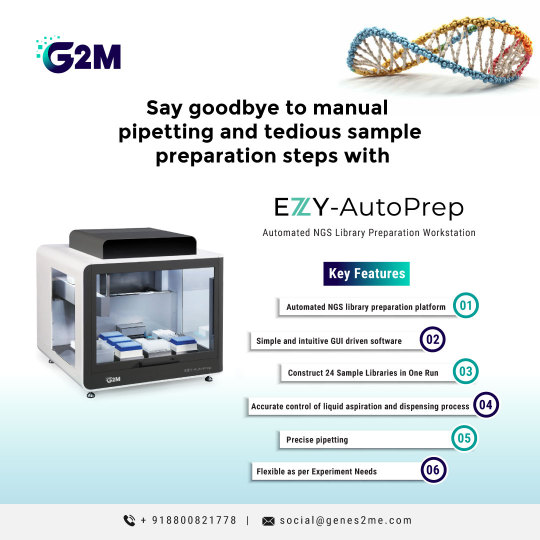
Introducing G2M’s newly launched EZY-AutoPrep—an automated #NGS library preparation workstation capable of constructing 24 sample libraries in a single run. With user-friendly software and supporting hardware, EZY-#AutoPrep ensures quick sample processing, delivering a seamless library preparation experience. Features include heating, cooling, #magnetic plate lifting, #PCR cycling, UV sterilization, and efficient purification which ensures precise, contamination-free library construction.
#Revolutionize your #NGS workflow with EZY-AutoPrep.
Visit our website for more information: https://genes2me.com/automated-ngs-library-sample-preparation
For more details, Call us at +91-8800821778 or drop us an email at [email protected]
#rtpcr #genes2me #nextgenerationsequencing #nextgeneration #sequencing #automated #ivd #dna #rna #protocols #nucleicacid
#rt-pcr#pcr#g2m#genes2me#ivd#ngs#revolutionize#AutoPrep#magnetic#dna solutions#rna solutions#automated#nucleic acid#nucleic acid extraction
0 notes
Text
The Growth of Forensic Technologies | Industry Forecast 2025-2032

In today’s world, forensic technologies play a pivotal role in solving crimes, collecting evidence, and upholding justice. From advanced DNA profiling to digital forensics and biometric systems, forensic technologies are revolutionizing the way criminal investigations are conducted. This evolution is fueling significant growth in the global forensic technologies market.
Let’s explore the factors driving this market, regional dynamics, top companies leading the industry, and the promising future of forensic technologies.
Understanding the Forensic Technologies Market
Forensic technology refers to the use of science and advanced technology to solve crimes and aid criminal investigations. These technologies enable forensic scientists, investigators, and law enforcement agencies to analyze evidence more efficiently, from physical evidence like blood samples to digital evidence like encrypted data on devices.
Forensic Technologies Market size is poised to reach USD 47.07 Billion by 2032, growing at a CAGR of 12.28% during the forecast period (2025-2032).
The forensic technologies market includes tools and services used in criminal investigations, ranging from crime scene investigation tools and DNA analysis systems to digital forensics software and automated fingerprint identification systems. This sector is booming due to technological advancements, the rising complexity of crimes, and the growing emphasis on scientific evidence in courtrooms.
Key Drivers of the Forensic Technologies Market
Several factors contribute to the market's growth:
Advancements in DNA Analysis Improved DNA sequencing and extraction technologies allow investigators to solve cold cases and conduct more accurate identifications. DNA profiling remains a cornerstone of modern criminal justice systems.
The Rise of Digital Forensics With cybercrimes on the rise, digital forensics is an essential tool for uncovering encrypted data and investigating online criminal activities.
Growing Use of Biometric Systems Fingerprint recognition, facial recognition, and iris scanning technologies are widely adopted for suspect identification and crime prevention.
Government Investments Many governments are increasing their budgets for law enforcement agencies, enabling them to adopt cutting-edge forensic tools and techniques.
Rising Crime Rates The global increase in crime has heightened the demand for advanced investigative solutions to handle complex cases.
For more insights, check out the sample report here: https://www.skyquestt.com/sample-request/forensic-technologies-market
Top Companies in the Forensic Technologies Market
Several key players dominate the forensic technologies market by innovating and offering advanced solutions tailored to meet law enforcement needs. Here are some of the leading companies shaping the industry:
Thermo Fisher Scientific Inc. A global leader in scientific instrumentation, Thermo Fisher offers advanced DNA analysis and genetic sequencing tools that are critical for forensic investigations.
Agilent Technologies Inc. Known for its expertise in chemical and biological analysis, Agilent provides tools for drug testing, toxicology, and trace evidence analysis.
3M Company 3M offers innovative solutions for crime scene analysis, including fingerprint analysis systems and evidence collection tools.
NEC Corporation NEC is a major player in biometric systems, offering facial recognition and fingerprint identification technologies widely used by law enforcement agencies.
Promega Corporation Promega specializes in DNA and RNA analysis kits, which are critical for forensic laboratories handling genetic evidence.
GE Healthcare GE Healthcare provides imaging and diagnostic tools used in forensic pathology and autopsy investigations.
Foster + Freeman This company focuses on developing advanced crime scene investigation tools, including document examination and fingerprint recovery technologies.
Eurofins Scientific Eurofins offers comprehensive forensic services, including DNA testing, toxicology, and environmental forensics.
Forensic Fluids Laboratories A leading company in oral fluid drug testing, Forensic Fluids Laboratories provides solutions for quick and reliable drug detection.
MSAB Specializing in mobile forensics, MSAB develops tools to extract and analyze data from mobile devices for criminal investigations.
Get Customized Reports with your Requirements - https://www.skyquestt.com/speak-with-analyst/forensic-technologies-market
Regional Insights: A Global Perspective
The forensic technologies market is thriving across various regions, each contributing uniquely to its growth.
North America: Leading the Market North America, particularly the United States, dominates the global market due to substantial government investments, the presence of leading forensic technology providers, and advanced criminal justice systems.
Europe: Pioneering Digital Forensics Europe is a hub for digital forensics, with countries like Germany, France, and the UK adopting state-of-the-art tools to combat cybercrimes and ensure data security.
Asia-Pacific: Fastest-Growing Market The Asia-Pacific region is experiencing rapid adoption of forensic technologies in countries like China, India, and Japan. Increasing crime rates and government initiatives to modernize law enforcement are driving this growth.
Latin America and the Middle East: Emerging Opportunities While still in developmental stages, these regions are witnessing growing demand for forensic tools as governments invest in public safety and law enforcement infrastructure.
Africa: Gaining Momentum Africa is gradually adopting forensic technologies, with a focus on DNA analysis and biometric systems to improve criminal investigations.
Is this report aligned with your requirements? Interested in making a Purchase - https://www.skyquestt.com/buy-now/forensic-technologies-market
Future Trends in Forensic Technologies
The future of forensic technologies is bright, with rapid advancements on the horizon. Emerging technologies such as artificial intelligence (AI), blockchain for secure data handling, and robotics for evidence collection will transform the field.
For example:
AI in Digital Forensics: AI-powered tools can analyze massive datasets quickly, identifying critical patterns and evidence.
Blockchain Integration: Blockchain ensures secure and tamper-proof evidence handling, critical for maintaining chain-of-custody in criminal cases.
Robotics: Robots can assist in gathering evidence from hazardous crime scenes, ensuring investigator safety.
Stay Ahead: Explore Comprehensive Market Insights
As forensic technologies continue to evolve, it’s essential for industry professionals, law enforcement agencies, and stakeholders to stay updated. Access the latest insights into market trends, key players, and regional dynamics in our comprehensive report.
View ToC and list of Companies and here: https://www.skyquestt.com/report/forensic-technologies-market
Take Action Now: Stay Ahead with Expert Insights
Stay informed about market trends, industry innovations, and the future of crime investigation. With the right tools and knowledge, businesses and law enforcement agencies can make more informed decisions and stay ahead in this rapidly evolving market.
#Asia Forensic Technologies Market#Europe Forensic Technologies Market#Middle East Forensic Technologies Market Size#North America Forensic Technologies Market
0 notes
Text
Clinfinite Solutions delivers superior tools for tissue sample analysis.

Introduction
Tissue samples are critical to medical research, disease diagnosis, and treatment development. Tissue samples, whether utilized in hospitals, laboratories, or research facilities, contribute to the understanding of the human body and provide vital insights into a variety of health issues. In this blog, we will look at what tissue samples are, why they are significant, the different varieties that exist, how they are collected, and how they are used in science and medicine.
What Are Tissue Samples?
A tissue sample, often called a biopsy specimen, is a small piece of tissue removed from the body for medical evaluation. These samples allow medical practitioners to investigate tissue shape and cellular makeup, which is crucial for illness diagnosis and therapy choices. Tissue samples can be taken from practically anywhere on the body and are frequently used to study microscopic abnormalities.
Why Are Tissue Samples Important?
Tissue samples are indispensable for understanding diseases at a cellular level and tailoring personalized treatments. By analyzing tissue samples, doctors can identify the presence of diseases, understand their progression, and choose the most effective treatments. For example, in cancer care, tissue samples help determine the type, stage, and genetic makeup of a tumor, which is vital for creating targeted therapies. Beyond diagnosis and treatment, tissue samples contribute significantly to medical research, helping scientists study disease mechanisms and develop new drugs.
Types of Tissue Samples
Tissue samples can vary based on their source, collection method, and intended use. Here are some common types:
Biopsy Samples: These are obtained during a biopsy procedure to diagnose diseases such as cancer or inflammatory conditions.
Surgical Samples: Tissues removed during surgeries, such as tumor resections, are often analyzed to confirm diagnoses and evaluate treatment effectiveness.
Frozen vs. Fixed Samples: Frozen samples are preserved at low temperatures for immediate analysis, while fixed samples are treated with chemicals to prevent decay and allow for long-term study.
Organ-Specific Samples: Specialized tissues, such as skin, liver, or brain samples, are often used for specific medical investigations.
How Are Tissue Samples Collected?
The method of tissue sample collection depends on the type of tissue and the purpose of the examination. Common collection techniques include:
Needle Biopsy: A thin needle is used to extract a small tissue sample, often guided by imaging techniques such as ultrasound or CT scans.
Endoscopic Biopsy: A flexible tube with a camera is inserted into the body to collect tissue samples from internal organs.
Excisional Biopsy: A surgical procedure where an entire lump or abnormal area is removed for analysis.
Regardless of the method, proper sterile techniques and patient consent are essential to ensure safety and ethical compliance.
Applications of Tissue Samples
Tissue samples have far-reaching applications across multiple disciplines of science and medicine. Some of the key applications include:
Disease Diagnosis: Pathologists use tissue samples to detect abnormalities and diagnose conditions such as infections, cancer, and autoimmune disorders.
Cancer Research: Tissue samples provide insights into tumor biology, helping researchers develop targeted cancer therapies and understand resistance mechanisms.
Drug Testing and Development: Researchers use tissue samples to test the efficacy and safety of new drugs, ensuring they work effectively on specific tissue types.
Genetic and Molecular Studies: Tissue samples are essential for studying DNA, RNA, and protein expressions, advancing fields such as genomics and proteomics.
Challenges in Handling and Storage of Tissue Samples
Handling tissue samples comes with challenges that can impact the accuracy of research and diagnosis. Proper preservation techniques, such as freezing or chemical fixation, are crucial to maintaining sample integrity. Contamination, degradation, and transport issues can compromise the quality of samples. Moreover, ethical considerations and adherence to regulations are essential for ensuring responsible use.
Ethical Considerations in Tissue Sample Use
The use of tissue samples raises ethical questions about consent, ownership, and privacy. Obtaining informed consent from patients is a fundamental step in ensuring ethical practices. Privacy concerns are addressed through regulations like the Health Insurance Portability and Accountability Act (HIPAA), which governs the use and sharing of personal health information. Biobanks, which store tissue samples for research, play a critical role in maintaining ethical standards while advancing medical science.
Future Perspectives in Tissue Sample Research
Advances in technology are unlocking new possibilities for tissue sample analysis and applications. Artificial intelligence (AI) and machine learning are transforming the way tissue samples are analyzed, enabling faster and more accurate diagnoses. Personalized medicine is becoming a reality through detailed tissue profiling, allowing treatments to be tailored to an individual’s unique biology. Emerging trends such as organ-on-a-chip models and synthetic biology are pushing the boundaries of what can be achieved with tissue samples.
Conclusion
Tissue samples are essential to modern medicine, enabling advances in diagnoses, therapy, and scientific knowledge. By examining tissues at the molecular level, we can gain a deeper understanding of diseases, create novel treatments, and enhance patient outcomes. As technology advances, the potential for tissue samples to advance healthcare and research grows exponentially. It is critical, however, to maintain ethical procedures and ensure the proper use of these priceless resources.
0 notes
Text
Biological Labs Tools are: 1- Beakers are containers use for mixing, stirring and heating chemicals, 2- Beaker Tongs use to move beakers containing hot liquids, 3- Erlenmeyer Flasks hold solids or liquids that may release gases during reaction, 4- Volumetric Flasks use to accurately prepare solutions, 5- Graduated Cylinder use to measure volumes of liquids, 6- Test Tubes use for holding small samples or for containing small scale reactions for microbial culture, 7- Test Tubes Holder useful for holding test tube, 8- Test Tubes Brush use for cleaning test tubes and narrow mouth glassware, 9- Test Tubes Racks use for holding and organize test tubes on the labs, 10- Glass Rod use to manually stir solutions and transfer single drop of solution, 11- Forceps or Tweezers use to pick up small objects, 12- Funnel use to aid in transfer of liquid between vessels, 13- Spatulas use to dispense solid chemicals from containers, 14- Graduates Pipet use to dispense liquids drop by drop in milliliters, 15- Micropipettes are accurately and precisely transfer volumes of liquids in microliter ranges, 16- Multichannel Micropipettes are accurately and precisely transfer volumes in microliter ranges in multi well plates, 17- Dropper use for drawing liquid in drops, 18- Mortar and Pestle use for crushing and grinding substances into fine powder, 19- Hot Mitt use to grip and transfer hot glassware, 20- Heat Resistant Gloves use to handle hot glassware or other, 21- Thermometer use to measure in degree Celsius or Fahrenheit, 22- Ruler use to calculate distances in centimeters and inches, 23- Wash Bottle use for washing glassware’s, 24- Bunsen Burner attached to gas line to provide heat for performing assay, 25- Goggles use to protect eyes when handling chemicals or glasses, 26- Stop Watch use to time how long takes to complete chemical reaction, 27- Petri Dishes use to hold specimens for observation and growing culture, 28- Reagent Bottles use for store chemicals in liquid of powder in cabinet or on shelves, 29- Scissors use to cut tissues, 30- Scalpel use to cut tissues, 31- Probe use to remove underlying tissues, 32- Dissection Tray use for store specimen for dissection, 33- Centrifuge Eppendorf and Flacon Tubes use in DNA, RNA and Protein extractions #geneticteacher
0 notes
Text
IndiMag2 (IndiMag2 Automated DNA/RNA Extraction System) is an advanced solution designed to enhance the accuracy and efficiency of veterinary diagnostics. The system offers cutting-edge technology that streamlines the process of extracting DNA and RNA from various animal samples, ensuring faster and more reliable results for veterinarians.

Here are some key features of the IndiMag2 system:
High Efficiency and Precision
IndiMag2 delivers quick and accurate extraction of nucleic acids, which are crucial for detecting infections, diseases, and other health conditions in animals. The system minimizes human error, providing consistent and reliable results every time.
User-Friendly Interface
With an easy-to-use interface, the IndiMag2 simplifies the process for veterinary professionals. It allows them to focus more on analyzing results rather than spending time on complex procedures.
Wide Application Range
The IndiMag2 is versatile and can be used to extract DNA and RNA from a wide variety of animal species. Whether it’s for companion animals, livestock, or wildlife, this system adapts to various diagnostic needs.
Time and Cost Saving
By automating the extraction process, IndiMag2 reduces the time needed for manual preparation. This efficiency translates into faster diagnostic results, helping veterinarians provide timely treatment to animals.
Support for Veterinary Research
IndiMag2 plays a crucial role in veterinary research, supporting advancements in understanding animal health and the development of new treatments and vaccines.
Conclusion The IndiMag2 Automated DNA/RNA Extraction System is transforming veterinary diagnostics, improving accuracy, speed, and convenience for animal health professionals. This technology helps ensure better care for animals by enabling faster disease detection and improving overall veterinary practices.
#veterinary solutions#veterinary diagnostics#ovacyte pet#veterinary diagnostic companies#improving veterinary diagnostics#animal veterinary diagnostics#animal testing kit#indimag 2
0 notes
Text
Cell Lysis & Disruption Market to Hit $6.2B by 2033, CAGR 5.1%
Cell Lysis and Disruption Market : Cell lysis and disruption are essential techniques in biotechnology, enabling the extraction of intracellular contents for research and industrial applications. These processes break open cell membranes to release valuable biomolecules, such as proteins, DNA, RNA, and metabolites. From enzymatic methods to mechanical approaches like sonication and homogenization, the choice of lysis technique depends on the cell type and the target molecule. Advances in this field have introduced precision methods that minimize damage to biomolecules, ensuring higher yields and improved quality for downstream applications like drug development, diagnostics, and molecular biology studies.
To Request Sample Report : https://www.globalinsightservices.com/request-sample/?id=GIS325712 &utm_source=SnehaPatil&utm_medium=Article
As the demand for bioprocessing solutions grows, innovations in cell lysis and disruption are focusing on automation, scalability, and efficiency. High-pressure homogenizers and bead mills are now integrated with automated systems to handle large volumes, meeting the needs of industries like biopharma and synthetic biology. Furthermore, chemical and enzymatic reagents are being fine-tuned to support specific cellular structures, making the process more targeted and efficient. These advancements are driving breakthroughs in research and production, accelerating discoveries in areas ranging from personalized medicine to sustainable bio-based manufacturing.
#CellLysis #BiotechnologyInnovation #MolecularBiology #CellDisruption #BioProcessing #BiotechTools #ProteinExtraction #DNAIsolation #BiopharmaTech #SyntheticBiology #LabAutomation #ResearchSolutions #LifeSciences #MolecularResearch #ScientificAdvancements
0 notes
Text
Role of integrin β1 in the inhibition of cell proliferation by phycocyanin in the leukemia cell line K562 cells
Abstract:In this study, we observed the effects of different concentrations of phycocyanin on the proliferation of K562 cells, and examined the changes in the expression of integrin β1 on the cell surface and the expression of intracellular adhesion plaque kinase (FAK) gene after treatment with phycocyanin, so as to investigate the possible mechanism of the inhibition of cell proliferation of K562 cells by phycocyanin.
Flow cytometry (FCM) was used to detect the changes in the expression of integrin β1 on the surface of K562 cells before and after treatment with phycocyanin; MTT method was used to determine the effects of different concentrations of phycocyanin on the proliferation of K562 cells; RT-PCR was used to detect the effects of different concentrations of phycocyanin on the expression of FAK gene in K562 cells. The results showed that integrin β1 was highly expressed in K562 cells and its expression could not be altered by phycocyanin. Different concentrations of phycocyanin inhibited the proliferation of K562 cells. Phycocyanin could up-regulate the intracellular FAK gene expression level and restore the cell proliferation inhibition signal mediated by integrin β1.
CONCLUSION: Phycocyanin may inhibit the proliferation of K562 cells by enhancing the binding between the cell matrix and integrin β1 on the surface of K562 cells, up-regulating the expression level of FAK gene in K562 cells, and restoring the proliferation inhibitory signal mediated by integrin β1.
Phycocyanin, an effective component of Spirulina, can improve the immunity of the body and resist the occurrence of diseases [1]. It has been found that phycocyanin has a strong inhibitory effect on the growth of HeLa, HL-60, K562 and U-937 cells cultured in vitro [2], but the mechanism of inhibition of cell growth is not clear.
The CML ph+ counts present in patients with chronic myelogenous leukemia (CML) may proliferate abnormally and evade apoptosis. It has been shown that these characteristics of CML ph+ cells are closely related to the dysfunction of integrin β1-mediated adhesion and proliferation inhibition [3].
In recent years, studies have shown that integrin β1-activating antibody 8A2 can significantly improve the adhesion function of CML ph+ cells; interferon can enhance integrin-mediated proliferation inhibition by decreasing the hyperphosphorylation of foCal adhesion kinase (FAK) in the integrin signaling pathway in ph+ cells, and by increasing the content of FAK in the normal functional state [4, 5]. and enhance integrin-mediated proliferation inhibition [4, 5]. In the present study, we took K562 cells, a chronic granulocytic leukemia cell line, as the target, and examined the effect of phycocyanin on the proliferation of K562 cells, and observed the role of integrin β1 and the change of FAK gene expression in this process, so as to find out whether phycocyanin could inhibit the proliferation of tumor cells by restoring the function of integrin β1.
Materials and methods
Cell Lines and Reagents
The CML acute phase cell line K652 cells were kindly donated by the Cell Biology Laboratory of Hebei Medical University, and the normal human bone marrow mononuclear cells (BMMNC) and BMMNC of CML acute phase patients were obtained from clinical specimens. Mouse anti-human integrin β1 monoclonal antibody and sheep anti-mouse fluorescent antibody FITC were purchased from Suzhou Coulter Company. Fetal bovine serum, RPMI 1640 culture medium and tetramethyl azole blue (MTT) were purchased from Huamei Company. Alginate was donated by Hengshui Bosheng Biological Company, and was diluted with sterile triple-distilled water to the required concentration before use. The mouse IgG isotype control antibody was purchased from Dingguo Biological Company, and the PCR systems, including TRIZOL RNA extraction solution, random primers, M-MLV reverse transcriptase, TaqDNA polymerase, and dNTP, were purchased from Huamei Bioengineering Company, and the FAK primer was designed by Primer 5.0 software, and designed by Beijing Saibai Sheng Bioengineering Company. The FAK primers were designed by Primer 5.0 software and synthesized by Beijing Sebastian Gene Technology Co.
Modeling of suspension culture
K562 cells were cultured in RPMI 1640 culture medium containing 10% fetal bovine serum at 37℃, 5% CO2 and saturated humidity. Logarithmic growth phase cells were taken and inoculated into 24-well plates at 5 × 10⁶/ml. The final concentrations of alginate were 40 μg/ml, 80 μg/ml and 160 μg/ml, respectively.
Flow Cytometry Determination of Integrin β1 Expression Levels in K562 Cells
Collect the logarithmic growth cells and adjust the cell density to 5 × 10⁶/ml, take 100 μl of cell suspension, add 10 μl of anti integrin β1 (CD29) monoclonal antibody to one group, add mouse IgG as isotype control to the other group, mix lightly and leave it at 4℃ for 30 minutes under light protection; centrifuge to remove the supernatant, and wash it twice with cold PBS, remove the excess antibody; add goat anti-mouse FITC antibody 10 μl and cold PBS 100 μl, pipette and mix well, leave it at 4℃ under light protection for 30 minutes; after washing with cold PBS, the cells were resuspended in 200 l cold PBS, mix well and transfer to cold PBS. 10 μl of sheep anti-mouse FITC secondary antibody and 100 μl of cold PBS were added respectively, and the cells were pipetted and mixed well, and placed at 4℃ for 30 minutes under light protection. After washing with cold PBS, the cells were resuspended in 200 μl of cold PBS, and then mixed well and transferred to flow tubes, and then the rate of integrin β1-positive cells was detected by flow cytometry using FACS Calibur, and data were analyzed by using the software of WANEQest.
Effect of different concentrations of phycocyanin on the proliferation of K562 cells determined by MTT colorimetric assay
The concentration of K562 cells in logarithmic growth phase was 5 × 10⁶/ml, 1 ml per well was inoculated into 24-well flat-bottomed culture plates, and three experimental groups of alginate concentration were set up (40 μg/ml group, 80 μg/ml group, 160 μg/ml group), each group was set up with 6 parallel wells, and the assay was conducted after 1-6 days of continuous culture. Pipette 50 ml of cell solution from each well of a 24-well plate into the corresponding well of a 96-well plate, and set up a negative control group. Four hours before the end of culture, add 10 μl MTT solution (5 mg/ml) to each well of the 96-well plate, centrifuge and discard the supernatant, add 200 μl DMSO to fully dissolve the crystals, and then measure the absorbance A value at 490 nm by using an enzyme counter, and then plot the growth curve of the cells. Cell proliferation inhibition rate = (1 - A value of treatment group/A value of control group) × 100%.
RNA extraction and concentration determination
The K562 cells were centrifuged separately and the total RNA was extracted by TRIZOL. The integrity of the extracted RNA was determined by 2% agarose gel electrophoresis and its degradation was observed, and the A260 and A280 ratios of the RNA samples were measured on a UV spectrophotometer. The A260 and A280 ratios of the RNA samples were measured on a UV spectrophotometer, and the total RNA of each group was adjusted to the same concentration with DEPC water.
RT-PCR of FAK mRNA expression levels in K562 cells of each group.
The 20 μl reverse transcription system contained 1 μl of 50 μg/ml random primer, 4 μl of 5 × reverse transcription reaction buffer, 2 μl of 10 mmol/L dNTP, 0.5 μl of 50 U/ μl RNasin, 15 U of M-MLV reverse transcriptase, and 20 μl of DEPC water. 5 μl, M-MLV reverse transcriptase 15 U, and the remaining 20 μl was made up with DEPC water. Reverse transcribe 2 μg of total RNA into CDNA at 37 ℃ for 60 min, and terminate the reaction at 95 ℃ for 5 min. Use 50 μl reaction system to amplify the target gene and GAPDH gene at the same time, the specific cycle parameters are as follows: 94 ℃ pre-denaturation for 2 minutes into the cycle, 94 ℃ denaturation for 40 seconds, 54 ℃ annealing for 1 minute, 72 ℃ extension for 1 minute, after 40 cycles of amplification, 72 ℃ extension for 10 minutes. GAPDH gene upstream primer: 5/-CGGGAAGCTTGTGATCTT TGG-3/; downstream primer: 5/GGCATGGATGGCATGGACT- GA-3/; the length of the primer amplified fragment was 358 bp.FAK upstream primer: 5/TTGCGGAGAATATGGGCTGACCTAA-3/; downstream primer: 5/TGGCATGGATGCTGACCTAA-3/. FAK upstream primer: 5/-TTGCGGAGAATATGGCTGACCTAA-3 /; downstream primer: 5/-TGGTATTGATGGGCAAAGCCCGTTC-3 /; the length of the primer amplicon was 116 bp. 10 μl of RT-PCR product and 10 μl of DNA marker were subjected to 1.5% agarose gel electrophoresis. 10 μl of RT-PCR product and 10 μl of DNA marker were subjected to 1.5% agarose gel electrophoresis. Absorbance was scanned using a MULIT IMAGE gel image analyzer to observe the intensity of the bands in shades of gray and the results were expressed as the ratio of the integral absorbance of the target gene to that of GAPDH.
statistical processing
Data were expressed as mean ± standard deviation (X- ± SD) and analyzed using SPSS 10 . 0 software for statistical analysis. Comparison of mean differences between groups was performed by one-way ANOVA with LSD, and P < 0.05 was considered statistically significant. 05 was considered statistically significant.
Results
K562 cells highly express integrin β1
The positive expression rate of integrin β1 (CD29) in normal human BMMNC and K562 cells as determined by FCM was 72 . 40 ± 3 . 56% and 99 . 24 ± 0 . 52%, with the latter significantly higher than the former (P < 0.05). (P < 0.05). The positive expression rate of BMMNC integrin β1 in patients with CML in the acute phase was 93 . 43 ± 1 . 67%, which was significantly higher than the normal group (P < 0.05). These results indicated that BMMNC integrin β1 expression was elevated in K562 cells and CML patients. The positive expression rate of integrin β1 in K562 cells treated with phycocyanin did not change significantly compared with that in the untreated group (Figure 1).
Inhibition of K562 cell proliferation by phycocyanin
MTT test results showed that the inhibition rate of K562 cell proliferation was significantly higher in all concentrations of phycocyanin treatment groups compared with the control group (P < 0.01), and showed a dose-dependent change. The inhibition rate of K562 cell proliferation in all concentration groups was significantly higher than that in the control group (P < 0.01) and showed a dose-dependent change; and the inhibitory effect on K562 cell proliferation was most obvious when incubated with phycocyanin for 4 days, with the inhibitory rate of cells in the 40, 80, and 160 μg/ ml groups on the fourth day being 24 . 76% ± 4 . 76% ± 4 . The cell inhibition rates of the 40, 80 and 160 μg/ml groups on day 4 were 24 . 95% ± 5 . 13% and 46.61% ± 2.64%, respectively (Table).
Total RNA extraction and characterization of K562 cells from each group
The A260 and A280 ratios of total RNA in each group were determined by UV spectrophotometry, and the results showed that the ratios were all in the range of 1.8 to 2.0. The results showed that the ratios were all in the range of 1.8 to 2.0. The integrity of the total RNA was confirmed by 1.5% agarose gel electrophoresis. The results showed that the ratio of A260 and A280 was in the range of 1.8 to 2.0, confirming the integrity of total RNA. 1.5% agarose gel electrophoresis showed three characteristic bands of 5S, 18S and 28S, and the content of the 28S band was about two times that of the 18S band.
Effect of phycocyanin on the expression level of FAK mRNA in K562 cells
As shown in Figure 2, the FAK/GAPDH optical density ratios of the PCR product bands were 0.40 (control), 0.88 (40 μg/ml), 1.03 (80 μg/ml), 0.91 (160 μg/ml), and 1.36 (normal human BMMNC), respectively. Compared with normal human BMMNC, the expression of FAK gene in K562 cells decreased significantly; the expression of FAK gene in K562 cells increased significantly after 4 days of treatment with different concentrations of phycocyanin, but there was no dose-dependence among the concentration groups.
discussion
Phycocyanin is one of the important components of Spirulina. Studies have shown that algal blue protein has high physiological activity and medicinal value, such as preventing and treating tumors, improving immunity and promoting the regeneration of animal blood cells [6]. In this experiment, the effect of algal blue protein in inhibiting the growth of tumor cells was confirmed again. We found that the inhibitory effect of phycocyanin on the proliferation of human leukemia cell line K562 is related to the function of integrin β1 on the cell surface. In order to elucidate the mechanism of its action, we conducted further studies.
FCM results showed that integrin β1 was highly expressed on the cell surface of BMMNC and K562 cells from patients with CML in the acute phase, whereas phycocyanin did not alter its expression. It has been shown that the high expression of integrin β1 is associated with the stimulation of P210 fusion proteins in CML progenitor cells and that P210 proteins inhibit their normal signaling. In addition, Lundell et al. [7] found that the integrin-mediated growth inhibitory signaling in ph+ cells was defective in clone formation experiments. Therefore, we hypothesized that phycocyanin may act by ameliorating the functional defects of integrin β1 rather than altering its expression. To confirm this, we examined the gene expression level of FAK in K562 cells from a phycocyanin-treated group.
It is well known that FAK is at the intersection of integrin signaling pathway and growth factor signaling pathway, and plays an important role in the growth factor receptor system and integrin synergistic regulation of cell activation, proliferation, apoptosis and other activities [8]. It is an intracellular non-receptor-type tyrosine protein kinase, which participates in various intracellular signaling pathways and plays an important role in signaling [9]. RT-PCR results showed that the expression level of FAK gene in K562 cells was significantly reduced compared with that in normal human BMMNC.
FAK gene expression was significantly increased in K562 cells after alginate treatment. Therefore, we hypothesized that the negative regulation of CML ph+ cell proliferation by the integrin β1 signaling pathway could be restored by inhibiting the overphosphorylation of FAK induced by the P210 fusion protein, and increasing the expression and activity of FAK with normal function in the cell. This result is consistent with the experimental results of Chang-Hyun et al [5], which confirms that restoring the proliferation inhibition of integrin β1 in CML ph+ cells is one of the effective ways to treat CML. Meanwhile, the present study also proved that phycocyanin does have anti-tumor effects and is a drug with great potential for development.
References
[1] Yao Baozhen. Nutritional value of spirulina and its health functions. Food Research and Development, 1998;6:2 - 5
[2] ZHANG Chengwu, LIU Yufeng, WANG Xixia et al. Effects of Spirulina platensis on the growth of human blood cancer cell lines HL-60, K-562 and U-937. Marine Science, 2000;24:45 - 48
[3] Salesse S , verfaillie CM. Mechanisms underlying abnormal traffic- king and expansion of malignant progenitors in CML: BCR/ABL-in- duced defects in integrin function in CML. oncogene, 2002; 21: 8605 - 8611
[4] verfaillie CM, Hurley R, Lundell BI, et #l. Integrin-madiated regu- lation of hematopoiesis : do BCR/ABL-induced defects in integrin
function underlie the abnormal circulation and proliferation of CML progenitors?Acta Haemaol, 1997; 97: 40 - 52
[5]Chang Hyun, Liu Xiaoli, Du Qingfeng, et al. Study on adhesion-related molecules of chronic myeloid leukemia, integrin β1 and local adhesion metabolic kinase. Journal of the First Military Medical University, 2003;23:1047 - 1049.
[6] PENG Wei-Min, SHANG Shu-Min. Studies on the properties of bile proteins of Spirulina obtususus. Journal of China Agricultural University, 1999; (4): 35 - 38
[7] Lundell BI, McCarthy JB, Kovach NL, et #l. Activation-dependent alpha5 beta1 integrin-madiated adhesion to fibronectin decreases pro- lixferation of chronic myelogenous leukemia progenitors and K562 cells. blood, 1996;87:2450 - 2458
[8] Ni J, Chen YL. Adhesion spot kinase. Advances in Physiological Sciences, 1999;30:367 - 372
[9] PENG Xu, XU Yaqin. Adhesion spot kinase. Overseas Medicine - Molecular Biology, 1999;21:65-69.
#phycocyanin #cphycocyanin #bluephycocyanin
0 notes
Text
Magnetic Levitation Centrifuges: The Future of High-Speed Laboratory Innovation

Market Overview and Report Coverage
The magnetic levitation centrifuge market is gaining momentum as advancements in laboratory and industrial equipment technology drive the need for more efficient and reliable centrifugation solutions. Magnetic levitation (maglev) centrifuges utilize magnetic fields to suspend the rotor, eliminating mechanical friction and significantly reducing wear and tear. This technology offers several advantages, including enhanced precision, lower maintenance costs, and a longer lifespan, making it an attractive option for applications in research, healthcare, and industrial settings.
According to Infinium Global Research, the global magnetic levitation centrifuge market is expected to witness robust growth from 2023 to 2030. The increasing demand for high-precision separation and purification processes in various industries, coupled with the rising need for advanced laboratory equipment, is driving market expansion. Additionally, the growing focus on reducing operational costs and improving efficiency in laboratories and manufacturing plants is further fueling the adoption of maglev centrifuges.
Market Segmentation
By Type:
Benchtop Magnetic Levitation Centrifuges: These compact units are designed for use in small laboratories and research facilities where space is limited. They are ideal for routine tasks such as cell separation, protein purification, and nucleic acid extraction.
Floor-Standing Magnetic Levitation Centrifuges: Larger and more powerful, these centrifuges are used in high-throughput laboratories, hospitals, and industrial applications where large volumes of samples need to be processed quickly and efficiently.
Industrial Magnetic Levitation Centrifuges: Tailored for heavy-duty applications, these centrifuges are utilized in industries such as biotechnology, pharmaceuticals, and food processing. They offer high capacity and are designed to handle continuous operations with minimal maintenance.
By Application:
Clinical and Medical Laboratories: In healthcare, magnetic levitation centrifuges are used for various diagnostic purposes, including blood separation, DNA/RNA analysis, and tissue culture. The technology’s precision and reliability make it essential in critical diagnostic procedures.
Biotechnology and Pharmaceuticals: Maglev centrifuges play a crucial role in the biotechnology and pharmaceutical industries for the separation and purification of biomolecules, including proteins, enzymes, and nucleic acids. Their ability to handle delicate biological samples without causing damage is a significant advantage.
Industrial Applications: In industrial settings, these centrifuges are used for the separation of solids and liquids, recovery of valuable materials, and waste management. Industries such as chemical processing, food and beverage, and environmental engineering rely on maglev centrifuges for efficient and cost-effective operations.
Sample pages of Report: https://www.infiniumglobalresearch.com/form/1025?name=Sample
Regional Analysis:
North America: The North American market, led by the United States, is expected to dominate the global maglev centrifuge market due to the presence of advanced healthcare infrastructure, robust R&D activities, and significant investments in biotechnology and pharmaceuticals.
Europe: Europe is another key market, with countries like Germany, France, and the UK leading in the adoption of maglev centrifuge technology. The region’s strong focus on innovation and technological advancements in healthcare and industrial applications is driving growth.
Asia-Pacific: The Asia-Pacific region is anticipated to experience the fastest growth, driven by expanding healthcare infrastructure, increasing investment in research and development, and the rising demand for advanced laboratory equipment in countries like China, Japan, and India.
Latin America and Middle East & Africa: These regions are also expected to witness significant growth, fueled by increasing healthcare expenditures, growing industrial activities, and a rising focus on improving laboratory and manufacturing efficiency.
Emerging Trends in the Magnetic Levitation Centrifuge Market
Several trends are shaping the future of the magnetic levitation centrifuge market. The integration of smart technologies and automation is enhancing the efficiency and user-friendliness of these devices, enabling remote monitoring and precise control over centrifugation processes. Additionally, there is a growing trend toward the miniaturization of centrifuges for use in point-of-care diagnostics and mobile laboratories. The demand for energy-efficient and environmentally friendly equipment is also pushing manufacturers to develop greener maglev centrifuges.
Report Overview : https://www.infiniumglobalresearch.com/market-reports/global-magnetic-levitation-centrifuge-market
Major Market Players
Thermo Fisher Scientific: A leader in the laboratory equipment market, Thermo Fisher offers a range of magnetic levitation centrifuges known for their reliability and precision. The company’s strong focus on innovation and customer service has helped it maintain a leading position in the market.
Beckman Coulter: Beckman Coulter is renowned for its advanced centrifugation technology, including maglev centrifuges designed for both clinical and research applications. The company’s products are widely used in laboratories and hospitals worldwide.
Eppendorf AG: Eppendorf is a key player in the life sciences industry, offering high-performance magnetic levitation centrifuges that cater to the needs of research and clinical laboratories. The company’s emphasis on quality and precision has made it a preferred choice among researchers.
Sigma Laborzentrifugen GmbH: A German company specializing in centrifuges, Sigma offers a range of maglev centrifuges known for their durability and efficiency. The company is expanding its global presence through strategic partnerships and product innovation.
Hitachi Koki: Hitachi’s magnetic levitation centrifuges are known for their advanced technology and high throughput capabilities. The company is a significant player in the Asian market, with a growing presence in Europe and North America.
0 notes