#ultrasoundexam
Text
Ultrasound in obstetrics and gynaecology
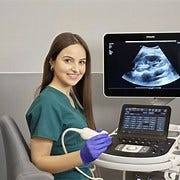
Ultrasound technology has revolutionized the field of obstetrics and gynecology (OB/GYN), offering a non-invasive and highly effective method for imaging and diagnosing various conditions. Here are some key applications of ultrasound in obstetrics and gynecology:
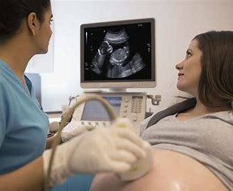
Prenatal Care: Ultrasound is routinely used during prenatal care to monitor fetal development, growth, and well-being. It helps determine gestational age, assess fetal anatomy, detect abnormalities, and evaluate the placenta and amniotic fluid levels.
Dating and Viability: Ultrasound is used to accurately determine the gestational age of the fetus, helping to confirm the due date and assess fetal viability in early pregnancy.
Anomaly Scan: Anomaly scans, typically performed around 18–20 weeks of pregnancy, use ultrasound to assess the fetal anatomy in detail. This helps detect structural abnormalities, congenital anomalies, and markers for genetic disorders.
Multiple Gestations: Ultrasound is essential for diagnosing and monitoring multiple pregnancies, including twins, triplets, or higher-order multiples. It helps determine chorionicity, amnionicity, and assess the growth and well-being of each fetus.
Fetal Growth Assessment: Serial ultrasound measurements of fetal biometry, including biparietal diameter, head circumference, abdominal circumference, and femur length, are used to assess fetal growth and detect intrauterine growth restriction (IUGR) or macrosomia.
Fetal Doppler Studies: Doppler ultrasound evaluates blood flow in the umbilical artery, middle cerebral artery, and ductus venosus to assess fetal well-being, detect signs of fetal distress, and monitor placental function.
Fetal Echocardiography: Specialized ultrasound techniques are used to evaluate the fetal heart, detect congenital heart defects, and assess cardiac function in cases of suspected cardiac abnormalities or high-risk pregnancies.
Amniocentesis and Chorionic Villus Sampling (CVS): Ultrasound guidance is often used during invasive prenatal procedures such as amniocentesis and CVS to ensure accurate placement of the needle and minimize the risk to the fetus.
Gynecological Disorders: Ultrasound is used to diagnose and monitor various gynecological conditions such as ovarian cysts, fibroids, endometrial abnormalities, and pelvic inflammatory disease (PID). It helps evaluate the size, location, and characteristics of these abnormalities.
Infertility Evaluation: Ultrasound plays a crucial role in evaluating infertility by assessing ovarian reserve, detecting structural abnormalities in the uterus or ovaries, and monitoring follicular development during assisted reproductive techniques (ART) such as in vitro fertilization (IVF).
Overall, ultrasound is an indispensable tool in obstetrics and gynecology, providing valuable diagnostic information, guiding interventions, and facilitating optimal care for pregnant women and gynecological patients. If you are preparing ultrasound courses. join StudyUltrasound.
0 notes
Text
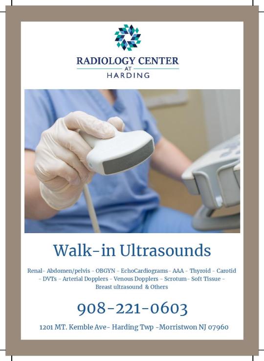
Walk-in Ultrasound
An ultrasound scan is a medical test that uses high-frequency sound waves to capture live images from the inside of your body.
https://bit.ly/2GlUqE0
0 notes
Text
Preparing for an ultrasound scan
Usually, when a doctor recommends an ultrasound scan, they will explain why the procedure is necessary and how best to get ready for it. Pre-examination preparation generally depends on the area of the body that will be examined; however, it is not always necessary to follow a set protocol.
Eating, drinking, and taking drugs are all perfectly acceptable both before and after an ultrasound examination, unless specifically directed otherwise.
In certain cases, an 8–12 hour fast will be necessary (before to an abdomen or vascular ultrasound). This is to prevent issues with sound waves coming into contact with undigested food in the stomach, which could cause fuzzy images.
Eat a fat-free and/or low-fiber meal the night before, followed by a brief period of fasting (for up to 6 hours before the scan), if the digestive system or organs such as the liver, pancreas, gallbladder, or spleen are to be checked. This is done to reduce the amount of gas in the digestive tract, which might interfere with scan results by producing images that are difficult to understand. Nonetheless, you can continue to drink water and other required medications during this pre-examination time.
In other cases, a doctor might advise a patient to stay hydrated in the hours before the scan and hold their urine in order to make sure their bladder is full when the scan happens (this might be necessary for a pelvic ultrasound, to see an unborn child, or to check on the health of the uterus and ovaries in general). This makes it easier to see certain areas during the examination, including the bladder. An enema may occasionally be administered before to a scan operation in order to cleanse the colon.
Before undergoing an ultrasound scan, it is crucial to disclose to the examining physician or radiographer all medications — prescription, over-the-counter, and supplement — that you are taking. Giving these away can be quite helpful in the diagnosis process.
Depending on the area of the body being inspected, a person may occasionally be required to take off certain articles of clothes and put on a medical gown that is supplied. To ensure comfort, a doctor could also advise dressing loosely on the day of the procedure.
If a patient needs medication to help them calm during the surgery, a sedative can be given (via an IV / intravenous line via the back of the hand or through a vein in the arm).
0 notes
Text
HOW Do Ultrasound Scans Work

Ultrasound scans, also known as sonography, work by using high-frequency sound waves to create images of the inside of the body. Here’s a step-by-step explanation of how ultrasound scans work:
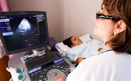
Generation of Sound Waves: A transducer, a handheld device, is used to generate high-frequency sound waves. These sound waves are typically inaudible to the human ear, with frequencies higher than the upper audible limit of human hearing.
Transmission into the Body: The transducer is placed against the skin on the area being examined. A gel is often applied to the skin to ensure proper contact and to eliminate air pockets that can interfere with the transmission of sound waves.
Propagation through Tissues: The generated sound waves travel through the body tissues. When they encounter different types of tissues (fluids, solids, air), some of the waves are reflected back to the transducer while others continue to penetrate deeper.
Echo Reception: The transducer not only sends out sound waves but also receives the echoes created as the waves bounce back from the internal structures. The timing and intensity of these echoes provide information about the density and composition of the tissues.
Conversion into Images: The received echoes are converted into electrical signals. These signals are then processed by a computer, which uses the information to create real-time images on a monitor. The images show the internal structures, such as organs or a developing fetus during pregnancy.
Doppler Ultrasound (optional): In some cases, a Doppler ultrasound may be used to assess blood flow. This involves evaluating the Doppler shift in the frequency of the echoes, allowing visualization of blood movement in vessels.
Recording or Capturing Images: The images can be recorded or captured in real-time during the examination. This helps healthcare professionals review and analyze the structures and functions of the internal organs or monitor the progress of a pregnancy.
Ultrasound scans are widely used in various medical applications, including monitoring pregnancies, imaging abdominal and pelvic organs, and assessing blood flow in vessels. They are non-invasive, safe, and do not involve ionizing radiation, making them a valuable diagnostic tool in medical practice.
if you are preparing Ultrasound exam. join ultrasound course in StudyUltrasound . StudyUltrasound is the best option.
0 notes
Text
CAREER OPTIONS IN ULTRASOUND
A career in ultrasound, also known as diagnostic medical sonography, offers various opportunities for individuals interested in medical imaging and healthcare. Here are some career options in ultrasound:
Diagnostic Medical Sonographer: These professionals operate ultrasound equipment to produce images of the body’s internal structures. They work closely with physicians to assist in the diagnosis and treatment of medical conditions. There are specialties within diagnostic medical sonography, such as abdominal, obstetric and gynecological, vascular, and cardiac sonography.
Cardiac Sonographer (Echocardiographer): Specializing in imaging the heart, cardiac sonographers use ultrasound to create detailed pictures of the heart’s chambers, valves, and blood vessels. This helps in assessing heart function and identifying cardiac issues.
Vascular Technologist: Vascular technologists specialize in imaging blood vessels and blood flow to detect issues such as blood clots, arterial blockages, or venous disorders. They may work in hospitals, clinics, or vascular laboratories.
Obstetric and Gynecologic Sonographer: These sonographers focus on imaging the female reproductive system, including monitoring fetal development during pregnancy. They play a crucial role in providing information about the health of the fetus and supporting obstetricians in managing pregnancies.
Musculoskeletal Sonographer: Specializing in imaging muscles, tendons, ligaments, and joints, musculoskeletal sonographers assist in diagnosing conditions such as injuries, arthritis, or tumors affecting the musculoskeletal system.
Pediatric Sonographer: Working with children and infants, pediatric sonographers specialize in imaging pediatric organs and tissues. They collaborate with pediatricians to diagnose and monitor various medical conditions in young patients.
Research and Development: Some professionals in ultrasound pursue careers in research and development, working for companies that develop and improve ultrasound technology. They may be involved in designing new equipment, enhancing imaging techniques, or conducting clinical trials.
Education and Training: Experienced sonographers may choose to become educators, teaching ultrasound techniques and principles to students in educational institutions, hospitals, or training programs.
Management and Administration: Sonographers with experience and leadership skills may transition into managerial or administrative roles within healthcare settings, overseeing ultrasound departments or clinics.
Mobile Sonography Services: Some sonographers work for mobile ultrasound services, providing imaging services at various locations, including hospitals, clinics, and patients’ homes.
To pursue a career in ultrasound, individuals typically need formal education and training in diagnostic medical sonography, usually through an accredited program. Certification by professional organizations, such as the American Registry for Diagnostic Medical Sonography (ARDMS) or Cardiovascular Credentialing International (CCI), is often required or preferred for employment. Additionally, staying updated on advancements in technology and techniques through continuing education is essential in this field. If you really interested to build a career in ultrasound. join ultrasound certification courses in StudyUltrasound is the best option.
0 notes
Text
To Become an Ultrasound Specialist in Gynaecology and Obstetrics.

In obstetrics and gynaecology, ultrasound is an essential tool for diagnosing, tracking, and treating a variety of problems that impact women’s reproductive health. A mix of academic understanding, practical expertise, and ongoing education is needed to become an expert in this field of ultrasound.
There are several steps you can take to gain the knowledge and experience you need to excel in this field.
• Get a solid education in obstetrics and gynaecology
The first step towards being an expert in ultrasound in obstetrics and gynaecology is to have a solid understanding of the anatomy, physiology, and pathophysiology of the female reproductive system. You will be able to identify normal and abnormal findings, comprehend the indications for ultrasound, and make well-informed clinical judgements using this knowledge.
• Obtain specialized training in ultrasound
The next stage after gaining a strong theoretical grasp is to practise doing and interpreting ultrasonography tests. The process include working under the guidance of seasoned radiologists and sonographers to hone your scanning methods and interpretation abilities. Understanding the various ultrasound equipment and imaging modalities — such as 2D, 3D, and 4D ultrasound — that are utilised in obstetrics and gynaecology is essential.
• Get hands-on experience.
When it comes to ultrasonography, this is especially true: practice makes perfect. Look for opportunities to practise using ultrasound equipment and procedures firsthand. You can accomplish this by looking for a mentor who can provide you opportunity to practise your abilities or by volunteering at a hospital or clinic that does ultrasounds.
• Stay up to date with the latest research and technology
Since ultrasound technology is always changing, it’s critical to keep up with the most recent findings and developments in the industry. Read the most recent research papers to stay up to date on best practices and attend conferences and seminars to learn about new methods and technology.
Finally, a combination of theoretical knowledge, practical skills, ongoing education, and strong interpersonal and communication abilities are needed to become an expert in ultrasound in obstetrics and gynaecology. Anyone can become an expert in this subject and significantly improve the lives of women and their families with hard work and dedication.
if you are preparing Ultrasound cetification course join StudyUltarasound.
0 notes
Text
Why should I select OBG’s Ultrasound option? What advantages exist?

Obstetrics and gynaecology together make up the medical specialty known as OBG. Here, obstetrics deals with pregnancy, childbirth, the postpartum period, and so on, whereas gynaecology addresses the health of the female reproductive system, including the vagina, uterus, ovaries, and so on. In obstetrics and gynaecology, ultrasound is frequently used to diagnose and track pregnancy as well as medical conditions like cancer, fibroids, and cysts. We’ll talk about the advantages and significance of a career in ultrasound OBG here.
How to become an expert in Ultrasound?
Understanding the principles and how they are used in the field is the first step towards becoming an expert in ultrasound technology. Researching the many ultrasound imaging modalities and their unique role throughout pregnancy is advised in order to become familiar with the various probes used, the anatomy of the ovaries and uterus, as well as patient placement rules.
It’s crucial to acquire clinical expertise in addition to ultrasonography in order to become an expert in foetal development monitoring, amniotic fluid evaluation, chorionic villus sampling, and ectopic pregnancy diagnosis.
Benefits of building a career in Ultrasound OBG?
Career Advancement
Learning how to use ultrasound in the medical field expands one’s professional growth options by providing access to a variety of opportunities. Ultrasonography experts can guarantee a better career in this profession by securing higher-paying roles and promotions.
Enhanced Diagnostic Abilities:
Accurate diagnosis and management of diseases such ovarian cysts, foetal development limitation, and ectopic pregnancy can be achieved by healthcare professionals who possess the knowledge and skills to perform and interpret ultrasound scans. Ultrasound-assisted early detection can reduce problems and enhance patient outcomes.
Skill Development
In addition to imparting technical knowledge, ultrasound courses cultivate important soft skills like critical thinking and effective communication. These qualities help healthcare workers become well-rounded individuals who can work more effectively with patients and other professionals.
Best Salary Packages
Professionals with ultrasound training are in high demand, which makes it possible to negotiate excellent compensation packages that include retirement plans, health insurance, and other perks. The quality of life is raised and overall compensation is increased by this recognition.
Are you hoping to pursue your ideal job in OBG ultrasonography? Recall that mastering ultrasound technology is a difficult but worthwhile endeavour for gynaecologists. You can receive the appropriate help from our team of professionals.
Get information about several short- and long-term courses in ultrasound OBG by getting in touch with the StudyUltrasound team.
0 notes
Text
Learn How Different Ultrasound Scans Work and What Their Use Is

A sonogram is another name for an ultrasound scan. During this process, high-frequency sound waves are used to create an image of a portion of the interior of the body.
There are numerous uses for ultrasound. It can be used to guide a surgeon during specific surgeries, diagnose a problem, and keep an eye on an unborn child.
Working Of Ultrasound
An ultrasonic probe, a tiny gadget, is employed. High-frequency sound waves are emitted by this. It is impossible for you to hear these waves of sound. The “echoes” that are produced when sound waves reverberate off various body parts are detected by the probe and converted into a moving picture.
While the scan is running, this image is shown on a monitor.
Different Kinds of Ultrasound Scans
Different types of ultrasound scanning exist. Which body part is being scanned and for what purpose determines the kind in large part.
The three primary categories are:
External Ultrasound Scan
The main purposes of an external ultrasound scanning are to look at an unborn child’s heart or womb. In addition, it can be used to evaluate other organs or tissues that are visible through the skin, like muscles and joints, as well as the liver, kidneys, and other organs located in the abdomen and pelvis.
Your skin is probed on the area of your body that is being checked with a tiny, portable device. Your skin is covered with a lubricating lubricant to facilitate the smooth movement of the probe. This guarantees that the probe and skin remain in constant contact.
Internal or Transvaginal Ultrasound Scan
An internal examination gives a physician a closer look at organs like the womb, ovaries, and prostate gland inside the body.
Meaning “through the vagina,” an ultrasound is called transvaginal. You will be instructed to lie on your side with your knees pulled up towards your chest, or on your back, during the treatment.
Images are then sent to a monitor using a tiny, sterile ultrasonic probe that is little wider than a finger. The probe is then gently inserted into the vagina or rectum.
Internal examinations typically don’t hurt and shouldn’t take too long, but they may cause some discomfort.
Endoscopic Ultrasound Scan
An endoscope is inserted into your body to perform an endoscopic ultrasound scan. In order to examine places like your stomach or food pipe, this is typically done through the mouth (oesophagus).
As the endoscope is gently lowered towards your stomach, you will be requested to lie on your side. The end of the endoscope is equipped with an ultrasonic instrument and a light. Similar to an external ultrasound, sound waves are employed to produce images once it is put into the body.
A local anaesthetic spray to numb your throat and a sedative to keep you calm are frequently administered because endoscopic ultrasound scans can be painful and even make you feel sick.
In the unlikely event that you bite the endoscope, a mouth guard might be provided to you to keep your mouth open and shield your teeth.
Hope you found this information helpful!!if you want to build a career in ultrasound you can join ultrasound courses in StudyUltrasound
Connect with us to know more
0 notes
Text
Scope of ultrasound in Obstetrics and Gynecology

Ultrasound plays a crucial role in Obstetrics and Gynecology, offering a wide range of applications for both routine care and diagnostic purposes. Here are some key aspects of the scope of ultrasound in Obstetrics and Gynecology:
Prenatal Imaging:
Fetal Monitoring: Ultrasound is widely used for monitoring fetal development during pregnancy. It helps assess the size, position, and overall health of the fetus.
Doppler Ultrasound: Measures blood flow in the umbilical cord and other fetal vessels, providing valuable information about fetal well-being.
2. Diagnosis of Pregnancy Complications:
Ectopic Pregnancy: Ultrasound can help diagnose ectopic pregnancies, where the embryo implants outside the uterus.
Miscarriage: It aids in confirming or ruling out miscarriages by visualizing the fetal heartbeat and development.
3.Detection of Birth Defects:
Anomaly Screening: Ultrasound is used for detailed anatomical surveys to detect any structural abnormalities in the developing fetus.
Nuchal Translucency Testing: Assesses the risk of chromosomal abnormalities by measuring the thickness of the nuchal fold in the fetus.
4.Guidance for Invasive Procedures:
Amniocentesis and Chorionic Villus Sampling (CVS): Ultrasound is used to guide these diagnostic procedures, helping to ensure accurate sampling without harm to the fetus.
5. Assessment of Uterine and Ovarian Conditions:
Uterine Abnormalities: Ultrasound can identify conditions such as uterine fibroids, polyps, or congenital anomalies.
Ovarian Cysts: Helps in visualizing and characterizing ovarian cysts, assessing their size and characteristics.
6. Evaluation of Pelvic Disorders:
Endometriosis: Ultrasound can be used to detect endometriosis lesions in the pelvis.
Pelvic Inflammatory Disease (PID): Helps in assessing inflammation and abnormalities in the pelvic organs.
7.Infertility Assessment:
Follicle Monitoring: Ultrasound is used to monitor ovarian follicle development during fertility treatments.
Uterine Abnormalities: Helps in assessing the uterine lining and detecting abnormalities that may impact fertility.
8.Gynecologic Cancer Diagnosis and Staging:
Ovarian, Uterine, and Cervical Cancer: Ultrasound aids in the initial diagnosis, staging, and monitoring of gynecologic cancers.
9.Postpartum Evaluation:
Postpartum Uterine Assessment: Ultrasound is used to assess the size and condition of the uterus after delivery.
Breast Imaging: In some cases, ultrasound may be used for breast imaging in lactating women.
10.Fetal Echocardiography:
Cardiac Evaluation: Ultrasound allows for detailed examination of the fetal heart, aiding in the detection of congenital heart defects.
The scope of ultrasound in Obstetrics and Gynecology continues to evolve with advancements in technology, providing healthcare professionals with valuable tools for prenatal care, diagnosis, and monitoring of reproductive health.
If you want to build a career in Ultrasound .StudyUltrasound is the best choice.
0 notes
Text
Learn All About This Cutting-Edge Technology: Ultrasound

Learn More About Ultrasound
Another name for an ultrasound scan is sonography. This medical test records live images from inside the human body using high-frequency sound waves. The technology bears a striking resemblance to the sonar and radar systems used by the military for ship and aircraft detection. With ultrasound, a physician can detect concerns with tissues, organs, and veins without having to make an incision. Continue reading to learn about the different preparation processes for ultrasounds.
The standard imaging methods use radiation. Conversely, ultrasound doesn’t use radiation. This makes it the best way to see how a fetus is developing when a woman is pregnant.
Reasons Why Ultrasound Is Performed
Pregnancy is commonly linked to ultrasound. It gives a pregnant woman her first glimpse of her unborn kid. And the test is not just useful for that. Ultrasound serves a multitude of purposes.
If a patient is experiencing discomfort, edema, or other symptoms that call for an interior look of the organs, a doctor is likely to order this treatment.
These organs are viewable using an ultrasound.
– Bladder
– Brain (in infants)
– Eyes
– Gallbladder
– Kidneys
– Liver
– Ovaries
– Pancreas
– Spleen
– Thyroid
– Testicles
– Uterus
– Blood vessels
Additionally, surgeons utilize it to direct their movements when doing medical operations like biopsies.
Preparation For an Ultrasound
The location or organ being studied usually determines how to prepare for an ultrasound. The doctor may advise you to fast for eight to twelve hours before to your ultrasound procedure if your abdomen is being evaluated. This is because food particles that haven’t fully digested can obstruct sound wavelengths. This will make it harder for the technician to capture a clean picture.
It is sometimes suggested that you fast the night before the surgery and have a low-fat dinner the previous evening. This is for the liver, pancreas, spleen, or gallbladder examination. On the other hand, you can keep taking your meds as directed and drinking water. To make your bladder appear fuller and easier to see, you could be encouraged to drink a lot of water and hold your pee.
Prior to the exam, make sure your doctor is aware of any prescription pharmaceuticals, over-the-counter medications, or herbal supplements you take. Prior to the surgery, make sure you ask any questions you may have and follow your doctor’s instructions.
The danger of this surgery is very low. It doesn’t include radiation, unlike CT scans or X-rays. I hope this information was helpful to you.
if you want to build a career in ultrasound StudyUltrasound is the best platform.
0 notes
Text
Examining a Potential Ultrasound Career: Your Road to Success

A career in ultrasonography offers a dynamic and fulfilling opportunity in the quickly evolving field of medicine today. The need for qualified ultrasound technicians is growing as technology advances. This blog will cover a range of opportunities, possible educational paths, and the thrilling process of developing a career in ultrasonography.
Ultrasound as a Career:
The use of ultrasound technology is essential for non-invasive medical condition diagnosis. It includes imaging interior body structures using high-frequency sound waves, which helps doctors diagnose conditions, keep an eye on pregnant patients, and more.
Educational Pathways:
Pursuing a specialized ultrasound school or program is usually the first step in starting a career in ultrasound. These programs, which provide thorough instruction in ultrasound methods, equipment operation, anatomy, patient care, and medical ethics, are offered by a number of universities. Certain courses may provide specialist knowledge in fields such as abdominal sonography, echocardiography, or obstetrics.
Career Opportunities:
Graduates of the ultrasound course can investigate a variety of ultrasound-related options. Ultrasound technologists frequently work in clinics, hospitals, DVI centers, and doctor’s offices. Because of the role’s flexibility, professionals can focus on particular areas of interest.
Advancements and Growth:
The ultrasound industry is impacted by the ongoing evolution of technology. Professionals need to keep up with the most recent developments and methods. Career progression requires obtaining credentials, continuing education, and keeping up with technology innovations.
An ultrasonography career involves more than just using equipment; it also involves helping with patient care and diagnosis. Stability, room for advancement, and the joy of improving healthcare are all offered by the field. Enrolling in ultrasound courses is a big step toward a fulfilling profession that opens doors to a plethora of options in the rapidly developing field of medical technology.
0 notes
Text
The Power of Ultrasound in Gynecology and Obstetrics: Progress and Uses
Using high-frequency sound waves, this non-invasive imaging method produces real-time images of the reproductive system, fetus, and pelvic organs. Ultrasonography continues to be a crucial component of all-encompassing healthcare for women globally, even as research and technology advance.
1.Advances in Prenatal Care through Ultrasound
During pregnancy, ultrasound equipment is essential for tracking the development of the fetus. Early ultrasounds determine the gestational age and validate the viability of the pregnancy. Mid-term ultrasounds monitor the location of the fetus inside the uterus, identify anomalies, and offer thorough evaluations of the essential organs.
2. Obstetrical Diagnostic Capabilities
Early intervention is aided by the ease with which prenatal abnormalities can be detected using ultrasound. By measuring blood flow, Doppler ultrasound makes that the placenta and umbilical cord are getting enough nutrients. Improved sight of the fetus with advanced 3D/4D imaging helps parents feel more emotionally connected to their pregnant child.
3. Ultrasound’s in Gynecological Applications
Ultrasound is essential in gynecology to diagnose diseases of the female reproductive system. Identification of conditions like endometriosis, fibroids, and ovarian cysts is aided by transvaginal ultrasounds. Before imaging, saline is injected into the uterus during a sonohysterogram to help discover anomalies in the uterus.
4. Fertility treatments and procedures guided by ultrasound
Targeting aberrant tissues accurately, ultrasound guides minimally invasive operations such as aspirations and biopsies. It tracks the development of follicles during fertility treatments, assisting techniques like IUI and IVF that increase the likelihood of conception.
5. Developments in Ultrasound Technology
Ultrasound technology is constantly evolving, with new developments including enhanced resolution, portable devices, and AI integration. AI algorithms and high-resolution imaging improve accuracy, increasing the dependability and accessibility of measurements and diagnostic interpretations.
6. Safety Factors and Restrictions
Although ultrasonography is generally safe, overuse should be avoided. Despite its advantages, not all fetal anomalies may be detected by it, and effective interpretation necessitates experience. Minimal dangers are guaranteed by safety protocols when using routine diagnostic ultrasound.
Prenatal care is still being revolutionized by ultrasound technology, which also helps with diagnosis and directs obstetric and gynecological procedures. Because to its non-invasiveness and continuous technological improvements, it guarantees better patient care, which helps women seeking gynecological exams and expectant mothers have healthier outcomes. If you are building a career in ultrasound StudyUltrasound is the best platform.
0 notes
Text
An Extensive Guide to Gynecology and Obstetrics Ultrasound Scanning Techniques

In obstetrics and gynecology, ultrasound is a non-invasive imaging method that is essential for understanding the health and development of the mother and the fetus during pregnancy. Accurate diagnosis and improved patient care in obstetric and gynecological ultrasound depend on an understanding of the various scanning techniques and planes.
Transabdominal Ultrasound
One prevalent procedure in both obstetrics and gynecology is transabdominal ultrasonography. To create images, a transducer is dragged over the skin after a gel has been placed to the abdomen. In the early stages of pregnancy, this method is frequently used to determine gestational age and confirm pregnancy.
Transvaginal Ultrasound
A specifically made transducer is inserted into the vagina to perform transvaginal ultrasonography. The uterus and ovaries, among other pelvic organs, can be seen more clearly and up close thanks to this technology. It’s frequently used to improve visualization in the early stages of pregnancy.
Doppler Ultrasound
Vascular blood flow is measured via Doppler ultrasonography. It supports the management of high-risk pregnancies in obstetrics by assisting in the assessment of blood flow to the fetus and placenta. It helps assess blood flow in the pelvic organs in gynecology, especially when there is unusual bleeding or there are possible vascular problems.
3D and 4D Ultrasound
Three-dimensional (3D) and four-dimensional (4D) ultrasounds provide three-dimensional images of the fetus or pelvic organs. These images offer a more detailed view, enhancing diagnostic accuracy and aiding in the assessment of fetal abnormalities.
Transperineal Ultrasound
By placing the transducer on the perineum, transperineal ultrasonography allows for a view of the muscles of the anal sphincter and pelvic floor. It is frequently used in obstetrics to assess certain pelvic abnormalities and in gynecology to evaluate pelvic floor diseases.
Healthcare providers must be aware of these ultrasound scanning procedures in order to make correct diagnoses and guarantee the health of the mother and the developing fetus. Improved imaging capabilities and better patient care in obstetrics and gynecology are the results of ongoing advancements in ultrasound technology. If you want to build a career in ultrasound StudyMEDIC is the best platform.
join ultrasound course for build a profession in ultrasound.
1 note
·
View note
Text
BENEFITS OF ULTRASOUND COURSES: What makes ultrasound in OBG the better option?

There are many different career options available in the realm of ultrasonic music. Improving one’s ultrasound skills will enable obstetricians and general practitioners (OGGs) to become experts in their field and advance their careers, as ultrasound scanning is an essential component of examining the foetus (within a woman’s uterus), uterus, and ovaries.
The advantages of using ultrasound in OBG are covered here.
Career Advancement
Healthcare workers can broaden their areas of practice and improve their employability by learning ultrasonography techniques. Ultrasound expertise can also result in promotions and better-paying jobs.
Enable them to better doctor in ULTRASOUND!
Learning how to conduct and interpret ultrasound scans also helps them become better doctors since they enable medical practitioners to correctly diagnose and treat diseases like ovarian cysts, fetal growth limitation, and ectopic pregnancy. Patient outcomes can be enhanced, and complications can be avoided with early diagnosis of certain disorders.
Skill development
You can also gain valuable skills from ultrasound courses that can aid you in your work. As an illustration, taking ultrasound classes can aid in the development of excellent communication skills, as you will need to interact with patients and other medical professionals in an efficient manner. They can also aid in the development of critical thinking abilities since successful diagnosis requires analysis and interpretation of ultrasonography images.
Best salary package
The compensation package is an additional perk. They can choose the greatest compensation package based on their location, expertise, and skills. A lot of firms provide paid time off, retirement plans, and health insurance.
Flexible and convenient learning experience.
Since many ultrasound courses are available online, you can study whenever and wherever it’s convenient for you. Those who are currently employed in the medical industry and might not have the time to attend regular lessons will particularly benefit from this.
In the end, enrolling in an ultrasound course can be a wise investment for your professional future. You will be able to pursue a rewarding career in Ultrasound the medical profession with the knowledge and abilities you acquire from these courses.
#ultrasound#ultrasoundcourse#ultrasoundexam#ultrasoundscanning#ultrasoundstudents#mbbs#education#medicaleducation
0 notes
Last Seen Blogs

oceanattic
Between ocean and mountain lies true meaning.

sweatymoneybagelhound
Untitled

artbyhoney
Big artistic Mess

my2feet09
My1Spot

mydrawsofsomething-blog
lookatmyart